L
ung cancer resection surgery carries a substantial risk of postoperative pulmonary complications. Postoperative pulmonary complications are associated with mortality,1,2 intensive care admission, longer hospital length of stay,3 and increased costs.4,5 Video-assisted thoracoscopic sur-gery has recently gained wide popularity for cancer resec-tion. Indeed, as compared to the standard thoracotomic approach, video-assisted thoracoscopic surgery decreases surgical complications, postoperative pain, and the overall incidence of postoperative pulmonary complications.6–8Postoperative diaphragmatic dysfunction has been doc-umented after thoracic surgery with the thoracotomic
approach within the first postoperative day.9 Surgical injury of the diaphragm or of the phrenic nerve and diaphragmatic fatigue due to postoperative respiratory mechanics impair-ment play an important role in its pathogenesis.10 However, postoperative diaphragmatic dysfunction is largely under-estimated as a contributing factor for postoperative pul-monary complications11 due to the lack of reproducible diagnostic methods. Furthermore, little is known on the impact of video-assisted thoracoscopic surgery, as compared
aBStract
Background: Postoperative diaphragmatic dysfunction after thoracic
sur-gery is underestimated due to the lack of reproducible bedside diagnostic methods. We used point of care ultrasound to assess diaphragmatic function bedside in patients undergoing video-assisted thoracoscopic or thoracotomic lung resection. Our main hypothesis was that the thoracoscopic approach may be associated with lower incidence of postoperative diaphragm dysfunc-tion as compared to thoracotomy. Furthermore, we assessed the associadysfunc-tion between postoperative diaphragmatic dysfunction and postoperative pulmo-nary complications.
Methods: This was a prospective observational cohort study. Two cohorts
of patients were evaluated: those undergoing video-assisted thoracoscopic surgery versus those undergoing thoracotomy. Diaphragmatic dysfunction was defined as a diaphragmatic excursion less than 10 mm. The ultrasound evaluations were carried out before (preoperative) and after (i.e., 2 h and 24 h postoperatively) surgery. The occurrence of postoperative pulmonary compli-cations was assessed up to 7 days after surgery.
results: Among the 75 patients enrolled, the incidence of postoperative
diaphragmatic dysfunction at 24 h was higher in the thoracotomy group as compared to video-assisted thoracoscopic surgery group (29 of 35, 83% vs. 22 of 40, 55%, respectively; odds ratio = 3.95 [95% CI, 1.5 to 10.3]; P = 0.005). Patients with diaphragmatic dysfunction on the first day after surgery had higher percentage of postoperative pulmonary complications (odds ratio = 5.5 [95% CI, 1.9 to 16.3]; P = 0.001). Radiologically assessed atelectasis was 46% (16 of 35) in the thoracotomy group versus 13% (5 of 40) in the video-assisted thoracoscopic surgery group (P = 0.040). Univariate logistic regression analysis indicated postoperative diaphragmatic dysfunction as a risk factor for postoperative pulmonary complications (odds ratio = 5.5 [95% CI, 1.9 to 16.3]; P = 0.002).
conclusions: Point of care ultrasound can be used to evaluate
postoper-ative diaphragmatic function. On the first postoperpostoper-ative day, diaphragmatic dysfunction was less common after video-assisted than after the thoracotomic surgery and is associated with postoperative pulmonary complications. (ANESTHESIOLOGY 2019; 131:266–78)
Point of Care Ultrasound
to Identify Diaphragmatic
Dysfunction after Thoracic
Surgery
S. Spadaro, M.D., Ph.D., S. Grasso, M.D., Ph.D., M. Dres, M.D., A. Fogagnolo, M.D., F. Dalla Corte, M.D., N. Tamburini, M.D., P. Maniscalco, M.D., G. Cavallesco, M.D., V. Alvisi, M.D., T. Stripoli, M.D., Ph.D., E. De Camillis, M.D., R. Ragazzi, M.D., C. A. Volta, M.D.
Anesthesiology 2019; 131:266–78
Supplemental Digital Content is available for this article. Direct URL citations appear in the printed text and are available in both the HTML and PDF versions of this article. Links to the digital files are provided in the HTML text of this article on the Journal’s Web site (www.anesthesiology.org).
Submitted for publication May 17, 2018. Accepted for publication April 4, 2019. From the Department of Morphology, Surgery and Experimental Medicine, Intensive Care Unit, Sant’Anna Hospital, Ferrara, Italy (S.S., A.F., F.D.C., N.T., P.M., G.C., V.A., E.D.C., R.R., C.A.V.), the Department of Emergency and Organ Transplant, Aldo Moro University of Bari, Bari, Italy (S.G., T.S.), and Sorbonne Universités, UPMC Université Paris 06, INSERM, UMRS1158 Neurophysiologie respiratoire expérimentale et clinique, Paris, France (M.D.). Copyright © 2019, the American Society of Anesthesiologists, Inc. All Rights Reserved. Anesthesiology 2019; 131:266–78. 10.1097/ALN.0000000000002774
editor’S PerSPective
What We Already Know about this topic
• Patients undergoing thoracic surgery are at high risk for postoper-ative pulmonary complications
• The feasibility of using point of care ultrasound to diagnose dia-phragmatic dysfunction is unclear
What this Article tells us that Is New
• Point of care ultrasound can be used to detect diaphragmatic dys-function after thoracic surgery
• Diaphragmatic dysfunction may be associated with postoperative pulmonary complications
Diaphragm Dysfunction after Thoracic Surgery
to the standard thoracotomic technique, on postoperative diaphragmatic dysfunction and postoperative pulmonary complications.
The reference methods to evaluate diaphragmatic func-tion are phrenic nerve stimulafunc-tion12 and transdiaphragmatic pressure assessment.13 Both of these techniques are invasive, require considerable expertise, and are often unavailable at the bedside. Furthermore, they do not allow one to distin-guish between bilateral and unilateral diaphragmatic dys-function. Recently, the ultrasound technique has emerged as a noninvasive point of care tool to assess diaphragmatic function.14,15 A decreased inspiratory diaphragmatic dome excursion has been recently validated as an index of dia-phragmatic dysfunction both in critically ill16 and in surgi-cal patients.17–19
In this study, we assessed the postoperative diaphragmatic function through the ultrasound technique after cancer resection surgery. Our main hypothesis was that the video- assisted thoracoscopic surgery technique, as compared to the thoracotomic approach, could decrease the postopera-tive diaphragm dysfunction. Our secondary endpoint was to assess the clinical variables associated with postoperative diaphragmatic dysfunction and the impact of postoperative diaphragmatic dysfunction on postoperative pulmonary complications within the first 7 postoperative days.
Materials and Methods
Patients
The study was performed at the Sant’Anna University Hospital of Ferrara, Italy. All patients receiving elective lung resection surgery for pulmonary neoplasm between February 2016 and November 2016 were included. The study was approved by the ethics committee of our institu-tion (February 23, 2016) and was recorded retrospectively on clinicaltrial.gov (NCT03347578). Written informed consent was obtained from each patient during the preop-erative visit.
We enrolled patients 18 yr or older, with an American Society of Anesthesiologists physical status classification score of II to III, scheduled for thoracotomy or video- assisted thoracoscopic surgery lung resection surgery for cancer. The surgical approach was nonrandomly assigned and decided upon technical and/or oncologic reasons by the surgeon. A standard posterolateral thoracotomy with mus-cle-sparing technique was adopted in the open technique; for the video-assisted thoracoscopic surgery approach, two small incisions (at the eighth intercostal space in midaxillary and posterior axillary line) and a 5-cm utility incision at the level of the fifth intercostal space (for lung tissue removal) were performed.
The exclusion criteria were body mass index greater than 35 kg/m2, contraindications for epidural catheter posi-tioning, history of neuromuscular disease, previous tho-racic surgery, and phrenic nerve palsy. Patients were further
excluded from the final analysis if correct diaphragm visu-alization was not achievable (such as in case of postoperative subcutaneous emphysema).
All patients admitted to the study underwent a preopera-tive physiologic assessment including cardiovascular evalua-tion (electrocardiogram and echocardiography), pulmonary function tests, and arterial blood gas analysis according to clinical practice guidelines.20 Pulmonary function tests con-sisted of spirometry performed according to the American Thoracic Society recommendations21 using a SpiroPro spi-rometer (SpiroPro, Jaeger, Germany).
On the first postoperative day, pulmonary function tests were repeated in the semirecumbent position (with the head of the bed elevated at an angle of 45°). Pulmonary function tests were performed by a technician and validated by a pul-monologist. Spirometric measurements on the first postop-erative day included forced vital capacity, forced expiratory volume in 1 s and tidal volume (VT) was performed through a portable spirometer (MicroLoop Spirometer; CareFusion Corp., USA) that meets the American Thoracic Society standards. For both the preoperative and postoperative pul-monary function test assessments, the highest value of three spirometric measurements was recorded.
Anesthesia
Before general anesthesia induction, a thoracic epidural catheter (Tuohy; Braun Laboratories, Melsungen AG, Germany) was placed at T3 to T6 for postoperative pain control under local anesthesia. The position of the catheter tip was verified by a test dose of 4 ml of 1% lidocaine.
Propofol (1.5 to 2 mg · kg−1) and fentanyl (3 μg · kg−1) were used to induce anesthesia. Muscle paralysis was obtained with rocuronium bromide (0.6 mg · kg−1) to facil-itate tracheal intubation. The trachea was intubated with an appropriately sized and side double lumen tube (Broncho-part; Rush, Germany). The correct position of the tube was checked by a fiberoptic bronchoscope in supine position after intubation and in lateral decubitus. Anesthesia was maintained with a continuous infusion of propofol (70 to 100 μg · kg−1 · min–1), remifentanil (0.1 to 0.2 μg · kg−1 · min–1), and rocuronium bromide (7 μg · kg−1 · min–1). The Bispectral Index (BIS) was used to monitor the depth of anesthesia. The BIS was calculated and displayed continu-ously using an Aspect A2000 electroencephalogram ana-lyzer (Aspect Medical System, USA) with the anesthetics titrated to maintain a BIS index between 40 and 60.
The lungs were ventilated through a Dräger Primus ventilator (Drägerwerk AG & Co. KGaA, Germany) with a square flow waveform with a VT of 6 to 8 ml · kg−1 ideal body weight in two-lungs ventilation and a protective one-lung ventilation with a VT of 4 to 5 ml · kg−1.22 Intraoperative positive end-expiratory pressure was selected by the treating physician. Patients in one-lung ventilation were ventilated using oxygen and air with a fraction of inspired oxygen (Fio2) set to maintain the oxygen saturation measured by
pulse oximetry (Spo2) 92% or greater. Intraoperative respi-ratory rates, positive end-expirespi-ratory pressure, and Fio2 were manually recorded, and the mean of all measurements for each phase was finally reported. As usual care in our depart-ment, recruitment maneuvers were used as a rescue therapy in case of intraoperative hypoxemia (defined as Spo2 less than 90%).22,23 Balanced crystalloid solutions were adminis-tered at a rate of 3 ml · kg−1 · h−1. Patients were monitored by electrocardiogram, pulse oximetry, end-tidal carbon diox-ide, and invasive arterial pressure using a Datex Ohmeda S/5 monitor (Datex-Ohmeda Division, Instrumentarium Corp., Finland).
At the end of surgery, train-of-four (TOF-Watch accel-eromyographs; Organon-Teknika, France) stimulations were used to assess the presence of a residual neuromuscular block, with a train-of-four ratio 0.9 or greater considered suitable for extubation. If a train-of-four ratio less than 0.9 was present, neuromuscular block was reversed with sugam-madex 4 mg/kg, and patients were extubated in the operat-ing room. The standard extubation criteria were as follows: (1) cooperative and alert patient; (2) smooth spontaneous ventilation; (3) train-of-four 0.9 or greater at the adductor pollicis; (4) Spo2 greater than 96% on Fio2 of 0.4 or less and end-tidal carbon dioxide less than 45 mmHg; (5) stable hemodynamic; (6) core temperature 36.5°C or greater; and (7) no evidence of early surgical complications.
ultrasonography
Ultrasonographic assessments were performed by a sin-gle well-trained anesthesiologist with 3 yr of certified experience (R.R.) by using an ultrasonography machine (M-Turbo; SonoSite, Inc., USA). All measurements were performed in spontaneously and resting tidal breathing patients lying in the semirecumbent position.
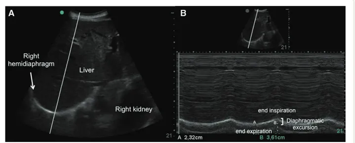
The liver and spleen were regarded as echographic windows for the right and left hemidiaphragm, respec-tively. Diaphragmatic excursion was evaluated using a 3.5- to 5-MHz convex ultrasound probe. The subcostal acoustic window described by Boussuges et al.24 for dia-phragm examination was used. For the visualization of right hemidiaphragm, the probe was placed between the midclavicular and anterior axillary lines, while for the left hemidiaphragm, a subcostal or low intercostal probe posi-tion was chosen between the anterior and midaxillary lines. This approach was chosen due to medications and chest tubes. The two-dimensional mode (B-mode) was used to select the hemidiaphragm exploration line. With the probe fixed on the chest wall, the ultrasound probe was medi-ally, cephalad, and dorsally directed so that the ultrasound beam reached the posterior part of the hemidiaphragmatic dome at an angle as close to 90° as possible.25,26 The ultraso-nography machine was then switched to the motion mode (M-mode). During inspiration, the normal diaphragm contracts and moves caudally toward the transducer; this is recorded as an upward motion of the motion mode tracing
and regarded as the diaphragmatic excursion during inspi-ration, which was measured on the vertical axis from the baseline to the point of maximum height of inspiration, on a frozen image (fig. 1).
Diaphragmatic excursion was recorded using a sweep speed set in order to detect at least three consecutive respi-ratory cycles on the same screenshot. Recordings measure-ments were considered suitable only if the diaphragmatic excursion pattern was regular in amplitude and frequency. To reduce the measurement error, the diaphragmatic excursion estimation was averaged over three consecutive respiratory cycles.
Analgesia and Postoperative Pain Assessment
Postoperative analgesia was administered through the epi-dural catheter. An epiepi-dural analgesia solution of 300 ml, consisting of 0.2% levobupivacaine, was delivered at rate of 5 to 7 ml/h. Intravenous nonsteroidal anti-inflammatory drugs were added to the aforementioned analgesic protocol upon the treating physician’s decision.
Postoperative pain was assessed by the same two phy-sicians throughout the study period using an 11-point numerical rating scale (0 = no pain; 10 = worst possible pain) at rest. Additional IV morphine 2 mg was adminis-tered when numerical rating scale was 3 or greater at rest.
Outcome Measures
The primary outcome of the study was the occurrence of postoperative diaphragmatic dysfunction through point of care ultrasound, defined as a diaphragmatic excursion less than 10 mm or negative. This cutoff value has been vali-dated in different patients (i.e., after abdominal surgery, in critically ill patients or in healthy volunteers).17,24,27 The diaphragmatic excursion is negative in case of unilateral or bilateral diaphragmatic palsy.28
Postoperative pulmonary complications were recorded from the first to the seventh postoperative day by the attend-ing physicians who were blinded to the diaphragmatic ultrasonographic parameters. We considered the follow-ing as postoperative pulmonary complications: hypoxemia, bronchospasm, atelectasis, new pulmonary infiltrates, sus-pected pulmonary infection, and pleural effusion. The post-operative pulmonary complications definitions details are reported in the Appendix.
study Protocol
The timeline of the protocol is summarized in figure 2. Diaphragmatic ultrasound was performed by the same anesthesiologist (R.R.) the day before operation (preoper-ative) and 2 and 24 h postoperatively. Pulmonary function tests were conducted on the same day of the preoperative evaluation and at 24 h postoperatively. The postoperative pain intensity was assessed at 2 h postoperatively and 24 h postoperatively. The occurrence and type of pulmonary
Diaphragm Dysfunction after Thoracic Surgery
complications were evaluated daily from the first to the sev-enth postoperative day.
statistical Analysis
This is the primary analysis of these data, and all the anal-yses reported are preplanned. Normal distribution of data was tested by the Shapiro–Wilk Normality Test. Data are reported as mean ± SD or median [interquartile range], as appropriate. Unpaired Student’s t tests or Mann–Whitney U tests were used to test the differences between groups (i.e., thoracotomy vs. video-assisted thoracoscopic sur-gery) for data with normal or not normal distribution,
respectively. Pearson’s chi-square test or the Fisher exact test was used to compare categorical data. Differences between diaphragmatic excursion at different times in the same sub-ject were assessed through the Friedman rank analysis or the Wilcoxon signed-rank test for matched data as appropri-ate. When multiple comparisons were made, P values were adjusted by the Bonferroni post hoc procedure.
The association between diaphragmatic dysfunction as expressed by a diaphragmatic excursion less than 10 mm in at least one measurement and clinically meaningful perioperative variables, including surgical access, chronic obstructive pulmonary disease history, length of surgery, age, American Society of Anesthesiologists physical status
Fig. 1. ultrasound images of the right hemidiaphragm. (A) A two-dimensional mode diaphragm picture: the bright curved line depicts the
diaphragm; motion mode selected beam was directed as perpendicular as possible to the posterior part of the diaphragm. (B) A motion mode image of diaphragmatic excursion: caliper A indicates the end of expiration, and caliper B the end of inspiration. the distance between the two points, measured as the difference between the two caliper lines linked to the bottom of the image, shows an excursion of 12.9 mm.
Fig. 2. study protocol timeline.
score, sex, body mass index, and smoking history, was modeled using binary logistic regression analysis and was reported as the estimated crude odds ratio and relative 95% CI. In the same fashion, logistic regression analysis was applied to investigate the possible risk factors for the occurrence of at least one postoperative pulmonary com-plication. Furthermore, the significance of the interaction between surgery type and diaphragmatic dysfunction was tested using the likelihood-ratio test.
A two-tailed P value less than 0.05 was considered statistically significant. The P values of the comparison between diaphragmatic excursion in the two groups during the study were adjusted by the Bonferroni post hoc procedure.
Statistical analysis was performed with using SPSS Statistics for Windows, Version 25.0 (IBM, USA).
The sample size was calculated according to the pri-mary endpoint, i.e., the occurrence of diaphragmatic dys-function after lung resection surgery. To detect a mean difference of 4.8 mm in diaphragmatic excursion after 24 h from operation (assuming a SD of 5 mm for preoperative values and 3 mm for postoperative values) using paired sample t tests with an α of 0.05 and 99% power, a mini-mum of 28 patients were required for each group. This was the observed difference in diaphragmatic excursion found in a previous study on the effect of upper abdominal sur-gery on diaphragmatic movements.17 Taking into account a loss to follow up of 20%, we decided to enroll at least 35 patients for each group. The sample size analysis was performed using MedCalc software (9.3.6.0; Mariakerke, Belgium).
results
Patient Population
During the study period, 101 patients were screened for eligibility. Of these, 79 met the inclusion criteria. Four were subsequently excluded due to a not achievable dia-phragmatic ultrasonographic visualization. We did not have any further missing or lost data. Accordingly, 75 patients (35 in the thoracotomy group and 40 in the video-assisted thoracoscopic surgery group) completed the study (fig. 3). Clinical and demographic characteris-tics of the patients are described in table 1. Two patients in thoracotomy group and one patient in video-assisted thoracoscopic surgery group received an intraoperative recruitment maneuver. Neuromuscular block was reversed through sugammadex in 14 patients (6 in the thoracotomy and 8 in the video-assisted thoracoscopic surgery group; P = 0.776). All patients met the standard extubation crite-ria at the end of the surgery. Preoperative and intraoper-ative variables did not differ significantly between groups (table 1 and Supplemental Digital Content 1, http://links. lww.com/ALN/B952).
Perioperative trend in Diaphragmatic Excursion and Pulmonary Function tests
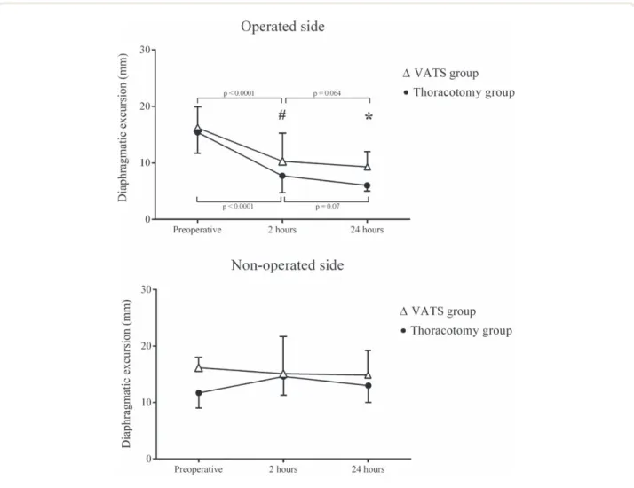
Fifty-one of 75 patients (68%) experienced diaphragm dysfunction in the operated side 24 h postoperatively. Diaphragm dysfunction was diagnosed in 22 of 40 patients (55%) in the video-assisted thoracoscopic surgery group and in 29 of 35 patients (83%) in the thoracotomy group (P = 0.019). Diaphragmatic excursion decreased in the oper-ated side by 56% [36 to 72%] in the thoracotomy group and by 43% [23 to 58%] in the video-assisted thoracoscopic surgery group (P = 0.033 for comparison between groups) 24 h postoperatively, as compared to preoperatively (fig. 4). None of the enrolled patients had a negative diaphragmatic excursion. In the nonoperated side, the diaphragmatic excursion remained unchanged, regardless of the surgical technique (fig. 4).
Diaphragm dysfunction 2 h postoperatively was detected in 17 of 40 patients (42%) in the video-assisted thoraco-scopic surgery group and 25 of 35 (71%) in the thoracot-omy group (P = 0.019). Decline in diaphragmatic excursion 2 h postoperatively in the operated side was 44% (18 to 69%) in the thoracotomy group and 32% (8 to 57%) in the video-assisted thoracoscopic surgery group (P = 0.202 for comparison between groups). Table 2 reports the periop-erative values of diaphragmatic excursions in the operated side, VT, and pulmonary function tests.
In the thoracotomy group, going from preoperatively to 24 h postoperatively, tidal volume (VT) decreased by 38% [25 to 49%], whereas in the video-assisted thoraco-scopic surgery group, it decreased by 27% [12 to 40%] (both P < 0.001 compared to baseline). The VT decline was more pronounced in the thoracotomy group (P = 0.044 vs. video-assisted thoracoscopic surgery group). Regardless of the surgical approach, VT at 24 h postoper-atively decreased more in the 51 patients with diaphrag-matic dysfunction (i.e., 39% [20 to 49%]), than in the 24 patients without diaphragmatic dysfunction (i.e., 26% [12 to 43%]; P = 0.034).
The postoperative decline of forced expiratory vol-ume in 1 s (FEV1) did not differ between the thoracotomy (–60% [–67 to –51%]) and the video-assisted thoracoscopic surgery group (–59% [–72 to –50%]; P = 0.941 for com-parison between groups). Patients with diaphragmatic dys-function experienced a more enhanced FEV1 reduction as compared to those without diaphragmatic dysfunction (–50% vs. –61%, respectively; P = 0.002).
As compared to preoperative values, the forced vital capacity decreased by 62% in the thoracotomy group and by 52% in the video-assisted thoracoscopic surgery group (P = 0.026 between groups at 24 h postoper-atively). A higher impairment in forced vital capacity was found in patients with diaphragmatic dysfunction where the forced vital capacity decreased by 58% com-pared to 42% in patients without diaphragmatic dys-function (P < 0.001).
Diaphragm Dysfunction after Thoracic Surgery
Factors Associated with Postoperative Diaphragm Dysfunction at 24 h
Table 3 shows that, according to the univariate analysis, patients receiving thoracotomy and being active smokers at the time of the operation were at higher risk of devel-oping diaphragmatic dysfunction 24 h postoperatively. Among them, the thoracotomic approach was the risk factor associated with the highest odds ratio (odds ratio = 3.95 [95% CI, 1.5 to 10.3]) for postoperative diaphrag-matic dysfunction.
Impact of Diaphragm Dysfunction on Postoperative Pulmonary Complications
The postoperative pulmonary complications are summa-rized in Supplemental Digital Content 2 (http://links. lww.com/ALN/B953). Overall, 39 of 75 (52%) patients
developed at least one postoperative pulmonary complica-tion. The occurrence of postoperative pulmonary compli-cations was 69% in the patients who had thoracotomy (24 of 35 patients) and 38% in the patients who had video- assisted thoracoscopic surgery (15 of 40 patients; P = 0.007; odds ratio = 3.63 [95% CI, 1.4 to 9.5]; table 4 and Supplemental Digital Content 2, http://links.lww. com/ALN/B953). Patients with diaphragmatic dysfunction at 24 h postoperatively had a higher percentage of postop-erative pulmonary complications compared with patients without diaphragmatic dysfunction (33 of 51 patients [65%] vs. 6 of 24 patients [25%]; P = 0.001; odds ratio = 5.5 [95% CI, 1.9 to 16.3]; table 4 and Supplemental Digital Content 3, http://links.lww.com/ALN/B954). Patients with dia-phragmatic dysfunction at 24 h postoperatively experienced more postoperative pulmonary complications (2 [0 to 3] vs. 0 [0 to 1.5]; P = 0.012).
Fig. 3. Flowchart of the study. BMI, body mass index; DE, diaphragmatic excursion; t, time; VAts, video-assisted thoracoscopy.
The surgical approach did not have any significant effect on the association between diaphragmatic dysfunction and postoperative pulmonary complication development (like-lihood-ratio test for interaction term: P = 0.515). The anal-ysis of standardized differences between groups is shown in Supplemental Digital Content 4 (http://links.lww.com/ ALN/B955).
Postoperative pain did not differ between the two groups, both at 2 h postoperatively (numerical rating scale thoracotomy 3 [1 to 3] vs. numerical rating scale video- assisted thoracoscopic surgery 2 [0 to 3], P = 0.171) and at 24 h postoperatively (numerical rating scale thoracotomy 3 [1 to 4] vs. numerical rating scale video-assisted thoraco-scopic surgery 3 [2 to 3], P = 0.634). Rescue IV opioids were given in 37% of the patients in the thoracotomy group and in 28% of the patients in the video-assisted thoracoscopic surgery group at 2 h postoperatively (13 of 35 patients vs. 11 of 40 patients, P = 0.518), while at 24 h postoperatively, patients receiving opioids were 43% in thoracotomy group and 28% in video-assisted thoracoscopic surgery group (15 of 35 patients vs. 11 of 40 patients; P = 0.249). None of patients received more than two rescue boluses per day.
discussion
The main result of the current study is that elective lung cancer resection frequently induces postoperative diaphrag-matic dysfunction on the operated side (68%). We found that, as compared with the standard thoracotomic tech-nique, video-assisted thoracoscopic surgery had a less detri-mental impact on diaphragmatic excursion. Diaphragmatic dysfunction 24 h postoperatively was associated with post-operative pulmonary complications occurring within the first 7 postoperative days.
Thoracic surgery29 as well as general anesthesia and mechanical ventilation, per se, may decrease diaphragmatic performance.30–32 Welvaart et al.29 showed a marked and selective diaphragm muscle fiber dysfunction occurring as early as 2 h after thoracic surgery. In our patients, the diaphragmatic excursion in the nonoperated side remained unchanged throughout the study, and the diaphragmatic dysfunction was more common after thoracotomy rather than after video-assisted thoracoscopic surgery (83% vs. 55%, P = 0.010). Our data seem to confirm and expand previous data obtained in patients undergoing lung biopsy,
table 1. Perioperative Characteristics of Patients Enrolled
variables total thoracotomy vatS P value
No. of patients 75 35 40 Age, yr 67 ± 11 66 ± 11 68 ± 10 0.323 BMI, kg · m–2 26.1 ± 4.2 26.0 ± 3.5 26.1 ± 4.7 0.953 Male 46 (61) 18 (51) 28 (70) 0.099 smoking history 62 (83) 30 (86) 32 (80) 0.622 Current smokers 24 (32) 13 (37) 11 (28) Pack-yr 40 [25–52] 40 [37–56] 36 [21–53] 0.202 Comorbidities
Chronic heart disease 49 (65) 19 (54) 30 (75) 0.060
COPD 23 (31) 10 (29) 13 (33) 0.713
Metabolic pathology 43 (57) 23 (66) 20 (50) 0.170
Chronic liver disease 7 (9) 3 (9) 4 (10) 0.832
NYHA classification
2 26 (35) 10 (29) 16 (40) 0.339
3 3 (4) 1 (3) 2 (5) 0.999
AsA physical status
II 24 (32) 15 (43) 9 (23) 0.101 III 51 (68) 20 (57) 31 (78) 0.101 surgical site Right 40 (54) 14 (41) 26 (65) 0.069 surgical procedures Lobectomy 46 (61) 30 (86) 16 (40) < 0.001 Wedge resection 25 (33) 2 (6) 23 (58) < 0.001 Bilobectomy 4 (5) 3 (6) 1 (3) 0.339
Intraoperative mechanical ventilation
tidal volume, tLV, ml/kg 6.5 ± 0.9 6.7 ± 0.9 6.3 ± 0.9 0.102
tidal volume, OLV, ml/kg 4.9 ± 0.5 5.0 ± 0.8 4.9 ± 0.5 0.814
Respiratory rate 15 ± 2 14 ± 2 16 ± 2 0.081
PEEP (cm H2O) 5 ± 2 5 ± 2 6 ± 2 0.182
Fio2 0.39 ± 0.09 0.40 ± 0.07 0.38 ± 0.12 0.265
Duration of operation, min 133 [100–201] 130 [100–190] 135 [95–210] 0.787
Normally distributed variables are reported as mean ± sD and nonnormally distributed variables as median [interquartile range]; percentage data are shown as No. (%). AsA, American society of Anesthesiologists; BMI, body mass index; COPD, chronic obstructive pulmonary disease; Fio2, fraction of inspired oxygen; NYHA, New York Heart Association; OLV, one-lung ventilation; PEEP, positive end-expiratory pressure; tLV, two-lungs ventilation; VAts, video-assisted thoracoscopy.
Diaphragm Dysfunction after Thoracic Surgery
Fig. 4. Diaphragmatic excursion data are shown as median and first and third quartiles (error bars). Preoperative, 2 h and 24 h after
oper-ation diaphragmatic excursion in the video assisted thoracoscophy (VAts) and in the thoracothomy group. #P = 0.007 between groups after 2 h; *P < 0.0001 between groups after 24 h. Pairwise comparisons were adjusted using Bonferroni correction.
table 2. Perioperative Diaphragmatic Excursion of the Operated side, tidal Volume, and Pulmonary Function tests in Patients with or
without Diaphragmatic Dysfunction 24 h Postoperatively
variables
diaphragmatic dysfunction
(n = 51) nondiaphragmatic dysfunction (n = 24) Preoperative 24 h Postoperatively Preoperative 24 h Postoperatively
Diaphragmatic excursion, operated side, mm 16 [12.0–18] 6 [5–8]*† 17 [12–21] 12 [11–14]*
Diaphragmatic excursion, nonoperated side, mm 15 [11–19] 13 [10–20] 16 [12–18] 16 [13–21]
tidal volume, ml 650 [480–650] 400 [250–450]*† 620 [510–675] 450 [352–510]*
FEV1/FVC 75.2 [69.9–82.0] 76 [64.5–86.0] 73.5 [68.0–73.5] 70.5 [65–79.2]
FEV1, l 2.4 ± 0.7 0.9 ± 0.3*† 2.3 ± 0.6 1.1 ± 0.2*
FVC, l 3.2 [2.5–3.6] 1.2 [0.9–1.6]*† 3.1 [2.7–3.5] 1.6 [1.1–1.7]*
Data are expressed as median [interquartile range] or mean ± sD. Diaphragmatic dysfunction was defined as a diaphragmatic excursion less than 10 mm or negative on the operated side.
*P < 0.05 versus preoperative. †P < 0.05 versus nondiaphragmatic dysfunction patients. FVC, forced vital capacity; FEV1, forced expiratory volume in the first second.
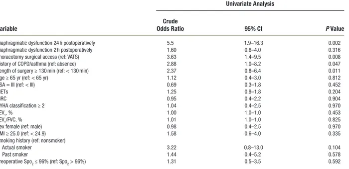
table 4. Association between Perioperative Variables and Development of at Least One Postoperative Pulmonary Complication According to Logistic Regression Analysis
variable
Univariate analysis crude
odds ratio 95% ci P value
Diaphragmatic dysfunction 24 h postoperatively 5.5 1.9–16.3 0.002
Diaphragmatic dysfunction 2 h postoperatively 1.60 0.6–4.0 0.316
thoracotomy surgical access (ref: VAts) 3.63 1.4–9.5 0.008
History of COPD/asthma (ref: absence) 2.88 1.0–8.2 0.047
Length of surgery ≥ 130 min (ref: < 130 min) 2.37 0.8–6.4 0.011
Age ≥ 65 yr (ref: < 65 yr) 1.12 0.4–3.0 0.812
AsA = III (ref: < III) 0.69 0.3–1.8 0.452
MEts 1.25 0.9–1.8 0.204
MRC 0.95 0.4–2.2 0.904
NYHA classification ≥ 2 1.04 0.4–2.5 0.970
FEV1, % 1.00 1.0–1.0 0.453
FEV1/FVC, % 1.01 1.0–1.0 0.825
sex female (ref: male) 0.98 0.4–2.5 0.970
BMI ≥ 25.0 (ref: < 24.9) 1.58 0.6–4.0 0.335
smoking history (ref: nonsmoker)
Actual smoker 3.22 0.8–13.0 0.104
Past smoker 1.44 0.4–5.2 0.578
Preoperative spo2 ≤ 96% (ref: spo2 > 96%) 1.31 0.5–3.5 0.592
Diaphragmatic dysfunction was defined as a diaphragmatic excursion less than 10 mm or negative on the operated side.
AsA, American society of Anesthesiologists physical status classification; BMI, body mass index; COPD, chronic obstructive pulmonary disease; FEV1, forced expiratory volume in the first second; FVC, forced vital capacity; MEt, metabolic equivalent; MRC, Medical Research Council scale for dyspnea; NYHA, New York Heart Association; spo2, oxygen saturation measured by pulse oximetry; VAts, video-assisted thoracoscopy.
table 3. Association between Diaphragmatic Dysfunction 24 h Postoperatively and Perioperative Variables According to Logistic
Regression Analysis
Univariate analysis crude
odds ratio 95% ci P value surgical access (ref: VAts)
thoracotomy 3.95 1.5–10.3 0.005
COPD/asthma (ref: absence)
Presence 0.89 0.4–2.4 0.847
Length of surgery (ref: < 130 min)
≥ 130 min 1.41 0.8–3.8 0.105
Age (ref: < 65 yr)
≥ 65 hr 1.06 0.4–2.7 0.952
AsA (ref: < III)
III 1.03 0.4–2.5 0.905
sex (ref: male)
Female 1.49 0.6–3.8 0.378
BMI (ref: < 24.9)
≥ 25.0 1.98 0.8–5.1 0.159
smoking history (ref: nonsmoker)
Actual smoker 2.78 1.0–7.6 0.044
Diaphragmatic dysfunction was defined as a diaphragmatic excursion less than 10 mm or negative on the operated side.
AsA, American society of Anesthesiologists physical status classification; BMI, body mass index; COPD, chronic obstructive pulmonary disease; ref, reference; VAts, video-assisted thoracoscopy.
Diaphragm Dysfunction after Thoracic Surgery
showing that, as compared to thoracotomy, video-assisted thoracoscopic surgery is associated with better recovery of respiratory muscle function.33 Overall, it is tempting to speculate that, in our patients, postoperative diaphragmatic dysfunction was caused more by surgery-induced variations in chest wall conformation and resting diaphragm length9 rather than by phrenic nerve inhibition due to the postop-erative pain or postoppostop-erative respiratory drive impairment. Indeed, we maintained an adequate postoperative pain control (numerical rating scale values between 2 and 3) by positioning a thoracic epidural catheter, a technique with minimal impact on the respiratory drive as compared to intravenous opioids.34
Diaphragmatic dysfunction affects both the lung and chest wall mechanics and favors the development of atel-ectasis.9,35,36 Interestingly, we found radiologic evidence of atelectasis in 18 patients among the 51 patients with diaphragmatic dysfunction at 24 h postoperatively (35%) and only in 3 patients among the 24 without diaphrag-matic dysfunction (13%, P = 0.040; Supplemental Digital Content 3, http://links.lww.com/ALN/B954). The rela-tionship between diaphragmatic disfunction and atelecta-sis can be explained by the decrease in transdiaphragmatic pressure.35 The diaphragmatic contraction provides a differ-ential pressure between the abdomen and chest. When the diaphragm is dysfunctional, it is less effective in maintaining distinct pressures in the two cavities. Furthermore, confirm-ing previous studies,14,15,37 we found that the diaphragmatic dysfunction decreased the ability to generate VT, another mechanism to explain the development of atelectasis.38
Maeda et al.39 showed a significant decrease in maxi-mal transdiaphragmatic pressure after thoracotomy, whereas Fratacci et al.9 observed a marked reduction in diaphragmatic contractility after pulmonary resection using electromyogra-phy. Takazakura et al.40 reported a decrease of 36% in the dia-phragmatic excursion on the operated side after thoracotomy, assessed by dynamic magnetic resonance. Most of these tech-niques are difficult to apply at the bedside. Conversely, we reported the incidence of diaphragm dysfunction after tho-racic surgery with a reliable, noninvasive, and widely available ultrasonographical examination. Given the increasing pop-ularity of diaphragmatic ultrasound in several postoperative contexts,41 our research might represent a starting reference for further studies. Since we found a significant association between diaphragmatic dysfunction and postoperative pul-monary complications (table 4), the ultrasound assessment of diaphragm motion could be a useful tool to identify patients at greater risk of postoperative pulmonary complications that should be managed more cautiously, for example, with con-tinuous positive airway pressure,36 noninvasive ventilation, or incentive ventilation and physiotherapy.
Several study limitations must be acknowledged. First, we did not exclude a direct phrenic nerve injury in our patients by measuring the phrenic nerve conduction time. However, a surgical trauma of the phrenic nerve is at least
improbable during lung cancer resection. Second, we did not randomize the surgical procedures; however, we rea-soned that a strict randomization to a more invasive surgical procedure such as thoracotomy would have probably been unethical. In our study, the surgical approach was decided upon technical and/or oncologic reasons by the surgeon. We should acknowledge that it was impossible to blind the ultrasonographer due to the easily discernible differences between the video-assisted thoracoscopic surgery and the thoracotomy incisions. However, the physician performing the ultrasound evaluation was not involved in the patient’s postoperative care and did not communicate the results to the treating physicians. Thus, we can exclude that the postoperative care was influenced by the result of diaphrag-matic ultrasonography. Third, we only took into account the diaphragmatic excursion to assess the diaphragmatic dysfunction; other techniques, such as the measurement of the diaphragmatic thickness, could have provided more information to our study. Nevertheless, the evaluation of the diaphragmatic thickness versus diaphragmatic excursion seems to be appropriate more in mechanically ventilated than in spontaneously breathing patients, as it was in our study.27 Fourth, the relatively small sample size did not allow an alternative statistical model, which would clarify the independent effect of each covariate in the outcomes investigated; this limitation is reflected by wide CIs for the observed relationship. Finally, even if we analyzed the occurrence of postoperative pulmonary complications for the first 7 postoperative days, we evaluated the diaphrag-matic excursion only after 2 and 24 h after surgery and, thus, we do not know what was the diaphragmatic excursion on the day of postoperative pulmonary complication diagnosis. However, the aim of our study was to investigate whether the ultrasound evaluation of the diaphragm could screen patients at high risk of postoperative pulmonary compli-cations; for this purpose, an early risk assessment seems appropriate.
In conclusion, point of care ultrasound, a noninvasive, bedside-available tool, can be used to detect diaphragmatic dysfunction after thoracic surgery. Confirming our main hypothesis, we found that the thoracotomic technique car-ries a higher risk of diaphragmatic dysfunction as compared to video-assisted thoracoscopic surgery. Also, postoperative pulmonary complications were more frequent in patients with diaphragmatic dysfunction at 24 h. Given the increas-ing popularity of diaphragmatic ultrasound in several post-operative contexts, our study might represent a starting reference for further studies designed to clarify whether ultrasonography assessment of diaphragm function could carry significant clinical advantages.
Acknowledgments
The authors thank Elisa Maietti, Department of Biostatistics, University of Ferrara, Ferrara, Italy, for her statistical advice and review of the article.
Research support
The study was funded by the University of Ferrara, Ferrara, Italy.
Competing Interests
The authors declare no competing interests.
Correspondence
Address correspondence to Dr. Spadaro: Department of Morphology, Surgery and Experimental Medicine, University of Ferrara, Anesthesia and Intensive Care Unit, Sant’Anna Hospital 8 Aldo Moro, 44125 Ferrara, Italy. [email protected]. Information on purchas-ing reprints may be found at www.anesthesiology.org or on the masthead page at the beginning of this issue. Anesthesiology’s articles are made freely accessible to all readers, for personal use only, 6 months from the cover date of the issue.
references
1. Lugg ST, Agostini PJ, Tikka T, Kerr A, Adams K, Bishay E, Kalkat MS, Steyn RS, Rajesh PB, Thickett DR, Naidu B: Long-term impact of developing a postop-erative pulmonary complication after lung surgery. Thorax 2016; 71:171–6
2. Agostini P, Cieslik H, Rathinam S, Bishay E, Kalkat MS, Rajesh PB, Steyn RS, Singh S, Naidu B: Postoperative pulmonary complications following thoracic surgery: Are there any modifiable risk factors? Thorax 2010; 65:815–8
3. Spadaro S, Caramori G, Rizzuto C, Mojoli F, Zani G, Ragazzi R, Valpiani G, Dalla Corte F, Marangoni E, Volta CA: Expiratory flow limitation as a risk factor for pulmonary complications after major abdominal surgery. Anesth Analg 2017; 124:524–30
4. Ploeg AJ, Kappetein AP, van Tongeren RB, Pahlplatz PV, Kastelein GW, Breslau PJ: Factors associated with perioperative complications and long-term results after pulmonary resection for primary car-cinoma of the lung. Eur J Cardiothorac Surg 2003; 23:26–9
5. Stéphan F, Boucheseiche S, Hollande J, Flahault A, Cheffi A, Bazelly B, Bonnet F: Pulmonary complica-tions following lung resection: A comprehensive anal-ysis of incidence and possible risk factors. Chest 2000; 118:1263–70
6. Waller DA, Forty J, Morritt GN: Video-assisted thora-coscopic surgery versus thoracotomy for spontaneous pneumothorax. Ann Thorac Surg 1994; 58:372–6; dis-cussion 376–7
7. Whitson BA, Groth SS, Duval SJ, Swanson SJ, Maddaus MA: Surgery for early-stage non-small cell lung cancer: A systematic review of the video-assisted thoracoscopic
surgery versus thoracotomy approaches to lobectomy. Ann Thorac Surg 2008; 86:2008–16; discussion 2016–8 8. Paul S, Altorki NK, Sheng S, Lee PC, Harpole DH,
Onaitis MW, Stiles BM, Port JL, D’Amico TA: Thoracoscopic lobectomy is associated with lower morbidity than open lobectomy: A propensity-matched analysis from the STS database. J Thorac Cardiovasc Surg 2010; 139:366–78
9. Fratacci MD, Kimball WR, Wain JC, Kacmarek RM, Polaner DM, Zapol WM: Diaphragmatic shortening after thoracic surgery in humans. Effects of mechan-ical ventilation and thoracic epidural anesthesia. Anesthesiology 1993; 79:654–65
10. Siafakas NM, Mitrouska I, Bouros D, Georgopoulos D: Surgery and the respiratory muscles. Thorax 1999; 54:458–65
11. Sasaki N, Meyer MJ, Eikermann M: Postoperative respi-ratory muscle dysfunction: Pathophysiology and pre-ventive strategies. Anesthesiology 2013; 118:961–78 12. Dubé BP, Dres M, Mayaux J, Demiri S, Similowski T,
Demoule A: Ultrasound evaluation of diaphragm func-tion in mechanically ventilated patients: Comparison to phrenic stimulation and prognostic implications. Thorax 2017; 72:811–8
13. Dres M, Goligher EC, Heunks LMA, Brochard LJ: Critical illness-associated diaphragm weakness. Intensive Care Med 2017; 43:1441–52
14. Zambon M, Greco M, Bocchino S, Cabrini L, Beccaria PF, Zangrillo A: Assessment of diaphragmatic dysfunc-tion in the critically ill patient with ultrasound: A sys-tematic review. Intensive Care Med 2017; 43:29–38 15. Spadaro S, Grasso S, Mauri T, Dalla Corte F, Alvisi V,
Ragazzi R, Cricca V, Biondi G, Di Mussi R, Marangoni E, Volta CA: Can diaphragmatic ultrasonography per-formed during the T-tube trial predict weaning fail-ure? The role of diaphragmatic rapid shallow breathing index. Crit Care 2016; 20:305
16. Kim WY, Suh HJ, Hong SB, Koh Y, Lim CM: Diaphragm dysfunction assessed by ultrasonography: Influence on weaning from mechanical ventilation. Crit Care Med 2011; 39:2627–30
17. Kim SH, Na S, Choi JS, Na SH, Shin S, Koh SO: An evaluation of diaphragmatic movement by M-mode sonography as a predictor of pulmonary dysfunction after upper abdominal surgery. Anesth Analg 2010; 110:1349–54
18. Oh YJ, Lee JR, Choi YS, Koh SO, Na S: Randomized controlled comparison of combined general and epidural anesthesia versus general anesthesia on dia-phragmatic function after laparoscopic prostatectomy. Minerva Anestesiol 2013; 79:1371–80
19. Lerolle N, Guérot E, Dimassi S, Zegdi R, Faisy C, Fagon JY, Diehl JL: Ultrasonographic diagnostic crite-rion for severe diaphragmatic dysfunction after cardiac surgery. Chest 2009; 135:401–7
Diaphragm Dysfunction after Thoracic Surgery
20. Colice GL, Shafazand S, Griffin JP, Keenan R, Bolliger CT; American College of Chest Physicians: Physiologic evaluation of the patient with lung cancer being con-sidered for resectional surgery: ACCP evidenced-based clinical practice guidelines (2nd edition). Chest 2007; 132:161S–77S
21. Miller MR, Hankinson J, Brusasco V, Burgos F, Casaburi R, Coates A, Crapo R, Enright P, van der Grinten CP, Gustafsson P, Jensen R, Johnson DC, MacIntyre N, McKay R, Navajas D, Pedersen OF, Pellegrino R, Viegi G, Wanger J; ATS/ERS Task Force: Standardisation of spirometry. Eur Respir J 2005; 26:319–38
22. Spadaro S, Grasso S, Karbing DS, Fogagnolo A, Contoli M, Bollini G, Ragazzi R, Cinnella G, Verri M, Cavallesco NG, Rees SE, Volta CA: Physiologic evalu-ation of ventilevalu-ation perfusion mismatch and respiratory mechanics at different positive end-expiratory pressure in patients undergoing protective one-lung ventilation. Anesthesiology 2018; 128:531–8
23. Rauseo M, Mirabella L, Grasso S, Cotoia A, Spadaro S, D’Antini D, Valentino F, Tullo L, Loizzi D, Sollitto F, Cinnella G: PEEP titration based on the open lung approach during one lung ventilation in thoracic sur-gery: A physiological study. BMC Anesthesiol 2018; 18:156
24. Boussuges A, Gole Y, Blanc P: Diaphragmatic motion studied by M-mode ultrasonography: Methods, repro-ducibility, and normal values. Chest 2009; 135:391–400 25. Ayoub J, Cohendy R, Dauzat M, Targhetta R, De la
Coussaye JE, Bourgeois JM, Ramonatxo M, Prefaut C, Pourcelot L: Non-invasive quantification of diaphragm kinetics using M-mode sonography. Can J Anaesth 1997; 44:739–44
26. Matamis D, Soilemezi E, Tsagourias M, Akoumianaki E, Dimassi S, Boroli F, Richard JC, Brochard L: Sonographic evaluation of the diaphragm in criti-cally ill patients. Technique and clinical applications. Intensive Care Med 2013; 39:801–10
27. Umbrello M, Formenti P, Longhi D, Galimberti A, Piva I, Pezzi A, Mistraletti G, Marini JJ, Iapichino G: Diaphragm ultrasound as indicator of respiratory effort in critically ill patients undergoing assisted mechani-cal ventilation: A pilot clinimechani-cal study. Crit Care 2015; 19:161
28. Gerscovich EO, Cronan M, McGahan JP, Jain K, Jones CD, McDonald C: Ultrasonographic evaluation of diaphragmatic motion. J Ultrasound Med 2001; 20:597–604
29. Welvaart WN, Paul MA, Stienen GJ, van Hees HW, Loer SA, Bouwman R, Niessen H, de Man FS, Witt CC, Granzier H, Vonk-Noordegraaf A, Ottenheijm CA: Selective diaphragm muscle weakness after con-tractile inactivity during thoracic surgery. Ann Surg 2011; 254:1044–9
30. Vassilakopoulos T, Mastora Z, Katsaounou P, Doukas G, Klimopoulos S, Roussos C, Zakynthinos S: Contribution of pain to inspiratory muscle dysfunc-tion after upper abdominal surgery: A randomized controlled trial. Am J Respir Crit Care Med 2000; 161(4 Pt 1):1372–5
31. Mrozek S, Jung B, Petrof BJ, Pauly M, Roberge S, Lacampagne A, Cassan C, Thireau J, Molinari N, Futier E, Scheuermann V, Constantin JM, Matecki S, Jaber S: Rapid onset of specific diaphragm weakness in a healthy murine model of ventilator-induced diaphrag-matic dysfunction. Anesthesiology 2012; 117:560–7 32. Hussain SN, Cornachione AS, Guichon C, Al Khunaizi
A, Leite Fde S, Petrof BJ, Mofarrahi M, Moroz N, de Varennes B, Goldberg P, Rassier DE: Prolonged con-trolled mechanical ventilation in humans triggers myofibrillar contractile dysfunction and myofilament protein loss in the diaphragm. Thorax 2016; 71:436–45 33. Bernard A, Brondel L, Arnal E, Favre JP: Evaluation of
respiratory muscle strength by randomized controlled trial comparing thoracoscopy, transaxillary thoracot-omy, and posterolateral thoracotomy for lung biopsy. Eur J Cardiothorac Surg 2006; 29:596–600
34. Simonneau G, Vivien A, Sartene R, Kunstlinger F, Samii K, Noviant Y, Duroux P: Diaphragm dys-function induced by upper abdominal surgery. Role of postoperative pain. Am Rev Respir Dis 1983; 128:899–903
35. Laghi F, Tobin MJ: Disorders of the respiratory muscles. Am J Respir Crit Care Med 2003; 168:10–48
36. Squadrone V, Coha M, Cerutti E, Schellino MM, Biolino P, Occella P, Belloni G, Vilianis G, Fiore G, Cavallo F, Ranieri VM; Piedmont Intensive Care Units Network (PICUN): Continuous positive airway pres-sure for treatment of postoperative hypoxemia: A ran-domized controlled trial. JAMA 2005; 293:589–95 37. Cohen E, Mier A, Heywood P, Murphy K, Boultbee
J, Guz A: Diaphragmatic movement in hemiplegic patients measured by ultrasonography. Thorax 1994; 49:890–5
38. Schindler MB: Treatment of atelectasis: Where is the evidence? Crit Care 2005; 9:341–2
39. Maeda H, Nakahara K, Ohno K, Kido T, Ikeda M, Kawashima Y: Diaphragm function after pulmonary resection. Relationship to postoperative respiratory failure. Am Rev Respir Dis 1988; 137:678–81
40. Takazakura R, Takahashi M, Nitta N, Sawai S, Tezuka N, Fujino S, Murata K: Assessment of diaphragmatic motion after lung resection using magnetic resonance imaging. Radiat Med 2007; 25:155–63
41. Bignami E, Guarnieri M, Saglietti F, Ramelli A, Vetrugno L: Diaphragmatic dysfunction following car-diac surgery: Is there a role for pulmonary ultrasound? J Cardiothorac Vasc Anesth 2018; 32:e6–7
appendix. definitions of Pulmonary Postoperative
complications
Postoperatively, a physician not aware of the patient study group and not involved in the patient’s ongoing care col-lected data on the occurrence of a symptomatic and clin-ically significant postoperative pulmonary complications during the hospital length of stay or within the first 7 post-operative days through review of clinical records, laboratory, and radiology data.
1. Hypoxemia
Pao2 less than 60 mmHg or oxygen saturation measured by pulse oximetry less than 90% in room air but responding to supplemental oxygen (excluding hypoventilation)
2. Severe hypoxemia
Need for noninvasive or invasive mechanical ventilation or a Pao2 less than 60 mmHg or oxygen saturation measured by pulse oximetry less than 90% despite supplemental oxy-gen (excluding hypoventilation)
3. Bronchospasm
Defined as newly detected expiratory wheezing treated with bronchodilators
4. Suspected pulmonary infection
Defined as new or progressive radiographic infiltrate plus at least two of the following: antibiotic treatment, tympanic
temperature greater than 38°C, leukocytosis or leucopenia (leukocyte count less than 4,000 cells/mm3 or greater than 12,000 cells/mm3), and/or purulent secretions
5. New pulmonary infiltrates
Chest radiograph demonstrating new monolateral or bilat-eral infiltrate without other clinical signs
6. Atelectasis
Chest radiograph demonstrating lung opacification with shift of the mediastinum, hilum, or hemidiaphragm toward the affected area, and compensatory overinflation in the adjacent nonatelectatic lung
7. Pleural effusion
Chest radiograph demonstrating blunting of the costo-phrenic angle, loss of the sharp silhouette of the ipsilateral hemidiaphragm in upright position, evidence of displace-ment of adjacent anatomical structures, or (in the supine position) a hazy opacity in one hemithorax with preserved vascular shadows
Source: PROVE Network Investigators for the Clinical Trial Network of the European Society of Anesthesiology, Hemmes SN, Gama de Abreu M, Pelosi P, Schultz MJ. High versus low positive end-expiratory pressure during general anesthesia for open abdominal surgery (PROVHILO trial): A multicentre randomised controlled trial. Lancet 2014; 384:495–503