E-Mail [email protected]
Brief Communication
Neurodegener Dis 2016;16:407–410 DOI: 10.1159/000446397
Using High-Field Magnetic Resonance
Imaging to Estimate Distensibility of the
Middle Cerebral Artery
Esther A.H. Warnert
a, b
Jasper Verbree
c
Richard G. Wise
a
Matthias J.P. van Osch
c
a Cardiff University Brain Research Imaging Centre, School of Psychology, Cardiff University, Cardiff , UK; b Department of Clinical Neurological Sciences, University of Western Ontario, London, Ont., Canada; c C.J. Gorter Center for High-Field MRI, Department of Radiology, Leiden University Medical Center, Leiden ,
The Netherlands
files. Conclusion: These results lead to the first noninvasive image-based estimation of distensibility of the MCA (approx. 5.8 × 10 –4 mm Hg –1 ) and demonstrate that ultra-high-field MRI could be a promising tool for investigating distensibility of intracranial arteries in relation to cerebrovascular pathology.
© 2016 The Author(s) Published by S. Karger AG, Basel
Introduction
Healthy cerebral arteries are able to smooth out the pulsatile blood flow originating from the heart into an almost continuous flow into the capillary bed of the brain [1] . If the cerebral arteries stiffen, the pulsatile blood flow propagates further into the arterial tree, where the pulsa-tile shear stress can induce damage to the walls of the small vessels [2, 3] . This process has been linked to severe pathologies, including cerebral small vessel disease and vascular cognitive decline [2, 3] , and highlights the poten-tial value of a measure of cerebral arterial stiffness as a marker of cerebrovascular health.
Key Words
Arterial stiffness · Arterial structure/compliance · Cerebral small vessel disease · Middle cerebral artery · Ultra-high-field MRI
Abstract
Background: Although cerebral arterial stiffness may be an important marker for cerebrovascular health, there is not yet a measurement that accurately reflects the distensibility of major intracranial arteries. Herein, we aim to noninvasively measure distension of the human middle cerebral artery (MCA). Methods: Ten healthy volunteers (age: 30.3 ± 10.8 years) underwent ultra-high-field (7-tesla) MRI scanning. Time-of-flight angiography and phase-contrast flow imaging were used to locate the M1 segment of the MCA and to determine the occurrence of systole and diastole. High-resolution cross-sectional cardiac triggered T 2 -weighted images of the M1 seg-ment of the MCA were acquired in systole and diastole.
Re-sults: The average distension of the MCA area from diastole to
systole was 2.58% (range: 0.08%–6.48%). There was no signifi-cant correlation between MCA distension and the pulsatility index, calculated from the phase-contrast flow velocity
Received: January 21, 2016 Accepted after revision: April 23, 2016 Published online: July 23, 2016
D i s e a s e s
Richard G. Wise
Cardiff University Brain Research Imaging Centre, School of Psychology Cardiff University, Maindy Road
Cardiff CF24 4HQ (UK) E-Mail wiserg @ cardiff.ac.uk
© 2016 The Author(s) Published by S. Karger AG, Basel 1660–2854/16/0166–0407$0/0
www.karger.com/ndd This article is licensed under the Creative Commons Attribution 4.0 International License (CC BY) (http://www.karger.com/Services/ OpenAccessLicense). Usage, derivative works and distribution are permitted provided that proper credit is given to the author and the original publisher.
Warnert/Verbree/Wise/van Osch
Neurodegener Dis 2016;16:407–410 DOI: 10.1159/000446397
408
Measuring stiffness (or its inverse: distensibility) in ex-tracranial arteries is commonly done by assessing chang-es in diameter occurring with changchang-es in prchang-essure at the same site [4] . The skull complicates such measurements for intracranial arteries, which explains the absence of an accurate measurement of cerebral arterial stiffness. How-ever, volume changes of approximately 5% occurring throughout the cardiac cycle in the middle cerebral artery (MCA) have recently been measured using computed to-mography (CT) angiography [5] .
In contrast to CT, magnetic resonance imaging (MRI) can be used noninvasively to measure the cross-sectional area of the MCA [6] . Based on the volume changes found by CT angiography [5] , distention of the MCA during the cardiac cycle is expected to be small (0.05–0.1 mm). Non-invasive imaging of such small geometrical changes re-quires high-resolution measurements, which are current-ly oncurrent-ly feasible with ultra-high-field MRI [6] .
In this proof-of-principle experiment, we aimed to measure distension of the MCA by using ultra-high-field MRI and by synchronizing image acquisition to the car-diac cycle. In addition, we assessed the pulsatility index (PI) as a combined measure of cerebrovascular resistance and stiffness. To the best of our knowledge, this is the first noninvasive assessment of cerebral arterial distension in humans.
Materials and Methods
Ten healthy participants were recruited for this experiment (6 females, all nonsmoking, average age 30.3 ± 10.8 years). Informed consent was obtained from all volunteers. This study was per-formed under approval of the Institutional Review Board of the Leiden University Medical Center according to the Declaration of Helsinki and in accordance with the guidelines for Good Clinical Practice (CPMP/ICH/135/95).
Image Acquisition
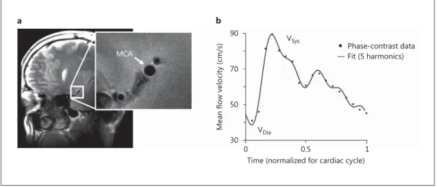
MRI scans were performed at 7 T (whole-body Philips Achie-va; Philips Healthcare, Best, The Netherlands) similar to a previ-ously described protocol [6] . All image acquisition parameters are stated in the online supplementary data (for all online suppl. material, see www.karger.com/doi/10.1159/000446397). A 3-di-mensional time-of-flight (TOF) angiogram was performed to identify the MCA and for planning of an imaging plane perpen-dicular to the M1 segment. Care was taken to select a straight portion of the MCA and exclude branching arteries. Planning of this imaging plane was guided by additional reconstructions of the TOF scan to better visualize the course of the MCA in all di-rections. The position and orientation of the imaging plane were copied to the quantitative flow scan to assess the flow velocity waveform through the MCA. Directly after acquisition, a circular region of interest in the center of the MCA was used to determine the flow velocity profile at the level of M1, which was used to de-termine the time points at which peak diastole and peak systole occurred and to calculate the post-trigger acquisition delay times for the cardiac triggered T 2 -weighted images. High-resolution and cardiac triggered T 2 -weighted images were acquired at 4 time points in the cardiac cycle in pseudo-random order: 200 (t 1 ) and 100 ms (t 2 ) preceding peak diastole, and 50 ms before (t 3 ) and 50
90 70 50 30 0 0.5 MCA
Time (normalized for cardiac cycle)
Mean flow v elocity (cm/s) Phase-contrast data Fit (5 harmonics) VDia VSys 1 a b
Fig. 1. a Example of T 2 -weighted high-resolution structural image of the cross-sectional area of the M1 segment
of the MCA. b Example of phase-contrast flow velocity data (dots) measured at the same location as in a . The flow velocity waveform was approximated by fitting the first five harmonics of a Fourier sequence (solid line), from which the pulsatility index was calculated. Sys = Systole; Dia = diastole.
Distensibility of the Middle Cerebral Artery
Neurodegener Dis 2016;16:407–410
DOI: 10.1159/000446397 409
ms after (t 4 ) peak systole. These time points were chosen because in the carotid artery peak flow velocities have been shown to closely follow systolic and diastolic pressure [7] . An example of a high-resolution structural image can be seen in figure 1 a. Throughout all scans, pulse oximetry at the finger was used to measure the cardiac pulse.
Image Analysis
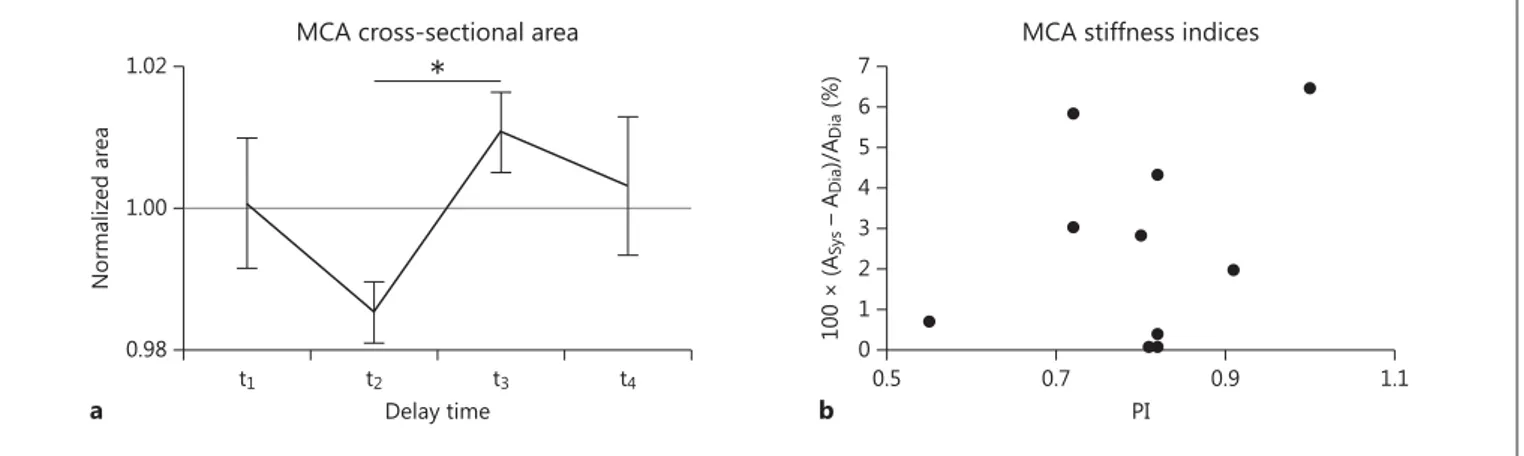
Two observers, blinded to participant and cardiac phase of the images, manually drew elliptical regions of interests on the T 2 -weighted images to delineate the internal wall of the MCA. Each observer repeated this process once, and the average of all 8 mea-surements (two scans per time point and two observers who delin-eated the MCA twice) per time point was used to calculate MCA cross-sectional area. Paired t tests were used to investigate wheth-er the MCA area at time points t 1 , t 3 , and t 4 were significantly dif-ferent than at t 2 (triggered closest to peak-diastolic velocity).
To investigate the consistency within and between observers, the intraclass coefficient of correlation for consistency [ICC(C,1), ICC(C,k)] was calculated with MATLAB (R2012b; MathWorks, Natick, Mass., USA).
The quantitative flow scan was also used to calculate a com-monly used cerebral arterial stiffness index based on the blood flow velocity waveform through the MCA, PI [8, 9] . A linear regression was performed ( robustfit in MATLAB) between the distention of the MCA between peak diastole and peak systole (t 2 and t 3 ) and PI.
Results
There was high consistency in MCA area measurements within observer [ICC(C,1) Obs.1 = 0.89, ICC(C,1) Obs.2 = 0.92]
and between observers [ICC(C,k) = 0.91], justifying the averaging of all 8 MCA area measurements per time point.
There was a significant increase in MCA area between diastole and systole of 2.58% (min–max range: 0.08%– 6.48%; t test, p < 0.01; fig. 2 a).
The group average PI was 0.80 ± 0.12 and was not sig-nificantly correlated with the change in MCA area be-tween systole and diastole (r 2 = 0.13, p = 0.35; fig. 2 b).
Discussion
In the current study, it is shown for the first time that ultra-high-field MRI can be used to noninvasively mea-sure distention of the MCA occurring through the car-diac cycle, which is a prerequisite for measurements of cerebral arterial stiffness. When combined with reference values for central pulse pressure in healthy participants (45 mm Hg [10] ), the estimated average MCA distensibil-ity is approximately 5.8 × 10 –4 mm Hg –1 .
This level of distensibility in the MCA is plausible, as the significant change in MCA area of 2.58 ± 2.4% through the cardiac cycle falls within the range of volume changes in the MCA measured with CT [5] . Furthermore, the re-sulting estimation of MCA distensibility follows the ex-pectation that intracranial arteries are less distensible than extracranial arteries (reported distensibility in the carotid artery ranges from 0.5 × 10 –3 to 5.8 × 10 –3 mm
Hg –1 [4] ).
There was no correlation between the distention of the MCA cross-sectional area and PI. Although in a small co-hort, this finding highlights that in addition to local
arte-7 6 5 4 3 2 1 0 0.5 0.7 PI
MCA stiffness indices
b 100 × (ASy s – A Dia )/A Dia (%) 0.9 1.1 1.02 1.00 0.98 t1 Delay time
MCA cross-sectional area
a Normalized ar ea t2 t3 t4 *
Fig. 2. a Group average (n = 10) cross-sectional area of the MCA
for 4 different delay times. Data were normalized per participant by dividing by the average MCA cross-sectional area of each indi-vidual. Note that t 2 is the delay time closest to peak diastole and t 3 the delay time closest to peak systole. There was a significant
dif-ference ( * p < 0.01, paired t test) between the diastolic and systolic area. Error bars = SEM. b Increase in area from diastole (Dia) to systole (Sys) plotted against the PI for all 10 participants (no sig-nificant correlation, r 2 = 0.13, p = 0.35).
Warnert/Verbree/Wise/van Osch
Neurodegener Dis 2016;16:407–410 DOI: 10.1159/000446397
410
rial stiffness, other factors such as stiffness and resistance of the downstream vascular bed shape the blood flow ve-locity waveform [11] . Furthermore, this illustrates the need for investigation of the relationship between cere-bral arterial distensibility and the formation of blood flow velocity waveforms, which is an important step in under-standing the mechanisms that link increased arterial stiff-ness to cerebrovascular pathologies, such as small vessel disease [9] and vascular cognitive decline [2] . Future studies investigating these mechanisms should include participants with a wide range of expected cerebral arte-rial distensibilities, e.g. young and elderly individuals or patients with small vessel disease.
A limitation of this study is that no blood pressure mea-surements were included, and, therefore, only an estima-tion of MCA distensibility was feasible. However, under-lying pulse pressures are not expected to show large de-viations from reference values [10] , because only healthy and nonsmoking volunteers were recruited. Future work should include blood pressure measurements such that cerebral arterial stiffness can be quantified in terms of
dis-tensibility or compliance [12] . Although local pulse pres-sure meapres-surements would be a requirement for accurate estimation of arterial distensibility [4] , we recommend noninvasive measurements of blood pressure (e.g. brachi-al blood pressure [12] ) in studies for which it is not feasible to invasively assess intracranial blood pressure.
In summary, we have shown that ultra-high-field MRI can be used to noninvasively measure cerebral arterial distensibility, and that this is a promising tool for future research into the relationship between cerebral arterial stiffness and cerebrovascular pathology.
Acknowledgment
This work was funded by Cardiff University’s Institutional Strategic Support Fund from the Wellcome Trust, UK.
Disclosure Statement
The authors have no conflict of interest to disclose.
References
1 Hall JE, Guyton AC: Vascular Distensibility and Functions of the Arterial and Venous Sys-tems. Textbook of Medical Physiology. Phila-delphia, Elsevier, 2006, pp 171–180. 2 O’Rourke MF, Safar ME: Relationship
be-tween aortic stiffening and microvascular dis-ease in brain and kidney: cause and logic of therapy. Hypertension 2005; 46: 200–204. 3 Mitchell GF, van Buchem MA, Sigurdsson S,
Gotal JD, Jonsdottir MK, et al: Arterial stiff-ness, pressure and flow pulsatility and brain structure and function: the Age, Gene/Envi-ronment Susceptibility–Reykjavik study. Brain 2011; 134: 3398–3407.
4 O’Rourke MF, Staessen JA, Vlachopoulos C, Duprez D, Plante GE: Clinical applications of arterial stiffness; definitions and reference values. Am J Hypertens 2002; 15: 426–444.
5 Kuroda J, Kinoshita M, Tanaka H, Nishida T, Nakamura H, et al: Cardiac cycle-related vol-ume change in unruptured cerebral aneu-rysms: a detailed volume quantification study using 4-dimensional CT angiography. Stroke 2012; 43: 61–66.
6 Verbree J, Bronzwaer ASGT, Ghariq E, Ver-sluis MJ, Daemen MJAP, et al: Assessment of middle cerebral artery diameter during hypo-capnia and hyperhypo-capnia in humans using ul-tra-high-field MRI. J Appl Physiol 2014; 117: 1084–1089.
7 Hirata K, Yaginuma T, O’Rourke MF, Kawakami M: Age-related changes in carotid artery flow and pressure pulses: possible im-plications for cerebral microvascular disease. Stroke 2006; 37: 2552–2556.
8 Bouvy WH, Geurts LJ, Kuijf HJ, Luijten PR, Kappelle LJ, et al: Assessment of blood flow velocity and pulsatility in cerebral perforating arteries with 7-T quantitative flow MRI. NMR Biomed 2015, Epub ahead of print.
9 Webb AJS, Simoni M, Mazzucco S, Kuker W, Schulz U, et al: Increased cerebral arterial pul-satility in patients with leukoaraiosis: arterial stiffness enhances transmission of aortic pul-satility. Stroke 2012; 43: 2631–2636.
10 Mitchell GF, Parise H, Benjamin EJ, Larson MG, Keyes MJ, et al: Changes in arterial stiff-ness and wave reflection with advancing age in healthy men and women: the Framingham Heart Study. Hypertension 2004; 43: 1239– 1245.
11 Bude RO, Rubin JM: Relationship between the resistive index and vascular compliance and resistance. Radiology 1999; 211: 411–417. 12 Warnert EAH, Murphy K, Hall JE, Wise RG:
Noninvasive assessment of arterial compli-ance of human cerebral arteries with short in-version time arterial spin labeling. J Cereb Blood Flow Metab 2015; 35: 461–468.