TRIASSIC SPECIES OF SAURICHTHYS (ACTINOPTERYGII)
ILJA KOGAN1,2, ANDREA TINTORI3 & MARTIN LICHT†
1TU Bergakademie Freiberg, Geological Institute, Department of Palaeontology, Bernhard-von-Cotta-Str. 2, 09599 Freiberg, Germany.
E-mail: [email protected].
2Kazan Federal University, Institute of Geology and Petroleum Technologies, Kremlyovskaya Str. 4/5, 420008 Kazan, Russia. 3TRIASSICA, Institute for Triassic Lagerstätten, 23828-Perledo, Italy.
To cite this article: Kogan I., Tintori A. & Licht M. (2020) - Locomotor function of scales and axial skeleton in Middle–Late Triassic species of Saurichthys (Actinopterygii) Riv. It. Paleontol. Strat., 126(2): 475-498.
Abstract. Starting in the Late Permian, the ‘Triassic osteichthyan revolution’ gave rise to several new morpho-types of actinopterygians, including the iconic barracuda-shaped predator Saurichthys. About 50 species, from 10 cm to over 1.5 m long, are known from mainly marine deposits worldwide. Despite current interest in Saurichthys, freshwater species and those from late Middle to early Late Triassic remain understudied. We document the postcranial morpho-logy of three small to mid-sized (15–45 cm) species from this timeframe represented by sufficiently complete indi-viduals: Saurichthys orientalis Sytchevskaya, 1999, from lacustrine deposits of the Madygen Formation (late Ladinian/ Carnian); S. striolatus (Bronn, 1858) from the fully marine Predil Limestone (early Carnian); and S. calcaratus Griffith, 1977, from the terrigenously influenced coastal environment of the Lunz Formation (middle Carnian). S. orientalis resembles early saurichthyids in having six rows of large, thick ganoid scales; fins with segmented lepidotrichia; and flank scales relating to dorsal vertebral elements as 1:2. S. calcaratus and S. striolatus share unsegmented fin rays and a reduced scale cover with well-ossified but narrow mid-dorsal and mid-ventral scales and small, thin flank scales, relat-ing to the dorsal arcualia as 1:1. Ventral arcualia are first described for S. calcaratus and S. striolatus, where they change in shape and number at the abdominal-caudal transition. In all three species, force transmission to the tail fin is enhanced by the caudal peduncle strengthened by a stiff structure arising from interlocking or fusion of the last enlarged mid-dorsal and mid-ventral scales (scutes), while the vertebral column remains rather lightly built.
Received: January 15, 2020; accepted: May 19, 2020
Keywords: Pike-like predators; vertebral column; functional morphology; aquatic locomotion; Saurichthyiformes; Saurichthyidae.
I
ntroductIonDue to its characteristic elongate body shape, its abundance and the fact that even small frag-ments can be determined at least at the genus level,
Saurichthys Agassiz, 1834, is one of the iconic Tri-assic actinopterygian taxa. Representatives of this ‘palaeopterygian’ genus were distributed globally already at the beginning of the Mesozoic (Beltan & Tintori 1981; Mutter et al. 2008) and mostly lived in shallow coastal marine environments and
intra-carbonatic platform lagoons, but also occurred in lacustrine and fluvial systems. About 50 species are considered valid and have been described from all continents, except South America and Antarc-tica, based on often abundant complete skeletons or large fragments (see Romano et al. 2012 for an overview of the fossil record and Tintori 2013; Tintori et al. 2014b; Werneburg et al. 2014; Maxwell et al. 2015, 2016; Wu et al. 2015, 2018 for newly described species).
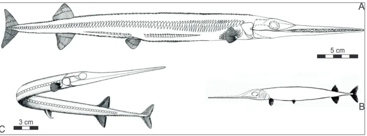
All Saurichthys species share a long, slender body with an abbreviate-diphycercal tail, poste-riorly shifted pelvics and almost symmetrical op-posed anal and dorsal fins (Fig. 1). The extremely
elongated head features a palaeoniscoid maxilla/ preopercular complex, as well as dentigerous lower and upper jaws of equal length tapering forward in a more or less elongated rostrum (e.g., Woodward 1888, 1890, 1895; Stensiö 1925; Gardiner 1960). In recent fishes, this body shape and fin disposition is characteristic for fast-start predators specialized on acceleration rather than on continuous swim-ming (e.g., Webb 1984; Kogan et al. 2015), a lifestyle probable for most saurichthyids as well (Rieppel 1985; Lombardo & Tintori 2005).
While the general saurichthyid bauplan (head and body shape, position of pelvic, dorsal and anal fins, dentition) remained constant throughout the whole Triassic, several internal and external charac-ters may show few remarkable variations. The ratio between the head and the body length varies a lot, being related to both the ontogenetic stages and the adult final size (Tintori 2013). Apart from the cranial osteology, considerable changes are documented in the patterns of squamation, vertebral column, and fin morphology and structure (Stensiö 1925; Beltan & Tintori 1981; Rieppel 1985, 1992; Tintori 1990, 2013; Wu et al. 2009, 2015). The earliest forms, for instance, possessed a nearly complete scale cover, which was subsequently, since the Spathian, reduced to an even number of longitudinal rows of scales, till just one mid-dorsal and one mid-ventral in latest species (see e.g. Schmid & Sánchez-Villagra 2010). Another structural transformation is reported for the lepidotrichia of the pelvic and especially the unpaired fins, which may vary from unsegmented to repeatedly segmented, though there is no clear temporal trend as originally supposed by Rieppel (1992). Fringing fulcra covered the leading edge of the fins of the earliest Triassic saurichthyids from Greenland, Madagascar and Spitsbergen (Sten-siö 1925; Piveteau 1944–45; Rieppel 1985; Kogan 2011; Kogan & Romano 2016a, b), but are absent in most later species. Finally, the axial skeleton exhibits considerable diversity. None of the known species had ossified vertebral centra, but up to 200 paired neural arches rested dorsolaterally on the persistent notochord. Additionally, ossified ventral elements, i.e. the paired haemal arches, are known in several species, where they are normally restricted to the posterior body portion and relate to respective dor-sal elements either as 1:1 or as 1:2. Among the well-known species, the shape of the neural and haemal arches shows a great variability. Especially the
neu-ral arches can be characterized by the presence or absence and the morphology of several projections (usually referred to as praezygapophyses, neural spines and postzygapophyses) that were needed for articulation with adjoining segments (e.g., Stensiö 1925), stiffening the vertebral column itself (Tintori 1990, 2013; Gozzi 2006), thus having a major im-pact on locomotor performance. So, not only seems the variability to be much greater in the postcranial skeleton, but it also appears to be more important because of its functional significance with respect to the way of swimming and chasing of the differ-ent species (e.g., Lauder 2006).
Despite the high number of nominal species of Saurichthys, the knowledge of their anatomy is very uneven due to the quality of available mate-rial and the researchers’ interests. Forms from the Early Triassic of Spitsbergen and Madagascar, for instance, have been studied in great detail (Sten-siö 1925; Piveteau 1944–45; Lehman 1952; Lehm-an et al. 1959; BeltLehm-an 1958, 1968), although often mainly skull characters were taken into account, just because of the preservation of the material. Detailed studies of the postcranium of Early Tri-assic saurichthyids have been published only re-cently (Mutter et al. 2008; Kogan 2011; Tintori et al. 2014b; Kogan & Romano 2016a, b). Extensive work on complete specimens has furthermore been done on the Anisian and early Ladinian (Middle Tri-assic) species of the Southern Alps (Rieppel 1985, 1992; Tintori 1990, 2013; Maxwell et al. 2013, 2015) as well as Southern China (Wu et al. 2009, 2011, 2015, 2018). Tintori (1990) addressed the body anatomy of Norian saurichthyids from Northern Italy while Gozzi (2006) revised the Norian species from the Alps. The few species described by earlier authors from late Ladinian and Carnian, in contrast, are quite poorly known, as are also the saurichthyids found in freshwater deposits (Kogan 2018). Diver-sity of saurichthyids and their abundance in fossil assemblages declined during this time (Romano et al. 2012; Tintori et al. 2014a), which, however, is documented by a much smaller number of fos-sil fish localities than in the Early/Middle Triassic (Tintori et al. 2014a); nonetheless, statistical analysis by Romano et al. (2016) indicates that osteichthy-an diversity estimates for the Late Triassic are not significantly biased. Recently, new Saurichthys finds have been reported by Hitij et al.(2019) from Upper Carnian in the Vrata Valley in the north-eastern part
of the Slovenian Julian Alps. This is probably the first record from Late Carnian marine rocks and, although comprising only two specimens, shows
Saurichthys of about 30 cm in length, thus relatively small compared with many other species, but simi-lar to the Carnian species herein considered.
The discovery of a nearly complete speci-men of Saurichthys orientalis Sytchevskaya, 1999 from the non-marine Ladinian/Carnian Madygen Formation of Kyrgyzstan, Central Asia (Kogan et al. 2009), enables to obtain new data from this time slice, especially in comparison with two Car-nian species, Saurichthys striolatus (Bronn, 1858) and
Saurichthys calcaratus Griffith, 1977. The latter two
species come from marine deposits of Southern and Northern Calcareous Alps and have not yet been subject to detailed anatomical analyses espe-cially for the postcranial skeleton. Thus, this com-parative study was designed to increase the mor-phological knowledge of the three nearly coeval species, which have been included in several phy-logenetic analyses based mainly on old literature data, and to further explore the various locomotor adaptations in Saurichthys.
M
aterIal andstratIgraphIcalsettIngSaurichthys orientalis Sytchevskaya, 1999 (Fig. 1A) is found in brownish to grey-shaded, lami-nated, lacustrine siltstones of the Middle to Late Triassic Madygen Formation (Fergana Valley, Kyr-gyzstan, Central Asia) and was first described by
Sytchevskaya (1999) on the basis of four fragmen-tary fossils. Sytchevskaya (1999) suggested a total body length of about 20 cm for these specimens, but examination of her material shows this esti-mate to be underrated. A nearly complete skeleton whose total length must have reached ca. 45 cm has been discovered in the Madygen Formation during the 2008 field season of the Freiberg work-ing group and attributed to the same taxon by Ko-gan et al. (2009). Due to the diversity, abundance and preservation of floral and faunal remains cluding various terrestrial and aquatic plants, in-vertebrate ichnia, insects of almost all Triassic lin-eages, microconchids, crustaceans and bivalves, a number of actinopterygian fishes, teeth and egg capsules of two freshwater sharks, articulated dip-noan skeletons, some coelacanth scales and several peculiar tetrapods, the fluvio-lacustrine deposits of the Madygen Formation are considered a con-servation/concentration fossil Lagerstaette (Voigt et al. 2006, 2017). Besides the faunal composition, the continental deposition environment of the Madygen Formation is supported by palaeogeo-graphic reconstructions placing the study area dur-ing Middle –Late Triassic at a minimum distance of about 600 km from the nearest marine shoreline (Fedorenko & Miletenko 2002) and by oxygen and strontium isotopy of shark and actinopterygian teeth (Fischer et al. 2011). Macrofloral correlations by Dobruskina (1995) constrained the age of the Madygen Formation as Ladinian/Carnian, where-as insect biostratigraphy by Shcherbakov (2008) indicated rather a Ladinian age. Preliminary results
Fig. 1 - Whole-body restorations of Late Ladinian/Carnian saurichthyids, drawn to scale. A) Saurichthys orientalis (from Kogan et al. 2009); B) Saurichthys striolatus (from Bronn 1858); C) Saurichthys calcaratus (from Griffith 1977). A and C used with kind permission from the publishers.
of radiometric dating (Voigt et al. 2017) reveal a placement near to the Ladinian/Carnian bound-ary.
Saurichthys striolatus (Bronn 1858; Fig. 1B) is quite common in black, laminated lime- and marl-stones (“fish shales”) in the basal/middle part of the Lower Carnian Predil Limestone in the area of Cave del Predil (Julian Alps, Northern Italy). The locality is known in palaeontological literature as Raibl (Tintori et al. 1985) as this was the Aus-trian name of the mining village up to the First World War, when the area was annexed to Italy and many topographical names were changed in ‘more Italian looking’ ones. The deposits have been ac-cumulated in a lagoonal environment, where fos-silization was favoured by anoxic bottom waters. The fossil assemblage comprises terrestrial plants, crustaceans, cephalopods, gastropods, bivalves, echinoderms and rare corals; vertebrates are repre-sented by diverse actinopterygian fishes and a coel-acanthid (Tintori et al. 1985; Dalla Vecchia 2008, Tintori 2018). The fishes are mostly preserved as complete skeletons of small size (as has already been noted by Kner 1866). Actually, S. striolatus can be considered one of the smallest species of
Saurichthys described so far, the longest specimen being no more than 18 cm in total length. Most fossils have been collected in the second half of the 19th century and were acquired by several Eu-ropean museums, even if most of the material is stored in Vienna. A few new specimens have been obtained since the 1990s by the Natural History Museum (Museo Friulano di Storia Naturale) of Udine and the Palaeontological Museum (Museo Paleontologico cittadino ‘A.d.F.’) in Monfalcone, even if the correlation with the original fossilifer-ous level is not totally achieved because most of the new material has been found in loose rocks in a large area surrounding Cave del Predil. Com-parison between the old collections and the recent ones suggests the existence of at least two/three different fish assemblages, the oldest one being possibly from the lowermost part of the unit (up-permost Ladinian?) yielding taxa not known in the XIX Century collections (A.T. pers. obs. 2018), the latter being probably achieved mainly around the old mine entrances.
Saurichthys calcaratus Griffith, 1977 (Fig. 1C)
is known only from the type series, which com-prises a nearly complete, but badly preserved,
skeleton and several fragments that represent at least two additional individuals. The length of the complete specimen is about 40 cm. The fos-sils come from the Reingraben Shale Member of the early–middle Carnian Lunz Formation (Dogu-zhaeva et al. 2007; Reingraben Formation of Pott et al. 2008) of Lunz am See (Northern Calcareous Alps, Lower Austria), characterized by dark, lami-nated, fine-grained, silicate-domilami-nated, supposed shelf deposits with a low calcium carbonate con-tent (Doguzhaeva et al. 2007). Faunal remains of the Reingraben Shale represent abundant cepha-lopods, crustaceans and bivalves, actinopteryg-ian fishes (Griffith 1977) including the flying fish
Thoracopterus (Tintori & Sassi 1992; Tintori et al. 2012), a coelacanthid, and the dipnoan Ceratodus
sturii (Griffith 1977; Doguzhaeva et al. 2007).
Con-chostracans, generally known as fresh water dwell-ers, are found abundantly in direct association with
Saurichthys specimens, on the same bedding plane. It must be pointed out that conchostracans are not recorded associated with the other fish specimens known from this locality. This possibly means that
Saurichthys calcaratus is not to be considered from the same horizon and palaeoenvironment as the other fish taxa are often found together with am-monoids, thus they can be considered as strictly marine. A diverse terrestrial flora is preserved in the overlying “Lunzer Sandstein” (Pott et al. 2008). Fossil finds were made in the end of the 19th and beginning of the 20th century; Saurichthys calcaratus and many other fish specimens are now housed in the Natural History Museum of Vienna. The fish assemblage from Lunz is similar to the XIX Century one from Raibl/Cave del Predil, at least for a few common genera, although the lack of detailed stratigraphic data about the levels yielding the fishes still prevents a sound stratigraphic cor-relation between the two faunas.
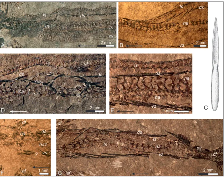
Vertebral terminology. The morphology and homology of the saurichthyid vertebral col-umn has been addressed by several authors (Sten-siö 1925; Lehman 1952; Rieppel 1985; Tintori 1990, 2013; Wu et al. 2009, 2015; Maxwell et al. 2013, 2015). The persistent notochord supports for the whole length pairs of ossified dorsal (neu-ral arches) and, in the posterior region, vent(neu-ral (haemal arches) elements (Fig. 2). It has been re-peatedly noted that the ventral elements, when
os-sified, relate as 1:1 or 1:2 to the dorsal ones, and both relationships can even occur in different body regions of one individual. Most authors now agree that saurichthyids have achieved a unique mode of axial elongation by developing, along the whole vertebral column, dorsal and ventral inter-calaries (interdorsals and interventrals sensu Sten-siö 1925) of equal or nearly equal morphology to the true neural and haemal arches (basidorsals and basiventrals sensu Stensiö 1925). This hypothesis is supported by osteological observations (Stensiö 1925; Wu et al. 2009, 2011, 2015; Maxwell et al. 2013, 2015, Tintori 2019) but also by soft-tissue preservation suggesting that two pairs of neural arch-like elements belong within one embryonic somite (Maxwell et al. 2013). Based on this conclu-sion, Maxwell et al. (2013, 2015) and Maxwell & Wilson (2013) divided the number of neural arch-like elements in saurichthyids by two in order to obtain anatomically correct vertebral counts com-parable with other actinopterygians. Although this interpretation is convincing, we were unable to find osteological differences between true neural arches and intercalaries in the specimens described in this paper, as it happens also for other species (Tintori 1990, 2013), and therefore cannot make this distinction in our material. We therefore refer to dorsal elements as neural arches and to ventral elements as haemal arches, irrespective of their os-teological identity.
According to most previous descriptions, each neural arch carries a posterodorsally directed neural spine portion and can additionally have
an-terior and posan-terior articular processes (prae- and postzygapophyses following Rieppel 1985, Tintori 1990, 2013; Fig. 2). As parts of the paired neural arches, both spines and zygapophyses are paired as well; their naming, used here for reasons of convention, does therefore not necessarily imply homology with equally named structures in tetra-pods. Whereas neural spines and praezygapophy-ses seem to be developed in most saurichthyid species, distinctive postzygapophyses have been described so far only by Stensiö (1925) in S.
orna-tus, S. elongatus and some specimens indetermin-able at species level, and by Wu et al. (2009, 2011, 2015) in S. dawaziensis, S. yangjuanensis and the
‘Si-nosaurichthys’ species group. Very short postzyg-apophyses are possibly present also in S. grignae (Tintori 2013, fig. 6). Praezygapophyses point an-teromedially and are thus overlapped by the dorso-caudally directed neural spines of the preceding neural arches (Tintori 1990, 2013), but overlap the postzygapophyses thereof, which are directed cau-domedially when developed (Wu et al. 2009, 2011, 2015; contrary to the statement of Stensiö 1925, p. 118, according to which praezygapophyses would be covered by postzygapophyses).
Institutional abbreviations: FG: TU Bergakademie Frei-berg, Germany; GBA: Geologische Bundesanstalt/Geological Sur-vey of Austria, Vienna; GM: Goldfuss Museum, Steinmann-Institut für Geologie, Mineralogie und Paläontologie, Universität Bonn, Germany; MFSN: Museo Friulano di Storia Naturale, Udine, Italy; NHM: Naturhistorisches Museum, Vienna, Austria; MPCM, Museo paleontologico Cittadino, A.d.F., Monfalcone, Italy; PIN: Palaeon-tological Institute of the Russian Academy of Sciences, Moscow, Russia.
and terminology used for its description. A–C) No-tochord and dorsal verte-bral elements in anterior (A), oblique (B) and lateral (C) view; D–E) dorsal and ventral elements in the ab-dominal body portion of S. madagascariensis (D; after Lehman 1952) and S. curionii (E; after Rieppel 1985); F) tentative interpretation of a dorsal vertebral element of S. striolatus. Abbreviations: ha: haemal arch, na: neural arch, not: notochord, ns: neural spine, poz: postzyga-pophysis, prz: praezygapo-physis.
s
ysteMatIcpalaeontologySubclass ACTINOPTERYGII Cope, 1887 Order Saurichthyiformes Aldinger, 1937
Family: Saurichthyidae Owen, 1860 [sensu Stensiö 1925]
Genus Saurichthys Agassiz, 1834
Type species: Saurichthys apicalis Agassiz, 1834
Morphological descriptions
Saurichthys striolatus (Bronn, 1858)
1858 Belonorhynchus striolatus, Bronn, p. 7, pl. 1, fig. 1,10, pl. 2, fig. 1. 1866 Belonorhynchus striolatus, Kner, p. 189, pl. 6.
1888 Belonorhynchus striolatus, Woodward, p. 354. 1887,1890 Belonorhynchus striolatus , Zittel, p. 265, fig. 270. 1882 Saurichthys striolatus, Reis, p. 148, fig. 5.
1895 Belonorhynchus striolatus, Woodward, p. 11. 1925 Saurichthys striolatus, Stensiö, p. 11.
1959 Saurichthys striolatus, Griffith, p. 588, figs. 1,6, pl. 1. 1981 Saurichthys striolatus, Beltan & Tintori, p. 53.
1985 Saurichthys striolatus, Tintori, Muscio & Nardon, p. 201. 1990 Saurichthys striolatus, Tintori, p. 100.
2012 Saurichthys striolatus, Romano et al., p. 558, fig. 8, tab. 2. 2013 Saurichthys striolatus, Tintori, p. 295, 296, 298, 299. 2013 Saurichthys striolatus, Wilson et al., p. 902. 2018 Saurichthys striolatus, Tintori, p. 59.
This Saurichthys species is a very stable one, from a taxonomical point of view, owing probably to the very small size and to the lack of collecting for more than a century, so that the available mate-rial was only that collected around the middle of the 19th century, before new interest for the ‘Raibl Fauna’ started again in the 80s of the last century. Most specimens of S. striolatus (Figs. 3–5) represent articulated individuals, sometimes in part and coun-terpart. They are often flattened dorsoventrally up to the level of the pelvic fins, whereas the posterior part of the body and the unpaired fins are preserved in lateral view. The vertebral column, the mid-ven-tral and the mid-dorsal scale rows can mostly be traced more or less continuously along the body; fins and fin supports are usually damaged, but well visible. The description is based on numerous spec-imens from several collections, which are addressed in the text and illustrations where appropriate. S.
striolatus appears to be quite common in the old col-lections, but rarer in the new ones from the Raibl-Cave del Predil area. This is consistent with the ra-tio found in several other Triassic fish assemblages, pointing to a gregarious way of life of Saurichthys. However, the fact that this species appears to be
present in all the different assemblages that are now under investigation around Raibl /Cave del Predil (A.T. pers. obs.), although in different proportion, is quite unusual for Saurichthys as its species seem to be quite short-ranging. Furthermore, S.
striola-tus is the only species of Saurichthys yielded by the Predil Limestone, which again is not common for this genus, usually present with two or more species in each assemblage (Tintori 1990, 2013; Wu et al. 2011, 2015;Maxwell et al. 2015).
Axial skeleton. Although many specimens of
Saurichthys striolatus are fairly complete, no accurate vertebral count can be provided because vertebral columns are never well enough preserved. Where-as several early authors such Where-as Kner (1866), Zittel (1887–90) and Reis (1892) gave numbers between 150 and 160, Bronn (1858) suggested the presence of 170–200 ‘vertebrae’ and Griffith (1959) reported a total of 180–190. Our observations rather con-firm the first mentioned estimate of about 160 dor-sal elements, thus equivalent to about 80 vertebral segments.
The neural arches of S. striolatus (Fig. 3) are typically developed as ‘T’-shaped structures, where the vertical axis of the ‘T’ is slender, strongly con-vex laterally and with concave anterior and poste-rior edges. The ventral region is enlarged to form a broad ‘foot’ or base. The upper (horizontal) rod of the ‘T’ is slender but its single branches are much longer than the width (anteroposterior length) of the vertical rod, and the anterior branch (praezyg-apophysis) is consistently longer than the posterior one. In some places, a small dorsal projection seems to occur between the anterior and posterior
branch-Fig. 3 - General morphology and details of the axial skeleton of Saurichthys striolatus (Bronn, 1858). A) Total view of speci-men FG 01/2011; B) close-up of the abdominal body part of FG 01/2011; C) neural arches of the abdominal body part in medial view, enlarged from B; D) close-up of the trunk anterior to the dorsal and anal fins of FG 01/2011 (anterior caudal part); E) interpretive drawing of the neural arches of the abdominal body part; F) interpretive drawing of the neural arches of the caudal body part in dorsal view. Abbreviations: ds: mid-dorsal scale row, ha: haemal arch, ls: mid-lateral scale row, na: neural arch, ns: neural spine, poz: postzygapophysis, prz: praezygapophysis, raf: radials of the anal fin, rdf: radials of the dorsal fin, vs: mid-ventral scale row.
es of the neural arch, just above its vertical rod (Fig. 3C). If this projection represents the remnant of a neural spine, the posterior neural branch should be referred to as the postzygapophysis. The bases of neighbouring neural arches are close to each other and sometimes in contact, whereas the space be-tween the narrowest neural arch points can be near-ly double their width (anteroposterior length). Sub-sequent neural arches articulate with one another by the anterior and posterior branches of the upper ‘T’ rod in a way that the praezygapophysis of the posterior neural arch lies parallel to the dorsal mar-gin of the preceding one and is crossed by the cau-dally or dorsocaucau-dally directed posterior branch of the latter. The preservation is seldom good enough (also because of the very small size of the elements) to clearly recognize the pattern of overlap, as the praezygapophyses are generally better visible than the posterior branches (postzygapophyses?) in both lateral and medial view. It seems, nevertheless, that they lie more medially and are overlapped by these posterior branches from the outside (Fig. 3E). As far as can be recognized, the length of the praezyg-apophyses does not extend farther anteriorly than the middle of the preceding neural arch. When pre-served in dorsal view, the neural arches from both right and left side of the notochord are mostly ex-posing their concave outside and keep connected with each other along their dorsal margin (Fig. 3F).
Posterior to the dorsal and anal fin, the neu-ral arches increase in width and their bases become broader and higher.
In larger specimens, a double row of small, elongated oval ossifications is present anterior to the pelvic fins. This double row lies parallel to the ventral margin of one or both series of neural arch-es and never splits into far separate rows. The oval elements are half as numerous as the neural arches and are interpreted as the paired haemal arches of the abdominal body portion (Fig. 3B). Posterior to the pelvic fins, the haemal arches become triangular to polygonal in outline and now correspond to the neural arches in number (Fig. 3D).
Squamation. At least four continuous rows of scales are distinguishable in all examined specimens: relatively thick and elongated dorsal and mid-ventral scales, and small, thin, rounded scales of the lateral line (Fig. 4). Since specimens of S. striolatus are seldom preserved in perfectly lateral position,
both rows of flank scales are visible near the out-lines of the body.
The mid-dorsal scale row begins immediately behind the skull and consists of fusiform to spine-like, elongated, slender, anteriorly pointed scales (Fig. 4A–E, G). The outer surface of each scale is convex in its posterior part and has a depression in the anterior part, where it is overlapped by the preceding scale (Fig. 4B, C). The inner surface is ex-cavated in the posterior portion where it overlaps the following scale. In general, the dorsal scales in each body portion are less numerous than the neu-ral arches.
The mid-ventral scale row starts about 1/6 of the trunk length behind the skull, at a distance cor-responding to approximately 20 –30 neural arches (Fig. 4A). Apart from its origin, it is similar to the mid-dorsal scale row, but the individual scales tend to be slightly longer than those of the dorsal row. Slightly anterior to the pelvic fins (at about the 90th neural arch), the mid-ventral row splits in two to form the anal loop, which is located between the pelvics and consists of 5–6 pairs of scales (Figs. 4D, 5C).
The course of the mid-dorsal and mid-ventral scale row is interrupted by the bases of the dorsal and anal fin.
Both dorsal and ventral mid-scales increase in size caudally. In measured specimens, their lengths range approximately between 0.8 and 1.5 mm in front of the pelvic fins, 2–3 mm in front of dor-sal and anal fin, and 3.5–5 mm between these and the caudal fin (Fig. 4G). In the caudal region, dorsal and ventral scales overlap to about three-quarters of their length, and so one scale can be overlapped by three others. Near the caudal fin, the scales sud-denly become broader (scute-like) and almost enve-lope the narrow caudal pedicle just in front of the first caudal lepidotrichia. Limits of individual scutes are hardly distinguishable, but at least three or four elements seem to be present. The scutes have the shape of caudally pointing ‘V’, at least for the ex-posed region, and their lateral margins are serrated. A longitudinal keel on the inside of each scute fits into a groove on the outside of the following scale, ensuring a very stiff connection. The keel on the anteriormost scute is much longer than the exposed surface of the scute and bifurcated in the anterior portion, suggesting a very long and stiff contact with the overlapping scale in front (Fig. 4G).
The lateral line scales (Fig. 4A, B, D, E) are very thin and fragile. Mostly, they are preserved as a series of rounded structures, sometimes show-ing a faint concentric striation. They correspond in number to the neural arches. In a few specimens, small longitudinal depressions near the posterior scale margin can be recognized; these could be impressions of the lateral line sensory canal. The scale row is often paralleled by a broad, dark line in which usually no structure can be detected; in some specimens, however, this second line seems also to consist of distinct, rounded elements, and could therefore be interpreted as an additional row of scales, in a ventro-lateral position. We sup-pose that those scales were very thin (also related
to the very small size of this species) but possibly pigmented in some way, so that the preservation could be very problematic. These rows, if present, are not complete but probably appeared at half of the trunk length.
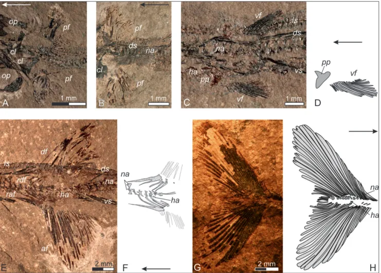
Paired fins. The pectoral fins of Saurichthys striolatus (Fig. 5A, B) are rather small and rounded but seldom completely preserved; about 33 lepi-dotrichia can be counted in specimen FG 01/2011 (Fig. 5A). This is in agreement with Griffith (1959) who gives a fin-ray number of 35 for the pectorals. The fins lie close to the cleithrum, which has the shape of a ‘T’ turned upside-down, as it is com-mon for most species of Saurichthys. The right and left cleithra are often preserved in contact along
Fig. 4 - Squamation of Saurichthys striolatus (Bronn, 1858). A) The anterior trunk portion as preserved in specimen GBA 2006/97/13; B) an-terior abdominal portion as preserved in specimen GBA 2006/97/27; C) drawing of a typical mid-dorsal or mid-ventral scale of S. striolatus; D) posterior abdominal portion and anal loop of the mid-ventral scale row as preserved in specimen GBA 2006/97/39; E) anterior caudal portion (posterior to the pelvic fins) as preserved in GBA 2006/97/39; F) trunk portion posterior to the dorsal and anal fins as preserved in GBA 2006/97/26; G) caudal body part of GBA 2006/97/39. Abbreviations: af: anal fin, cf: caudal fin, df: dorsal fin, ds: mid-dorsal scale row, es: enlarged scutes of the caudal peduncle, ha: haemal arches, ls: mid-lateral scale row, na: neural arches, pf: pectoral fin, vf: pelvic fin, vs: mid-ventral scale row.
their ventral margin (Fig. 5A). The lepidotrichia are unsegmented and unbranched again as in al-most every Saurichthys species. The anterior lepi-dotrichia are broad and lie close to each other, forming a nearly continuous surface (as in all the other fins of this species); the posteriormost fin rays are thinner and probably more round in cross-section. The pectoral fins are often preserved at the right and left side of the dorsoventrally flat-tened skull.
The rather weak pelvic fins (Fig. 5C, D) are positioned on both sides of the cloaca, slightly behind the middle of the trunk, which equals a distance of about 95–105 neural arches from the skull. They comprise about 20 rather short, un-segmented and unbranched lepidotrichia. In some specimens, a pair of polygonal plates is visible
be-tween the bases of the fins, which can be inter-preted as the pelvic plates.
There is no trace of fringing fulcra in any of the paired fins of S. striolatus.
Unpaired fins. The opposing dorsal and anal
fins (Fig. 5E) are placed roughly in the middle of the distance between the pelvic and the caudal fins (slightly closer to the pelvics), beginning at approximately 125–135 neural arches from the skull. Up to 35 unsegmented lepidotrichia have been counted in the dorsal fin, up to 40 in the anal fin. The fin rays increase in length up to the 7th or 8th ray and become gradually shorter subsequently. Most of them are clearly unbranched but the lon-gest rays may exhibit a small area of bifurcation just at their distal end. The anterior rays are broad
Fig. 5 - The fins of Saurichthys striolatus (Bronn, 1858). A) Pectoral fins as preserved in specimen FG 01/2011; B) pectoral fins as preserved in specimen GBA 2006/97/13; C) anal loop and pelvic fins as preserved in specimen FG 01/2011; D) interpretive drawing of the right pelvic fin of FG 01/2011; E) dorsal and anal fins as preserved in specimen GBA 2006/97/39; F) interpretive drawing of the endoske-letal radials of the dorsal and anal fins of GBA 2006/97/39; G) caudal fin as preserved in specimen GBA 2006/97/12; H) interpretive drawing of G. Abbreviations: af: anal fin, cl: cleithrum, df: dorsal fin, ds: mid-dorsal scale row, ha: haemal arches, ls: mid-lateral scale row, na: neural arches, op: operculum (=suboperculum following Argyriou et al. 2018), pf: pectoral fin, pp: pelvic plate, raf: radials of the anal fin, rdf: radials of the dorsal fin, vf: pelvic fin, vs: mid-ventral scale row.
pearance of continuous planes. Only five to eight obliquely trending, ossified radials have been ob-served for both fins, at least in larger specimens. The anterior two seem to be fused distally in both the dorsal and the anal fin (Fig. 5F). Each radial seems to correspond to a segment of the verte-bral column (pair of neural or haemal arches).
A structure seen in specimen GBA 2006/97/26 (Fig. 4F) suggests a more complex ar-chitecture in the posterior part of the fin base, as it has been recently described also in other species. It is located close to the posterior margin of the anal fin and appears paired or just deeply forked, broadening and flattening anteriorly. Thus, this element might be interpreted as a pair of ventral scales enclosing/supporting the fin base (see Wu et al. 2015, Maxwell et al. 2015, Kogan & Romano 2016a), a modified single mid-ventral scale sup-porting the caudal part of the fin (see Tintori et al. 2014b) or a modified radial (see Tintori 2013). Any interpretation remains speculative since in no oth-er specimen a similar structure has been obsoth-erved. The caudal fin (Fig. 5G, H) consists of up to 35 lepidotrichia in both the epaxial and the hyp-axial lobe. The fin rays are unsegmented and in the largest specimens, the longest rays bifurcate dis-tally. The vertebral column continues straight to the posterior margin of the tail fin, and the lepido-trichia are inserted close to the neural and haemal arches although the connection is not precisely visible. Both lobes are semi-oval in outline and their anterior lepidotrichia are placed close to each other, forming nearly continuous surfaces. How-ever, about ten posteriormost rays of each lobe deviate from this appearance, being distinctly thin-ner and shorter. Whereas the last lepidotrichia of the ‘regular’ caudal lobes insert in a very low angle to the vertebral column, the first of these terminal rays seem to insert at some distance behind the ‘regular’ ones and in a higher angle. Considered to-gether, these thinner (softer?) rays may be referred to a third, median or terminal lobe in the caudal fin. In many specimens, the number of caudal fin rays appears lower because these thinner rays are missing; this can be a further indication for the ex-istence of a terminal lobe that got lost as a whole during decay before embedding.
Fringing fulcra are absent in all unpaired fins of S. striolatus.
1925 Saurichthys, Stensiö, p. 5
1977 Saurichthys calcaratus, Griffith, p. 4, figs. 1-3, pl. 1. 1981 Saurichthys calcaratus, Beltan & Tintori, p. 53
Saurichthys calcaratus (Figs. 6–7) is represented only by the type series, stored under the collection number NHM 2007z0170/0001 to /0017. The ho-lotype 2007z0170/001 is a nearly complete but badly preserved skeleton with an elongate, possibly phos-phatized cone placed in the body axis anterior to the pelvic fins that might represent the infilling of the gastrointestinal tract (Fig. 6A). Its partial counter-part has the number 2007z0170/0011, and the in-complete skull 2007z0170/002 has been selected as paratype (Griffith 1977). Additionally, this descrip-tion is based on the specimens 2007z0170/0003 and /0013 (trunk portion between pelvic fins and caudal peduncle in part and counterpart), /0009 and /0014 (trunk portions), /0004–0005 and /0010 (tail in part and counterpart), and /0007 (caudal fin).
Axial skeleton. Due to its preservation, no vertebral count or detailed description of the ver-tebral elements can be provided for the holotype of S. calcaratus (Fig. 6A). The vertebral morphol-ogy is therefore described mainly based on other body fragments. In general, the neural arches are roughly ‘T’-shaped in lateral outline, consisting of a ventrally broad main portion more or less narrow-ing dorsally (long axis of the ‘T’), and two upper branches directed cranially (praezygapophysis) and caudally (postzygapophysis? neural spine?). These branches ensure ‘articulation’ of subsequent neural arches, as every anterior branch lies medially to the posterior one of the proceeding element. However, it is not clear if they were in contact or not when in life. Both branches vary in shape depending on their position along the body. Anterior to the dor-sal and anal fins, the praezygapophyses are shorter and more horizontally directed (Fig. 6B–E), where-as behind these fins they become longer and point more dorsally (Fig. 6F–H). The posterior branches are generally short and nearly horizontal, except for the region of the dorsal fin, where they are longer and posterodorsally directed. The space between two subsequent neural arches is comparable to the width of one vertical rod of a neural arch.
Ventral vertebral ossifications seem to have been restricted to the caudal body part. Specimen
NHM 2007z0170/0013 exhibits relatively large, oval to polygonal structures ventral to the neural arches (Fig. 6D, F). These non-overlapping structures re-late to the neural arches as 1:2 anterior to the dorsal and anal fins and apparently also posterior to them, but their outline is very hard to determine. A medi-an opening recognisable in some of them might be the foramen for intersegmental vessels (see Stensiö 1925; Kogan & Romano 2016b). These elements, not preserved in any other specimen of the type series, have been interpreted by Griffith (1977) as the lateral line scales.
Squamation. S. calcaratus possessed four rows of scales in mid-ventral, mid-dorsal and mid-lateral position.
The mid-ventral and the mid-dorsal scales are spindle-shaped and much elongated, and in life po-sition, they must have overlapped each other to a considerable extent. They are less numerous than the neural arches, but due to the fragmentary pre-servation, the exact relationship cannot be deter-mined. It can be assumed based on specimen NHM 2007z0170/0014 (Fig. 6C) that the mid-ventral scale row started at some distance behind the skull, whereas the mid-dorsal row began more anteriorly. In the abdominal body part, the mid-ventral scales tend to be longer than the mid-dorsal ones (Fig. 6B). Farther caudally, the scales of both these rows increase in size (Fig. 6D, F). In front of the caudal fin, the last nine or ten mid-ventral and mid-dorsal scales are modified to huge, ‘V’-shaped scutes that enclosed the caudal peduncle more or less com-pletely from below and above (Fig. 6H). Rail-like processes that are seen below a damaged scute in the specimen NHM 2007z0170/0010 indicate a strong articulation between subsequent scutes. We assume that these ‘processes’ border a deep groove in which fitted a median keel on the inside of the preceding scute whose posterior part is lacking (Fig. 6I, J).
The mid-lateral scale row is represented by a line of small oval ossified elements present on each side of the precaudal scutes of specimen NHM 2007z0170/0010 (Fig. 6H-J). It seems probable that in life, these elements were placed on the flanks of the caudal peduncle, between the dorsal and the ventral series of scutes. Similar elements can be recognized on the caudal peduncle of the holotype and its counterpart /0011; small oval ossifications,
arranged in one or two longitudinal lines, can be observed in nearly all other specimens, where they sometimes lie ventrally to the neural arches (as in spec. /0003) and also correspond to them in num-ber. This could suggest their interpretation as hae-mal arches; however, the position of one of these lines in specimens /0014 and /0009 (Fig. 6B) where it lies far removed from the vertebral column but still parallel to it, as well as the supposed symmetri-cal disposition of the lines on the flanks of the cau-dal peduncle, indicates that they are rather related to the lateral line than to the vertebral column. In specimen /0009, the oval ossifications seem to con-stitute only the center of larger structures rhombic or circular in outline. Vague circular impressions be-hind the skull in the holotype probably document the true shape of these very thin mid-lateral scales.
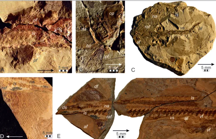
Paired fins. Pectoral fins are preserved in the
ho-lotype NHM 2007z0170/0001 (Fig. 7A) and the para-type /0002 (Fig. 7B). The pectoral fins are small and fan-shaped and consist of at least 24 unsegmented and unbranched lepidotrichia. The cleithrum has the typical saurichthyid shape of an inverted ‘T’.
The pelvic fins, preserved in the holo-type (Fig. 7C) and less well in specimen NHM 2007z0170/0003 and its counterpart /0013, are larger than the pectorals and are placed roughly in the middle between those and the caudal fin. They consist of 18–20 unsegmented and unbranched lepidotrichia and exhibit an unusual modification that has already been described by Griffith (1977). The longest five or six rays of each pelvic fin fuse distally “to form a solid, stout, curved, rod-like structure that projects beyond the general level of
Fig. 6 - Saurichthys calcaratus Griffith, 1977. A) Holotype NHM 2007z0170/0001; B) abdominal body portion as preser-ved in specimen NHM 2007z0170/0009; C) detail of the abdominal body portion as preserved in specimen NHM 2007z0170/0014; D) region anterior to the dorsal and anal fins as preserved in specimen NHM 2007z0170/0013; E) schematic drawing of neural arches from D (in lateral view); F) anterior part of caudal peduncle as preserved in NHM 2007z0170/0013; G) schematic drawing of neural arches from F (in medial view); H) caudal peduncle and tail fin as preserved in NHM 2007z0170/0010; I, J) detail of H show-ing the articulation of precaudal scutes (I, schematic dra-wing and J, enlarged photograph). Abbreviations: cf: caudal fin, df: dorsal fin, ds: mid-dorsal scales, ha: haemal arches, ls: mid-lateral scales, na: neural arches, ns: neural spine, prz: praezygapophysis, vs: mid-ventral scales.
the fin” (Griffith 1977: 8). This structure is devel-oped in both individuals in which the pelvic fins are present and has been interpreted as a gonopodium. No fringing fulcra are observed on any of the paired fins.
Unpaired fins. Both dorsal and anal fin, placed
roughly in the middle between the pelvics and the caudal fin (NHM 2007z0170/0001, Fig. 7F), con-sist of 31–32 unsegmented lepidotrichia, some of which are distally bifurcated. The dorsal fin is sup-ported by at least five to six elongated radials (axo-nosts), the anal fin by seven or eight. Especially the radials of the anal fin are nearly horizontal. Distally, the first two radials of both fins seem to be fused (Fig. 7D, E). A distal series of radials (baseosts) is not preserved, except for some remnants of inde-terminable shape that may be recognized vaguely in the anal fin of specimen /0013.
Three caudal fins of S. calcaratus are known, constraining the minimal number of individuals belonging to the type series (Figs. 6H, 7F). Their epaxial and hypaxial lobes comprise about 30 un-segmented lepidotrichia each, and the longer fin rays show at least one distal bifurcation. The caudal fins seem to be slightly forked in general shape, al-though they are too imperfectly preserved to allow for an exact description.
Fringing fulcra are not developed in any of the unpaired fins.
Saurichthys orientalis Sytchevskaya, 1999
1981 Saurichthys, Minikh, p. 105
1999 Saurichthys orientalis, Sytchevskaya, p. 465, fig. 16, 17. 2009 Saurichthys orientalis, Kogan et al., p. 142, fig. 4, 5A. 2012 Saurichthys orientalis, Romano et al., p. 556, fig. 8, tab. 3.
The description (Figs. 8, 9) is based on the near-complete specimen FG596/III/43 (Fig. 8A) and supplemented by data from the type series, cu-rated in the PIN. The type series consists of the holotype PIN 3267/172a, an incomplete skull with the anterior trunk portion attached, a further cranial and three postcranial fragments, representing more than one individual.
Axial skeleton. Saurichthys orientalis is known only from incomplete specimens, which means that no vertebral count can be reliably estimated.
In FG 596/III/43, the best-preserved individual, the vertebral column can be traced discontinuously from anterior to the pelvic fins until the level of the dorsal and anal fins. The neural arches (Fig. 8B, C) have the general shape of a ‘Y’, with a middle por-tion that is convex laterally and concave anteriorly and posteriorly, an expanded base or ‘foot’ and two distinct branches in the dorsal part. The anterior branches (praezygapophyses) point anterodorsally, mostly extending slightly over the preceding neu-ral arch. Their shape is similar in all preserved ele-ments anterior except the most posterior ones. The posterior branches (neural spines) are best seen in the region anterior to the dorsal fin, where they are developed as long posterodorsally ascending struc-tures, cylindrical proximally and flattening distally, and lie lateral to the praezygapophyses. They form a higher angle to the notochord than the anterior branches and are distinctly shorter and broader than these farther anteriorly. In the region of the dor-sal and anal fin, both branches are short and nearly horizontal, but are still very closely connected with each other.
There is no evidence for the presence of ven-tral ossifications, but this may be a preservational effect.
Squamation. S. orientalis exhibits six rows of thick, ornamented, ganoid scales: a mid-ventral, a mid-dorsal, a pair of mid-lateral and a pair of ven-tro-lateral rows.
The mid-dorsal scales (Figs. 8B, I, 9E) prob-ably begin immediately behind the head and retain a
Fig. 7 - Fins of Saurichthys calcaratus Griffith, 1977. A) Pectoral fins as preserved in the holotype NHM 2007z0170/0001; B) pectoral fin as preserved in the paratype NHM 2007z0170/0002; C) pelvic fins as preserved in the holotype NHM 2007z0170/0001; D) dorsal and anal fins as preser-ved in NHM 2007z0170/0003; E) schematic drawing of the fin endoskeleton from D; F) dorsal, anal and caudal fins as preserved in the holotype NHM 2007z0170/0001. Abbre-viations: af: anal fin, cf: caudal fin, cl: cleithrum, df: dorsal fin, ds: mid-dorsal scales, na: neural arches, op: operculum (=suboperculum following Argyriou et al. 2018), pf: pecto-ral fin, raf: radials of the anal fin, rdf: radials of the dorsal fin, vs: mid-ventral scales.
constant general shape, being ovoid to drop-shaped in outline. However, being broader than long in the anterior region (5 mm width to 3 mm length), they soon become longer and narrower towards the dor-sal fin (3 vs. 5 mm). The scales possess a prominent median keel on their ventral side, that seems nearly continuous along the scale row, and overlap each other in longitudinal direction to a considerable ex-tent. The first dorsal scale behind the dorsal fin is split in two symmetrical elements that probably en-closed the last lepidotrichia of the dorsal fin (Fig. 8K).
The mid-ventral scales start at some distance from the skull (corresponding to about 10–11 mid-lateral scales) as small, elongated, oval elements (ca. 1 mm width to 1.5 mm length) with a median keel on the inside (Fig. 8I). They rapidly increase in size and attain nearly the same shape as the mid-dorsal scutes anterior to the pelvic fins. At the level of the pelvic fins, the mid-ventral row splits to form the oval anal loop consisting of at least four paired, curved, elongated elements (Figs. 8D, J, 9C). Be-hind the pelvics, the mid-ventral scales are again of similar shape to the mid-dorsal scales, as far as preserved. Three small mid-ventral scales preceding the anal fin can be referred to as basal fulcra (Fig. 9C); it is likely that the mid-dorsal scale row had the same specialization in front of the dorsal fin, but this portion is not preserved in the available mate-rial.
In external view, the exposed portions (free field) of the mid-dorsal and mid-ventral scales show an ornamentation consisting of coarse ganoine tu-bercles (Fig. 8J).
Rare isolated scutes (FG 596/III/122) are re-ferred to the caudal peduncle of S. orientalis on the basis of their overall resemblance in size and or-namentation with precaudal scutes in several other
Saurichthys species (Fig. 8E–H). They may belong to the mid-dorsal or to the mid-ventral row, which are indistinguishable in shape. The three-dimensional preservation of these scutes allows a detailed de-scription of the scute-to-scute articulation system. The outside of a scute is subdivided in a ‘V’-shaped free field, ornamented with tubercles, and an unor-namented anterior portion that was overlapped by the preceding scute. The area of overlap is cleft in the middle by a deep longitudinal groove (Fig. 8E, F). A deep, narrow median keel running on the in-side of the preceding scale (Fig. 8G, H) must have
fitted in this groove, making rotational movements between successive scutes more or less impossible. In S. orientalis, this type of scute connection is ob-served all the way along the dorsal and mid-ventral scale rows, but is less pronounced anteriorly, where the scales rotate more freely.
Posteriorly adjoining the pectoral girdle, the mid-lateral scales (Figs. 8B, I, J, 9A, E) are triangu-lar to rhombic in general shape, anterodorsally in-clined, and have narrow projections in anterodorsal and posteroventral directions. These projections ap-pear at about the third scale behind the skull, soon achieving a maximum length nearly equal to the height of the main scale body, but disappear gradu-ally in front of the pelvic fins. The scales themselves do not change much in their dimensions, ranging from 6 to 7 mm in height and from 2 to 2.5 mm in length in the preserved body portion. Each scale slightly overlaps the following with its posterior edge. The lateral line canal runs as a median hori-zontal ridge on the internal surface of the scales and pierces from one scale to another in the region of overlap. The mid-lateral scales are about half as nu-merous as the neural arches in the respective body portion. All mid-lateral scales of FG596/III/43 are preserved in internal view, but where they are dam-aged, it can be recognized that they are ornamented with ganoine tubercles in the dorsal half and appar-ently with vertical striae in the ventral half.
The ventro-lateral scale rows probably start behind the beginning of the mid-ventral row and cannot be traced posterior to the pelvic fins. The
Fig. 8 - Saurichthys orientalis Sytchevskaya, 1999. A) Best-preserved specimen FG 596/III/43; B) squamation and vertebral co-lumn as preserved in the counterpart of FG 596/III/43, region between the pelvic and dorsal fin; C) schematic dra-wing of neural arches from B; D) scales of the pelvic fin re-gion as preserved in the counterpart of FG 596/III/43; E) cast of a scute from the caudal peduncle, FG 596/III/122a, in external view; F) interpretive drawing of E; G) scute from the caudal peduncle, FG 596/III/122b, preserved in internal view; H) interpretive drawing of G; I) anterior body part of FG 596/III/43; J) abdominal body part of FG 596/III/43; K) mid-dorsal scales posterior to the dor-sal fin, FG 596/III/43. Abbreviations: al: anal loop of the mid-ventral scale row; df: dorsal fin; ds: mid-dorsal scales; ls: mid-lateral scales; na: neural arches; ns: neural spine; prz: praezygapophysis; vf: pelvic fin; vls: ventro-lateral scales; vs: mid-ventral scales.
scales are trapezoid in general shape (about 2.6–3 mm length to 2 mm height) and have a marked pro-jection in dorso-caudal direction. They are orna-mented with tubercles.
Scales of all rows roughly correspond to each other in number within a given body segment.
Very small arrays of ganoine tubercles are preserved on the trunk, indicating the possible
pres-ence of additional minute scales in the dorsal body half. Furthermore, ventrolateral fields of oblique bony plates are developed immediately behind the pectoral fins, but their exact shape and extension remains unclear (Fig. 9B).
Paired fins. The pectoral fins (Fig. 9A, B) are
fan-shaped and consist of at least 26–28 lepido-trichia. A few pectoral fin rays of the holotype PIN 3267/172a (Fig. 9B) show displacement of their distal parts, which might be due to distal segmen-tation. The cleithrum has the saurichthyid-typical shape of an inverted ‘T’ (Fig. 9A).
The pelvic fins (Fig. 9C, E) begin at a distance corresponding to about 43 mid-lateral scales behind the skull and comprise about 24 lepidotrichia. As documented in specimen PIN 3267/180 (Fig. 9C), the longer pelvic fin rays consisted of no less than four segments, the proximal ones being distinctly longer than the distal.
Fringing fulcra are not developed on the paired fins of S. orientalis.
Unpaired fins. The dorsal and anal fin begin at
a distance corresponding to 20-22 mid-lateral scales or about 42 neural arches from the beginning of the pelvic fins; the anal fin seems to begin slightly in front of the dorsal fin (Fig. 9E). The dorsal fin of FG 596/III/43 consists of about 38 lepidotrichia, the preserved portion of which is segmented twice; from comparison with the anal fin it can be con-cluded, however, that the dorsal 2/3 of the fin are missing.
The anteriormost part of the anal fin is miss-ing as well. At least 32 lepidotrichia are preserved, the longest of them showing five or six segmenta-tions and bifurcating distally (Fig. 9D).
Both fins are supported by at least six oblique radials (axonosts). No fringing fulcra are observed.
The caudal fin of S. orientalis is unknown.
Fig. 9 - Fins of Saurichthys orientalis Sytchevskaya, 1999. A) Pectoral fins as preserved in FG 596/III/43; B) pectoral fin as preserved in the holotype PIN 3267/172a; C) pelvic and anal fins as preserved in PIN 3267/180; D) anal fin as preserved in the counterpart of FG 596/III/43; E) pelvic, dorsal and anal fins as preserved in FG 596/III/43. Anterior to the right in all figures except D. Abbreviations: al: anal loop of the mid-ventral scale row; af: anal fin; bp: bony plates; cl: cleithrum; df: dorsal fin; ds: mid-dorsal scales; ls: mid-lateral scales; na: neural arches; op: operculum (=suboperculum following Argyriou et al. 2018); pf: pectoral fin; raf: radials of the anal fin; vf: pelvic fin; vs: mid-ventral scales.
Present reappraisal of the three late Ladin-ian/early Carnian Saurichthys species greatly enlarges the knowledge of their morphology. Although a few publications (Bronn 1858; Kner 1866; Zittel 1887–90; Reis 1892; Woodward 1895; Griffith 1959) have dealt in the far past with the morphology of
S. striolatus, anatomic details of the axial skeleton and the endoskeleton of the pelvic, anal and dorsal fins are described here for the first time. This is also the case for the supposed terminal tail lobe, hitherto discussed only for Saurichthys curionii and S.
(Costa-saurichthys) costasquamosus from the Middle Triassic of Monte San Giorgio by Rieppel (1985). Whereas mid-dorsal and mid-ventral scales of S. striolatus are well-ossified, the lateral line scales are very thin; the presence of a further, similarly developed, ventro-lateral scale row is indicated in some specimens, as suggested earlier by Zittel (1887–90).
Although the type series of S. calcaratus was known already to Stensiö (1925), this species was de-scribed only by Griffith (1977), whose observations were precise in some points such as the exoskeleton of the fins but vague in others. We were able to cor-rect the squamation pattern reconstructed by this author and to provide some more information on the endoskeletal morphology. Two individuals show a modification of pelvic fins, whose longest fin rays form stout hook-like structures, which Griffith (1977) interpreted as copulatory organs. This was a daring interpretation, taking into account that evi-dence for viviparity in saurichthyids was first pre-sented by Rieppel (1985) for the Middle Triassic
S. curionii and S. macrocephalus and further substan-tiated by Bürgin (1990), Renesto & Stockar (2009) and Maxwell et al. (2018). Furthermore, Tintori et al. (1998, p. 15) identified arrays of coarse tubercles ornamenting the lower jaw as a sexually dimorphic character in some Middle Triassic Saurichthys. Vivi-parity would require internal fertilization, accom-plished by an unpaired gonopodium probably de-rived from modified mid-ventral scales in males of
S. curionii and S. macrocephalus (Rieppel 1985; Bürgin 1990). In other fossil and extant actinopterygians, sexual dimorphism of usually anal fins has been re-ferred to their reproductive function (e.g., Bürgin 1990; Lombardo 1999). However, as in Saurichthys the cloaca opened well anterior to the anal fin, actu-ally just between the pelvic fins, Griffith (1977) and
form a channel directing the seminal fluid into the female’s genital opening.
S. orientalis is the most ‘primitive’ of the three species studied, closely resembling the early saurichthyid morphotype as proposed by Romano et al. (2012) and Kogan & Romano (2016a). The thick ganoid squamation and the fins with seg-mented and branched lepidotrichia are in contrast with the reduced scale cover and unsegmented fin rays of S. striolatus and S. calcaratus, but in agree-ment with several early species such as S. aff. dayi or
S. madagascariensis (Kogan 2011; Kogan & Romano 2016a). S. orientalis furthermore shares with these species the 2:1 ratio of neural arches to mid-lateral scales, whereas these elements relate as 1:1 in both
S. striolatus and S. calcaratus (an ‘advanced’ character according to Maxwell et al. 2013). It mainly devi-ates from the early morphotype by the absence of fringing fulcra, a character shared with the other two Carnian forms. Ventral vertebral elements of
S. orientalis are unknown; otherwise, our description supports the basal phylogenetic placement of S.
ori-entalis recovered by Maxwell et al. (2013, 2015). In all three species, the neural elements of the vertebral column possess distinct anterior branches (praezygapophyses) that articulate with the preced-ing neural arch. These processes are shorter, but still considerable, in S. striolatus, longer and variable in shape in S. calcaratus, where they overlap the pre-ceding neural arch more or less completely in the posterior trunk portion, and longest in S. orientalis where they are generally longer than one neural arch.
S. orientalis also has distinct posterodorsally ascend-ing neural spines, as it is supposed to be typical for the genus. In S. striolatus, in contrast, the posterior branches of the neural arches are small and nearly horizontal, and it cannot be ruled out whether they are neural spines or postzygapophyses. The fact that a median vertical projection is developed in some neural arches supports the latter homologization. The posterior neural arch branches of S. calcaratus are variable in shape and direction, some of them being nearly horizontal or even posteroventrally inclined, while the more anterior ones point pos-terodorsally; they may again be homologized with neural spines. In terms of articulation, the connec-tion between neural arches was probably stiffest in
the preservation pattern of most specimens), thus increasing with body size. This is consistent with Tintori’s (2013) hypothesis that the development of the vertebral ‘grid structure’ formed by intercalation of very elongated praezygapophyses and neural spines depends on a species’ size and lifestyle.
Besides body size and segmentation of fin rays, the most obvious deviation in the morphology of the three species lies in their squamation, which is reduced in both S. striolatus and S. calcaratus. In these species, the mid-dorsal and mid-ventral scales are spindle-shaped and more or less elongated and have no ganoine ornamentation, and the mid-lateral scales (as well as the possible ventro-lateral ones in
S. striolatus) are small and thin. In the case of S.
stri-olatus, the flank scales seem to have completely lost
the ganoine. The scales of the dorsal and mid-ventral rows overlap each other to an increasingly large portion backwards and in the caudal peduncle, they form solid structures in S. striolatus and are de-veloped as interlocking scutes in S. calcaratus. In S.
orientalis, in contrast, all scales are ornamented with ganoine tubercles and striae, and the mid-dorsal and mid-ventral scales have the shape of more or less broad scutes all the way along the body. ‘Keel and groove’-articulation is present in all these scutes, but it is reasonable to assume that it was weaker anteri-orly and stronger in the caudal peduncle.
The caudal fins are symmetrical in S.
striola-tus and S. calcaratus, while no information is avail-able on S. orientalis. However, in all saurichthyids in which the caudal fin is preserved, the epaxial and hypaxial lobes are fully or nearly fully symmetrical. Furthermore, the symmetry is high also between the dorsal and the anal fins, including the species studied herein. The fin-ray counts of the dorsal and anal fins are only slightly higher than those of each caudal lobe in both S. striolatus and S. calcaratus, which again seems to be typical for saurichthyids. Most noteworthy, in forms with highly elongated median fins, such as Saurichthys (Sinosaurichthys)
longi-medialis, the fin ray elongation is comparable in the
dorsal, anal and both lobes of the caudal fin (Wu et al. 2011). The distances between the pelvic, dorsal/ anal and caudal fins vary slightly among the species, but as a general rule, they subdivide the posterior half of the trunk in nearly equal portions.
To describe the function of the symmetri-cal median fins, Weihs & Webb (1983) introduced the concept of a double-tail configuration. They
regard the symmetrical dorsal and anal fins as an ‘anterior tail’ that would interact with the caudal fin to produce optimal propulsion. According to Wei-hs (1989), tens of percents of energy lost by the movement of the ‘anterior tail’ can be utilized when the caudal fin has about the same height. Distance between both ‘tails’ is interpreted in the context of swimming style: so, about 0.4 of the body length is considered the optimal tail distance for fast-swim-ming fishes. In contrast, the dorso-ventral fin pair positioned close to the true tail constitutes an opti-mization towards rapid acceleration, as the ‘anterior tail’ stabilizes flow over the caudal fin that, in turn, can therefore increase thrust using higher ampli-tudes (Weihs 1989). In this context, a general ten-dency of larger saurichthyids towards anal and dor-sal fins positioned closer to the tail than in smaller forms (pers. obs.) points to an increased fast-start performance in the former.
On the example of recent mackerel (Scomber
scombrus) and longfin tuna (Thunnus alalunga), Videler
(1985) has shown that transmission of propulsive force towards the tail fin is optimized by strength-ening the connection between fin and vertebral column. In teleosts, this can be accomplished by a tight intercalation of the ural plate and the caudal fin rays. In saurichthyids, the same effect may have been achieved by the direct insertion of the lepi-dotrichia on both sides of the vertebral column, and especially by the caudal peduncle stiffened by stronger articulation between following neural and haemal elements and by the development of spe-cialized dorsal and ventral enlarged scutes. Force transmission may further have been increased in species with unsegmented fin rays, as stiffness of lepidotrichia decreases with increasing segmenta-tion (Lauder 2015), although usually unsegmented rays are thinner than the segmented ones, at least in Saurichthys.
c
onclusIonSaurichthys was the first actinopterygian fish
with the typical design of an acceleration special-ist. Fast-start performance was optimized by the streamlined body shape minimizing drag force, by the position, shape and structure of the unpaired fins, by weight loss through the reduction of squa-mation, and by endo- and exoskeletal structures
body undulations (e.g., Long & Nipper 1996) and drag that would be caused by lateral recoil (e.g., Do-menici & Blake 1997).
With increasing knowledge of saurichthyid species, several phylogenetic trends postulated by earlier authors (Rieppel 1985, 1991) become in-creasingly questionable (Tintori 2013; Tintori et al. 2014b). One tendency, although not linear, that can be observed after the Spathian, is reduction of squamation, in terms of the number, shape and thickness of the scales (Schmid & Sánchez-Villagra 2010; Romano et al. 2012; Tintori 2013; Tintori et al. 2014b). This is generally seen in the context of weight reduction (e.g., Rieppel 1985; Lombardo & Tintori 2005), which would enhance fast-start per-formance of the fish (Webb et al. 1992). Present study, however, highlights the functional impor-tance of dorsal and ventral mid-scales even in spe-cies with otherwise reduced squamation, such as
S. striolatus and S. calcaratus. Especially the caudal peduncle is stabilized by specialized mid-dorsal and mid-ventral scutes in nearly all saurichthyid species. Furthermore, in some species of Saurichthys, mid-dorsal and mid-ventral scales are also involved in supporting the dorsal and anal fins (Tintori et al. 2014b; Maxwell et al. 2015). The patterns of over-lap and of interlock demonstrated here widen the range of known locomotor adaptations in
Saurich-thys and may help explaining why the mid-dorsal and mid-ventral scales persist in all species of this genus.
The fact that smaller Saurichthys species, such as S. striolatus and S. calcaratus, but also S. minimahleri,
S. diannae, S. spinosa (Werneburg et al. 2014; Max-well et al. 2016; Wu et al. 2018) and some yet unde-scribed small forms from the Middle Muschelkalk (Anisian) of Germany (Schneider et al. 2012; I.K. pers. obs.) exhibit no ‘grid structure’ (sensu Tintori 2013) in their vertebral column and tend to have a comparatively higher distance between their ‘an-terior’ and ‘pos‘an-terior’ tails, points to a somewhat different swimming style, perhaps involving more sustained swimming. We conclude that, apart from a general trend towards reducing squamation, mor-phological adaptations seen in the fins, caudal pe-duncle and vertebral column of different species of Saurichthys correlate with size, diet and mode of locomotion, and not necessarily reflect phylogeny.
contributed to the discussion, and performed most of the original drawings used herein. He unexpectedly passed away in September 2015. We retain Martin in the list of authors and dedicate the paper to his memory.
Following persons and institutions are thanked for the ac-cess to specimens (in temporal order of collection works): Birgit Gaitzsch, FG, Germany; Georg Heumann, GM, Bonn, Germany; Florian Witzmann, Museum für Naturkunde Berlin, Germany; Adriana López-Arbarello and Markus Moser, Bayerische Staatssam-mlung für Paläontologie und Geologie Munich, Germany; Giuseppe Muscio and Luca Simonetto, MFSN, Udine, Italy; Maurizio Tentor, MPCM, Monfalcone, Italy; Irene Zorn, GBA, Vienna, Austria; Ur-sula Göhlich, NHM, Vienna, Austria; Evgeniya Sytchevskaya, PIN, Moscow, Russia. Work in collections has been supported by grants from the MFSN Udine and the “Verein der Freunde und Förderer der TU Bergakademie Freiberg” to I.K.
We owe the discovery of the new specimen of Saurichthys orientalis to field work in the Madygen Formation by Sebastian Voigt, Jan Fischer, Katharina Schönberger (formerly FG), supported by the grant VO 1466/1 from the Deutsche Forschungsgemeinschaft and a travel grant from the DAAD.
We received further information from and enjoyed valuable discussions with: Kristina Eck (Museum für Geowissenschaften, Heidelberg, Germany); Carlo Romano (Palaeontological Institue and Museum, University of Zurich, Switzerland); Barbara Meller (NHM); and Jörg W. Schneider and Frank Scholze (FG).
Thoughthful reviews by Carlo Romano and Fei-Xiang Wu (Institute for Vertebrate Paleontology and Paleoanthropology, Bei-jing, China) helped to improve the manuscript; many thanks to them and the editors, especially to C. Lombardo for her help and and sug-gestions.
I.K. benefited from a PhD scholarship of the State of Sax-ony, acknowledges funding by SYNTHESYS and the ESF. This work was performed according to the Russian Government Program of Competitive Growth of Kazan Federal University.
RefeRences
Agassiz L. (1834) - Abgerissene Bemerkungen über fossile Fische. Neues Jahrbuch für Mineralogie, Geognosie, Geologie
und Petrefaktenkunde, 1834: 379-390.
Aldinger H. (1937) - Permische Ganoidfische aus Ostgrön-land. Meddelelser om Grønland, 102: 1-392.
Argyriou T., Giles S., Friedman M., Romano C., Kogan I. & Sánchez-Villagra M. (2018) - Internal cranial anatomy of Early Triassic species of †Saurichthys (Actinopterygii: †Saurichthyiformes): implications for the phylogenetic placement of †saurichthyiforms. BMC Evolutionary
Biol-ogy,18(1): 161, doi: 10.1186/s12862-018-1264-4.
Beltan L. (1958) - Remarques concernant la variabilité du nombre des pariétaux chez le genre Saurichthys. Comptes
rendus des séances de l’Académie des Sciences, 247: 1634-1636. Beltan L. (1968) - La faune ichthyologique de l’Eotrias du
N.W. de Magagascar: le neurocrâne. Cahiers de Paléon-tologie, Paris, 135 pp.
Beltan L. & Tintori A. (1981) - The genus Saurichthys (Pisces, Actinopterygii) during the Gondwana Period. In: Cress-well M.M. & Vella P. (Eds) - Gondwana five. Selected