Tutor: Prof. Antonella Pantaleo
Co Tutor: Alessandro Dessì
Roberto Dallocchio
University of Sassari
Department of Biomedical sciences
PhD School in Life Sciences and Biotechnologies
XXXI cycle
In vitro and in silico studies of Syk inhibitors as
new antimalarial drugs
PhD candidate
Giuseppe Marchetti
Coordinator: Prof. Leonardo Sechi
Contents
ABBREVIATIONS 5 SUMMARY 6 1. INTRODUCTION 8 1.1. Background and epidemiology of Malaria 9 1.2. Malaria parasite life cycle and P. falciparum 11 1.3. Protection from malaria infection 14 1.3.1. Genetic factors 14 1.3.2. Acquired immunity 14 1.4. Diagnosis of Malaria 16 1.4.1. Uncomplicated Malaria 16 1.4.1.1. Incubation Period 17 1.4.2 Severe malaria 18 1.4.3 Cerebral malaria 18 1.5. Erythrocytes plasmatic membrane 20 1.5.1. Red blood cells plasmatic membrane structure 20 1.6. The spleen tyrosine kinase (Syk) 23 1.7. Treatments for Malaria 27 1.7.1. Artemisinin and Artemisinin derivatives 27 1.7.1.1. The artemisinin mechanism of action 28 1.7.1.2. Artemisinin derivatives 28 1.8. Artemisinin-based combination therapies (ACTs) 29 1.9. Drugs resistance development 30 1.9.1. State of partial artemisinin resistance around the world 31 1.9.2. Current state of ACTs failures 31 1.10. Computational studies to explain the biological phenomenon 33 1.11. Syk inhibitors as treatment for malaria 38 1.11.1 Kinase inhibitors mainly used in therapy 39 2. AIM OF THE PROJECT 42 3. MATERIALS & METHODS 44 3.1. Experimental In vitro studies 45 3.1.1. ICEstimator 1.2 software 46 3.1.2. Treatment of red blood cells (RBCs) 463.1.3. RBCs membrane preparation 47 3.1.4. SDS-PAGE preparation 47 3.1.5. Western blot analyses 47 3.2. Computational studies 48 3.2.1. Molecular Mechanics (MM) and Quantum Mechanics (QM) 48 3.2.2. Molecular docking 48 3.2.2.1. Docking validation protocol (close conformation of Syk) 49 3.2.3. Quantum Mechanics molecular descriptors 51 3.2.3.1. Homo-Lumo 51 3.2.3.2. Molecular Electrostatic Potential (MEP) 51 3.2.4. Molecular dynamics (MD) 52 3.2.4.1. RMSD maps in Molecular dynamics 53 3.2.5. Identification of receptor for the Virtual Screening 54 3.2.5.1. 3D alignment superposition of Syk 54 3.2.5.2. UCSF Chimera software 55 3.2.6. Docking validation protocol with 43 crystal structures of Syk 55 3.2.6.1. PyMOL software 55 3.2.7. Residues assessment providing Syk specificity in the human kinome 56 3.2.7.1. Alignment method: ClustalX software 56 3.2.8. Virtual screening (VS) 57 4. RESULTS & DISCUSSION 58 4.1. In vitro results 59 4.1.1. IC50 evaluation of Syk inhibitors in parasitized erythrocytes 59 4.1.2. Proteomic profile of Tyr phosphorylation in treated RBCs 60 4.2. Computational results 63 4.2.1. Results of docking validation protocol (close conformation of Syk) 64 4.2.2. Docking results 65 4.2.3. Assessment of Syk inhibitors efficacy 70 4.2.4. Identification of residues as new sites for Syk specificity 70 4.2.5. Docking validation to select the target for VS (open conformation of Syk) 71 4.2.6. Molecular dynamic results 75 4.2.7. RMSD maps results in Molecular Dynamics 78 4.2.8. Virtual Screening results 79
4.2.9. Compounds to test in vitro selected from Virtual screening 81 5. CONCLUSION & PERSPECTIVES 86 6. REFERENCES 89 Publications & communications 99
ABBREVIATIONS WHO : World Health Organization RBCs : Red Blood Cells SYK : Spleen Tyrosine Kinase Hb : Haemoglobin PKs : Protein Kinases ACTs : Artemisinin Combination Therapies ART : Artemisinin DHA : Dihydroartemisinin AS : Artesunate ATH : Artemether LMF : Lumefantrine PPQ : Piperaquine Hz : Hemozoin MDR : Multidrug resistance IC50 : Inhibitory Concentration of 50% Fe-PPIX : Ferriprotoporphyrin IX SDS-PAGE : Sodium Dodecyl Sulphate - PolyAcrylamide Gel Electrophoresis MM : Molecular Mechanics QM : Quantum Mechanics HOMO : highest occupied molecular orbital LUMO : lowest unoccupied molecular orbital MEP : Molecular Electrostatic Potential VS : Virtual Screening LBVS : Ligand-Based Virtual Screening SBVS : Structure-Based Virtual Screening MD : Molecular Dynamics RMSD: Root mean square deviation Fig. : Figure Tab : Table
Summary
Malaria remains one of the most devastating infectious diseases and despite the current therapies are efficient, the WHO recommends Artemisinin Combination Therapies (ACTs) as the frontline treatments against P. falciparum malaria to limit the artemisinin resistance. Unfortunately, in the Greater Mekong subregion, the efficacy of artemisinin-based combined therapies (ACTs) has recently been questioned by resistance to both artemisinin derivatives and to the partner drugs. Recently, a new mechanism of action based on the release of denatured haemoglobin products (haemichromes), bound to erythrocyte membrane through the cytoplasmic domain of the AE1 (Band 3 protein) has been characterized. More specifically phosphorylated band 3 becomes uncoupled from the cytoskeleton, leading to the formation of membrane clusters containing haemichromes – band 3 aggregates that can either be released from red blood cells as microvesicles or picked by spleen macrophages by a mechanism known as erythrocyte pitting. This process is mediated by erythrocytic Syk kinase that carries out the Tyr phosphorylation of band 3.
The consequent destabilization of the membrane is thought to be essential for parasite egress from the red cell at the end of the parasite’s life cycle, since inhibitors of the Syk tyrosine kinase block band 3 tyrosine phosphorylation, membrane weakening and parasite reinfection.
In vitro and in silico studies have been performed in order to investigate the inhibitory activity of these compounds and acquire as much as possible information about the ligands and protein structure, in order to add more evidence to the process occurring in infected erythrocytes.
The aim of this study was to examine the effects of the Syk inhibitors on critical events during parasite growth and maturation within the human erythrocyte through in vitro experiments and explore current molecular docking strategies used in drug discovery and medicinal chemistry, considering the advances in the field and the role played by the integration of structure- and ligand-based methods. In vitro studies have involved the treatment of parasitized erythrocytes cultures with different concentrations of Syk inhibitors and we evaluated the Tyr phosphorylation levels in Band 3 residues by proteomic approach.
In silico studies were based on different approaches of molecular modelling (docking, molecular dynamics and virtual screening). In presence of Syk inhibitors we observed a marked decrease of band 3 phosphorylation, proportional to the increase of drug dosage. The proteomic data about the IC50 values follow the same trend in relation to the computational analyses results. These studies enabled us to better analyse the structure of different Syk inhibitors and to possibly discover new molecules through virtual screening analysis. In the long term, we aim to further investigate the efficacy of these compounds by evaluating their inhibitory activity through proteomic and in vitro studies.
1. INTRODUCTION
1.1 Background and epidemiology of Malaria
Malaria is a parasitic disease caused by a protozoa emosporidae belonging at genus of plasmodium. This infective disease is transmitted by the female Anopheles mosquito bite, which lives mainly in the region with a temperate and hot climate. Among all studied mosquitoes species, only few of them are responsible of malaria disease, whereas the others are harmless, as they prefer animal blood compared to human blood. The main vector responsible of malaria in the Afrotropical region is Anopheles Gambiae specie (Fig.1). In the past few years, the disease has spread through the western continents even though it has been eradicated
in these regions few years ago. The migrants fluxes have been claimed as a possible cause of this unexpected event.
Malaria is an infective disease that causes a high level of deaths in the world. The 2017 Malaria report of registered 216 million new cases and 455 thousands dead. Over 77 % of children die with severe cerebral damages and convulsions under 5
years old (around 300 thousands), for this reason malaria is also known as “disease of childhood” [1]. The sub-Saharan region is the most malaria endemic area in the world registering around 90% of cases in the world. The number of Plasmodium falciparum malaria cases has rapidly decreased in the last five years [2]. In the period 2011-2016 have been registered 3600 cases in Italy mainly due to the migratory fluxes. Malaria remains one of the most devastating infectious diseases in the world, thereby the main effort consists in eradicate it. This disease is endemic in 103 nation therefore many people are exposed to this kind of pathology. Malaria affects also pregnant women and infants, who Fig. 1. Female Anopheles mosquito
contract the pathology directly from the mother, through the blood exchange [3,4].
Annually around 100 thousand of infants die for malaria infected from their mother [5] and 25 million of pregnant women are at risk of infection around the world [6].
Exist five protozoa species able to cause Malaria in humans. They belong to Plasmodium genus: • P. falciparum • P. vivax • P. ovale • P. malariae • P. knowlesi (humans and macaques) These species of Plasmodium cause different kinds of malaria and is important to differentiate them, because the mortality, incidence and distribution are completely different between them.
Among the Plasmodium species, two are the most severe, P. vivax (endemic region) and P. falciparum (Africa, Asia, latin America).
The most common cause of death is due to Plasmodium falciparum. The immune system plays an important role in the attack of the illness. The first time that our organism makes contact with the infective agent is crucial, dangerous and potentially, because the appropriate immune response is not yet present to prevent the infection. For this reason, people who are physiologically immunosuppressed such as kids and breastfeeding women are more prone to die.
1.2 Malaria parasite life cycle and P. falciparum
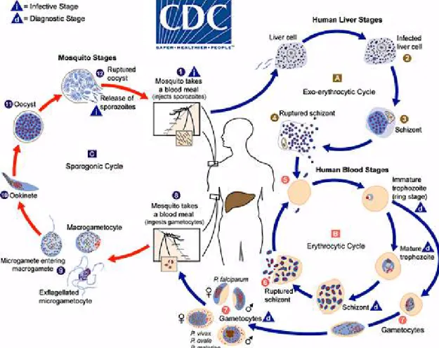
The malaria parasite life cycle involves two hosts. As shown in figure 3, during a blood meal, a malaria-infected female Anopheles mosquito inoculates sporozoites into the human host 1. The sporozoites infect liver cells 2 and mature into schizonts 3, with consequent rupture of membrane and release of merozoites 4. In P. vivax and P. ovale a dormant stage [hypnozoites] can persist in the liver and cause relapses by invading the bloodstream weeks, or even years later.
After this initial replication in the liver (exo-erythrocytic schizogony A), the parasites undergo asexual multiplication in the erythrocytes (erythrocytic schizogony B). Merozoites infect red blood cells 5. The ring stage (1-24 hours) and trophozoites (24-36 hours) mature into schizonts (36-48 hours), with lysis of red blood cells (RBCs) membrane (Fig.2) and release of merozoites 6. Some parasites differentiate into sexual erythrocytic stages (gametocytes) 7. Fig. 2. Intraerythrocytic cycle of parasite
Blood stage parasites are accountable for the clinical manifestations of the disease. The gametocytes, male (microgametocytes) and female (macrogametocytes), are ingested by an Anopheles mosquito during a blood meal 8. The parasites proliferation in the mosquito is known as the sporogonic cycle C. At the same time, in the mosquito’s stomach, the microgametes penetrate the macrogametes generating the zigotes 9. The zygotes in turn become motile and elongated (ookinetes) 10 which invade the midgut wall of the mosquito where they develop into oocysts 11. The oocysts grow, rupture, and release sporozoites 12, which make their way to the mosquito’s salivary glands. Inoculation of the sporozoites 1 into a new human host perpetuates the malaria life cycle (Fig.3) [7].
The activation of parasite metabolic processes inside the RBCs, triggers alterations of host cell energetic metabolism, deep proteins and membrane structural changes. An example in P. falciparum consists of molecules
expression on the erythrocytes external surface that mediates the infected RBCs process of adhesion to the endothelial cells in capillary of some organs. This phenomenon, known as “abduction”, represents an essential pathogenic mechanism in P. falciparum severe malaria. Malaria parasites use haemoglobin (hereafter referred to as Hb) as a major nutrient source in the intraerythrocytic stage, during which the heme group is converted to hemozoin (hereafter referred to as Hz), which is essential for parasite survival [8]. Mature trophozoites digest the haemoglobin and metabolize the glucose, through the anaerobic glycolysis. An infected RBC increases of 50-100% the consumption of glucose with the production of lactic acid. The degradation of Hb occurs in a specialized parasite organelle called the food vacuole. A number of studies have suggested that Hb degradation is a cooperative process that involves proteases of multiple catalytic classes, including cysteine, aspartic, and metallo proteases [9]. These proteases produce short peptides that are further degraded to amino acids, probably by aminopeptidases [10]. During the process of Hb degradation, heme group released in the food vacuole is toxic to Plasmodium, as it induces oxygen-derived free radical formation, lipid peroxidation and protein and DNA oxidation. Organisms such as Plasmodium, Schistosoma, and Rhodnius, which use Hb as a nutrient source, have evolved different strategies to detoxify this free heme group. Plasmodium spp. converts the heme group to β-hematin, which is a dark brown pigment also known as Hz, through a process that is essential for the life cycle of these organisms [14,15]. Hz is a cyclic dimer of ferriprotoporphyrin IX [Fe(III)PPIX] in which the propionate group of each Fe(III)PPIX molecule coordinates the Fe(III) centre of its partner [15].
The formation of mature schizonts containing a variable number of merozoites (24-32), entails their release and the lysis of erythrocytes. This process occurs every 48h corresponding with the parasite life cycle. During this event, the first clinic symptoms of the disease are shown: the characteristic fever ‘malignant tertian’ in P. falciparum, ‘benign tertian’ in P. vivax and P. ovale and ‘quartan’ in P. malariae (Fig 4.).
1.3. Protection from Malaria infection 1.3.1. Genetic Factors Biologic characteristics present from birth can protect against certain types of malaria. Two genetic factors, both associated with human red blood cells, have been shown to be epidemiologically important [13]. People who have the sickle cell trait (heterozygotes for the abnormal haemoglobin gene HbS) are relatively protected against P. falciparum malaria and therefore, hold a biologic advantage. As P. falciparum malaria has been a leading cause of death in Africa since remote times, the sickle cell trait is now more frequently found in this region and in people of African ancestry than in other population groups. In general, the prevalence of haemoglobin-related disorders and other blood cell dyscrasias, such as Hemoglobin C, the thalassemia and G6PD deficiency, are more prevalent in malaria endemic areas and are thought to provide protection from malarial disease.
Other genetic factors related to red blood cells also influence malaria but with a minor impact. Various genetic determinants (such as the “HLA complex,” which plays a role in control of immune responses) may equally influence an individual’s risk of developing severe malaria.
1.3.2. Acquired Immunity
Acquired immunity greatly influences how malaria, affects an individual and a community [13]. After repeated malaria infections a person may develop a partially protective immunity (Fig.5). Such “semi-immune” persons often can
still be infected by malaria parasites but may not develop a severe form of this disease, lacking any typical malaria symptoms indeed.
In areas with high P. falciparum transmission (most of Africa south of the Sahara), newborns will be protected during the first few months of life presumably by maternal antibodies transferred to them through the placenta. As these antibodies decrease with time, these young children become vulnerable to disease and death by malaria. In high transmission areas, young children are at major risk group and are targeted preferentially by malaria control interventions. In areas with lower transmission (such as Asia and Latin America), infections are less frequent and a larger proportion of the older children (2-5 years old) and adults have no protective immunity. In such areas, malaria disease can be found in all age groups, and epidemics can occur. Fig. 5. Immune response with P. falciparum infection
1.4. Diagnosis of Malaria
Diagnosis of malaria depends on the demonstration of parasites in the blood, usually by microscopy. Additional laboratory findings may include mild anaemia, mild decrease in blood platelets (thrombocytopenia), higher bilirubin and aminotransferases. Depending on the malaria parasites, it may be present a variety of symptoms [14], ranging from absent or very mild symptoms to severe disease and even death. For this reason, malaria disease can be classified as uncomplicated or complicated (severe) [15]. 1.4.1. Uncomplicated Malaria The classical (rarely observed) malaria attack lasts 6-10 hours. It consists of • a cold stage (sensation of cold, shivering) • a hot stage (fever, headaches, vomiting; seizures in young children)
• and finally a sweating stage (sweats, return to normal temperature,
tiredness).
Usually attacks occur every second day with the “tertian” parasites (P. falciparum, P. vivax, and P. ovale) and every third day with the “quartan” parasite (P. malariae).
More commonly, the patient presents a combination of the following symptoms (Fig. 6): • Fever • Chills • Sweats • Headaches • Nausea and vomiting • Body aches • General malaise
In countries where cases of malaria are infrequent, these symptoms may be attributed to influenza, cold, or other common infections, especially if malaria is
not suspected. On the other hand, in countries where malaria is frequent, Fig. 6. Main symptoms of malaria
residents often recognize the symptoms as malaria and treat themselves without seeking diagnostic confirmation (“presumptive treatment”). Physical signs may include: • Elevated temperatures • Perspiration • Weakness • Enlarged spleen • Mild jaundice • Enlargement of the liver • Increased respiratory rate 1.4.1.1. Incubation Period Following the infective bite by the Anopheles Mosquitos a period of time (the “incubation period”) elapses before the first symptoms appear (Tab.1). The incubation period in most cases varies from 7 to 30 days. The shortest periods are observed most frequently with P. falciparum and the longest ones with P. malariae. Antimalarial drugs used for prophylaxis purposes by travellers can delay the appearance of malaria symptoms by weeks or months, long after the traveller has left the malaria-endemic area. (This can happen particularly with P. vivax and P. ovale, both of which can produce dormant liver stage parasites; the liver stages may reactivate and cause disease months after the infective mosquito bite.) Plasmodium species
P. vivax P. ovale P. malariae P. falciparum
Pro-erythrocytic
phase (days) 6-8 9 14-16 5-7
Erythrocytic cycle
(hours) 48 50 72 48
Incubation period
(days) 12-17 or even 6-12 months 16-18 or more 18-40 or more 9-14
Sporogony (days) 8-10 12-14 14-16 9-10
Such long delays between exposure and development of symptoms can result in misdiagnosis or delayed diagnosis due to reduced clinical suspicion by the health-care provider. Returned travellers should always remind their health-care providers of any travel in areas where malaria occurs during the past 12 months. 1.4.2. Severe Malaria
Severe malaria occurs when infections are complicated by serious organ failures or abnormalities in the patient’s blood or metabolism [16]. The manifestations of severe malaria include: • Cerebral malaria, with abnormal behaviour, impairment of consciousness, seizures, coma, or other neurologic abnormalities • Severe anaemia due to haemolysis (destruction of the red blood cells) • Haemoglobinuria (haemoglobin in the urine) due to haemolysis • Acute respiratory distress syndrome (ARDS), an inflammatory reaction in the lungs that inhibits oxygen exchange, which may occur even after the parasite counts have decreased in response to treatment • Abnormalities in blood coagulation • Low blood pressure caused by cardiovascular collapse • Acute kidney failure
• Hyperparasitemia, where more than 5% of the red blood cells are
infected by malaria parasites
• Metabolic acidosis (excessive acidity in the blood and tissue fluids), often
in association with hypoglycaemia
• Hypoglycaemia (low blood glucose). Hypoglycaemia may also occur in
pregnant women with uncomplicated malaria, or after treatment with quinine.
•
1.4.3. Cerebral malaria
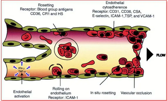
Despite decades of research, cerebral malaria remains one of the most serious complications of Plasmodium infection [17]. The erythrocytes infected by parasite in the microcirculation, especially cerebral circulation have a tendency to accumulate, forming “rosette” (on the surface are present
plasmodium antigens that bond to the endothelial receptors) causing the formation of clots, blocking the bloodstream (Fig. 7).
This process is not a classic thrombosis, because is caused by infected RBCs. Since the brain and the hematic microcirculation cannot be infected, immediately the immune system induces an acute inflammatory response leading to a degeneration of cerebral tissue.
The process involves the release of the cerebral TNF and INF-γ, causing the increase of fever and definitely the death. During the lysis process, there is the release of a substance, known as hemozoin (the heme group is an amphipathic and toxic molecule, able to enter the membranes). In order to detoxify from the heme, the parasite forms the crystals through the specific enzymes. The macrophages recognize the accumulation of heme and phagocyte it. Once phagocytize the heme, they cannot play their role and the consequence is an immunosuppression due to the complete blocking of phagocytosis functions of macrophages. Instead, the erythrocytes are seized in the microcirculation of some organs, as lung, heart, spleen and brain. Once seized, they release more heme causing a big inflammatory state that lead to the patient death.
1.5. Erythrocytes plasmatic membrane
The RBCs plasmatic membrane is constituted for 50% of protein (mainly intrinsic), 40% of lipids and 10% of carbohydrate. The erythrocyte is different from the other cells since the cytoskeleton forms a shell to support the plasmatic membrane. 1.5.1. Red blood cells plasmatic membrane structure Protein Band 3 (Anion Exchange 1) is the main protein (25%) in the red blood cells membrane, involved in different cell process. Its molecular weight is 95 KDa and it can exist in monomeric, dimeric, tetrameric form or aggregates [18]. In the red blood cells, the Band 3 mediates the anionic exchange between the bicarbonate ion (HCO3 -) presents in cytoplasm and ion chloride (Cl-) presents in the plasma (Fig. 8).
The bicarbonate ion is involved in the following reaction catalysed from the carbonic anhydrase.
CO2 +H2O ! H2CO3 ! H+ + HCO3 –
The presence of bicarbonate ion activates the Band 3 that through an anion exchange allows the diffusion of chloride ion. The reaction among a proton and anion chloride leads to the formation of hydrochloric acid that modify the pH to lower values (acidic). The presence of acidic cytoplasm, decrease the affinity between the haemoglobin and the oxygen in order to regulate the transfer of it to the tissues. O2 Fig. 8. Role of Band 3 in RBCs anion exchange
The protein Band 3 (Fig. 9) is composed of a transmembrane domain and two cytoplasmic domains [19]. Each protein domain has precise and different cellular functions:
- The transmembrane hydrophobic domain is a region of 52KDa, includes the aminoacidic residues 360-911, dipped in the bilayer phospholipidic forming a series of 12-14 folding. This domain lead to the formation of an anionic channel that allows the Exchange of Cl - and HCO3 – among the external and internal of cell.
- The N-terminal hydrophilic domain (cytoplasmic domain), of 43KDa, includes the aminoacidic residues 1-359, known as cytosolic domain of Band 3 (cdb3). It penetrates in the cytosol playing a role of anchorage to the cytoskeleton and for some protein as haemoglobin and aldolase.
- The C-terminal hydrophilic domain includes the last 33 aminoacidic residues directed towards the cytoplasm.
At the N-terminal domain can bind different kind of proteins including the ankyrin and Band 4.1 and 4.2 that characterize the erythrocyte shape.
Fig. 9. Model of human erythrocyte band 3. The protein contains 2 structurally and functionally distinct domains: a cytoplasmic binding domain (amino acids 1-359) and a
The cytoskeleton is constituted from the Ankyrin complex and Actin junctional complex (Fig.10). The cytoskeletal proteins are situated in the internal surface of erythrocyte membrane and it forms a fibrillar skeleton with the function of holding the red blood cell structure (Fig.11).
The Ankyrin protein has the role to bind the Band 3 with high molecular weight protein as the α and β spectrins, belonging to the erythrocytes membrane [20]. Fig. 10. Ankyrin and Actin junctional complex representation Fig. 11. Organization of the skeleton and the location of RBCs proteins in mycroscpy.
These proteins create few anchorage points between the β subunit of spectrin and the integral proteins of membrane as Band 3 and glycophorin.
The spectrin dimers associate in tetramers, forming with other proteins as actin, adducin, 4.9 and 4.1 protein and tropomyosin, a fibrillar net (Fig.12) that contribute to stabilize the erythrocytes structure. Furthermore in the junctional complex are present three more proteins as adducin, P55 and dematin [21,22]. 1.6. The spleen tyrosine Kinase (Syk) Spleen tyrosine kinase (Syk, EC 2.7.10.2) is a cytosolic non-receptor tyrosine kinase (Fig.13) that performs its function downstream of antigen receptors in immune cells such as mast cells, B-lymphocytes, or macrophages. Syk is a crucial signal transducer of activated immunoreceptors to multiple events depending on the cell types including proliferation, differentiation, and phagocytosis [23-26]. Inhibiting this protein is possible to avoid all cascade of events causing the onset of many disorders. Furthermore Syk is one of the principal proteins involved in oxidative stresses processes. The therapeutic intervention on mechanisms in which Syk is involved might provide an attractive target for autoimmune or inflammation diseases [27].
Syk belongs to the same subfamily as the closely related Zeta-chain-associated protein kinase 70 (ZAP-70). The Spleen tyrosine kinase (72KDa) is composed of two Src homology tandem domains defined N-SH2 and C-SH2; these are important for the activity regulation and to localize this kinase in cell membrane (Fig.14).
The protein consisting of:
- a tandem SH2 module (N-SH2 and C-SH2), that presents an α-helix which has an important role in the interaction protein-protein and serves as a docking platform for immune receptor tyrosine-based activating motifs (ITAMs) which are displayed on the cytosolic side of the plasma membrane
Fig. 13. 3D beta-sheet structure of Spleen tyrosine kinase (Syk) (PDB ID: 4FL2). Represented with yellow is the N-SH2 domain; cyan the interdomain linker SH2; orange the C-SH2 domain; grey the interdomain linker; magenta and water green the catalytic domain or SH1 and with red the activation loop.
- an interdomain linker SH2, between N-SH2 and C-SH2 constituted of 50 amino acids; it represents the most conserved region in family kinase with 65% of homology sequence. - an interdomain linker of 80-100 amino acids located between C-SH2 and catalytic domain and it is important to regulate the kinase activity and for the presence of phosphotyrosine residues.
- a catalytic domain or SH1 constituted of 300 amino acids follows the interdomain linker. It contains the binding sites for ATP and two autophosphorylation sites (Tyr525 and Tyr526). Syk protein ends with a C-terminal tail with unidentified function yet.
The comparison of inactive and phosphorylated Syk structures reveals significant movement of the tandem SH2 domains region that could disrupt the interaction with the kinase domain characteristic of the inactive state. These data with that reported by different authors permitted the proposal of a model for the regulation of Syk kinase. Syk is maintained in auto-inhibited conformation by the interaction involving the tandem and kinase domains. Fig. 14. Graphical abstract showing Syk protein (A) and its closed and open conformation (B), [28].
Recent studies have previously observed that the erythrocytes possess a mechanism finalized to the expulsion of denatured haemoglobin requiring the activation of Syk kinase [29]. Syk kinase could play a role in the process of asexual P. falciparum growth [30,31] as malaria parasite exerts oxidative stress in erythrocytes causing the denaturation of haemoglobin, with formation of aggregates known as haemichromes in which the Fe2+ has been oxidized to Fe3+. The haemichromes binding to band 3 cause its oxidation and its subsequent phosphorylation by Syk kinase especially in two aminoacidic residues of Band 3 (Tyr 8 and 21). The Anion exchange 1 (AE1) is the more abundant protein of the red cell membrane constituting the major linkage between the lipid bilayer and the cytoskeleton. Under steady conditions, this linkage confers to the membrane the required elasticity and mechanical stability. The oxidation of band 3 and its phosphorylation by Syk protein, cause its detachment from the cytoskeleton and the destabilization of the membrane with the release of microvesicles containing band 3 (Fig.15), membrane and haemichromes. [32-36].
Fig. 15. Graphical abstract of the mechanism, which leads to the release of microparticles
1.7 Treatments for Malaria
1.7.1. Artemisinin and Artemisinin derivatives
Artemisinin (ART) and its semi-synthetic derivatives are a group of drugs used against Plasmodium falciparum
malaria [37]. Treatments containing an artemisinin derivative actually represent the standard therapies worldwide for P. falciparum. Artemisinin is isolated from the plant Artemisia annua, (Fig.16) sweet wormwood, an herb employed in Chinese traditional medicine.
Chemically, artemisinin is a sesquiterpene lactone containing an unusual peroxide bridge (Fig.17). This peroxide is believed to be responsible for the drug's mechanism of action. Few other natural compounds with such a
peroxide bridge are known [38].
Artemisinin and its endoperoxide derivatives have been used for the treatment of P. falciparum related infections but low bioavailability, poor pharmacokinetic properties and high cost of the drugs are a major drawback of their use.
Use of the drug by itself as a monotherapy is explicitly discouraged by the World Health Organization, which declared that malarial parasites are developing resistance to the drug [39].
Therapies that combine artemisinin or its derivatives with some other antimalarial drug are the preferred treatment for malaria and are both effective and well tolerated in patients. Fig. 16. Artemisia Annua Fig. 17. Structure of Artemisinin (ART)
1.7.1.1. The artemisinins mechanism of action
The mechanism of action of artemisinins is not known, but the most widely accepted theory is that they are first activated through cleavage after reacting with heme and iron (II) oxide, which results in the generation of free radicals that in turn damage susceptible proteins, resulting in the death of the parasite [40,41]. In 2016 artemisinin was shown to bind to a large number of targets suggesting that it acts in a promiscuous manner [42]. 1.7.1.2. Artemisinin derivatives
Because the physical properties of artemisinin itself, such as poor bioavailability, limit its effectiveness, semisynthetic derivatives of artemisinin have been developed. These include:
• Artesunate (AS), (water-soluble: for oral, rectal, intramuscular, or
intravenous use) • Artemether (ATH), (lipid-soluble: for oral, rectal or intramuscular use) • Dihydroartemisinin (DHA). • Artelinic acid • Artemotil Dihydroartemisinin (Fig.18) in particular (also known as dihydroqinghaosu, artenimol or DHA) is the active metabolite of
all artemisinin compounds (artemisinin, artesunate, artemether, etc.) and is also available as a drug in itself. It is a semi-synthetic derivative of artemisinin and is widely used as an intermediate in the preparation of other artemisinin-derived antimalarial drugs [43]. It is sold
commercially in combination with piperaquine (PPQ) and has been shown to be equivalent to artemether/lumefantrine (LMF) [44].
The proposed mechanism of action of artemisinin involves cleavage of endoperoxide bridges by iron, producing free radicals (hypervalent iron-oxo species, epoxides, aldehydes, and dicarbonyl compounds), which damage
biological macromolecules causing oxidative stress in the cells of the parasite [45]. Malaria is caused by apicomplexans, primarily Plasmodium falciparum, which largely reside in red blood cells and itself contains iron-rich heme-groups (in the form of hemozoin) [46]. In 2015 artemisinin was shown to bind to a large number targets suggesting that it acts in a promiscuous manner. Recent studies discovered that artemisinin targets a broad spectrum of proteins in the human cancer cell proteome through heme-activated radical alkylation [47].
1.8. Artemisinin-based combination therapies (ACTs)
The artemisinin-combination therapies (ACTs) have been integrated to the recent success of global malaria control, and protecting their efficacy for the treatment of malaria is a global health priority. The World Health Organization (WHO) recommends ACTs for the treatment of uncomplicated malaria caused by P. falciparum.
The main advantage of ACTs is that the artemisinin quickly reduces most of the malaria parasites and the partner drug clears the remaining ones. However, the efficacy of ACTs is threatened by the emergence of both artemisinin and partner drug resistance. Partial resistance to artemisinin causes delayed parasite clearance following treatment with an ACT. Such resistance does not usually lead to treatment failure; however, if the artemisinin component is less effective, the partner drug has to clear a greater parasite mass, jeopardizing the future efficacy of the partner drug.
In addition, partner drug resistance can arise independently of artemisinin resistance. Given that an effective partner drug is essential for clearing all remaining parasites, partner drug resistance carries a high risk of treatment failure. Because of their different roles, the efficacy of the artemisinin and the partner drug must be monitored concomitantly but separately.
1.9. Drug resistance development Resistance and treatment failures to antimalarial medicines can be defined as follows: • Antimalarial resistance is defined as the ability of a parasite strain to survive and/or multiply despite the administration and absorption of a drug given in doses equal to or higher than those usually recommended but within tolerance of the subject; • Artemisinin partial resistance is defined as delayed parasite clearance following treatment with an artesunate monotherapy or with an ACT – this represents partial resistance; • Multidrug resistance (MDR) is resistance to more than 2 antimalarial compounds of different chemical classes. This term usually refers to P. falciparum resistance to chloroquine, sulfadoxine-pyrimethamine, and a third antimalarial compound;
• Treatment failure is the inability to clear parasites from a patient’s
blood or to prevent their recrudescence after the administration of an antimalarial. Many factors can contribute to treatment failure, including incorrect dosage, poor patient compliance, poor drug quality, and drug interactions and resistance. Most of these factors are addressed by therapeutic efficacy studies.
In reporting the findings of therapeutic efficacy studies, the term "ACT resistance" is imprecise. ACT treatment failure (defined as treatment failure following treatment with an ACT, regardless of the presence of artemisinin partial resistance) is a more appropriate term that notes the specific ACT and the nature of the resistance if confirmed (i.e. artemisinin partial resistance or partner drug resistance, or both). The problem of antimalarial drug resistance is compounded by cross resistance, in which resistance to one drug confers resistance to other drugs that belong to the same chemical family or which have similar modes of action.
1.9.1. State of partial artemisinin resistance around the world
Artemisinin partial resistance likely emerged prior to 2001, and prior to the widespread deployment of ACTs in the GMS. To date, it has been confirmed in 5 countries of the GMS: Cambodia, the Lao People’s Democratic Republic, Myanmar, Thailand and Viet Nam.
In late 2013, researchers identified a new molecular marker: mutations in the Kelch 13 (K13) propeller domain were shown to be associated with delayed parasite clearance in vitro and in vivo [48]. The molecular marker allows for a more precise mapping and monitoring of the geographical distribution of resistance. It could also be a mechanism for retrospective mapping of resistance in a large number of settings.
Parasites carrying mutations in the K13 propeller domain have been reported in all 5 GMS countries listed above as well as in Guyana, where studies are ongoing to evaluate impact of this mutation on delayed clearance and ACT efficacy and its potential spread within and outside South America.
Molecular studies have shown that partial artemisinin resistance has emerged independently in several locations in the GMS and spread within the subregion. The K13 mutation identified in South America has also emerged independently. Artemisinin partial resistance has not been confirmed in Africa. Partial artemisinin resistance has occurred as a consequence of several factors: poor treatment practices, inadequate patient adherence to prescribed antimalarial regimens, and the widespread availability of oral artemisinin-based monotherapies and substandard forms of the drug.
1.9.2. Current state of ACT failures
Artemisinin resistance alone rarely leads to treatment failure. However, resistance of malaria parasites to ACT partner drugs can lead to treatment failure (regardless of the presence of artemisinin partial resistance). As a consequence, WHO in the 2017 Malaria report showed the maps of several ACTs that are failing (Artesunate-Amodiaquine) in the African regions (Fig.19) and Greater Mekong area (Artemether-Lumefantrine), (Fig.20).
The geographic scope of the problem could widen quickly and have important public health consequences: the spread or independent emergence of partner
drug resistance or multidrug resistance globally could pose a public health threat, as no alternative antimalarial medicine is available at present with the same level of efficacy and tolerability as ACTs [49].
The efficacy of WHO-recommended ACTs is assessed through therapeutic efficacy studies (TES). Such studies at regular intervals at the same sites allow for the early detection of declines in drug efficacy, providing evidence for guiding national malaria treatment policies.
Taking in consideration the resistance increasing, the monotherapy with artemisinin and ART derivatives is strongly not recommended and for this
Fig. 20. Map indicating the % of patients with ACT treatment failure (Artemether- Lumenfantrine)
2017-2018
Fig. 19. Map indicating the % of patients with ACT treatment failure (Artesunate-Amodiaquine)
reason, the ACT is substituted from the standard therapy. The development and research of new antimalarial drugs is necessary to avoid the increasing of parasite resistance. A promising approach in the antimalarial therapy consists in utilization of Syk kinase inhibitors with the purpose to block the expulsion of denatured haemoglobin and its accumulation inside the parasitized erythrocytes [50] and not allow the growth and proliferation of plasmodium. Moreover, an innovative method as molecular modelling appears an interesting way to better understand and explain the biological mechanism and discover some molecules able to inhibit the protein target.
1.10. Computational studies to explain the biological phenomenon
In the last years, several researches about the in vitro experiments on biological mechanism supplemented of computational sciences have led to the development of different molecular modelling approaches. Actually is possible to determine several molecules conformation, to evaluate the free energy and compounds energy of binding and calculate the molecular descriptors (lipophilicity, molecular electrostatic potential, molecule volume, accessible surface to the solvent and molecular orbitals). Moreover, different software has been developed in order to simulate the ligand-protein interaction, protein-protein with or without solvent aimed to a rational drugs design. These techniques, in a multidisciplinary approach, represent the interface among chemistry, biology and computational science with the purpose to study and deepen the interaction between the proteins and the ligand with low molecular weight and the different mechanism of action.
Many studies demonstrated that the molecular modelling can be a powerful methodology for biomedical research, useful for analysing the three dimensional structure of biological macromolecules with the aim to better understand and explain the cell processes as well as for drug discovery in pharmaceutical science and research in bionanotechnology [51-53].
The main approaches used in molecular modelling for studying the biological systems and the molecule interacting with the human protein, regard:
- Conformational analysis is necessary to determine the protein and ligands conformations with the lowest energy and protein flexibility,
namely the more stable and with a better interaction in the biological environment. For this reason, have been used techniques of Molecular Mechanics (MM) that are involved in most of Computational Structure-Based Drug Discovery (CSBDD) projects [54].
The conformational research has been carried out using the compound of interest seeking the minimum conformational (the different disposition of that structure in the space) depending from the number of different simple bond that are present in the molecule. This phase is even known as “minimization”. The following step consist on the computation of the best energy (the lowest value) using the Quantum Mechanics (QM), which is the science that explains the behaviour of matter and its interactions with energy on the scale of atoms and subatomic particles. This method allows the computation of bond length, binding angles and charge density with short time for small molecules and longer time with more atoms in compounds [55].
- Virtual screening (VS) is a computational technique used in drug discovery to search libraries of small molecules in order to identify those structures, which are most likely to bind to a drug target, typically a protein receptor or enzyme [56-58]. Virtual screening has been defined as the "automatically evaluating very large libraries of compounds" using computer programs. As this definition suggests, VS has largely been a numbers game focusing on how the enormous chemical space of over 1060 conceivable compounds can be filtered to a manageable number that can be synthesized, purchased, and tested [59]. Although searching the entire chemical universe may be a theoretically interesting problem, more practical VS scenarios focus on designing and optimizing targeted combinatorial libraries and enriching libraries of available compounds from in-house compound repositories or vendor offerings. As the accuracy of the method has increased, virtual screening has become an integral part of the drug discovery process [60].
Virtual Screening can be used to select in house database compounds for screening, choose compounds that can be purchased externally, and to choose which compound should be synthesized next.
Ligand-Based Virtual Screening (LBVS)
Given a set of structurally diverse ligands that binds to a receptor, a model of the receptor can be built by exploiting the collective information contained in such set of ligands. These are known as pharmacophore models. A candidate ligand can then be compared to the pharmacophore model to determine whether it is compatible with it and therefore likely to bind [61]. A popular approach to ligand-based virtual screening is based on searching molecules with shape similar to that of known actives, as such molecules will fit the target's binding site and hence will be likely to bind the target. Structure-Based Virtual Screening (SBVS)
Structure-based virtual screening involves docking of candidate ligands into a protein target followed by applying a scoring function to estimate the likelihood that the ligand will bind to the protein with high affinity [62-64]. Web servers oriented to prospective virtual screening are available to everyone [65,66].
Structure-based VS for drug discovery (SBDD) is becoming an essential tool in assisting fast and cost-efficient lead discovery and optimization. The application of rational, structure-based drug design is proven to be more efficient than the traditional way of drug discovery since it aims to understand the molecular basis of a disease and utilizes the knowledge of the three-dimensional structure of the biological target in the process. - Docking is a detailed study of protein-ligand or protein-protein interaction
aimed to explain their chemical behaviour and biological activity or give more information about unknown mechanism of action. The disadvantage of this method consists in the fact it cannot take in consideration the concentration of compounds such as in the in vitro studies. For this reason, the in silico obtained results may not be related to in vitro data. The protein is blocked in a position derivate from the X-ray crystal structure while the
ligand is examined to see how it interacts with the protein once is established the binding site. The obtained results are analysed in basis on how many times a single conformation of compound interacts with the macromolecule in a precise site with a lowest value of Energy. In this phase, the molecule can freely move in the protein changing its conformation to better adapt to binding site of protein but is not possible changing the bonds length. For this reason is important to carry out the quantum mechanics calculation in order to give more information about the specific bonds length [67].
- Molecular dynamics (MD) is a computational method for studying the physical movements of atoms and molecules over time in which the simulation is implemented from nanoseconds (ns) up to a maximum of seconds (s) depending from the facilities used for the computation. The interaction of atoms and molecules is allowed for a fixed period of time, giving a view of the dynamic evolution of the system. In the most common version, the trajectories of atoms and molecules are determined by numerically solving Newton's equations of motion for a system of interacting particles, where forces between the particles and their potential energies are often calculated using interatomic potentials with molecular mechanics force fields. Using the molecular dynamic we observe as the molecules move into the protein site of interaction surrounded by thousands of water molecules in order to simulate the biological environment [68].
Actually, the molecular modelling represents one of the fastest growing fields in science. It may vary from building and visualizing simple molecules in three dimensions (3D) to performing complex computer simulations on large proteins and nanostructures. Molecular modelling is a collection of computer-based techniques for driving, representing and manipulating the structures and reactions of molecules, and those properties that are dependent on these 3D structures. The techniques in silico cover several issues among them computational chemistry, drug design, computational biology, nanostructures, and material science [69].
The so far carried out studies for the development of new in silico drugs and the laboratory tests have been focused on different molecules that can be used as possible drugs for the protein complex inhibition needed for the growth and proliferation of malaria parasite [70-72].
Computational studies conducted on protein kinases (Pks) of Plasmodium falciparum are of particular interest since they catalyse a reaction of phosphorylation to control the parasite growth and differentiation throughout its life cycle.
In 2011 started a new project known as GO (Global Online) Fight Against Malaria belonging at the IBM (International Business Machines) World Community Grid. At this project participated the Art Olson Lab of TSRI (The Scripps Research Institute) in San Diego, CA, USA. This laboratory is one of the leaders in this sector and its effort consists to discover promising drug candidates that could be developed into new drugs that cure drug resistant forms of malaria. The computing power of World Community Grid has been used to perform computer simulations of the interactions between millions of chemical compounds and certain target proteins, to predict their ability to eliminate malaria. The best compounds were tested and further developed into possible treatments for the disease.
Following these ideas, we developed and took in consideration some chemical compound. The host target Drug resistance arguably constitutes the biggest problem faced in the field of infectious diseases today and is a major obstacle to the development of effective strategies to combat infection. Pathogens, particularly those with an intracellular habitat, exploit and subvert various host factors for survival and growth in an otherwise hostile environment. As such, one possible way to circumvent the emergence of a pathogen’s resistance is to develop drugs that target non-essential host factors hijacked by the pathogen rather than the pathogens own molecules. Indeed, host proteins are generally well conserved, when compared with the genetic variability of many pathogens [74].
1.11. Syk inhibitors as treatment for malaria
The human genome contains more than 500 PKs that are implicated in nearly all the signalling pathways. The attractiveness of PKs as possible drugs targets is enhanced by the fact that they are enzymes whose biological activity can be turned off by drugs that block their catalytic site. Indeed, although PKs are encoded by less than 2% of the human genome, they represent more than 20% of the druggable genome.
In a number of diseases, specific inhibitors of individual PKs have proved beneficial to cure tumours or other pathologies, and have entered clinical practice or advanced clinical trials [75].
In recent years, a great number of ATP-competitive kinase inhibitors have been isolated from the natural source or synthesized. These compounds are small molecules that interact with the ATP-binding site with high affinity blocking the phosphorylation process of the protein (Fig.21).
A characteristic and drawback of the more active compounds is the lack of selectivity towards the ATP-binding site present in different proteins and especially in kinase enzymes: this reduces or makes pointless the possibility of their clinical application. Despite exists a high sequence similarity in the kinases aminoacidic sequence, several research groups have been able to develop highly potent and selective Syk ATP-competitive inhibitors. Fig. 21. The biological activity of Syk inhibitors in infected erythrocytes.
1.11.1. Kinase inhibitors mainly used in therapy
Imatinib: (4-[(4-Methyl-1-piperazinyl)methyl]-N-[4-methyl-3-[[4-(3-pyridin-yl)-2-pyrimidinyl]amino]-phenyl]benzamide (Fig.22). It is known as Gleevec in United States and Glivec in Europe, in commerce from pharmaceutical company Novartis. It is drug non-specific for Syk protein (IC50 of 5µM); it is a well-tolerated tyrosine kinase inhibitor that is FDA-approved for use in children, prevents parasite-induced tyrosine phosphorylation of band 3 and terminates P. falciparum parasitemia in vitro by blocking parasite egress at clinically relevant concentrations [76]. This Drug is also already used for different cancer as Chronic Myeloid Leukaemia (CML), Acute Lymphoblastic Leukaemia (ALL) and gastrointestinal stromal tumour (GIST). R406(tamatinib):6-(5-fluoro-2-(3,4,5-trimethoxyphenylamino) pyrimidin-4-ylamino)-2,2-dimethyl-2H-pyrido[3,2-b][1,4]oxazin-3(4H)-one (Fig.23). It is an active metabolite of prodrug R788 (fostamatinib) specific, ATP-competitive inhibitor of spleen tyrosine kinase (Syk), which plays a key role in the signalling of activating Fc receptors and the B-cell receptor. It presents an IC50 of 41 nM [77] and it is already used in clinical trial for rheumatoid arthritis [78], autoimmune thrombocytopenia [79], autoimmune haemolytic anaemia, IgA nephropathy [80] and Lymphoma [81]. M.W. = 493.6 Fig. 22. Structure of Imatinib (Gleevec) M.W. = 470.4 Fig. 23. Structure of R406 (tamatinib)
P505-15:4-((3-(2H-1,2,3-triazol-2-yl)phenyl)amino)-2-(((1R,2S)-2-amino-cyclohexyl)amino)pyrimidine-5-carboxamide-hydrochloride (Fig.24). It is a novel, highly selective Syk inhibitor with IC50 of 1 nM in cell-free assays, >80-fold selective for Syk than Fgr, Lyn, FAK, Pyk2 and Zap70. Candidate drug already used in vivo studies in mices for rheumatoid arthritis and used for the treatment of non-Hodgkin lymphoma (NHL) and chronic Lymphocytic Leukaemia (CLL) [82,83]. Syk-inhibitor-II:2-(2-Aminoethylamino)-4-(3-trifluoromethylanilino)-pyrimi-dine-5-carboxamide (Fig.25). It is a cell-permeable compound that acts as a potent, selective, reversible, and ATP-competitive inhibitor of Syk with IC50 41 nM already used for inhibition of serotonin (5-HT) release in rat basophilic leukaemia (RBL) cells and to treat allergic diseases [84,85]. Fig. 24. Structure of P505-15 M.W. = 429.9 Fig. 25. Structure of Syk inhibitor II M.W. = 340.3
Another Syk inhibitor has been taken in consideration but with less importance considering the low inhibition efficacy in vitro tests, although the IC50 concentration of enzymatic assay reported in literature shows a highly potent compound. Syk-inhibitor-IV:2-(7-(3,4-Dimethoxyphenyl)-imidazo[1,2-c]pyrimidin-5-ylamino)-nicotinamide (Fig.26). Cell permeable imidazopyrimidine compound that acts a potent, ATP-competitive, reversible, and highly selective inhibitor of Syk tyrosine kinase activity (IC50 = 10 nM) with no inhibitory effect against Btk, Fyn, Itk, Lyn, and Src even at concentrations as high as 4.7 µM. This compound showed to inhibit Syk-mediated cellular functions in vitro [86] and exhibit good oral bioavailability and in vivo efficacy in the treatment of various allergy and asthma conditions in rat models [87].
Fig. 26. Structure of Syk inhibitor IV M.W. = 390.4
2. Aim of the project
Recently, a new phosphorylative pathway involved in the release of denatured haemoglobin products (haemichromes), bound to erythrocyte membranes through the cytoplasmic domain of the AE1 (band 3 protein), has been characterized. Tyr phosphorylation of the band 3 protein carried out mainly by the erythrocytic Syk kinase mediates this process. Phosphorylated band 3 becomes uncoupled from the cytoskeleton, leading to the formation of membrane clusters containing haemichromes – band 3 aggregates that can either be released from red blood cells as microvesicles or picked by spleen macrophages by a mechanism known as erythrocyte pitting. These mechanisms may be responsible for the elimination of noxious aggregates of denatured haemoglobin; indeed, during their 120-day lifespan, erythrocytes undergo dramatic changes, eliminating approximately 30% of the initial haemoglobin content of the membrane surface. Moreover, it has been observed that those modifications are an essential prerequisite for parasite egression from the host cell constituting the theoretical background of the observed anti-plasmodial activity of Syk inhibitors. Due to the mechanism describe above, in the present thesis the main purpose consists in studying and understanding the effects of different Syk inhibitors on critical events during parasite growth and maturation within the human erythrocyte.
In particular, the central concept was to leverage in vitro and computational tools to allow efficient therapy using Syk inhibitors and targeting the host red blood cell instead of parasite, in order to interfere with the growth and reinfection capability of parasite. Taking in consideration that Syk inhibitors block the phosphorylation events, is necessary to have more knowledge on the mechanisms responsible of the protein-ligand recognition and binding, in order to facilitate the design, development and discovery of new promising class of antimalarial drugs. A detailed analysis of Syk /ligand interactions is, therefore the central focus in order to understanding the biological process at the molecular level, also obtaining useful information on the pharmacophore structure of Syk inhibitors. Finally, this research has the purpose to prevent as much as possible the drug resistance by Plasmodium, improving and modifying the compounds structure or screen new molecules with a specific
diversity set selected from database to obtain the highest efficacy in infected RBCs. In vitro experimental methods are mainly focused on the evaluation of proteomic profile of Tyr phosphorylation related to biological tests on Plasmodium falciparum cultures, whereas in silico methods were based on different molecular modelling approaches as the molecular descriptors study, docking, virtual screening (VS) and molecular dynamics (MD).
The inhibition of Syk kinase by small molecules, competing with ATP-binding site, represents a certain approach already approved in the last years for the successful treatment of allergic and antibody-mediated autoimmune diseases, depending on Syk kinase activity, as well as for the treatment of lymphoma and leukaemia. A very interesting way to develop new drugs consists in the use of Quantum Mechanics (QM) method and molecular descriptors as electronic density, molecular electrostatic potential (MEP) and the charge distribution in order to characterize the ligands.
3. MATERIALS & METHODS
In this work, we performed a series of in vitro experiments using infected RBCs in order to investigate the biological activity of different Syk inhibitors on P. falciparum cultures and evaluate their IC50 concentrations. We measured their activity at different drug concentrations and on different parasite stages. Moreover, in proteomic studies have been quantified the levels of Tyr phosphorylation in oxidized RBCs using Diamide (sulfhydryl reagent which oxidizes sulfhydryl groups to the disulphide form). These analyses have been followed by the molecular modelling approach based on the identification of amino acidic interaction and the energy of binding in catalytic site of Syk protein through the molecular docking and molecular dynamic studies. The chemical characteristics of compounds have been analysed using the molecular descriptors. Furthermore, accurate studies on the human kinases crystal structures available on public database RCSB PDB allowed us to gain more information about the ligands and the protein target. Virtual screening was carried out in order to find new compounds having the same pattern of interaction in the catalytic site of Syk, useful to be tested in vitro.
3.1. Experimental in vitro studies
Experiments were performed using P. falciparum (Palo Alto strain) cultures (37°C and CO2 5%) in Growth Medium (RPMI, 10% H. plasma, 20mM HEPES pH 7.4, 2% SAGM (Sodium Chloride, Adenine, Glucose, Mannitol) pH 7.4, 10mM Glucose, 40mg/ml Gentamicin). Both haematocrit and parasitemia were at 2%. Before each experiment, cultures have been synchronized at rings stage with 5% Sorbitol. Parasitized red blood cells with 500 μl of Growth Medium contained in each well (24 multiwell plate) were treated with Syk inhibitor II, R406 (Tamatinib), Gleevec (Imatinib) and P505-15 (PRT062607) at concentration (0.2, 0.8, 1, 2, 4, 8, 10 μM) for 24h and 48h. The parasitemia was evaluated by optic microscopy after smears preparation. The smears have been stained using a specific protocol containing, methanol to fix the erythrocytes samples, eosin for the cytoplasm and thiazine in phosphate to stain the parasites. The IC50 of each compound has been calculated using ICEstimator 1.2 software [88].
3.1.1. ICEstimator 1.2 software
ICEstimator carries out a nonlinear regression on the relative effect - concentration points. The relative effects in % are used; they are derived from the raw effects entered by the user. The raw effect(s) for null concentration are used to derive the 100% relative parasite effect. The raw effect(s) for the maximal concentrations are used to derive the 0% relative parasite effect. The resulting curve depicts an inhibitory sigmoid that shows a decrease in the relative effect of the parasite from 100% at the null concentration towards zero when the drug concentration increases to infinity. ICEstimator applies the following model: RE: relative effect of the parasite in %. It defines the Y-axis. C: concentration of the tested drug. It defines the X-axis. IC50: drug concentration inhibiting 50% of parasite’s activity. IC90: drug concentration inhibiting 90% of parasite’s activity. IC99: drug concentration inhibiting 99% of parasite’s activity. γ: sigmoidicity factor, which expresses the steepness of the curve. 3.1.2. Treatment of Red Blood Cells. Venous blood was drawn from healthy volunteers following informed consent and pelleted at 1000 g for 10 minutes at room temperature. After removal of the buffy coat, RBCs were again pelleted and washed 3 times with Phosphate Buffered Saline (127 mM NaCl, 2.7 mM KCl, 8.1 mM Na2HPO4, 1.5 mM KH2PO4, 20 mM HEPES, 1 mM MgCl2, and pH 7.4) in 5 mM glucose (PBS glucose) to obtain packed cells. RBCs were suspended at 30% haematocrit in PBS glucose and it were pre-treated in different experiments with Syk inhibitor II, R406, Gleevec and P505-15 at concentration (0.2, 0.8, 1, 2, 4, 8, 10 μM) for 1 hour at 37°C in the dark, and then treated with oxidant diamide at 2 mM concentration for 45 minutes. For all protocols described, untreated controls and treated controls with only diamide 2 mM were processed identically. To prevent
100 x C
γC
2+ IC
50γfurther phosphorylation of band 3, after incubation we washed the cells with cold buffer and membranes were immediately prepared. 3.1.3. RBCs Membrane Preparation. Membrane proteins were prepared at 4°C on ice and for each sample, 150 μL of packed RBCs was diluted into 1.5 mL of cold haemolysis buffer (HB) (5 mM disodium phosphate, 1 mM EDTA, pH 8) containing a protease and a phosphatase inhibitor cocktail. Thereafter they have been washed up to 4 more times in the same buffer (until membranes became white) in a refrigerated eppendorf microfuge at 25000 g. The samples were stored frozen at −20°C until use. Membrane protein content was quantified using the CD Protein Assay (Bio-Rad).
3.1.4. SDS-PAGE Preparation
To perform one-dimensional electrophoresis (Sodium Dodecyl Sulphate - PolyAcrylamide Gel Electrophoresis), membrane proteins were solubilised in Laemmli Buffer [89] in a volume ratio of 1: 1. 30 µg of proteins for anti-phosphotyrosine; then samples were placed in thermomixer at 1400 rpm, 28°C for 30 minutes. After that, the samples were put at 95°C for 5 minutes and then were separated on 8% polyacrylamide gel under reducing and no-reducing conditions. The electrophoretic run was performed using the Bio-Rad mini-protean 3 setup.
3.1.5. Western blot analyses.
Proteins separated by SDS-PAGE were transferred to nitrocellulose membranes as previously described using Trans-blot turbo Bio-Rad and then probed with antibody anti-phosphotyrosine (sc7020, Santa Cruz, CA). This is produced in mouse from Santa Cruz, CA, and it is diluted to 1 : 2000. Secondary antibodies conjugated to infrared fluorescent dyes excitable at 680 nm or 800 nm (IRDye: anti-mouse 800 CW 926-32210, Li-COR, USA) were then used to visualize the desired antigens with an high performance laser scanner (Odyssey, Licor, USA). The quantitative analyses of tyrosine phosphorylation levels were carried out and were measured analysing western blot images by