Invited critical review
Cardiac biomarker testing in the clinical laboratory: Where do we stand?
General overview of the methodology with special emphasis on
natriuretic peptides
Aldo Clerico
⁎
, Claudio Passino, Maria Franzini, Michele Emdin
Scuola Superiore Sant'Anna and Fondazione G. Monasterio CNR— Regione Toscana, Pisa, Italy
a b s t r a c t
a r t i c l e i n f o
Article history: Received 6 May 2014
Received in revised form 28 May 2014 Accepted 4 June 2014
Available online xxxx Keywords: Natriuretic peptides Cardiac endocrine function Troponins
Immunoassay methods Heart failure Cardiovascular risk
Diagnosis of heart failure (HF) is not based on a single test, but on a combination of history, physical examination and appropriate investigations. For these reasons, the accuracy of diagnosis by clinical means alone is often inad-equate, especially in the early, asymptomatic stages of the HF. Thus, there is an increasing interest in the devel-opment of new cardiovascular biomarkers and, consequently, a great number of laboratory tests have recently been proposed for their assay. The aim of this article is to provide a general overview on the biomarkers, recom-mended by international guidelines, for the diagnosis, risk stratification, and follow-up of patients with HF. Car-diac natriuretic peptides and in particular the B-type related peptides, which are considered to be thefirst line biomarker for HF by international guidelines, will be discussed with special emphasis.
© 2014 Elsevier B.V. All rights reserved.
Contents
1. Introduction . . . 0
2. The clinical relevance of biochemical biomarkers in heart failure . . . 0
3. The multi-markers (MM) approach to cardiovascular risk evaluation . . . 0
4. Cardiac biomarker testing in the clinical laboratory: where we stand . . . 0
5. Standardization or harmonization . . . 0
6. The B-type cardiac natriuretic peptide system . . . 0
7. What B-type-related peptide should we measure and why? . . . 0
References . . . 0
1. Introduction
Estimates on the prevalence of symptomatic heart failure (HF) in the general European and North American population range from 0.4% to 2%[1–5]; with age, HF incidence and prevalence increase steeply, approaching 1 in 1000 among people over the age of 65
[1–5]. From an economic point of view, compared to other diagnoses and treatments, HF is the primary expenditure in Medicare in the US
[4], and in healthcare setting across European countries[1–3].
Despite the remarkable advances made during the past 50 years in understanding and treating the disease[6,7], HF continues to have a poor prognosis: approximately up to 40% of patients diagnosed with se-vere heart failure (NYHA class III–IV or ACC/AHA stage D) in the European and North American population die within one year, with survival rates similar to those of colon cancer, and worse than those of breast or prostate cancer[1–5].
About 20 years ago, Braunwald and Bristow[8]suggested the in-triguing hypothesis that it may be possible to reverse the process of HF, which had long been considered to be irreversible and amenable only to palliative therapy. According to this hypothesis, the intrinsic de-fects in myocardial contraction featured by some patients with chronic HF could be partially reversed by connecting the patient to a ventricular
Clinica Chimica Acta xxx (2014) xxx–xxx
⁎ Corresponding author at: Dept. of Laboratory Medicine, Fondazione G. Monasterio CNR— Regione Toscana, Via Giuseppe Moruzzi 1, 56124 Pisa, Italy. Tel.: +39 0585 493569; fax: +39 0585 493652.
http://dx.doi.org/10.1016/j.cca.2014.06.003
0009-8981/© 2014 Elsevier B.V. All rights reserved.
Contents lists available atScienceDirect
Clinica Chimica Acta
assist device for several months[9]and/or using an appropriate pharmacological treatment[8]. In particular, it is now well docu-mented that patients with chronic HF, treated withβ-adrenergic blocking agents, added to background therapy with ACE inhibitors, improve the systolic function and may reverse cardiac remodeling, leading a better clinical outcomes, including prolonged survival and reduced hospitalizations[1–5]. Thus, the view of chronic HF as an irre-versible, end-stage process is being replaced by the concept that intrin-sic defects of function and structure afflicting the chronically failing heart can be addressed through appropriate therapy[6]. From a theo-retical point of view, we can indeed assume that it is easier to arrest– or even reverse– a progressive process such as HF if action is taken in the earliest phase of the cardiac alteration.
In order to emphasize both the development and progression of the disease, the ACC/AHA guidelines for the diagnosis and management of chronic HF in the adult recommend a classification of HF based on 4 stages from A to D (Fig. 1)[4]. Thefirst two stages (A and B) do not in-clude symptomatic patients in an attempt to underscore to healthcare providers the importance of an early identification of patients who are at risk for developing HF. In particular, patients in stage A have only risk factors without structural or functional alterations of ventricular myocardial, while those in stage B show cardiac structural (such as hypertrophy) and/or functional (such as impaired left ventricular
dysfunction) alterations. The last two stages C and D identify instead symptomatic patients. Since early identification of individuals and risk stratification and diagnosis can be achieved today through the measure-ment of specific disease or risk markers, an increasing number of new cardiovascular biomarkers have been proposed, as previously reviewed in detail[9–17].
The aim of this review article is to provide a general outline on the methodology of the biomarkers recommended by international guide-lines for the diagnosis, risk stratification, and follow-up of patients with HF, with special emphasis on natriuretic peptides, which are considered to be the most useful biomarker for HF.
2. The clinical relevance of biochemical biomarkers in heart failure HF is defined as a syndrome, resulting from any structural or func-tional cardiac disorder that impairs the ability of the heart to function as a pump to support a physiological circulation[1–5]. The diagnosis of HF is not based on one single test[1,2]. Positive history and some physical signs (such as orthopnea, rales, third heart sound or jugular vein distension) share a good diagnostic specificity, but also a poor sensitivity in diagnosing acute congestive HF (Table 1)[16,17]. There-fore, the diagnosis of both acute and chronic HF relies on clinical judg-ment based on a combination of history, physical examination and
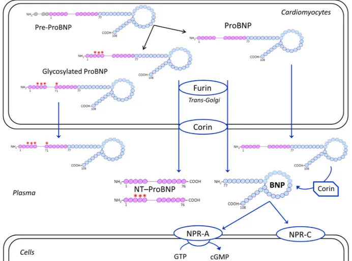
Fig. 1. Schematic representation of biosynthesis, secretion and distribution of B-type related natriuretic peptides. Human BNP is synthesized as a 134-amino acid (aa) precursor protein (pre-proBNP), including a signal peptide of 26 amino acids (grey), and is subsequently processed to form a 108-aa pro-peptide, named proBNP. The proBNP can be enzymatically cleaved by pro-protein convertases produced in the cardiomyocytes, such as corin and furin, mainly located in the trans-Golgi network and on the plasma membrane, respectively[116]. ProBNP is thus processed to form the 76-aa N-terminal peptide (NT-proBNP, violet) and the biologically active 32-aa C-terminal peptide (BNP, light blue), which are both secreted into plasma. Some of the proBNP is O-glycosylated within the Golgi apparatus. Proteolytic cleavage occurs either on or not O-glycosylated proBNP. But, if O-glycans bind to the threonine at position 71 the proBNP will not be processed by furin and corin, thus glycosylated proBNP will be secreted into circulation. Finally, also not glycosylated proBNP can be released as unprocessed peptide. However, the latter can be cleaved into NT-proBNP and BNP by plasmatic corin[117–120]. Only BNP1–32, which is the active hormone, is able to bind the specific receptors, A and
NPR-C. NPR-A is a guanylate cyclase-coupled receptor, which mediates the biological effects of cardiac natriuretic peptides. NPR-C, not coupled to a guanylate cyclase, has essentially a clearance function for all natriuretic peptides.
appropriate investigations, as recommended by all international guide-lines[1–5,18,19].
According to the most recent international guidelines[18,19], na-triuretic peptides, and in particular the peptides related to the B-type cardiac peptide hormone (such as BNP and NT-proBNP), are recom-mended as thefirst line biomarker for the diagnosis of both acute and chronic HF. The measurement of BNP and NT-proBNP is useful in supporting clinical judgment for the diagnosis or exclusion of HF in the setting of chronic ambulatory HF or acute decompensated HF (with the maximum degree of class of recommendation I and level of evidence A)[19]. The value of natriuretic peptide testing is partic-ularly significant when the etiology of dyspnea is unclear. Indeed, all international guidelines, starting from thefirst years of this century, state that lower values of BNP or NT-proBNP actually exclude the presence of HF and higher values have reasonably high positive pre-dictive value to diagnose HF[1–4,18,19].
From a pathophysiological point of view, it is important to underline that the cardiac endocrine function is an essential component of the integrated systems of the body and, thus, plays a pivotal role influid, electrolyte and hemodynamic homeostasis[20,21]. A continuous and intense information exchangeflows from the endocrine heart system to nervous and immunological systems and to other organs, including the kidney, endocrine glands, liver, adipose tissue, immunocompetent cells, and vice versa. This close link between cardiac natriuretic peptide sys-tem and counter-regulatory syssys-tems could explain the increase in circu-lating levels of BNP/NT-proBNP in some noncardiac-related clinical conditions[20,21](Table 2).
As far as the follow-up of HF patients is concerned, BNP and NT-proBNP levels mirror the effectiveness of the treatment of acute or
chronic HF, with lowering of levels over time associated with better clinical outcomes[22,23]. Even if most trials examining the strategy of biomarker“guided” HF management were small and underpowered, at present time, 3 comprehensive meta-analyses concluded that BNP-guided therapy reduces all-cause mortality and cardiovascular hospital-ization in patients with HF compared with usual clinical care, especially in patientsb75 years of age with co-morbidities[19,21,22,24–27].
As far as the stratification of cardiovascular risk in HF patients is concerned, measurement of natriuretic peptides is recommended by the most recent guidelines with the maximum degree of evidence (class I and level A) also for the prognosis in HF patient[19]. Several studies have demonstrated that increased circulating levels of cardiac troponin I (cTnI) and T (cTnT) – especially using high-sensitivity methods[28,29]– are found in patients with HF, who often do not present obvious myocardial ischemia or underlying coronary artery dis-ease[30–39]. Thesefindings[30–39]suggest that increased cTnI and cTnT in these patients could be caused by cardiomyocyte injury or ne-crosis. In chronic or acute decompensated HF, elevated cardiac troponin levels are associated with worse clinical outcomes and mortality. In-deed, HF patients, showing a significant and lasting decrease in troponin levels after appropriate pharmacological treatment have a better prog-nosis compared to those did not show any or only transient decrease
[31,38]. Based on these results[30–39], the latest guidelines recom-mend that troponin I or T be routinely measured, in addition to natri-uretic peptides, in patients presenting with acutely decompensated HF for evaluating risk stratification, with the maximum degree of evi-dence (class I and level A)[19].
In addition to natriuretic peptides and troponins, a huge number of other biomarkers have been suggested for the prognostic value in HF; for example, those related to pro-inflammatory mechanisms, ox-idative stress, cachexia, neuro-hormonal dysfunction, and myocardial remodeling, as previously reviewed in detail[9–17]. As an example, a list of some of these suggested biochemical biomarkers is reported in
Table 3. In particular, several studies have suggested that biomarkers of myocardialfibrosis, such as galectin-3[40–53]and soluble ST2
[54–64], are predictive of hospitalization and death in patients with HF. Accordingly[40–64], the most recent guidelines[19]also suggest the use of biomarkers of myocardialfibrosis for additive risk stratifica-tion, although with a lower degree of evidence compared to natriuretic peptides and troponins, in both ambulatory (class IIb, level B) and acute (class IIb, level A) HF patients. InTable 4, we summarized
Table 1
Accuracy of history and physicalfindings in diagnosing acute congestive HF (modified from references[16,17]).
Variable Sensitivity Specificity Accuracy
History of HF 62 94 80
Dyspnea 56 53 54
Orthopnea 47 88 72
Rales 56 80 70
Third heart sound 20 90 66
Jugular vein distension 39 94 72
Edema 67 68 68
Table 2
Selected causes of elevated natriuretic peptide concentrations (according to references
[19–21]). Cardiac
• Heart failure, including right ventricular syndromes • Acute coronary syndrome
• Heart muscle disease, including LVH • Valvular heart disease
• Pericardial disease • Atrial fibrillation • Myocarditis • Cardiac surgery • Cardioversion Noncardiac • Advancing age • Anemia • Renal failure • Liver disease
• Pulmonary: obstructive sleep apnea, severe pneumonia, pulmonary hypertension • Endocrine diseases (such as hyperthyroidism and primitive or secondary
hyperaldosteronism)
• Chronic inflammatory diseases (such as amyloidosis) • Critical illness
• Bacterial sepsis • Severe burns
• Toxic-metabolic insults, including cancer chemotherapy and envenomation
Table 3
Suggested prognostic biomarkers for HF. Neuro-hormones
Natriuretic peptides (ANP, BNP, CNP and related peptides) Thyroid hormones Renin–angiotensin–aldosterone system Catecholamines Endothelins Adrenomedullin Urocortin Leptin Adiponectin
Cardiac damage biomarkers Cardiac troponins, cTnI and cTnT Remodeling and inflammatory biomarkers Matrix metalloproteinases (MMPs) Adhesion molecules (ICAM, VICAM, selectin-P) C-Reactive Protein (CRP)
Cytokines and related receptor family (IL-2, IL-6, IL-8, TNF-α, ST2) Pentraxin 3
Galectin 3
Oxidative stress molecular biomarkers Gamma-glumatyltransferases (GGT)
Reactive oxygen species (ROS), Plasma oxidized LDL Cachexia biomarkers
Alterations of hypophyseal–suprarenal axis IGF-1 and GH
some pathophysiological and methodological characteristics of bio-markers, which are currently available and recommended by the most recent international guidelines for HF according to evidence based medicine principles[19].
3. The multi-markers (MM) approach to cardiovascular risk evaluation
Based on the knowledge acquired so far, it is likely that HF thera-py in the future will rely on strategies that combine multiple bio-markers[9,13,16]. This methodological known as the multi-marker (MM) approach or global risk model, is today considered the best model for risk prediction in the individual patient with cardiovascu-lar disease[13,65–67]. Unfortunately, the setup of an adequate MM model is currently complicated by some theoretical and methodo-logical difficulties[68]. According to the MM approach, each bio-marker should contribute independently to the diagnostic and prognostic accuracy in a multiple regression model, and ultimately lead to a better outcome for the patient.
In 2010, an expert panel from the American Heart Association established the criteria for the evaluation of novel markers of cardio-vascular risk[69]. The panel stated that an adequate evaluation of a novel risk marker should require (i) a sound research design, (ii) a representative at-risk population, and (iii) an adequate number of outcome events. Studies of a novel marker should report the degree to which it adds to the prognostic information provided by standard risk markers. Because no single statistical measure can provide all the information needed to assess a novel marker, studies should also report on measures of both discrimination and accuracy. Fur-thermore, the clinical value of a marker should be assessed by its ef-fect on patient management and outcomes.
In general, a novel risk marker should be evaluated in several phases according to Evidence-Based Laboratory Medicine principles[70], including initial proof of concept, prospective validation in indepen-dent populations, documentation of incremental information when added to standard risk markers, assessment of effects on patient management and outcomes, and ultimately, cost-effectiveness. Bio-markers that do not change the management of a disease are proba-bly unable to significantly affect patient outcome and are thus very seldom cost-effective (judged in terms of quality-adjusted life-years gained)[9,13,71]. Randomized trials are the gold standard for establishing the effectiveness of biomarker-guided strategies[72]. Unfortunately, there are few examples of such trials in cardiology, particularly in the primary prevention setting[72]. Indeed, the lack of well-designed randomized clinical trials explains the relatively low degree of evidence (i.e., class IIa, level B) assigned to the BNP-guided therapy in patients with chronic HF even by the most recent guidelines[19]. However, some pivotal randomized clinical trials on BNP-guided therapy are now in progress[27,73]. The results of these studies will hopefully spread more light on the real usefulness
of this strategy in HF patients, pushing the adoption of BNP-guided therapy in the management of HF patients.
4. Cardiac biomarker testing in the clinical laboratory: where we stand
Cardiovascular biochemical biomarkers are usually measured by means of non-competitive immunometric assay methods, using a combination of two (or more) antisera or monoclonal antibodies specific for separate epitopes of tested biomarkers[9,74]. However, the set-up of a reliable and robust measurement for cardiovascular biomarkers actually presents a very difficult challenger for the ex-perts in laboratory medicine. The desirable characteristics for an “ideal” circulating cardiovascular biomarker, measured by laboratory test, are reported inTable 5.
Cardiovascular biomarkers usually are peptides or proteins (includ-ing natriuretic peptides, cardiac troponins, galectin-3 and ST2), which are present in tissues and bodyfluids at very low concentrations in healthy subjects (i.e., in the range of ng/L). As a result, immunoassay methods with very high analytical sensitivity (i.e., low detection limit, LoD, of about 1 pg/tube or even lower) are necessary to measure with an acceptable analytical imprecision the circulating levels of some car-diac biomarkers (such as troponins and natriuretic peptides) in healthy subjects, especially in pediatric age[28,29,75–78].
Furthermore, some peptide biomarkers (such as BNP) usually share a family of related peptides in vivo and in vitro[21,79–81], while some protein biomarkers (such as troponins) exhibit considerable chemical and structural heterogeneity in blood of both healthy subjects of HF pa-tients[82–84]. These heterogeneous peptides and proteins can differ-ently cross-react with the antibodies used in immunoassay systems, affecting the accuracy of the measurement. As a result, it is not surpris-ing that there are large systematic differences between the circulatsurpris-ing
Table 4
Pathophysiological and methodological characteristics of biomarkers, which are currently available and recommended by international guidelines[19]. Biomarker type Pathophysiological relevance in HF Clinical relevance in HF
(Level of evidence)
Analytical methods
ANP and BNP and their related peptides Cardiac endocrine response to changes in hemodynamics, and to neuro-hormonal and immune systems disturbances
Diagnosis of exclusion of HF (IA) Prognosis of HF (IA)
Risk stratification (IA) Follow-up of HF patients (IIA)
Several validated immunoassay methods using automated platforms
Cardiac troponins I and T Specific marker of injury and damage of cardiomyocytes
Additive risk stratification (IA) Several validated immunoassay methods using automated platforms
Galectin−3 Noncardiac specific biomarker related to myocardial remodeling andfibrosis
Additive risk stratification (IIB) Some immunoassay methods using automated platforms
sST2 Noncardiac specific biomarker related to myocardial remodeling andfibrosis
Additive risk stratification (IIB) ELISA method
Table 5
Desirable features of an ideal circulating cardiovascular biomarker measured by a laboratory test.
− Laboratory test acceptable to patient − Stability in vivo and in vitro of the biomarker
− Adequate analytical sensitivity (functional sensitivity) of laboratory test − Good degree in reproducibility and accuracy of laboratory
− Easy to perform
− Complete automation of assay
− International standardization of the laboratory test − Low cost
− Low biological variation of the biomarker − Cardiac specificity
− Circulating levels of biomarker closely related to the pathophysiological mecha-nisms of cardiac disease
− Circulating levels of the biomarker closely related to the therapeutic interventions − Reference range and cut off values tested for gender, age, and ethnicity
dependence
− Diagnostic and prognostic accuracy tested by randomized clinical trials − Cost–benefit ratio favorable tested by randomized clinical trials
levels of biomarkers measured by immunoassay methods for both cTnI
[83–85]and BNP[79,86–91].
For example, Wu et al.[92]showed since 1998 that commercial immunoassays generated different results for a given protein con-centration due to multiple complex forms of cTnI. According to Apple[93], these data actually demonstrated that it was not possible to compare absolute concentrations obtained with assays from dif-ferent manufacturers. Commercially available cTnI assays use vari-ous standard materials and antibodies with different epitope specificities[83–85]. Hence, troponin I assays may yield results that are unique to a certain method or instrument to the point that values for a same patient sample may differ depending on the assay and platform used [84,93]. Due to the heterogeneity between cTnI methods and without an adequate standardization, reference values and decision limits should be determined separately for each meth-od and not be extrapolated from other assays. In terms of clinical practice, this situation is obviously confusing, especially when pa-tients are referred to different laboratories that use different cTnI methods. Starting 2001 a study group began on the behalf of some international organizations (such as AACC and IFCC) a process for standardization of cTnI immunoassay methods in order to establish a reference measurement procedure and materials[84,95].
5. Standardization or harmonization
Standardization of peptide and protein immunoassays, such as cTnI methods, is a very complicated task[95]. A complete standardization approach needs an accepted reference measurement procedures (RPM) and reference materials for the TnI, which however at present are still not available. Indeed, the term standardization can be used only when comparable results among measurement procedures are based on calibration traceability to SI unit using a RMP[83,84,94–96].
After over 10 years of efforts, some Authors think that cTnI assays is unlikely to become standardized[93]. Fred Apple suggests laboratorians and clinicians not get“bogged down” with cTnI standardization, en-couraging to aim efforts toward developing a clear understanding of the clinical and analytical evidence for cTnI immunassays and“to be happy” that the technological improvements that have led to the precise detection of low cTnI concentrations also will lead to better patient care[93].
Although we completely agree with Apple that the standardization of some critical immunoassay methods truly appears to be a“mission impossible”[97], we do however believe that achieving better har-monization (i.e., a reduction of heterogeneity) among the results provided by different methods is possible. In particular, we hypothesize that a better understanding of biochemical characteristics and the path-ophysiological role of a candidate biomarker may promote a harmoni-zation process indicating to manufacturers and laboratorians specific targets (i.e., epitopes) for the set-up of more accurate immunoassay methods. A good example for a possible harmonization process is repre-sented by immunoassay methods for cardiac B-type-related natriuretic peptide system.
6. The B-type cardiac natriuretic peptide system
The human BNP gene encodes for a pre-proBNP molecule of 134 amino acid residue, including a signal peptide of 26 amino acids. BNP is cleaved out of a prohormone molecule of 108 amino acids, the proBNP1–108(proBNP). According to the“classical” scenario of BNP pro-duction and secretion from cardiomyocytes, before being secreted from cardiomyocytes into the bloodstream, proBNP is split by some proteo-lytic enzymes (such as corin and/or furin) into two peptides: the biolog-ically inactive NH2-terminal peptide fragment proBNP1–76 (NT-proBNP), and the COOH-terminal peptide fragment proBNP77–108[21] (Fig. 1). The latter is a peptide of 32 amino acids (BNP1–32) and is usually indicated as BNP. This is the active hormone, that is the only able to bind
to the specific natriuretic peptide receptors (named NPR-A, B, C, respec-tively)[21].
Some recent studies open a new and more complex scenario regard-ing the pathophysiological and clinical relevance of circulatregard-ing B-type natriuretic peptides[22]. In addition to the peptide hormone BNP and the inactive peptide NT-proBNP, a huge numbers of circulating proBNP-derived fragments can be identified by chromatographic proce-dures in human plasma, including the intact and glycosylated forms of the precursor proBNP[98–116](Fig. 1). Several studies have also dem-onstrated that intact or glycosylated forms of proBNP constitute a signif-icant portion of immunoreactive B-type-related peptides circulating in plasma of patients with heart failure[98–116]. According to these find-ings, it is theoretically conceivable that the active hormone (i.e., BNP) may be produced even in vivo from the circulating intact precursor proBNP through enzymatic cleavage by some plasma proteases (such as corin)[117–119]. Indeed, a recent study using an in vivo rat model demonstrated that processing of human proBNP to active BNP can actu-ally occur in the circulation[120]. The peripheral processing of circulat-ing proBNP could likely be submitted to regulatory mechanisms, which might be impaired in patients with heart failure, opening new perspec-tives in the treatment of heart failure[80,121]. Indeed, a novel pharma-cological target may be the pharmacodynamic action of drugs inducing and/or modulating the maturation of the prohormone into active hor-mone (i.e., BNP)[122].
From a methodological and analytical points of view, the large het-erogeneity of B-type natriuretic peptides circulating in human blood seems to explain the systematic differences among the results provided by immunoassay methods considered specific to the peptide hormone BNP[87,90,91,123,124]. In particular, a recent study, using standard protocols and quality control materials, demonstrated that the IRMA method (by Shionogi's Diagnostic Division, Japan), the ADVIA method for the Centaur platform (by Siemens Health Care Diagnostics) and ST AIA-PACK method for the AIA platform (by TOSOH Corporation, Tokyo, Japan) measured greatly lower (up to the half) BNP values in comparison with other immunoassays, such as the POCT Triage method (by Alere Diagnostics), the BNP Triage Biosite for Access and UniCell DxI platforms (by Beckman Coulter Diagnostics), the MEIA method for the AxSYM platform and the chemiluminescent microparticle immunoas-say for ARCHITECT platform (both by Abbotts Diagnostics)[124]. It is in-teresting to note that the IRMA method by Shionogi, the ADVIA method for Centaur platform, the ST AIA-PACK method (personal communica-tions from TOSOH EUROPE N.V., Tessenderlo, Belgium) use the same an-tibodies and standard materials supplied by Shionogi's Diagnostic Division[18].
According to the study by Luckenbill et al.[79], a great part of these systematic differences between the different BNP immunoassay sys-tems should be due to the cross-reaction with the glycosylated or not glycosylated proBNP. Liang et al.[98]demonstrated that proBNP consti-tutes a substantial portion of immunoreactive BNP measured in plasma of HF patients. More recently, Macheret et al.[108], in study using a spe-cific immunoassay method for proBNP[101], demonstrated that this precursor peptide of BNP was detectable in all subjects studied, and its levels were dependent of gender, age, heart rate, and body mass index. Furthermore, these Authors found that the degree of clinical sen-sitivity and specificity of proBNP assay for the detection of left ventricu-lar dysfunction was comparable to two commercial assays for BNP and NT-proBNP[108]. The results of this study[108]confirm previous re-ports[101–107]suggesting that the intact precursor of biologically ac-tive BNP (i.e., the proBNP) circulates in plasma of both healthy subjects and HF patients.
7. What B-type-related peptide should we measure and why? According to this new scenario regarding the circulating levels of B-type cardiac natriuretic peptides, there are at least 3 different peptides that could be measured in human plasma samples: the active peptide
hormone, BNP, the inactive N-terminal fragment, NT-proBNP, or the pro-hormone peptide, proBNP[21,121]. These 3 peptides have different biochemical characteristics and pathophysiological relevance (Table 6). From an analytical point of view, the inactive peptide NT-proBNP and proBNP, are more stable in vivo and in vitro, with a longer plasma half-life and a lower intra-individual biological variation, than active peptide BNP (Table 4)[18,21,81]. From a pathophysiological point of view, several studies indicate that the inactive peptides, especially proBNP, show an incremental increase of their circulating levels re-lated to progression of HF greater than the active peptide hormone BNP[102,103,110,115,125]. In particular, two studies[115,125], which identified and quantified the individual cardiac natriuretic peptides by means of mass spectrometry, reported that the real levels of the peptide hormone BNP1–32in patients with severe HF are much lower than the BNP concentrations measured by commercially available immunoassay methods. Furthermore, results of another study using mass spectrometry determination actually provide specific evidence for the absence (i.e., concentration below the analytical sensi-tivity of the measurement) of circulating active peptide BNP1–32in advanced-stage HF patients[126].
According to the analytical characteristics and clinical results discussed above, we would assume that the inactive peptide NT-proBNP and NT-proBNP would be a better biomarker for the progression of HF than the active hormone BNP. However, at present, all the com-mercially available immunoassay methods considered specific to ac-tive peptide BNP significantly cross-react with proBNP [79]. In agreement with these data[79], a recent study[127]has found a good correlation between the BNP and proBNP values measured with commercially available immunoassay methods considered spe-cific for these two peptides in patients with severe HF. Moreover, all the international guidelines state that the commercially available BNP and NT-proBNP immunoassays usually give clinically compara-ble results when used for diagnosis, prognosis and follow-up of HF patients[18,21,80,88,89,128].
At present, we must realize that the commercially available im-munoassay methods considered specific for the active form of B-type cardiac natriuretic peptides present an obvious paradox. From a pathophysiological point of view, it would be better to measure the active peptide BNP (instead of inactive peptide NT-proBNP and proBNP) when we are interested in evaluating the“true biologically ac-tive status” of the cardiac endocrine function[21,22]. However, to date, none of the commercially available methods is able to provide such in-formation, accurately, as these methods are greatly affected by inactive peptides the concentrations of which are higher than active hormone in the blood samples of HF patients. In other words, at present time, all the commercially available immunoassay methods for BNP assay are not completely specific for the active form of the peptide.
In conclusion, BNP immunoassay methods show large systematic differences due to the interferences of some inactive peptides, especially the glycosylated and non-glycosylated forms of the precursor peptide, proBNP. The setup of more specific methods for the active peptide BNP1–32could reduce these systematic differences resulting a better harmonization among results.
As a future perspective, a more accurate estimation of both production/secretion of B-type related peptides from cardiomyocytes and overall activity of the cardiac endocrine function could be achieved by the testing plasma samples using simultaneously two methods: one specific for the intact precursor proBNP1–108,and the other for active peptide BNP1–32[80,121]. Information obtained by the con-temporaneous measurement of proBNP and BNP with specific assays may likely extend our present understanding of pathophysiological mechanisms linking together disease progression and cardiac endo-crine dysfunction[80]. However, the clinical usefulness of these new and more specific methods will have to be accurately evaluated by ran-domized clinical trials in comparison with NT-proBNP and proBNP methods according to the evidence-based medicine principles[69–72].
References
[1]NICE (National Institute of Clinical Excellence). Clinical Guideline 5. Chronic heart failure. Management of chronic heart failure in adults in primary and secondary care; July 2003 1–44 [London].
[2]Swedberg K, Cleland J, Dargie H, et al. Guidelines for the diagnosis and treatment of chronic heart failure: executive summary (update 2005): The Task Force for the Di-agnosis and Treatment of Chronic Heart Failure of the European Society of Cardiol-ogy. Eur Heart J 2005;26:1115–40.
[3]Nieminen MS, Böhm M, Cowie MR, et al. Executive summary of the guidelines on the diagnosis and treatment of acute heart failure: the Task Force on Acute Heart Failure of the European Society of Cardiology. Eur Heart J 2005;26:384–416.
[4]Hunt SA, American College of Cardiology, American Heart Association Task Force on Practice Guidelines (Writing Committee to Update the 2001 Guidelines for the Evaluation and Management of Heart Failure). ACC/AHA 2005 guideline update for the diagnosis and management of chronic heart failure in the adult: a re-port of the American College of Cardiology/American Heart Association Task Force on Practice Guidelines (Writing Committee to Update the 2001 Guide-lines for the Evaluation and Management of Heart Failure). J Am Coll Cardiol 2005;46:e1-82.
[5]Adams KF, Lindenfeld J, Arnold JMO, Baker DW, Barnard DH, Baughman KL. Execu-tive summary: HFSA 2006 Comprehensive Heart Failure Practice Guideline. J Card Fail 2006;12:10–38.
[6]Braunwald E, Bristow MR. Congestive heart failure:fifty years of progress. Circula-tion 2000;102(20 Suppl. 4):IV14–23.
[7]Dipla K, Mattiello JA, Jeevanandam V, Houser SR, Margulies KB. Myocyte recovery after mechanical circulatory support in humans with end-stage heart failure. Circu-lation 1998;97:2316–22.
[8]Eichhorn EJ, Bristow MR. Medical therapy can improve the biologic properties of the chronically failing heart: a new era in the treatment of heart failure. Circulation 1996;94:2285–96.
[9]Vittorini S, Clerico A. Cardiovascular biomarkers: increasing impact of laboratory medicine in cardiology practice. Clin Chem Lab Med 2008;46:748–63.
[10]Panteghini M. Role and importance of biochemical markers in clinical cardiology. Eur Heart J 2004;25:1187–96.
[11]Macabasco-O'Connell A, Miller PS. Biomarkers for heart failure. Prog Cardiovasc Nurs 2006;21:215–8.
[12]De Virginy DR. Novel and potential future biomarkers for assessment of the se-verity and prognosis of chronic heart faille: a clinical review. Heart Fail Rev 2006;11:333–4.
[13]Vasan S. Biomarkers of cardiovascular disease: molecular basis and practical con-siderations. Circulation 2006;113:2335–62.
[14]Lainscak M, von Haehling S, Springer J, Anker SD. Biomarkers for chronic heart fail-ure. Heart Fail Monit 2007;5:77–82.
[15]Braunwald E. Biomarkers in heart failure. N Engl J Med 2008;358:2148–59.
[16]Emdin M, Vittorini S, Passino C, Clerico A. Old and new biomarkers of heart failure. Eur J Heart Fail 2009;11:331–5.
[17]Maisel AS, McCullough PA. Cardiac natriuretic peptides: a proteomic window to cardiac function and clinical management. Rev Cardiovasc Med 2003;4(Suppl. 4): S3–S12.
[18]Thygesen K, Mair J, Mueller C, et al. Recommendations for the use of natriuretic peptides in acute cardiac care: a position statement from the Study Group on Bio-markers in Cardiology of the ESC Working Group on Acute Cardiac Care. Eur Heart J 2012;33:2001–6.
[19]Yancy CW, Jessup M, Bozkurt B, et al. 2013 ACCF/AHA guideline for the manage-ment of heart failure: a report of the American College of Cardiology Foundation/ American Heart Association Task Force on Practice Guidelines. J Am Coll Cardiol 2013;62:e147–239.
[20]Clerico A, Recchia FA, Passino C, Emdin M. Cardiac endocrine function is an essen-tial component of the homeostatic regulation network: physiological and clinical implications. Am J Physiol Heart Circ Physiol 2006;290:H17–29.
[21]Clerico A, Giannoni A, Vittorini S, Passino C. Thirty years of the heart as an endo-crine organ: physiological role and clinical utility of cardiac natriuretic hormones. Am J Physiol Heart Circ Physiol 2011;301:H12–20.
[22]Clerico A, Fontana M, Ripoli A, Emdin M. Clinical relevance of BNP measurement in the follow-up of patients with chronic heart failure. Adv Clin Chem 2009;48:163–79.
Table 6
Biochemical and physiological characteristics of BNP, NT-proBNP and proBNP peptides.
BNP NT-proBNP proBNP
Molecular mass 3462 Da 8457 Daa
11900 Daa
Amino acids 32 76 108
Biological function Active hormone Inactive Pro-hormone
Half life 15–20 min N60 min N60 min
Glycosylation Not glycosylated Highly glycosylated in vivo
Highly glycosylated in vivo
a
The molecular mass (MM) of NT-proBNP and proBNP depends to the degree of glyco-sylation of the peptide; in the Table are reported the MM of not glycosylated peptides.
[23]Troughton R, Michael Felker G, Januzzi Jr JL. Natriuretic peptide-guided heart fail-ure management. Eur Heart J 2014;35:16–24.
[24]Felker GM, Hasselblad V, Hernandez AF, O'Connor CM. Biomarker-guided therapy in chronic heart failure: a meta-analysis of randomized controlled trials. Am Heart J 2009;158:422–30.
[25]Porapakkham P, Porapakkham P, Zimmet H, Billah B, Krum H. B-type natriuretic peptide-guided heart failure therapy: a meta-analysis. Arch Intern Med 2010;170:507–14.
[26]Troughton RW, Frampton CM, Brunner-La Rocca HP, et al. Effect of B-type natri-uretic peptide-guided treatment of chronic heart failure on total mortality and hos-pitalization: an individual patient meta-analysis. Eur Heart J 2014;35:1559–67.
[27]Januzzi JL, Troughton R. Are serial BNP measurements useful in heart failure agement? Serial natriuretic peptide measurements are useful in heart failure man-agement. Circulation 2013;127:500–7.
[28]Clerico A, Giannoni A, Prontera T, Giovannini S. High-sensitivity troponin: a new toll for pathophysiological investigation and clinical practice. Adv Clin Chem 2009;49:1–30.
[29]Apple FS, Collinson PO. Analytical characteristics of high-sensitivity cardiac tropo-nin assay. Clin Chem 2012;58:54–61.
[30]Horwich TB, Patel J, MacLellan WR, et al. Cardiac troponin I is associated with im-paired hemodynamics, progressive left ventricular dysfunction, and increased mor-tality rates in advanced heart failure. Circulation 2003;108:833–8.
[31]Sato Y, Yamada T, Taniguchi R, et al. Persistently increased serum concentrations of cardiac troponin t in patients with idiopathic dilated cardiomyopathy are predic-tive of adverse outcomes. Circulation 2001;103:369–74.
[32]Setsuta K, Seino Y, Takahashi N, et al. Clinical significance of elevated levels of cardiac troponin T in patients with chronic heart failure. Am J Cardiol 1999;84:608–11 [A9].
[33]Hudson MP, O'Connor CM, Gattis WA, et al. Implications of elevated cardiac tropo-nin T in ambulatory patients with heart failure: a prospective analysis. Am Heart J 2004;147:546–52.
[34]Fonarow GC, Peacock WF, Horwich TB, et al. Usefulness of B-type natriuretic pep-tide and cardiac troponin levels to predict in-hospital mortality from ADHERE. Am J Cardiol 2008;101:231–7.
[35]Peacock WFIV, De Marco T, Fonarow GC, et al. Cardiac troponin and outcome in acute heart failure. N Engl J Med 2008;358:2117–26.
[36]Ilva T, Lassus J, Siirila-Waris K, et al. Clinical significance of cardiac troponins I and T in acute heart failure. Eur J Heart Fail 2008;10:772–9.
[37]Missov E, Calzolari C, Pau B. Circulating cardiac troponin I in severe congestive heart failure. Circulation 1997;96:2953–8.
[38]Ather S, Hira RS, Shenoy M, et al. Recurrent low-level troponin I elevation is a worse prognostic indicator than occasional injury pattern in patients hospitalized with heart failure. Int J Cardiol 2011;301:H2351–61.
[39]Januzzi Jr JL, Filippatos G, Nieminen M, et al. Troponin elevation in patients with heart failure: on behalf of the third Universal Definition of Myocardial Infarction Global Task Force: heart failure section. Eur Heart J 2012;33:2265–71.
[40]Tang WH, Shrestha K, Shao Z, et al. Usefulness of plasma galectin-3 levels in systolic heart failure to predict renal insufficiency and survival. Am J Cardiol 2011;108:385–90.
[41]de Boer RA, Lok DJ, Jaarsma T, et al. Predictive value of plasma galectin-3 levels in heart failure with reduced and preserved ejection fraction. Ann Med 2011;43:60–8.
[42]Lok DJ, van der Meer P, de la Porte PW, et al. Prognostic value of galectin-3, a novel marker offibrosis, in patients with chronic heart failure: data from the DEAL-HF study. Clin Res Cardiol 2010;99:323–8.
[43]Shah RV, Chen-Tournoux AA, Picard MH, et al. Galectin-3, cardiac structure and function, and long-term mortality in patients with acutely decompensated heart failure. Eur J Heart Fail 2010;12:826–32.
[44]Felker GM, Fiuzat M, Shaw LK, et al. Galectin-3 in ambulatory patients with heart failure: results from the HF-ACTION study. Circ Heart Fail 2012;5:72–8.
[45]Fermann GJ, Lindsell CJ, Storrow AB, et al. Galectin 3 complements BNP in risk strat-ification in acute heart failure. Biomarkers 2012;17:706–13.
[46]Lopez-Andrès N, Rossignol P, Iraqi W, et al. Association of galectin-3 andfibrosis markers with long-term cardiovascular outcomes in patients with heart failure, left ventricular dysfunction, and dyssynchrony: insights from the CARE-HF (Cardi-ac Resynchronization in Heart Failure) trial. Eur J Heart Fail 2012;14:74–81.
[47]Anand IS, Rector TS, Kuskowski M, et al. Baseline and serial measurements of galectin-3 in patients with heart failure: relationship to prognosis and effect of treatment with valsartan in the Val-HeFT. Eur J Heart Fail 2013;15:511–8.
[48]Carrasco-Sánchez FJ, Aramburu-Bodas O, Salamanca-Bautista P, et al. Predictive value of serum galectin-3 levels in patients with acute heart failure with preserved ejection fraction. Int J Cardiol 2013;169:177–82.
[49]Chen K, Jiang RJ, Wang CQ, et al. Predictive value of plasma galectin-3 in patients with chronic heart failure. Eur Rev Med Pharmacol Sci 2013;17:1005–11.
[50]de Boer RA, Edelmann F, Cohen-Solal A, Mamas MA, Maisel A, Pieske B. Galectin-3 in heart failure with preserved ejection fraction. Eur J Heart Fail 2013;15:1095–101.
[51]Lok DJ, Klip IT, Lok SI, et al. Incremental prognostic power of novel biomarkers (growth-differentiation factor-15, high-sensitivity C-reactive protein, galectin-3, and high-sensitivity troponin-T) in patients with advanced chronic heart failure. Am J Cardiol 2013;112:831–7.
[52]Motiwala SR, Szymonifka J, Belcher A, et al. Serial measurement of galectin-3 in pa-tients with chronic heart failure: results from the ProBNP Outpatient Tailored Chronic Heart Failure Therapy (PROTECT) study. Eur J Heart Fail 2013;15:1157–63.
[53]van der Velde AR, Gullestad L, Ueland T, et al. Prognostic value of changes in galectin-3 levels over time in patients with heart failure: data from CORONA and COACH. Circ Heart Fail 2013;6:219–26.
[54]Januzzi Jr JL, Peacock WF, Maisel AS, et al. Measurement of the interleukin family member ST2 in patients with acute dyspnea: results from the PRIDE (Pro-Brain Na-triuretic Peptide Investigation of Dyspnea in the Emergency Department) study. J Am Coll Cardiol 2007;50:607–13.
[55]Mueller T, Dieplinger B, Gegenhuber A, Poelz W, Pacher R, Haltmayer M. Increased plasma concentrations of soluble ST2 are predictive for 1-year mortality in patients with acute destabilized heart failure. Clin Chem 2008;54:752–6.
[56]Rehman SU, Mueller T, Januzzi Jr JL. Characteristics of the novel interleukin family biomarker ST2 in patients with acute heart failure. J Am Coll Cardiol 2008;52:1458–65.
[57]Manzano-Fernandez S, Mueller T, Pascual-Figal D, et al. Usefulness of soluble con-centrations of interleukin family member ST2 as predictor of mortality in patients with acutely decompensated heart failure relative to left ventricular ejection frac-tion. Am J Cardiol 2011;107:259–67.
[58]Aldous SJ, Richards AM, Troughton R, Than M. ST2 has diagnostic and prognostic utility for all-cause mortality and heart failure in patients presenting to the emer-gency department with chest pain. J Card Fail 2012;18:304–10.
[59]Henry-Okafor Q, Collins SP, Jenkins CA, et al. Soluble ST2 as a diagnostic and prognostic marker for acute heart failure syndromes. Open Biomark J 2012;2012:1–8.
[60]Bayes-Genis A, Zamora E, de Antonio M, et al. Soluble ST2 serum concentration and renal function in heart failure. J Card Fail 2013;19:768–75.
[61]Breidthardt T, Balmelli C, Twerenbold R, et al. Heart failure therapy-induced early ST2 changes may offer long-term therapy guidance. J Card Fail 2013;19:821–8.
[62]Chen LQ, de Lemos JA, Das SR, Ayers CR, Rohatgi A. Soluble ST2 is associated with all-cause and cardiovascular mortality in a population-based cohort: the Dallas Heart Study. Clin Chem 2013;59:536–46.
[63]Wang YC, Yu CC, Chiu FC, et al. Soluble ST2 as a biomarker for detecting stable heart failure with a normal ejection fraction in hypertensive patients. J Card Fail 2013;19:163–8.
[64]Gruson D, Lepoutre T, Ahn SA, Rousseau MF. Increased soluble ST2 is a stronger predictor of long-term cardiovascular death than natriuretic peptides in heart fail-ure patients with reduced ejection fraction. Int J Cardiol 2014;172:e250–2.
[65]Kattan MW. Evaluating a new marker's predictive contribution. Clin Cancer Res 2004;10:822–4.
[66]Assmann G, Cullen P, Schulte H. Simple scoring scheme for calculating the risk of acute coronary events based on the 10-year follow-up of the prospective cardio-vascular Munster (PROCAM) study. Circulation 2002;105:310–5.
[67]Conroy RM, Pyorala K, Fitzgerald AP, et al. Estimation of ten-year risk of fatal car-diovascular disease in Europe: the SCORE project. Eur Heart J 2003;24:987–1003.
[68]Hense HW. Observations, predictions and decisions— assessing cardiovascular risk assessment. Int J Epidemiol 2004;33:235–9.
[69]Hlatky MA, Greenland P, Arnett DK, et al. Criteria for evaluation of novel markers of cardiovascular risk: a scientific statement from the American Heart Association. Circulation 2009;119:2408–16.
[70]Price PC, Christensen RH. Evidence-based laboratory medicine: principles, practice, and outcome. 2nd ed. Washington DC: AACC Press; 2007 1–545.
[71]Marshall DA, O'Brien BJ. Economic evaluation of diagnostic tests. In: Price PC, Christensen RH, editors. Evidence-based laboratory medicine— from principles to outcomes. Washington DC: AACC Press; 2003. p. 159–86.
[72]Wang TJ. Assessing the role of circulating, genetic, and imaging biomarkers in car-diovascular risk prediction. Circulation 2011;123:551–65.
[73] Guiding Evidence Based Therapy Using Biomarker Intensified Treatment (GUIDE-IT).http://clinicaltrials.gov/ct2/show/NCT01685840?term=GUIDE+IT&rank=1. [Accessed September 12, 2012].
[74]Clerico A. The increasing impact of laboratory medicine on clinical cardiology. Clin Chem Lab Med 2003;41:871–83.
[75]Clerico A, Zucchelli GC, Pilo A, Passino C, Emdin M. Clinical relevance of biological variation: the lesson of brain natriuretic peptide (BNP) and NT-proBNP assay. Clin Chem Lab Med 2006;44:366–78.
[76]Clerico A, Fortunato A, Ripoli A, Prontera C, Zucchelli GC, Emdin M. Distribution of plasma cardiac troponin I values in healthy subjects: pathophysiological consider-ations. Clin Chem Lab Med 2008;46:804–8.
[77]Cantinotti M, Storti S, Parri MS, Prontera C, Murzi B, Clerico A. Reference intervals for brain natriuretic peptide in healthy newborns and infants measured with an automated immunoassay platform. Clin Chem Lab Med 2010;48:697–700.
[78]Giannoni A, Giovannini S, Clerico A. Measurement of circulating concentrations of cardiac troponin I and T in healthy subjects: a tool for monitoring myocardial tissue renewal? Clin Chem Lab Med 2009;47:1167–77.
[79]Luckenbill KN, Christenson RH, Jaffe AS, et al. Cross-reactivity of BNP, NT-proBNP, and proBNP in commercial BNP and NT-proBNP assays: preliminary observations from the IFCC Committee for Standardization of Markers of Cardiac Damage. Clin Chem 2008;54:619–21.
[80]Clerico A, Vittorini S, Passino C. Measurement of the pro-hormone of brain type na-triuretic peptide (proBNP): methodological considerations and pathophysiological relevance. Clin Chem Lab Med 2011;4:1949–54.
[81]Goetze JP. Biosynthesis of cardiac natriuretic peptides. Results Probl Cell Differ 2010;50:97–120.
[82]Katrukha AG, Bereznikova AV, Filatov VL, et al. Degradation of cardiac troponin I: implication for reliable immunodetection. Clin Chem 1998;44:2433–40.
[83]Panteghini M. Assay-related issues in the measurement of cardiac troponins. Clin Chim Acta 2009;402:88–93.
[84]Tate JR, Bunk DM, Christenson RH, et al. Standardisation of cardiac troponin I mea-surement: past and present. Pathology 2010;42:402–8.
[85]Panteghini M, Pagani F, Yeo KT, et al. Evaluation of imprecision for cardiac troponin assays at low-range concentrations. Clin Chem 2004;50:327–32.
[86]Clerico A, Del Ry S, Giannessi D. Measurement of natriuretic cardiac hormones (ANP, BNP, and related peptides) in clinical practice: the need for a new generation of immunoassay methods. Clin Chem 2000;46:1529–34.
[87]Rawlins ML, Owen WE, Roberts WL. Performance characteristics of four automated natriuretic peptide assays. Am J Clin Pathol 2005;123:439–45.
[88]Apple FS, Panteghini M, Ravkilde J, et al. Quality specifications for B-type natriuretic peptide assays. Clin Chem 2005;51:486–93.
[89]Apple FS, Wu AH, Jaffe AS, et al. National Academy of Clinical Biochemistry and IFCC Committee for Standardization of Markers of Cardiac Damage Laboratory Medicine practice guidelines: analytical issues for biomarkers of heart failure. Circulation 2007;116:e95–8.
[90]Prontera C, Zaninotto M, Giovannini S, et al. Proficiency testing project for brain na-triuretic peptide (BNP) and the N-terminal part of the propeptide of BNP (NT-proBNP) immunoassays: the CardioOrmoCheck study. Clin Chem Lab Med 2009;47:762–8.
[91]Clerico A, Zaninotto M, Prontera C, et al. State of the art of BNP and NT-proBNP im-munoassays: the CardioOrmoCheck study. Clin Chim Acta 2012;414:112–9.
[92]Wu AHB, Feng YJ, Moore R, et al. Characterization of cardiac troponin subunit re-lease into serum after acute myocardial infarction and comparison of assays for tro-ponin T and I. Clin Chem 1998;44:1198–208.
[93]Apple FS. Standardization of cardiac troponin I assays will not occur in my lifetime. Clin Chem 2012;58:169–71.
[94]Christenson RH, Duh SH, Apple FS, et al. Standardization of cardiac troponin I as-says: round robin of ten candidate reference materials. Clin Chem 2001;47:431–7.
[95]Christenson RH, Bunk DM, Schimmel H, Tate JR, IFCC Working Group on Standardization of Troponin I. Point: put simply, standardization of cardiac tropo-nin I is complicated. Clin Chem 2012;58:165–8.
[96]Gantzer ML, Miller WG. Harmonization of measurement procedures: how do we get it done? Clin Biochem Rev 2012;33:95–100.
[97]Iervasi G, Clerico A. Harmonization of free thyroid hormone test: a mission impos-sible? Clin Chem Lab Med 2011;49:43–8.
[98]Liang F, O'Rear J, Schellenberger U, et al. Evidence for functional heterogeneity of circulating B-type natriuretic peptide. J Am Coll Cardiol 2007;49:1071–8.
[99]Goetze JP. Biochemistry of pro-B-type natriuretic peptide-derived peptides: the en-docrine heart revisited. Clin Chem 2004;49:1503–10.
[100]Goetze JP. ProBNP-derived peptides in cardiac disease. Scand J Clin Lab Invest 2004;64:497–510.
[101]Giuliani I, Rieunier F, Larue C, et al. Assay for measurement of intact B-type natri-uretic peptide prohormone in blood. Clin Chem 2006;52:1054–61.
[102]Seferian KR, Tamm NN, Semenov AG, et al. The brain natriuretic peptide (BNP) pre-cursor is the major immunoreactive form of BNP in patients with heart failure. Clin Chem 2007;53:866–73.
[103]Hammerer-Lercher A, Halfinger B, Sarg B, et al. Analysis of circulating forms of proBNP and NT-proBNP in patients with severe heart failure. Clin Chem 2008;54:858–65.
[104]Goetze JP, Rehfeld JF. Peptide hormones and their prohormones as biomarkers. Biomark Med 2009;3:335–8.
[105]Dries DJ, Ky B, Wu A, Rame JE, Putt M, Cappola T. Simultaneous assessment of unprocessed ProBNP 1–108 in addition to processed BNP32 improves risk stratification in ambulatory patients with systolic heart failure. Circ Heart Fail 2010;3:220–7.
[106]Goetze JP, Kastrup J, Rehfeld JF. The paradox of increased natriuretic hormones in congestive heart failure patients: does the endocrine heart also fail in heart failure? Eur Heart J 2003;24:1471–2.
[107]Goetze JP, Kastrup J, Pedersen F, Rehfeld JF. Quantification of pro-B-type natriuretic peptide and its products in human plasma by use of an analysis independent of precursor processing. Clin Chem 2002;48:1035–42.
[108]Macheret F, Boerrigter G, McKie P, et al. Pro-B-type natriuretic peptide 1–108 circu-lates in the general community: plasma determinants and detection of left ventric-ular systolic dysfunction. J Am Coll Cardiol 2011;57:1386–95.
[109]Shimizu H, Masuta K, Aono K, et al. Molecular forms of human brain natriuretic peptide in plasma. Clin Chim Acta 2002;316:129–35.
[110]Shimizu H, Masuta K, Asada H, Sugita K, Sairenji T. Characterization of molecular forms of probrain natriuretic peptide in human plasma. Clin Chim Acta 2003;334:233–9.
[111]Schellenberger U, O'Rear J, Guzzetta A, Jue RA, Protter AA, Pollitt NS. The precursor to B-type natriuretic peptide is an O-linked glycoprotein. Arch Biochem Biophys 2006;451:160–6.
[112]Seferian KR, Tamm NN, Semenov AG, et al. Immunodetection of glycosylated NT-proBNP circulating in human blood. Clin Chem 2008;54:866–73.
[113]Crimmins DL, Kao JL. A glycosylated form of the human cardiac hormone pro B-type natriuretic peptide is an intrinsically unstructured monomeric protein. Arch Biochem Biophys 2008;475:36–41.
[114]Semenov AG, Postnikov AB, Tamm NN, et al. Processing of pro-brain natriuretic peptide is suppressed by O-glycosylation in the region close to the cleavage site. Clin Chem 2009;55:489–98.
[115]Miller WL, Phelps MA, Wood CM, et al. Comparison of mass spectrometry and clin-ical assay measurements of circulating fragments of B-type natriuretic peptide in patients with chronic heart failure. Circ Heart Fail 2011;4:355–60.
[116]Semenov AG, Tamm NN, Seferian KR, et al. Processing of pro-B-type natriuretic peptide: furin and corin as candidate convertases. Clin Chem 2010;56:1166–76.
[117]Jiang J, Wu S, Wang W, et al. Ectodomain shedding and autocleavage of the cardiac membrane protease corin. J Biol Chem 2011;286:10066–72.
[118]Knappe S, Wu F, Masikat MR, Wu Q. Functional analysis of the transmembrane do-main and activation cleavage of human corin: design and characterization of a sol-uble corin. J Biol Chem 2003;278:52363–70.
[119]Dong N, Chen S, Yang J, et al. Plasma soluble corin in patients with heart failure. Circ Heart Fail 2010;3:207–11.
[120]Semenov AG, Seferian KR, Tamm NN, et al. Human pro-B-type natriuretic peptide is processed in the circulation in a rat model. Clin Chem 2011;57:883–90.
[121]Emdin M, Passino C, Clerico A. Natriuretic peptide assays revisited: do we need pro-B-type natriuretic peptide? J Am Coll Cardiol 2011;57:1396–8.
[122]Del Ry S, Cabiati M, Clerico A. Recent advances on natriuretic peptide system: new promising therapeutic targets for the treatment of heart failure. Pharmacol Res 2013;76:190–8.
[123]Clerico A, Prontera C, Emdin M, et al. Analytical performance and diagnostic accu-racy of immunometric assays for the measurement of plasma BNP and NT-proBNP concentrations. Clin Chem 2005;51:445–7.
[124]Franzini M, Masotti S, Prontera C, et al. Systematic differences between BNP immu-noassays: comparison of methods using standard protocols and quality control ma-terials. Clin Chim Acta 2013;424:287–91.
[125]Niederkofler EE, Kiernan UA, O'Rear J, et al. Detection of endogenous B-type natri-uretic peptide at very low concentrations in patients with heart failure. Circ Heart Fail 2008;1:258–64.
[126]Hawkridge AM, Heublein DM, Bergen III HR, Cataliotti A, Burnett Jr JC, Muddiman DC. Quantitative mass spectral evidence for the absence of circulating brain natri-uretic peptide (BNP-32) in severe human heart failure. Proc Natl Acad Sci U S A 2005;102:17442–7.
[127]Miller WL, Grill DE, Jaffe AS. Comparison of novel pro-BNP1–108 and standard BNP assays in heart failure patients. Clin Chim Acta 2012;413:920–6.
[128]Emdin E, Clerico A, Clemenza F, et al. Consensus document. Recommendations for the clinical use of cardiac natriuretic peptides. J Cardiovasc Med (Ital Heart J) 2005;6:430–46.