0
UNIVERSITA’ DEGLI
STUDI DI ROMA
“TOR VERGATA”
FACOLTA’ DI MEDICINA E CHIRURGIA
DOTTORATO DI RICERCA IN
MICROBIOLOGIA MEDICA E IMMUNOLOGIA
XXII CICLO
Activation of Human Endogenous Retrovirus K and
cellular modifications in human melanoma cell lines:
gene expression analysis
Dottoranda: Reem Ali Ahmed Al Dossary
Tutor: Dott.ssa Emanuela Balestrieri
Docente guida: Prof.ssa Paola Sinibaldi Vallebona
Coordinatore: Prof. Enrico Garaci
1
L I S T O F C O N T E N T
ABSTRACT 7
CHAPTER 1: INTRODUCT ION 8
1.1. RETROVURUSES 8
1.1.1. General properties of Retroviruses 8
1.1.2. Structure of retroviruses 8
1.1.3. Genomic composition of retroviruses 11
1.1.4. Replication of retroviruses 12
1.1.5. Classification of Retroviruses 14
1.2. HUMAN ENDOGENOUS RETROVIRUSES (HERVs) 16
1.2.1. General properties of HERVs 16
1.2.2. Classification of HERVs 18
1.3. HERV-K AND DISEASES 21
1.3.1. Pathological role of HERVs 21
1.3.2. Virological etiology of cancer 25
1.4. HERVs AND HUMAN MELANOMA 26
1.4.1. HERV-K particles and human melanoma 26
1.4.2. HERV-K mRNA and protein expression in human melanoma 27
1.4.3. HERV-K and human melanoma: immunological evidences. 27
1.4.4. Biological events in the progression of human melanoma 29
1.4.5. Molecular changes in the progression of human melanoma 30
CHAPTER 2. AI M O F TH E ST UDY 36
CHAPTER 3 . MAT ERIALS AND MET H ODS 39
3.1. MATERIALS 39
3.1.1. Cell cultures 39
3.1.2. Microarray 39
2
3.2. METHODS 40
3.2.1. Cell cultures 40
3.2.2. RNA extraction and quantification 40
3.2.3. Microarray 41
3.2.4. Reverse transcription and Real-time PCR 42
CHAPTER 4. RES ULTS 4 5 4.1. BACKGROUND 45
4.2. GENE EXPRESSION ANALYSIS USING MICROARRAY 47
4.3. REAL TIME PCR 64
CHAPTER 5. DIS CUSSIO N 66
5.1. M IC RO AR RA Y -B ASE D G EN E EX PR ESS IO N AN ALY S IS OF AD HE RE NT T V M -A12 1% 6 6 5.2. M IC RO AR RA Y -B AS ED GE NE EX P RES S IO N A NAL YS IS O F NO N AD HE RENT T V M -A1 2s p A ND CL ON Es p 67
5.3. CO NF IRM AT IO N OF GEN E EX P RES S IO N U S IN G REA L -T IME P CR 68
3
L I S T O F T A B L E S
Table 1.1. Genetic composition of retroviruses 11
Table 1.2. Classification of retroviridae according to ICTV (International committee of taxonomy of viruses 15
Table 1.3. HERV families 19
Table 1.4. HERV transcripts detected in various human tissues 24
Table 3.1. Primer pairs designed for real-time PCR confirmation 43
Table 4.1. Number of modulated genes using microarray technology 49
Table 4.2. Comparison of gene expression of TVM-A12sp versus Clonesp 50
Table 4.3. Up regulated genes in TVM-A12 1% FBS prior to detachment 51
Table 4.4. Down regulated genes in TVM-A12 1% FBS prior to detachment 53
Table 4.5. Up regulated genes in suspended TVM-A12sp 56
Table 4.6. Down regulated genes in suspended TVM-A12sp 56
Table 4.7. Fold change in gene expression in comparison to adherent TVM-A12 obtained using real-time PCR 64
4
LIST OF FIGURES
Figure 1.1. Diagrammatic representation of retrovirus structure 9 Figure 1.2. Morphological forms of retroviruses 10 Figure 1.3. Mechanism of cDNA synthesis and formation of
long terminal repeat (LTR) 13
Figure 1.4. Progression of melanocyte transformation 29 Figure 1.5. Biological; events and molecular changes in the
Progression of melanoma 32 Figure 4.1. The association between peak rise in HERV-K env
mRNA expression and morphological modification 46
Figure 4.2. Number of up regulated and down regulated genes in TVM-A12 1% FBS, TVM-A12sp, and
Clonesp in comparison to TVM-A12 obtained
using microarray 50
5
Acknowledgments
It is my pleasure to thank my dear Prof.ssa. Paola Sinibaldi-Vallebona
for her continuous guidance and her endless kindness and cooperation. I
would also like to thank Prof. C.F. Perno for giving me this opportunity.
Many thanks to Dr. Claudia Matteucci and Dr. Emanuela Balestrieri for
their excellent guidance, throughout the whole project and their valuable
ideas, suggestions and interpretation. To my dear friend Dr. Roberta
Sorrentino for all the help she gave me and to all my Italian friends for
the great time we spent together
6
Dedication
I would like to dedicate this work to my loving parents Ali and Haya, my
dear husband Waleed and to my little angel Lyan.
7
A B S T R A C T
HERV-K has been linked extensively with human melanoma supported by the oncogenic ability of other human and animal retroviruses and by the detection of HERV-K particles and transcripts in human melanoma. In an attempt to approach better understanding of human melanoma pathogenesis, a cellular system has been developed at the University of Rome, Tor Vergata, that consist of original adherent melanoma cell line, named TVM-A12, and two non-adherent cell line obtained either by cloning, named Clonesp, or by stressful cultural condition
i.e. 1% FBS instead of 10%, named TVM-A12sp. It was observed that transition of melanoma
cell line TVM-A12 from adherent to non adherent phenotype under stressful cultural condition was accompanied by decrease expression of melanocyte differentiating antigen (Melan A/MART-1) and HLA class I and higher colony forming ability in soft agar assay. Furthermore, this transition was associated with increased expression of HERV-K env mRNA and protein and could be inhibited by down regulation of HERV-K expression using RNA interference. Therefore, the purpose of this study was to provide further characterization for better understanding of the genetic bases of increased malignant behaviour and HERV-K activation. Methods used include microarray technology followed by real-time PCR confirmation. Results of microarray test showed modulation in gene expression that favours increased malignant potential in non-adherent cells compared to parental cells. In addition it showed very similar gene expression profile in Clonesp and TVM-A12sp. Real-time PCR
confirmation showed transient up regulation of transcription factor BHLHB2 and MYC prior to detachment. TVM-A12 grown in stressful cultural conditions started to show up regulation of PTEN, VEFGA, CSK, PITCH1, FOXG1A, and TP53 which continue to rise in non adherent cells and in Clonesp. Once detached, they showed up regulation of WNT3, MYCN,
MYCL1, BTK, CCND2, WNT2, TIMP3, IRF3, GTF2I, CTNNB1, E2F1, ARHGAP5, ARHGEF5, GPR39, and ITGB4. On the other hand, adherent TVM-A12 1%, TVM-A12sp and
Clonesp showed down regulation of ANXA7, CTNNA1, NME1, RRM1, CDKN1A, XRCC6,
HDAC, TRAM1, CD59 and TOB1 in comparison to parental TVM-A12 cells. This study describes, for the first time, a unique cellular system that demonstrate the association between peak rise in HERV-K env expression and acquisition of a more malignant phenotype thus providing a useful tools for better understanding of human melanoma pathogenesis and for studying the effect of pharmacological agents and genetic modulators on HERV-K expression and melanoma progression.
8
C H A P T E R 1 . I N T R O D U C T I O N
1.1 RETROVIRUSES
1.1.1. General properties of Retroviruses
Retroviridae is a big and rapidly growing virus family, characterized by two main
features: the ability to convert genetic information from genomic viral RNA into DNA and to integrate into the host genome the DNA copy of viral RNA. In fact, genomic RNA is converted into a complementary copy (cDNA) in a process called reverse transcription, using the enzyme reverse transcriptase (RT), and the DNA copy of viral RNA is integrated in the host genome, using the enzyme integrase.
The ability of the virus to be integrated within the host genome and to replicate as a cellular gene, using cellular machinery, is a key factor for its persistence and evasion from the immune mechanisms.
1.1.2. Structure of retroviruses
Retroviruses are composed of an outer envelope made of lipid bilayer, derived from the membrane of infected cells, in which virally encoded glycoproteins were embedded. The number of these glycoproteins varies in different retroviruses and among different isolates of the same virus but the significance of this variation is not clearly understood. Viral glycoproteins play a significant role in the initial stage of infection through cellular receptor recognition and therefore in the determination of host range and of target cells and tissues within infected host. Matrix proteins separate viral envelope from viral capsid. Capsid is made of proteins and it encloses viral RNA in associated with nucleoproteins. Viral capsid also contains the viral enzymes RT, protease and integrase (Wagner et al. 2004) (Figure1.1).
9
Figure 1.1. Diagrammatic representation of retrovirus structure.
CA=capsid, NC=nucleocapsid, IN=integrase, RT=reverse transcriptase, TM= transmembrane, SU= surface component of envelope protein, MA= matrix protein, and PR= protease.
The morphology of the viral core is an important feature used in classifying retroviruses in to four main morphological forms: type A, B, C, and D particles (Goff 2001) (Figure 1.2).
Type A particles represent the immature form of the virus, which is mainly seen in mutant viruses with defective proteolysis of viral protein precursors and in infectious viruses, early in infection, prior to budding and maturation. They have doughnut like appearance due to the electron lucent center, surrounded by one or two concentric rings. Mature viruses on the other hand displays one of three morphological forms: type B particle has an eccentrically placed rounded core, type C particle has a centrally located rounded or slightly angular core and type D particle has a characteristic cylindrical or bar shaped core. Some retroviruses display morphology not belonging to any of these morphological forms like HIV-1 (Human Immunodeficiency Virus type one), which has a cone shaped core.
10 Figure1.2. Morphological forms of retroviruses.
(A) immature intracisternal type A particle, (B) type B particle with eccentric core seen in mouse mammary tumor virus, (C) and (D) type C particle with central core seen in murine leukemia virus and avian leucosis virus, (E) type D particle of mason Pfizer monkey virus, (F) type C particles seen in bovine leukemia virus, (G) rod or cone shaped core in bovine immunodeficiency virus, (H) spumavirus (Goff 2001).
11
1.1.3. Genomic composition of retroviruses
Retroviruses are RNA viruses composed of diploid genome of two copies of positive sense non-segmented single stranded RNA, carries 7-13 kilo bases and joined at 5` end by a self complementary region called dimmer linkage structure. The significance of having two copies is not clearly understood but it is thought to act as a biological buffer that compensate for the high rate of error of reverse transcriptase (Wagner et al. 2004). Retrovirus genome is composed of the following genes and functional elements:
R - U5 - PBS - gag - pol - env – PPT - U3 - R - An
Description of each gene and gene segment is shown in Table 1.1.
Table 1.1. Genome composition of retroviruses (Wagner et al. 2004).
Genes and
functional elements
Description
R 20-250 bases sequence repeated at both ends
U5 75-200 bases unique sequence at the 5` end with transcription signal and is involved in viral integration
PBS Primer binding site, which is the site for binding of cellular tRNA for reverse transcription
Gag Encodes for capsid, matrix and nucleoprotein
Pol Encode the enzymes reverse transcriptase and integrase
Env Encodes the surface and transmembrane component of envelope protein
PPT Polypurine tract which is a short stretch of A and G nucleotides that is resistant to degradation and is used as primer during cDNA synthesis
U3 Unique sequence at the 3` end involved in gene expression
12
Replication cycle of retroviruses start with receptor recognition on target cell followed by receptor mediated membrane fusion. Within the cytoplasm, partial uncoating of the virus occurs and reverse transcription of the viral genomic RNA start within the partially uncoated virus. Replication of the virus genome involves a step of reverse transcription to synthesize negative sense complementary DNA, followed by the synthesis of positive sense complementary DNA and the formation of long terminal repeat (LTR) made of U3, R, and U5 at both ends as shown in Figure 1.3. Following that, cDNA migrates into the nucleus by different mechanisms depending on retrovirus type: oncornaviruses require nuclear membrane breakdown, which occurs during cell division, while lentiviruses enter even in a non-dividing cell. Within the nucleus, cDNA integrate into the cellular genome, using the enzyme integrase, forming a provirus. Full length viral RNA can be produced from the provirus, by cellular machinery. Newly produced viral RNA migrates to the cytoplasm, where it can be either spliced to generate mRNA encoding various virus proteins or encapsidated to generate a new virus particle. Viral envelope proteins are incorporated in the cell membrane and immature viral particle buds through the plasma membrane taking an outer envelope of cellular lipid bilayer, in which virally encoded proteins are incorporated. After budding, maturation of the virion occurs by cleavage of precursor proteins (Wagner et al. 2004; Goff 2001).
13
Figure 1.3. Mechanism of cDNA synthesis and formation of LTR (long terminal repeat) PBS= primer binding site, R= repeat sequence, PPT= Polypurine tract (Wagner et al. 2004).
14
1.1.5. Classification of Retroviruses
Different classification systems have been used to describe retroviruses. The simplest one classifies retroviruses according to genome composition: simple retroviruses have genome encoding only the proteins essential for replication (gag, pol and env gene products) while complex retroviruses encode also an array of regulatory proteins.
Other classification system is based on the pathogenesis, distributing retroviruses in to three categories: Oncornaviruses, Lentiviruses and Spumaviruses. Oncornaviruses, as the name implies, have oncogenic potential but some members cause only benign diseases. Lentiviruses cause infections with characteristic long incubation periods and potentially fatal diseases while Spumaviruses have been shown to cause benign infections (Goff 2001).
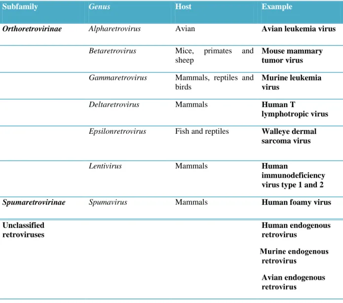
The most recent classification system, described by the international committee on taxonomy of viruses (ICTV), classifies retrovirus in to two subfamilies,
Orthoretrovirinae and Spumaretrovirinae. Orthoretrovirinae includes six genera: Alpharetroviruses, Betaretroviruses, and Gammaretroviruses, considered as simple
retroviruses, and Deltaretrovirus, Epsilonretrovirus and Lentivirus, described as complex retrovirus. Subfamily Spumaretrovirinae contains only the genus Spumavirus, which includes complex retroviruses. All genera display C type morphology except three genera: Betaretrovirus which has B or D type morphology, Lentivirus which has cylindrical or conical core, and Spumavirus which displays immature morphological form, with characteristic central uncondensed core.
It must be stressed that classification of Retroviridae is still incomplete with many retroviruses being unclassified including human endogenous retroviruses (Table 1.2)
15
Table 1.2. Classification of Retroviridae according to ICTV*.
Subfamily Genus Host Example
Orthoretrovirinae Alpharetrovirus Avian Avian leukemia virus
Betaretrovirus Mice, primates and
sheep
Mouse mammary tumor virus
Gammaretrovirus Mammals, reptiles and
birds
Murine leukemia virus
Deltaretrovirus Mammals Human T
lymphotropic virus
Epsilonretrovirus Fish and reptiles Walleye dermal
sarcoma virus
Lentivirus Mammals Human
immunodeficiency virus type 1 and 2
Spumaretrovirinae Spumavirus Mammals Human foamy virus Unclassified retroviruses Human endogenous retrovirus ruoneMenni eniruM ueruioouru Avian endogenousM ueruioouru
* This table is not meant to be comprehensive of all retroviruses. A complete comprehensive listing of retroviruses is available on The International Committee on Taxonomy of Viruses website.
16
1.2. HUMAN ENDOGENOUS RETROVIRUS (HERV)
1.2.1 General properties of HERVs
Retroviruses usually infect somatic cells. When the cell is proliferating, the insertion is present at the same chromosomal location in all progeny cells and vanishes with the last cell of the clone, which might not occur until the host dies. Occasionally however, a retrovirus can infect a cell belonging to the germ line. Any offspring, which develop from an infected germ-line cell, carry the provirus in every single nucleated cell of the body (at the same chromosomal position) and this proviral element is then passed to the descendants according to Mendelian laws. Retroviruses that became part of the host genome in this way are referred to as endogenous retroviruses (ERVs), and the transmission mode, from one host generation to the next, has been termed “vertical” to distinguish it from the “horizontal” spread of exogenous retroviruses. Endogenous retroviruses closely resemble infectious retroviruses integrated into the genome of our ancestors at least 25 million years ago and are detected in all vertebrates studied to date
(Bannert and Kurth 2006).
The obligatory integration step of the retroviral replication cycle allows the incorporation of the viral genome (provirus) into the host genome and retro transposition or reinfection of the germline can generate further insertions augmenting the number of a particular lineage in the genome (Gifford and Tristem 2003).
A complete ERV “provirus” (i.e., the retroviral genome integrated into the host cell genome) shares the same genomic structure of an exogenous retrovirus: four viral genes (gag, pro, pol and env) flanked by two long terminal repeats (LTRs). The gag gene encodes for the major viral structural protein, while pro and pol encode for the viral enzymatic machinery, necessary for the viral replication cycle. The env gene encodes for the envelope glycoprotein that is inserted in the lipid bilayer of the cell membrane to form the viral envelope and mediates entry of the virus into susceptible cells. The LTRs contain enhancer elements that direct expression of the viral genes. ERVs are destined to extinction if their expression brings deleterious consequences for the host. Thus, their
17
persistence in the host genome is the result of a fine balance reached throughout evolution which usually renders them replication defective due to the accumulation of mutations, deletions, rearrangements and methylation (Boeke and Stoye 1997).
The recent sequencing of the human genome has revealed that over 40% of human
DNA sequences belong to the category of repeated or retrotransposable elements
including endogenous retroviruses (Weiss 2006). Retroviral sequences (HERVs)
comprise up to 8% of the host genome (International Human Genome Sequencing Consortium 2001), most of which are defective, due to mutations, deletions or termination signals within coding sequences (Lower 1999) and only very few have been shown to express functional proteins (Bannert and Kurt 2004).
It is unclear whether they were retained because they performed or perform a useful biological function and thus persisted during evolution. Even if the exact function and the importance of HERV in human genome is not very well understood, it is thought to play an important role in genome modeling and plasticity through gene transposition and cellular protection from other retrovirus infection, by receptor interference. In
addition, HERV-W envelop gene on chromosome 7 encode the protein syncytin, which
is thought to play a key role in placenta formation (Ruprechta et al. 2008).
HERV sequences have the potential to be integrated in any location of the genome and consequently may alter the structure and function of other genes. Therefore, they can obviously be involved in genetic disorders. Mainly, they can disrupt genes at their integration site and generate truncated proteins or other isoforms by alternative splicing, or they may even regulate in an abnormal way the expression of particular transcripts by means of the new inserted LTRs, which may harbour promoter sequences.
Epigenetic control is notably involved in silencing most of these genetic elements but certain environmental factors such as viruses are known to dysregulate their expression in susceptible cells. More particularly, embryonal cells with limited gene methylation are most susceptible to uncontrolled activation of these mobile genetic elements by, e.g., viral infections (Perron and Lang 2009).
In contrast to humans, several animals contain ERVs highly related to exogenous retroviruses (Boeke and Stoye 1997; Arnaud et al. 2008) including mouse mammary
18
tumour virus (MMTV), feline leukaemia virus (FeLV), and avian leukaemia virus (ALV), which are currently active and infect mice, cats and chickens, respectively. The most striking example is represented by the koala, in which a retrovirus is currently undergoing the process of endogenisation since it still displays many of the features of an exogenous virus such as the ability to produce infectious viral particles and
variability in proviral copy number and sequence(Tarlinton et al. 2006).
1.2.2. Classification of HERVs
Approximately 30 HERV families have been identified in the human genome (Katzourakis and Tristem 2005)all composed of gag, pro, pol and env genes, flanked
by two long terminal repeat (LTR) regions (Boeke and Stoye 1997).They are present in
a variable copy number and are classified according to the single letter amino acid code for the tRNA specificity of the primer binding site used to initiate reverse transcription (Bannert and Kurth 2004). Up to date, HERV have not been included in the retrovirus taxonomy since most sequences are fragmented or incomplete.
Classification of endogenous retroviruses is still troublesome and currently HERV are classifies according to similarity to exogenous retroviruses in to three classes (Gifford and Tristem 2003) (Table 1.3). Class I shows great homology to Gammaretroviruses and Epsilonretrovirus and contains 18 HERV families, with large copy number in the human genome, almost all, are defective. Class II (also called HERV-K superfamily): shows homology to Betaretroviruses, includes 4 HERV families and is present in a copy number less than the other two classes. Class III: shows homology to
19
Table 1.3. HERV families (Gifford and Tristem 2003)
HERV family Primer Copy number
Class I
HERV Z69907 ND* 30
HERV-ADP tRNA Thr 60
HERV-E tRNA Glu 85
HERV-F tRNA Phe 15
HERV-F type b tRNA Phe 30
HERV-FRD tRNA His 15
HERV-H tRNA His 660
HERV-H49C23 No LTRs 70
HERV-I tRNA Ile 85
RRHERV-I tRNA Ile 15
ERV-9 tRNA Arg 70
HERV-F type c tRNA Phe ND
HERV-P tRNA Pro 70
HERV-R tRNA Arg 15
HERV-R type b tRNA Arg 15
HERV-T tRNA Thr 15
HERV-W tRNA Trp 115
HERV-XA tRNA Phe 15
Class II
HERV-K.HML1-4 tRNA Lys 170
HERV-K.HML5 tRNA Ile 45
HERV-K.HML6 tRNA Lys 70
HERV-K.HML9 ND* ND
Class III
HERV-L tRNA Leu 575
HERV-S tRNA Ser 70
HERV-U2 ND* ND
HERV-U3 ND* ND
20
The endogenous retrovirus HERV-K is the most recent (Barbulescu et al. 1999; Turner
et al. 2001) and transcriptionally active endogenous retroviral family (Ruda et al. 2004;
Seifarth et al. 1998; Wang-Johanning et al. 2001; Yi et al. 2001) and the members of HML-2 subgroup are human specific (Turner et al. 2001).
Genome-wide screening has revealed high levels of insertional polymorphism in the
HERV-K (HML-2) family (Belshaw et al. 2005) demonstrated by the presence of
insertionally polymorphic HERVs proviruses only in a proportion of the human population.
Only members of the HERV-K (HML-2) provirus family have open reading frames (ORFs) for all viral genes, and have therefore received attention in relation to diseases (Tonjes et al. 1996; Mayer et al. 1999; Bannert and Kurth 2004). Some of them have retained the capacity to form retroviral-like particles, as demonstrated in human teratocarcinoma, germ-cell tumours, melanoma, breast cancer and in megakaryocytes from patients with essential thrombocythemia (Boller et al. 2008; Buscher et al. 2005; Lower et al. 1993; Bieda et al. 2001; Morgan and Brodsky 2004; Muster et al. 2003; Seifarth et al. 1998), in the plasma of lymphoma patients (Contreras-Galindo et al. 2008) and possibly in human placenta (Kalter et al. 1973; Dirksen and Levy 1977, Wilkinson et al. 1994). However, up to date, the infectivity of these retroviral-like particles are not yet been demonstrated.
The HERV-K proviruses exist in two forms: type I and type II (Lower et al. 1993; Lower et al. 1995). Type I proviruses have a 292-bp deletion at the boundary of the pol and env genes and the viruses encode the Np9 protein (Armbruester et al. 2002) whereas type 2 proviruses are complete and the viruses produce Rec (an HIV-1 Rev protein equivalent) and Env proteins, which both have been implicated in tumorogenesis (Denne et al. 2007).
21
1.3. HERV AND DISEASES
1.3.1. Pathological role of HERVs
In humans, exogenous retroviruses are known to cause a number of diseases, such as immunodeficiency, neurological disease and cancer.
While endogenous retroviruses are firmly established pathogens in other species, the human endogenous retroviruses may well be considered as emerging pathogens.
The vast majority of the HERVs sequences are defective, although mRNAs from
several families are differentially but ubiquitously expressed in a number of cell types
and tissues (Forsman et al. 2005; Seifarth et al. 2005; Yi et al. 2006).HERV expression
levels vary even between individuals (Andersson et al. 1996) and they may cause
disease in some individuals and not in others due to the existence of HERV
polymorphisms (Moyes et al. 2007). Some HERV mRNAs and a few HERV-encoded proteins are expressed in placental or embryonic tissues and may have essential functions here (Muir et al. 2004).
HERVs family have been also implicated in the pathogenesis of human diseases including type 1 diabetes (Marguerat et al. 2004), autoimmune diseases such as rheumatoid arthritis, (Herrmann et al. 1998; Reynier et al. 2009),psoriasis(Molès et al.
2005) andsystemic lupus erythematosis (Adelman and Marchalonis 2002; Pullmann et
al. 2008). Recently, an increasing number of reports suggested the association of the
HERVs with neuropathological conditions such as multiple sclerosis (MS) and schizophrenia. In particular HERV-H/F (Christensen et al. 1998; Christensen et al. 2000) and HERV-W (Perron et al. 1997; Garson et al. 1998) virions have been detected in plasma, sera and cerebrospinal fluid (CSF) of MS patients and increased levels of
HERV-H, HERV-K and HERV-W RNA in MS brain (Johnston et al. 2001). HERV-W
env and gag proteins have also been found in brain tissue from MS patients and HERV antibodies reactive against specific HERV epitope have also been detected in sera and CSF samples of MS patients and cell-mediated immune responses have been reported
22
(Christensen et al. 2005). Furthermore, HERV-W mRNA has been detected in CSF and post-mortem samples obtained from schizophrenic patients (Karlsson et al. 2001). HIV-1 infection has been also associated with increased HERV-K expression. HERV-K RNA has been detected in the plasma of HIV-1 infected individuals but not in hepatitis C virus (HCV) infected patients or in seronegative controls (Contreras-Galindo et al. 2006). The level of HERV-K titre was found to correlate significantly with that of HIV-1, and was found to be high in HIV-1 patients with non-suppressive HAART therapy and undetectable or low in HIV-1 patients with effective suppressive HAART therapy (Contreras-Galindo et al. 2006). ). Moreover, a longitudinal study following HERV-K titer in HIV-1 infected individuals has shown a correlation between HERV-K expression level and treatment failure and viral rebound. More specifically, the increase in HERV-K expression was detected before the onset of treatment failure and viral rebound indicating that HERV-K expression might be a useful predictor of HIV-1 treatment failure (Contreras-Galindo et al. 2007). The increase in HERV-K expression in HIV-1 patients might have a role in the disease pathogenesis and might also be related to HIV-associated cancer (Contreras-Galindo et al. 2007)
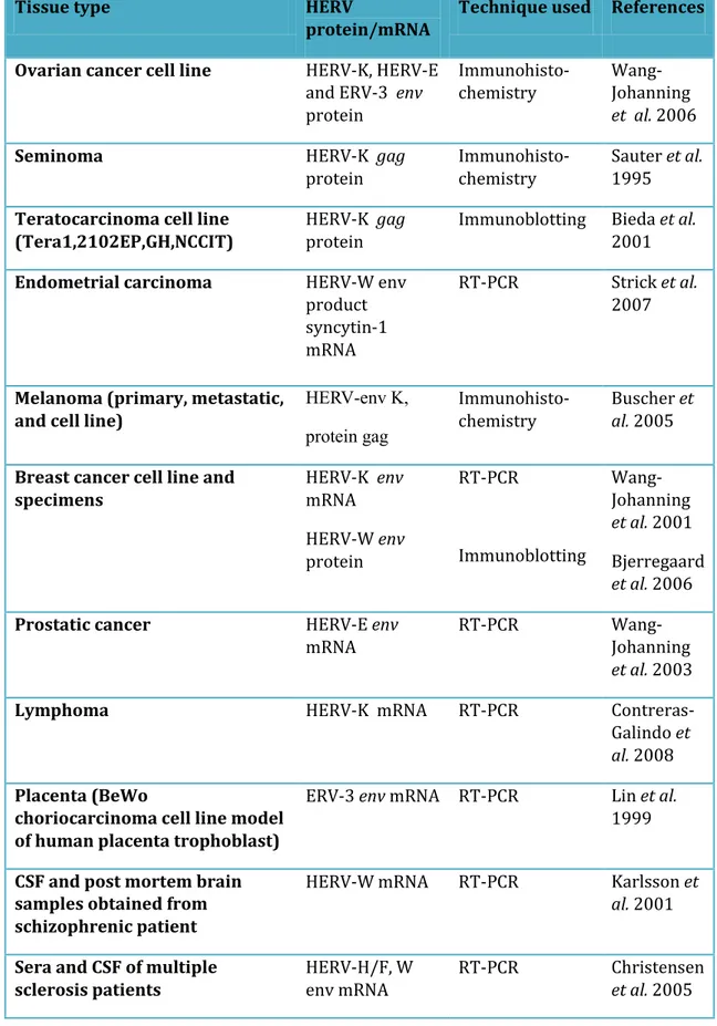
The HERV-K is the most recent (Barbulescu et al. 1999) and transcriptionally active endogenous retroviral family (Wang-Johanning et al. 2001; Tönjes et al. 1996) and therefore receivedparticular attention in relation to diseases (Bannert and Kurth 2004). Different HERV families have been found to be expressed in various human pathologies (Stauffer et al. 2004) (Table1.4.). HERV-E mRNA was detected by reverse transcription PCR in 38.8% of prostatic cancer but not in normal specimens (Wang-Johanning et al. 2003). HERV-W mRNA encoding env gene product syncytin 1 was found to be over expressed in endometrial cancer specimens and is thought to play an important role in cell-cell fusion in endometrial carcinoma (Strick et al. 2007) and in cell-endothelial fusion in breast cancer (Bjerregaard et al. 2006). In addition, HERV-K, HERV-E, and ERV-3 env mRNAs were detected in ovarian cancer and not in control and anti HERV antibodies were also detected in a high percentage of patients sera (55%, 40%, and 30% respectively) (Wang-Johanning et al. 2006). HERV-K transcripts were detected at high levels in the plasma of patients with lymphoma and breast carcinoma, which decreased with treatment and HERV-K like particles were also
23
observed using electron microscopy (Contreras-Galindo et al. 2008). Furthermore, HERV-K rec, which originate from env transcript, were detected in germ cell tumors and it was found that the induction of rec expression in transgenic mice induced developmental disturbances and carcinoma in situ (Galli et al. 2005) giving the first evidence that the induction of HERV-K rec expression might have an essential role in tumorogenesis in transgenic mice.
The mere expression of retroviral transcripts in these pathologies does not implicate a causal association and their exact roles in these processes need to be further evaluated.
24
Table 1.4. HERV transcript detected in various human tissues.
Tissue type HERV
protein/mRNA
Technique used References
Ovarian cancer cell line HERV-K, HERV-E
and ERV-3 env protein Immunohisto-chemistry Wang-Johanning et al. 2006
Seminoma HERV-K gag
protein
Immunohisto-chemistry
Sauter et al. 1995 Teratocarcinoma cell line
(Tera1,2102EP,GH,NCCIT)
HERV-K gag protein
Immunoblotting Bieda et al. 2001
Endometrial carcinoma HERV-W env
product syncytin-1 mRNA
RT-PCR Strick et al.
2007
Melanoma (primary, metastatic, and cell line)
, K v e -VREH gag Ki prp Immunohisto-chemistry Buscher et al. 2005
Breast cancer cell line and specimens HERV-K env mRNA HERV-W env protein RT-PCR Immunoblotting Wang-Johanning et al. 2001 Bjerregaard et al. 2006
Prostatic cancer HERV-E env
mRNA
RT-PCR
Wang-Johanning
et al. 2003
Lymphoma HERV-K mRNA RT-PCR
Contreras-Galindo et
al. 2008
Placenta (BeWo
choriocarcinoma cell line model of human placenta trophoblast)
ERV-3 env mRNA RT-PCR Lin et al.
1999
CSF and post mortem brain samples obtained from schizophrenic patient
HERV-W mRNA RT-PCR Karlsson et
al. 2001
Sera and CSF of multiple sclerosis patients
HERV-H/F, W env mRNA
RT-PCR Christensen
25
1.3.2. Virological etiology of cancer
The etiology of cancer can be summarized in three words: radiation, chemical agents and viruses.
Approximately 20% of human cancers have been attributed to viral infection, and other cancers may also have a viral component (Weiss 2001).
A number of human viruses have been implemented in the causation of a number of human cancers including hepatitis B and C virus as a causative agent of hepatocellular carcinoma (Koike 2007), EBV (Epstein Barr Virus) as a causative agent of Burkitt’s lymphoma, nasopharyngeal carcinoma and some of the lymphomas that complicate AIDS (Young and Murray 2003) and HPV (Human Papilloma Virus) as a cause of cervical cancer (Schiffman et al. 2007). Furthermore, retroviruses have been found to be causative agents in a number of animal as well as human cancers.
Exogenous retroviruses are carcinogenic throughout the animal kingdom, including marine invertebrates, birds, marsupials and a wide variety of placental mammals (Romalde et al 2007; Martin et al 1997).
These retroviruses are usually oncogenic through common indirect pathways, such as integration of the virus adjacent to a cellular oncogene, or incorporation of a host oncogene within the retroviral genome, or through more complex interactions involving viral LTRs and host regulatory pathways, such as tumour suppression genes (Lower et al. 1999; Hayward et al. 1981). In human HTLV-1 (human T-cell lymphoma/leukemia-1) virus is the only exogenous virus which has been proven with no doubt to be involved in the causation of human T-cell lymphoma/leukaemia (Huang et al. 2009) In addition HIV-1 (human immunodeficiency virus-1) has been associated with an increase incidence of opportunistic cancers which could be due to impaired immune control of cancer or due to insertional mutagenesis seen in non-Hodgkin’s lymphoma (Carbone 2002).
26
1.4. HERV-K AND HUMAN MELANOMA
1.4.1. HERV-K particles and human melanoma
The association between HERV and human melanoma gained attention since 1968 with the discovery of the possibility of transmitting hamster melanoma using cell free ultra filtrate along with the detection of virus like particle (Epstein et al. 1968). This discovery drew the attention toward the causal association between hamster melanoma and the still unknown virus particle. This was followed briefly in 1972 by the first published study on human melanoma in which virus particles were detected in primary malignant melanoma and in metastasis to regional lymph nodes (Birkmayer et
al. 1972). The particles were described as spherical or slightly ovoid measuring
90-120 nm in diameter with an electron dense core measuring 50-70 nm surrounded by three layered membrane with occasional projections. The observation of the similarity between these particles and those found in hamster melanoma strongly suggested a role of these particles in human melanoma.
Thereafter, two different study groups (Birkmayer et al. 1974; Balda et al. 1975)have
detected viral particles in primary and metastatic melanoma tissues, which were characterized by high-molecular-weight RNA and RNA-dependent DNA polymerase activity, suggestive of retroviruses. Since then, no infectious virus particles were isolated and the exact nature of the virus was not known. In an attempt to rescue the virus using a helper virus, Zavada et al. 1986 used mouse NIH-3T3 cells carrying molony mouse leukemia virus (MSV and MLV complex) and fused them with human melanoma cell carrying the virus to be rescued. The filtrate of the fused cells was infectious to both mouse and human cells indicating the rescue of an unknown human virus. More recently, virus-like particles have been demonstrated in melanoma cell line supernatants, by electron microscopy observation. They were shown to have RT activity, package sequences with 95.5-97% homology to HERV-K 108 (HML-2 HOM) and 93-95% homology to HERV-K 113 and to contain gag and env proteins (Muster et al. 2003, Buscher et al. 2005). No RT activity was detected in supernatants from cultured human neonatal melanocytes, indicating that the production of particles containing RT is exclusive to melanoma. However, these viral particles were found to be defective and noninfectious.
27
Further sequencing studies showed that particles derived from different melanoma cell lines and even those produced by the same cell line have sequence variability (Hirschl
et al. 2007).
1.4.2 HERV-K mRNA and protein expression in human melanoma
High copy numbers of the HERV-K 108 pol sequence was demonstrated in primary lymph node metastases and cutaneous metastases, by in situ hybridization. Retroviral proteins gag, Rec and env were found to be expressed in melanoma cell line and all primary melanoma, lymph node and cutaneous metastases, by immunofluorescence. The expression of HERV-K transmembrane envelope (TM), Rec and Np9 proteins has also been studied (Buscher et al. 2006) because of the particular interest of Rec and Np9 as potentially oncogenic and TM as putatively immunosuppressive. HERV-K Rec and Np9 mRNA were also found to be expressed in melanoma (39% and 29% respectively) and melanoma cell line (40% and 21% respectively) and Rec protein were detected in 14% of melanoma and no Np9 protein were detected (Buscher et al. 2006). More importantly, the interaction of Np9 and Rec with Promyelocytic Leukaemia Zink Finger (PLZF) tumour suppressor has been found to remove the inhibitory effect of PLZF on c-myc proto-oncogene leading to it’s over expression and subsequent carcinogenic effect (Denne et al. 2007). The expression of TM protein was detected in approximately 50% and HERV-K gag in approximately 70-80% of primary and metastatic melanoma samples. Env, Rec and Np9-specific antisera were also generated, characterized and used to study protein expression in metastatic melanoma and melanoma cell lines.
1.4.3. HERV-K and human melanoma: immunological evidences
Interest in the potential association of retrovirus with human melanoma was reignited when a HERV-K antigen, presented in association with the HLA class I molecule on autologous tumor cells of a patient with melanoma, was shown to be targeted by CD8+ T-cytotoxic lymphocytes (Schiavetti et al. 2002). The antigenic peptide, comprising nine amino acids, was found to be encoded by a short ORF in the env
28
region of a defective HERV-K provirus. The gene encoding the antigen, designated HER-K-MEL, was expressed in most samples of cutaneous and ocular melanoma tested, but also in the majority of benign naevi and in some normal skin samples. Furthermore the causal association between HERV and human melanoma was highly supported by a multicentre case-control study which revealed significant associations between reduced risk of melanoma and history of certain severe infections and/or history of vaccinations with bacille Calmette-Guerin (BCG) and/or vaccinia virus in childhood (Krone et al. 2005; Krone et al. 2003)). This can be explained by the fact that BCG and vaccinia virus have antigenic determinants homologous in their amino acids sequences with the melanoma antigen HERV-K-MEL, encoded by a human endogenous retrovirus K, which is expressed in about 95% of malignant melanocytes. In addition, yellow fever vaccine, with antigenic determinant homologous to HERV-K MEL antigen was found to reduce the risk of melanoma 10 years after vaccination (Mastrangelo et al. 2008).
Furthermore, animal experimental studies have shown that knocking down the melanoma-associated retrovirus (MelARV), using RNA interference (RNAi), resulted in the rejection of tumor cells in immunocompetent mice. Re-expression of the MelARV env gene in the knocked-down cells partially ablated tumor rejection, thus indicating that the env gene contributes to tumor immune escape (Mangeney et al. 2005).
Regarding human melanoma, there is evidence that the expression of HERV-K proteins may induce humoral immune response, with diagnostic and prognostic value. Antibodies specific for HERV-K TM and env proteins were detected in some sera from patients with melanoma, however HERV-K Rec and Np9-specific antibodies were not detected in the same patients, by Western blot analyses (Buscher et al. 2006; Humer et al. 2006)
29
1.4.4. Biological events in the progression of human melanoma
Melanoma is a complex genetic disease, the management of which requires an in-depth understanding of the biology underlying its initiation and progression.
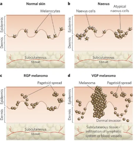
Malignant melanoma arises from epidermal melanocytes, specialized pigmented cells, predominantly present in the skin. Cutaneous melanocytes originate from highly motile neural-crest progenitors that migrate to the skin during embryonic development and their homeostasis is regulated by epidermal keratinocytes (Slominski et al. 2004). The Clark model (Clark et al. 1989), proposed for the progression of melanoma, describes the histological changes that accompany the progression from normal
melanocytes to malignant melanoma (Figure 1.4).
Figure 1.4. The Clark model: progression of melanocyte transformation (Gray-Schopfer et al. 2007)
30
Melanomas are histologically classified into five distinct stages: common acquired and congenital nevi without dysplasia, dysplastic nevi, Radial-Growth-Phase (RGP) melanoma, Vertical Growth Phase (VGP) melanoma and metastatic melanoma. RGP melanoma grow laterally and remains largely confined to the epidermis, while VGP melanoma invades the upper layers of the epidermis and penetrates into the underlying dermis and subcutaneous tissues forming expansile nodules of malignant cells. It is believed that the crucial step in the evolution to malignant melanoma is the transition from RGP to VGP melanoma.
Metastaticmelanoma, on the other hand, develops when tumour cells dissociate from
the primarylesion, migrate through the surrounding stroma, and invade bloodstream or lymphatic system to form a tumour at a distant site (Haass et al. 2005).
1.4.5. Molecular changes in the progression of human melanoma
The histological changes described in the Clark model have been correlated with particular gene mutations that affect molecular signalling, which in turn contribute to the progression from normal melanocyte to melanoma.
Molecular biological bases of human malignant melanoma development can be classified as either genetic, which include mutation, deletion, amplification or translocation, or epigenetic transcriptional modulation, which does not involve modification of DNA sequence. For the last years, the study of the genetic modulation that overlies melanoma development and progression has been facilitated by the availability of commercial microarray technique.
Between 1996 and 2006, more than 129 DNA microarrays have been reported. These studies were used in an attempt to differentiate early melanoma stages (RGP), from advanced stages (VGP) and to provide novel diagnostic and prognostic markers. However, solid conclusions could not be based on all of these studies, due to the chance of false positive results and the fact that not all results were reproducible. Nevertheless, many reviewers have adopted more rigorous approach to validate gene
31
expression analysis data, using multiple testing controlled analyses and have highlighted the most common genetic events that underline either the development of melanoma from melanocyte or the progression of melanoma from RGP into VGP which is thought to be the most critical step in predicting prognosis.
Comprehensive strategies, such as comparative genomic hybridization and mutation analysis by gene resequencing, have identified some of the crucial cell-signalling pathways in this disease.
In accordance with other cancers, human melanoma demonstrates a wide range of genetic modification that favors increase cellular proliferation, inhibition of apoptosis, epithelial mesenchymal transmission and cell migration and interference with cell senescence (Bennett 2008). Figure 1.5 Demonstrates the connections between molecular pathways and risk factors for melanoma, the different steps of neoplastic
transformationand the patterns of molecular changes.
Stimulation of melanocyte proliferation was found to be associated with activating mutation of oncogenic Ras or BRAF or increase copy number of CCND1. The Ras/Raf/MEK/ERK pathway is a key regulator of melanoma cell proliferation, with ERK being hyperactivated in up to 90% of human melanomas by the production of autocrine growth factors or in rare cases by mutational activation of growth- factor receptors such as c-Kit (Willmore-Payne et al. 2005).
BRAF is a proto-oncogene that link Ras to MAPK (Mitogen Activated Protein Kinase) pathway and therefore leads to activation cascade that ultimately leads to cellular proliferation and survival. The MAPK pathway is dysregulated in most melanomas and current understanding of the MAPK pathway in melanoma biology support the concept of distinct groups of molecular and genetic abnormalities in melanomas, related to type of sun exposure and body site.
BRAF and Ras activating mutations are thought to be one of the most common
mutations, which are found in around 70% of melanoma(Quatresooz et al. 2009).
Interestingly, BRAF mutation is seen mostly in melanoma arising from skin with
32
occur at a similar frequency in benign nevi andin primary and metastatic melanomas
(Pollock et al. 2003).
Figure 1.5. Biologic events and molecular changes in the progression of melanoma (Miller and Mihm 2006). At the stage of the benign nevus, BRAF mutation
and activation of the mitogen-activated protein kinase (MAPK) pathway occur. The cytological atypia in dysplastic nevi reflect lesions within the cyclin-dependent kinase inhibitor 2A (CDKN2A) and phosphatase and tensin homologue (PTEN) pathways. Further progression of melanoma is associated with decreased differentiation and decreased expression of melanoma markers regulated by microphthalmia-associated transcription factor (MITF). The vertical-growth phase and metastatic melanoma are notable for the striking changes in the control of cell adhesion. Changes in the expression of the melanocyte-specific gene melastatin 1 (TRPM1) correlate with metastatic propensity, but the function of this gene remains unknown. Other changes include the loss of E-cadherin and increased expression of N-cadherin, V3 integrin, and matrix metalloproteinase 2 (MMP-2).
33
In addition, activation of catenin oncogenic pathway via activating mutation of β-catenin itself, methylation or mutation of APC or activation of AKT3 can transcriptionally activate MYC and CCND1, inhibit apoptosis and activate microphthalmia-associated transcription factor (MITF), which is a key regulator of melanocyte gene expression. Suppression of retinoblastoma protein pathway (RB) via CCND1, CDK4, or RB1 mutation could by itself activate clonal expansion regardless of MAPK pathway (Bennett 2008).
Microphthalmia-associated transcription factor (MITF) is a transcription factor considered the master regulator of melanocyte biology, partly because it regulates the expression of melanogenic proteins such as tyrosinase, silver homologue (GP100) and melanoma-associated antigen recognized by T cells-1 (MART-1, also known as melan-A). MITF is expressed in most human melanomas and its target genes are diagnostic markers for this disease. Furthermore, continued MITF expression is essential for melanoma cell proliferation and survival (Levy et al. 2006; Widlund et
al. 2003). MITF is expressed at significantly lower levels in melanoma cells than in
melanocytes, and increased MITF levels reduce melanoma cell proliferation even in the presence of oncogenic BRAF. High MITF levels reduce melanoma cell tumorogenicity (Selzer et al. 2002).
Decreased or absent pigmentation accompany the progression from nevus to
melanoma.Expression of the melastatin 1 (TRPM1) gene, whose functionis unknown,
is also controlled by MITF (Miller et al. 2004). The mechanism of decreased
expression of these genes is a puzzle because MITFis present in nearly all melanomas
(Granter et al. 2002). Although MITF causes differentiation and cell-cycle arrest in normal melanocytes, melanoma cells do not have these characteristics. Recently, a
large-scale search for genomic changes in melanomafound an increased copy number
(4 to 119 copies per cell)of a region of chromosome 3 that includes the MITF locus.
This increase was accompanied by the increased expression of MITF protein. The
over expression of both MITF and BRAF couldtransform primary cultures of human
melanocytes, implicatingMITF as an oncogene. Notably, MITF amplification occurs
mostfrequently in tumours that have a poor prognosis and is associatedwith resistance
34
On the other hand, anti-apoptotic effect in human melanoma can be achieved by loss of apoptosis effecter APF1, PI3K activation via Ras, or NFKB activation via BRAF (Bennett 2008). NFKB can be also activated by cytokines produced by activated white blood cells and melanocyte or by hypoxia inducible factor α (HIFα) induced by hypoxic skin condition.
Cell senescence, which is seen in nevi, is unfavourable to human melanoma development and overcoming it is a key factor in melanoma development. Two main pathways control cell senescence: P16-RB and P53. P16-RB was found to be more relevant in human melanoma. P16, which facilitate cell senescence by acting on telomerase, is encoded by CDKN2A, which in turn is thought to be one of the two genes most commonly affected by inactivating mutation in cancer in general along with P53. Unlike BRAF, which is commonly mutated in melanoma arising from skin with intermittent sun exposure, CDKN2A is commonly mutated in mucosal and acral melanomas along with CDK4 (Quatresooz et al. 2009).
In human melanocytes, mutant BRAF proteininduces cell senescence by increasing the expression of thecell-cycle inhibitor of kinase 4A (INK4A) (Michaloglou et al. 2005). INK4A limits hyperplasticgrowth caused by a BRAF mutation. The arrest of
the cell cyclecaused by INK4A can, however, be overcome by mutations in INK4A
itself, as well as other cell-cycle factors.Thus, BRAF is implicated in several aspects of melanoma induction and progression.
Acquisition of potential for invasion and distant metastasis has been an interesting object of study of many researchers and although a big list of genes has been found differentially expressed in RGP and VGP melanoma, only few have been found to be significant. Non-canonical WNT pathway has a high correlation with invasive and metastatic melanoma. Furthermore, NFKB was found to play a role in activating transcription factor SNAI1 which repress E-cadherin and induce N-cadherin, which in turn activate proteases (matrix metalloproteinase -2 (MMP2) and tissue plasminogen activator (t-PA)) facilitating epithelial mesenchymal transition, cell migration and invasion (Bennett 2008).
Cadherins are multifunctional transmembrane glycoproteins that sustain cell-to-cell contacts, form connections with the actin cytoskeleton, and influence intracellular
35
signalling. Cadherins are divided into three subtypes: E (epithelial), present in
polarized epithelialcells in the epidermis, including melanocytes and keratinocytes;P
(placental) and N (neural), found in mesenchymal cells inthe dermis. The intracellular
domain is associated with a largeprotein complex that includes β-catenin and forms
structurallinks with bundles of actin filaments (Miller and Mihm 2006).
The major adhesion mediator between keratinocytes and normal melanocytes in the epidermis is E-cadherin, which disappears during melanoma progression (Tang et al. 1994). Instead of E-cadherin, melanoma cells express N-cadherin, characteristic of invasive carcinoma, which allows them to change cellular partners. Melanoma cells adhere through N-cadherin to fibroblasts and endothelial cells by gap junctions (small channels for electrolyte transport) (Hsu et al. 2000; Danen et al. 1996; Hsu et al. 1996). Progression from the radial-growth phase to the vertical-growth phase of melanoma is therefore marked by the loss of E-cadherin and the expression of N-cadherin (Danen et al. 1996). Besides these changes in cell adhesion, decreased
E-cadherinexpression (Gottardi et al. 1997) and aberrant N-cadherin expression increase
thesurvival of melanoma cells by stimulating β-catenin signalling (Qi et al. 2005). Cell that have lost epithelial differentiation, as manifested by the loss of functional E-cadherin, show increased mobility and invasiveness. Keratinocytes can no longer control melanoma cells that have lost E-cadherin and when melanoma cells are forced to express E-cadherins and are co-cultured with keratinocytes, they dramatically change: melanomas adhere to keratinocytes, no longer express invasion-related molecules and lose their invasive capacities (Hsu et al. 2000).
N-cadherin is also a survival factor for melanoma cells as they migrate through the dermis and a major adhesion receptor, when melanoma cells adhere to each other (Li
et al. 2001; Perils et al. 2004).
Similarly integrins are cell surface adhesion molecules playing an important physiologic role in cell adhesion, growth, proliferation, migration, and apoptosis (Tucker 2006).
36
C H A P T E R 2 . A I M O F T H E S T U D Y
According to the Clark model of tumorogenesis, (Clark et al. 1989) the most critical moment in the development of a melanoma would be the moment at which clonal evolution generates a cell with “vertical-growth phase” (VGP) properties i.e. a melanoma cell whose progeny can enter the deeper dermis rather than growing only in or near the epidermis. That is because VGF melanomas are believed to be competent of metastasis so the tumour would then proceed to spread and kill the patient if untreated.
Human melanoma has been linked to HERV-K expression but the timing of HERV-K expression and its significance in cancer progression is still unclear. The family of HERV-K has been subdivided into type 1 and type II proviruses. Type I proviruses are characterized by a 292 bp deletion at the pol-env boundary and an ORF for the nonstructural protein Np9. Type II, the undeleted prototype, has an intact env sequence and encodes the accessory protein Rec. Both proteins were found expressed in tumours and transformed cell lines and evidence has accumulated that they play a role in cancer development. Rec and Np9 directly bind to the promyelocytic leukemia zinc finger (PLZF) protein and inhibit the transcriptional inhibitory function of PLZF on MYC promoter thus promoting cellular proliferation (Denne et al. 2007). Furthermore, the presence of anti-HERV-K antibodies in early stages of melanoma
was shown to be a marker of reduced survival (Hahn et al. 2008).In a published work,
done at the University of Rome, Tor Vergata, HERV-K was reported to support the in
vitro transition of melanoma cells from adherent to a more malignant, non adherent
phenotype when exposed to stress conditions (Serafino et al. 2009).
In that paper a cellular system was described including 3 types of cells: original adherent human melanoma cell line (TVM-A12), and two derived anchorage independent rounded cells, growing in suspension, obtained by limiting dilution (Clonesp) and by serum starvation (1% of foetal calf serum in the culture medium)
(TVM-A12sp). Anchorage independent suspended cells (Clonesp and TVM-A12sp)
have been characterized, showing a more malignant phenotype in comparison to original adherent cells (TVM-A12) in the form of decreased expression of melanoma differentiation antigen (Melan A/MART-1), loss of HLA-I expression, and increased
37
ability to grow in semi-solid agar. More importantly, these phenotypic and functional modifications were accompanied by the activation of HERV-K expression and massive production of viral-like particles. Down-regulation of HERV-K expression by RNA interference prevented the transition from the adherent to the non-adherent growth phenotype in low serum condition.
Aim of this study is to provide further characterization of the cellular system at the genetic level for better understanding of the genetic modification associated with the phenotypically increased malignant potential and also to look for genes modification that might have a role in the induction of HERV-K expression. Such identification of basic genetic modification provide better understanding of the process of carcinogenesis and shed light on possible drug targets that can be used in the management of human malignant melanoma.
To understand the transcriptional activity of HERV-K in melanoma and their role in cancer progression we investigated the gene expression profile of melanoma cells derived from the same parental cell line (TVM-A12) by different stress conditions, i.e. limiting dilution (Clonesp) and serum starvation (1% FBS in the culture medium)
(TVM-A12sp). Cells obtained by both conditions were characterized by high levels of
expression of HERV-K, corresponding to a phenotype of more malignant cells. With this in mind the objectives were to:
1. Perform an initial screening of gene expression of a large number of genes commonly modified in human cancer using commercially available microarray tests that include tumor suppressor genes involved in apoptosis, cell expression cycle, cell growth and differentiation, cell mobility and signal transduction.
2. Determine the general theme of genes modification in the cellular system and identify genes with special importance to human melanoma progression and HERV-K expression.
3. Select genes, which show significant modification either up regulation or down regulation and perform real-time PCR confirmation.
38
4. Determine whether phenotypically more malignant suspended cells (TVM-A12sp
and Clonesp) have a more malignant genotype in comparison to the original adherent
39
C H A P T E R 3 . M A T E R I A L S A N D
M E T H O D S
3.1. MATERIALS
3.1.1. Cell cultures
The human melanoma cell line TVM-A12 was established as monolayers from
a melanoma lesion of a patient with metastatic melanoma, obtained at the presentation of the disease (Melino et al. 1993).
RPMI 1640 medium, Trypsin 0.05% EDTA 0.02% in PBS, Dulbecco’s
Phosphate buffered saline (PBS), Penicillin-streptomycin 100X and L-glutamine solution 100X (EuroClone, Milan, Italy)
Fetal bovine serum (FBS, EuroClone, Milan, Italy)
Forma direct heat CO2 Incubator (Thermo Fisher, USA)
3.1.2. Microarray
Nucleospin RNA II isolation kit (MACHEREY-NAGEL, Germany)
EuroGold total RNA kit (EuroClone, Milan, Italy)
Smartspec plus Spectrophotometer (Bio-Rad, Rome, Italy)
True labelling-AMP 2.0 cRNA synthesis and labelling kit (GA-030),
ArrayGrade™ cRNA Cleanup Kit, SuperArray Oligo GEArray® Microarrays, GEAhyb Hybridization solution (H-01), Chemiluminescent Detection Kit (D-01) and Microarray Membranes, Oligo GEArray® Human Cancer Microarray, OHS-802 (SuperArray Bioscience Corporation, USA)
Biotin-11 UTP (Perkin Elmer, Germany)
3.1.3. Reverse transcription and Real-time Polymerase chain reaction
(PCR)
I Script cDNA synthesis kit, IQ SYBR Green Supermix, and C1000 Thermal
Cycler (CFX96 Real-time System, Bio-Rad, Rome, Italy)
40
Primer pairs (Table 3.1.) (PRIM, Milan Italy)
3.2. METHODS
3.2.1. Cell culture
TVM-A12, a human melanoma adherent continuous cell line, obtained at the
University of Rome Tor Vergata from metastatic melanoma lesion was maintained and grown in RPMI 1640 with 10% (v/v) heat inactivated FBS (fetal bovine serum) supplemented with penicillin (100 IU/ml), streptomycin (100mg/ml) and L-glutamine (2 mM) and incubated at 37oC in a humidified
5% CO2 atmosphere. Adherent cells were passed every other day using trypsin
and 0.02% EDTA solution in PBS.
Clonesp was isolated by limiting dilution techniques, at 1 cell/well in 96 well
plate, from the TVM-A12 human melanoma cell line, maintained in RPMI 1640 medium supplemented with 10% (v/v) FBS (fetal bovine serum), penicillin (100 IU/ml), streptomycin (100mg/ml) and L-glutamine (2 mM). Clonesp was characterized by a completely lost of adherence growth and a
stably modified phenotype as big suspended clumps, even when maintained in RPMI 1640 complete medium supplemented with 10% (v/v) FBS.
TVM-A12sp suspended cell line was obtained by shifting the parental
TVM-A12 melanoma cell line from optimal growth factor conditions (10% FBS) to low-serum condition (i.e.,1% FBS). TVM-A12 were seeded at density of 2X10⁵ cells in T25 cm2 flask and grown in 10% FBS. After 24 hours the medium was changed to 1% FBS and cells were observed for detachment for one week.
3.2.2. RNA extraction and quantification
Cell pellets obtained by washing with PBS and subsequent sedimentation
using centrifugation 1000g, were used for RNA extraction using Nucleospin RNA II extraction kit according to manufacturer’s instruction. RNA was extracted from original adherent A12 (A12 10%), adherent TVM-A12 growing in 1% serum TVM-A12 1%), suspended TVM-TVM-A12
(TVM-41
A12sp), and clone cells growing in suspension in 10%FBS culture medium
(Clonesp).
RNA quantification using spectrophotometry at wavelength 260nm was done.
The ratio of absorbance at A260:A280 was used to determine the quality of
isolated RNA. Isolated RNA was stored at -80oC.
3.2.3. Microarray
Microarray membranes testing 440 genes involved in human cancer, including
genes involved in apoptosis, cell cycle, cell growth and differentiation, cell mobility and signal transduction, were used to test all cells in the cellular system (including original adherent TVM-A12, adherent TVM-A12 in 1% FBS, suspended TVM-A12sp, and Clonesp, obtained by limiting dilution
technique). A total of 3µg of isolated RNA with A260:A280 ratio in the range (1.6-1.9) was used for cRNA synthesis and labelling with biotin using true labelling-AMP 2.0 cRNA synthesis and labelling kit. It must be emphasised that 4 hours incubation was used for cRNA synthesis, amplification and labelling. This was followed by cRNA purification using clean up kit as instructed by manufacturers. Purified cRNA was used for hybridization overnight with subsequent chemiluminescence staining and imaging on chemiluminesence film according to manufacturer’s instructions.
Images obtained were uploaded in to an online image data acquisition and
analysis suite (GEArray Expression analysis suite,
http://www.superarray.com) for analysis. Spot intensity, for each gene, was PUC18 plasmid (negative control) DNA background subtracted and normalized by the software using Glyceraldehyde-3-phosphate dehydrogenase (GAPDH) and beta actin (ACTB) as housekeeping control genes. Gene expression modification in all cells of the cellular system in comparison to original adherent TVM-A12 was determined as fold change (ratio of spot signal detected in test sample over control sample). Genes with fold change ≤0.6 were considered down regulated and those ≥ 1.5 were considered up regulated.
Microarray analysis was done in triplicate for each cell condition, using samples from three different experiments.
42
3.2.4. Reverse transcription and Real time PCR
A total of 500 ng of isolated RNA from each sample was reverse-transcribed,in a total volume of 20l, using High-Capacity cDNA Reverse Transcription kit (Applied Biosystems, CA, USA) according to the manufacturer’s instructions.