SAPIENZA University of Rome
Faculty of Pharmacy and Medicine
PhD in Morphogenesis and Tissue Engineering XXIX Cycle
(A.A. 2015/2016)
CD44-NEGATIVE PROSTATE CANCER CELLS
GIVE RISE TO CD44
HIGHCELLS THAT DISPLAY
PHENOTYPICAL AND FUNCTIONAL
STEM-LIKE TRAITS
PhD Student Chiara Di Stefano
Tutor Coordinator
Alla mia famiglia
Molla gli ormeggi, esci dal porto sicuro
e lascia che il vento gonfi le tue vele.
Esplora. Sogna. Scopri.
Index
THE THESIS EXPLAINED ... 3
1. INTRODUCTION ... 7
1.1 Prostatic carcinoma ... 7
1.2 Tumor heterogeneity ... 11
1.3 Prostate cancer stem cells: markers and methods for their enrichment ... 15
1.4 CD44 and its splicing variants: molecular structure and functions ... 17
1.5 CD44s and CD44v isoforms as putative CSCs markers ... 20
1.6 Toll-Like receptors: properties and functions in health and disease ... 22
1.7 The dual role of Toll-Like receptor 3 in cancer ... 25
2. AIMS ... 27
3. RESULTS ... 29
3.1 Poly (I:C) treatment selects highly expressing CD44 and CD44-negative subpopulations ... 29
3.2 Poly (I:C)-enriched CD44-negative cells give rise to CD44-positive cells ... 33
3.3 CD44-negative cells from parental PC3 and DU-145 spontaneously convert to highly positive CD44 cells ... 34
3.4 Cells derived from CD44-negative subpopulation show higher clonogenic potential than cells derived from
CD44high population producing mainly holoclones ... 36 3.5 Clones derived from CD44-negative cells show a
greater clonogenic potential than clones generated by sorted CD44high population in secondary
clonogenic assay ... 38 3.6 CD44-negative-derived population specifically
re-expresses CD44 v8-v10 splice variant isoform ... 41 4. DISCUSSION ... 45 5. MATERIALS AND METHODS ... 51 5.1 Cell lines ... 51 5.2 Flow cytometry... 51 5.3 Fluorescence-activated cell sorting analysis ... 52 5.4 Single-cell cloning by limiting dilution ... 52 5.5 Establishment of GFP-tagged PC3 cells ... 53 5.6 RNA isolation, RT PCR and qRT-PCR ... 53 5.7 Statistical analysis ... 54 REFERENCES ... 55 LIST OF PUBLICATIONS ... 69
THE THESIS EXPLAINED The biological issue:
Prostate cancer (PCa) is initially treated with removal of androgens; however, androgen depletion is usually associated with the recurrence of the metastatic and more aggressive PCa termed castration-resistant prostate cancer (1). Small cell neuroendocrine carcinoma (SCNC), an extremely aggressive variant form of prostate cancer that contain all neuroendocrine (NE) cells, are often seen as recurrent tumors in patients with a history of adenocarcinoma and treated with hormonal therapy (2). NE cells are hormone-insensitive cells and express selectively the putative prostate cancer stem cell (CSC) marker CD44 (3). Our group has previously demonstrated that Toll-Like receptor 3 (TLR3) agonist poly (I:C), that mimics viral genome, induces apoptosis in the androgen-dependent PCa cell line LNCaP, that resemble prostatic adenocarcinoma, in vitro (4) and in vivo (5).
On the contrary, only a weak apoptotic effect is observed in the more aggressive and androgen-independent PCa cells PC3 and DU-145 (6), the prototype of SCNC composed by neuroendocrine highly aggressive cells.
It has been recently reported that stimulation of TLR3 by poly (I:C) promotes breast cancer cells toward a CSC phenotype in vitro and in vivo, evaluated as CD44high/CD24-negative expression (7). To ascertain if poly (I:C) treatment is able to select or to target CSCs, in this study we intend to determine whether the apoptosis resistance to poly (I:C) can be correlated to CD44 expression in the androgen independent PCa cell lines PC3 and DU-145.
Results:
Unexpectedly, we found very small CD44-negative cells, resistant to poly (I:C), that gave rise to a CD44-positive population in PC3 cells after just 48 hours of culture post sorting. The same rate of conversion was observed in the 2-4% of sorted CD44-negative very small in size cells contained in parental PC3 and DU-145 cells. In addition, CD44-negative-sorted PC3 cells converted in CD44high cells, expanded in vitro culture, displayed an higher clonogenic potential than sorted CD44high -derived population, probably because they produced mainly holoclones, described to be enriched in stem-like cells, whereas CD44high population generated mainly meroclones, a mixture of cells of different proliferative potential. Moreover, in secondary clonogenic assay, the clones generated by CD44-negative-sorted cells exhibited a significantly higher clonogenic potential than clones derived by CD44high -sorted
population. All types of the CD44high and CD44-negative-derived
clones became positive for CD44 suggesting that this molecule is probably expressed in both cancer stem, progenitor and more differentiated cells.
Although our data indicate that the cellular population generated by CD44-negative-sorted cells were indistinguishable from the CD44high -derived cells in terms of CD44 expression, we found that CD44 transcript variant 3, containing variant exons v8–v10, was more highly expressed in the population generated by CD44- negative-sorted cells at the mRNA and protein level in both the androgen-independent cell lines PC3 and DU-145.
Conclusions:
It is becoming apparent that characterization of prostate cancer stem cells (CSCs) should be based not only on cell surface markers because they are often subjected to dynamic expression changes. Our data are in accordance with this theory because we demonstrated that CD44high cells, described to be stem-like and more tumorigenic in prostate cancer, may arise from a CD44-negative population, displaying functional stem-like traits, enriched following poly (I:C) and docetaxel, the canonical chemotherapeutical treatment of PCa.
Moreover CD44-negative population isolated from PC3 and DU-145 cells, enriched of self-renewing cells, re-expressed specifically CD44 v8-v10 splice variant isoform. Given that many CD44v isoforms are preferentially expressed on cancer cells and involved in tumor progression, the identification of new CD44v isoforms in CSCs will open new field of investigation to find innovative targeted therapies against CSCs in PCa. To conclude, our data, support the concept that, under certain conditions, some tumors exhibit plasticity by reversibly transitioning between a stem and non-stem-cell state expressing microenvironment-dependent variable markers. Of note, the therapeutic potential of TLR3 agonist on prostate cancer, shown by us and others groups, should be carefully evaluated with consideration of its possible role in mediating nuclear reprogramming in the induction of pluripotency in embryonic fibroblasts (8) and CSC phenotypes (7) thus potentiating cancer recurrences, as demonstrated in breast cancer.
1. INTRODUCTION 1.1 Prostatic carcinoma
Prostate cancer (PCa) is the most common malignancy in men and the second leading cause of cancer-related deaths (9).
PCa develops over a long period of time from normal prostate to prostatic intraepithelial neoplasia (PIN) then to early- and late-stage PCa and finally to metastatic PCa (10). If localized, PCa can be cured by radical prostatectomy or irradiation. However, more than 30% of patients may relapse after surgery (11).
Although early detection through serum testing for prostate specific antigen (PSA) has significantly reduced the number of fatalities, its widespread use has caused overdiagnosis and overtreatment of PCa diagnosed during an indolent phase that could be avoided by using an active surveillance. On the contrary, use of PSA alone as a decision-point for identifying prostate biopsy can also lead to underdiagnosis or non-diagnosis of cancers that are histologically-aggressive but have PSA levels below routine thresholds (12). Therefore, in order to improve prostate cancer detection, decision-making and care, much research has needed to discern aggressive from indolent PCa.
Advanced PCa is mainly treated with androgen-deprivation therapy (ADT), but it is widely accepted that androgen ablation can promote the progression of the tumor to the castration resistant stage (1). Both castration resistant prostate cancer (CRPC) and primary PCa are able to metastasize to various organs such as the bone, lung and liver. Once metastasis occurs the disease becomes incurable (13). Over 95% of prostate tumors are adenocarcinomas that arise from the epithelia of the prostatic gland composed of three differentiated epithelial cell types: luminal, basal and neuroendocrine (NE) cells that are scattered throughout the prostate constituting ˜1% of the
total epithelial cell population. The luminal cells secrete proteins including PSA and express high levels of androgen receptor (AR) and low molecular weight keratins (cytokeratins 8 and 18).
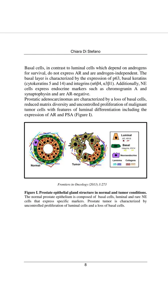
Basal cells, in contrast to luminal cells which depend on androgens for survival, do not express AR and are androgen-independent. The basal layer is characterized by the expression of p63, basal keratins (cytokeratins 5 and 14) and integrins (α6β4, α3β1). Additionally, NE cells express endocrine markers such as chromogranin A and synaptophysin and are AR-negative.
Prostatic adenocarcinomas are characterized by a loss of basal cells, reduced matrix diversity and uncontrolled proliferation of malignant tumor cells with features of luminal differentiation including the expression of AR and PSA (Figure I).
Frontiers in Oncology (2013) 3:273
Figure I. Prostate epithelial gland structure in normal and tumor conditions.
The normal prostate epithelium is composed of basal cells, luminal and rare NE cells that express specific markers. Prostate tumor is characterized by uncontrolled proliferation of luminal cells and a loss of basal cells.
However, tumor cells also express basal integrins and often co-express basal and luminal keratins, such as cytokeratins 5 and 8 that are restricted to their distinct cell types in normal tissue (14;15). The presence of “intermediate cells” that co-express luminal and basal markers sustains the hypothesis that PCa develops from the disruption of differentiation pathways which normally restrict basal and luminal marker expression to their respective cell types. The cell of origin, defined as the cell type within a normal tissue that undergoes oncogenic transformation giving rise to the tumor, has not been clearly identified in PCa.
The classic androgen ablation and replacement experiment demonstrates the presence of prostate stem/progenitor cells within the basal layer. Androgen withdrawal leads to apoptosis of the luminal cells and the survival of the basal cells. In the adult prostate, luminal cells are regularly shed and replaced through differentiation (16).
A simplistic view of this observation is that a basal progenitor or stem cell gives rise to both basal and luminal populations through a transient differentiation process (17;18). However, findings from mouse models support the idea that there are at least three different prostate epithelial progenitor populations (basal, luminal and bipotent progenitors), but which ones initiate PCa still remains unresolved (19-21). Several studies demonstrate either basal or luminal progenitors can be the initiating cancer cell (22;23).
Better understanding of prostate differentiation will be required to identify the cell of origin in PCa.
Adenocarcinoma cells, like the normal prostatic gland, rely on androgens for their development and progression and contain rare quiescent androgen-independent NE cells. This is the basis for the use of androgen-deprivation therapy for patients with advanced and recurrent diseases (24). This treatment rarely eradicate all prostate cancer cell populations inducing apoptosis of the androgen-dependent luminal tumor cells and an increase in NE cells that, through their secreted products, can promote androgen-independent growth of prostate cancer cells contributing to the progression of the
tumor to the castration resistant stage. Small cell neuroendocrine carcinoma (SCNC) in which the tumor cells are all NE cells highly aggressive, are often seen as recurrent tumors in patients with a history of adenocarcinoma and treated with hormonal therapy (2). Only 1 to 2% of prostate cancers are diagnosed originally as neuroendocrine prostate tumors. These tumors can be pure or admixed with prostatic adenocarcinomas. SCNC is defined as a poorly differentiated NE cancer characterized by NE tumor cells highly proliferative, metastatic, and resistant to most conventional therapies. To date, the origin of these tumors remains uncertain. Mounting evidence support the idea that NE-like tumor cells could transdifferentiate from adenocarcinoma in response to treatment. Accumulation of new genetic alterations such as RB loss, AURKA, and MYCN amplifications may promote rapid cell expansion and subsequent emergence of a prevailing NE component in the form of small-cell carcinoma that rapidly proliferates. Adenocarcinoma and small-cell carcinoma components may also derive from transformation of a common multipotential prostatic progenitor that accumulated molecular alterations driving epithelial or NE phenotypes (25). In fact, NE cells have some features of cancer stem cells (CSCs), such as the expression of CD44 and CD133 (3;26).
1.2 Tumor heterogeneity
The highly heterogeneous nature of prostate cancer poses many obstacles to therapy.
PCa striking cellular heterogeneity can give rise to therapeutic resistance and disease progression.
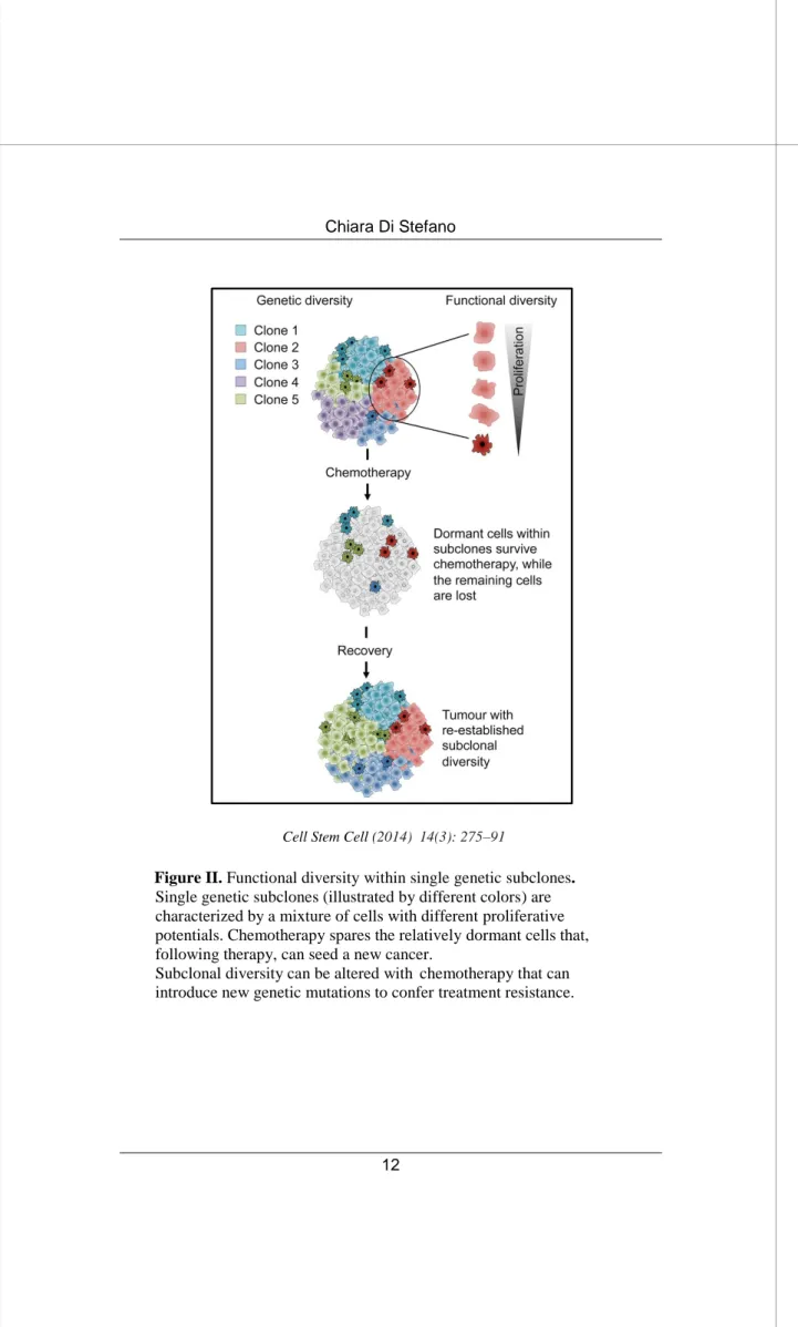
There are different theories to explain the origin of tumor cellular heterogeneity. In the stochastic model, tumor cells have a random probability of developing mutations to permit tumor maintenance and progression. It is becoming increasingly clear that a tumor does not have one single tumor genome, but consists of multiple genomes that belong to distinct subclones. Under selective pressures, such as chemo- or radiotherapy, clones that have acquired resistant properties survive and give rise again to a heterogeneous population. These subclones may evolve in parallel over the lifetime of a cancer and contribute to intratumoral heterogeneity. However, even within single genetic subclones, the cells behave differently: some cells retain self-renewal capacity and long-term clonal maintenance and most tumor cells are postmitotic and destined for clearance (Figure II)
Cell Stem Cell (2014) 14(3): 275–91
Figure II. Functional diversity within single genetic subclones.
Single genetic subclones (illustrated by different colors) are characterized by a mixture of cells with different proliferative potentials. Chemotherapy spares the relatively dormant cells that, following therapy, can seed a new cancer.
Subclonal diversity can be altered with chemotherapy that can introduce new genetic mutations to confer treatment resistance.
The cancer stem cell (CSC) model in which sustained tumor growth is restricted to selected subpopulations called cancer stem cells, is based upon the hypothesis that tumors, like adult tissues, originate from cells that can self-renew and give rise to more differentiated progeny that comprises the bulk of the tumor (27). Tumor heterogeneity, consisting of genetic and phenotypic diversity, is thus referable to a hierarchy of differentiation that includes CSCs. Both these hypothesis tend to identify a single cell population as the therapeutic target: the prevailing clone over time in the first case and the CSC in the latter.
Although the initial hierarchical theory for existence of cancer stem cells assumed a stable population that always give rise to progeny cells lacking, or reduced for, the tumor initiating property, is emerging that, some tumors, under certain conditions, exhibit plasticity by reversibly shifting between stem-like and more committed cells.
The notion that there are multiple hierarchies guided by similar but not identical CSCs in a tumor might help explain the highly complex nature of intratumoral heterogeneity and resistance to targeted therapies. Then integration of the CSC model with the stochastic model may be necessary (28) (Fig. III).
Modified by Stem Cells (2016) 34(8): 1997-2007
Figure III. Cancer stem cell model integrated with the stochastic model.
The cancer stem cell theory focuses on the internal heterogeneity within individual clonal subpopulations of a tumor due to a hierarchy of differentiation. The stochastic model allow the clonal outgrowth of novel cell populations arising from the acquisition of accumulating mutations.
1.3 Prostate cancer stem cells: markers and methods for their enrichment
The cancer stem cell model represents a rebirth of an old idea. In the early 1858 Rudolf Virchow proposed that cancers arise from embryonic-like tissue noting the histologic similarities between developing normal tissues and poorly differentiated cancers such as teratocarcinomas. Although this concept is not new, the first experimental evidence for the actual existence of CSCs came from hematological malignancies in 1997 (29). Subsequently, the existence of CSCs has been identified in different solid tumors such as breast cancer (30), gliomas (31), lung cancer (32), colon cancer (33;34) and prostate cancer (35).
CSCs have been identified by developing new culture methods and functional assays to enrich a subpopulation with self-renewal ability in vitro and tumor initiating and propagating ability in vivo. Prostate cancer stem cells (PCSCs) can be enriched by selecting cells that have the ability to efflux Hoechst dye (36), to form prostatespheres in a serum-free suspension culture system (37) or by sorting cells that exhibit high levels of ALDH activity (38). Most of PCSCs have been isolated by using cell surface markers such as CD24, CD44 (39), CD49f (40), CD133 (26;35), CD166 (41) and α2β1 integrins
(35). Those markers have been tested alone and in several combinations but there is currently no universally accepted collection of CSC markers for isolation of a “pure” population of PCSCs which could clearly lead in the all cases to distinct cancer stem cells. The cause of this situation is the considerable diversity in the tumor histotypes and their genetic heterogeneity.
Several studies demonstrate the utility of CD133 as an enrichment marker for cells with stem-like properties. Collins et al. used CD133 marker to identify the prostate cancer stem cell population with a CD44+ / α2β1high / CD133+ phenotype characterized by a significant
capacity for self-renewal. On the other hand, a large body of evidence narrows down its use as a stem cell marker. Although CD133-positive glioma cells were initially considered to be CSCs
(31), many proofs support the existence of CD133-negative glioma stem cells able to initiate tumors as well as generate CD133-positive progeny (42).
Patralawa and coworkers, in one of the studies performed in vitro and in vivo on cell lines and xenograft tumor models, showed that CD44-positive (CD44+) PCa cells are more tumorigenic and metastatic than the corresponding CD44-negative (CD44-) PCa cells and possess many intrinsic traits of tumor progenitors (39). On the other hand, they also observed tumor development in CD44- PCa cells and clonal analyses revealed the emergence of the CD44+ clones from the pure CD44- cell population. It has, therefore, been proposed that characterization of CSCs be based not only on cell surface markers that are often subjected to dynamic expression changes.
The formation of holoclones has been adopted as a surrogate stem cell assay especially in the study of prostate cancer. An increased number of holoclones is considered as enrichment for CSCs. The existence of a cellular hierarchy reflecting the differentiation and proliferative capacities of stem cells within normal tissue was first characterized over 20 years ago (43). Using primary cultures of human keratinocytes, the Authors found that single cells gave rise to 3 types of clones classified as holoclones, meroclones and paraclones with different proliferative capacities. These colonies are believed to be derived respectively from stem, transit-amplifying cells and differentiated cells. It has since been shown that the existence of a stem cell hierarchy persists in cancer cell lines, including prostate cancer (44).
The PC3 prostate cancer cell line was shown to contain an highly heterogeneous cell population of holoclones, meroclones and paraclones. Li and colleagues demonstrated that, like keratinocytes holoclones, PC3-derived holoclones contained self-renewing stem-like cells and were enriched for expression of the stem cell markers CD44 and α2β1 integrin. (44).
1.4 CD44 and its splicing variants: molecular structure and functions
The hyaluronic acid receptor CD44 is a single pass transmembrane glycoprotein involved in cell-cell and cell-matrix adhesion and in cell signaling. It has a relevant role in lymphocyte homing, inflammation, cell migration, signaling and tumor metastasis (45;46).
CD44 is encoded by the highly conserved CD44 gene located on chromosome 11 in humans (47).
Alternative splicing of 9 out of 19 exons in human CD44 pre-mRNA results in expression of different transcript variants (CD44v isoforms), leading to variation in the length and function of the extracellular domain (Figure IV A). The predominant form of CD44, expressed on most vertebrate cells, is named standard isoform (CD44s) and is translated to a polypeptide of 85-95 kD from exons 1-5 joined to exons 15-19. Most noticeable, CD44v isoforms are expressed in a broad range of cancers especially those in advanced stages (48).
CD44 protein contains three functional domains: an extracellular domain (or ectodomain), a transmembrane (TM) domain and an intracellular domain (ICD) (47) (Figure IV B). The N-terminal domain, encoded by the first five exons of CD44, acts as ligand-binding receptor for the main ligand hyaluronan (46) and also for extracellular matrix (ECM) proteins such as osteopontin (49), collagen, laminin (50), and fibronectin (51).
The introduction of new exons in the extracellular domain modulates HA-binding affinity by inducing conformational changes or providing CD44 isoforms to have new functions as a coreceptor by producing new binding sites for many growth factors and receptors (52).
The transmembrane domain is responsible for lymphocyte homing and mediates CD44 activation signaling by coupling CD44/CD44v to cofactors, adaptor proteins, receptor tyrosine kinases, or non-receptor protein-tyrosine kinases (53).
CD44 ICD has also a pivotal role for the function of CD44 in signaling transduction. CD44 undergoes sequential proteolytic cleavages in the extracellular domain, mediated by membrane-associated metalloproteases (MMPs), and transmembrane domain. The first cleavage produces a soluble fragment released into the culture supernatant (soluble CD44) and a membrane-bound product (CD44 EXT, CD44 extracellular truncation).
Then, CD44 EXT is cleaved by the presenilin-γ-secretase, resulting in the release of a CD44 intracellular domain (CD44 ICD) into cytoplasm that translocates into the nucleus where it can transcriptionally regulate the expression of target genes such as CD44 itself, thus promoting the rapid turnover of CD44 that is required for efficient cell migration (54).
Therefore, the sequential proteolytic cleavage is crucial for CD44-mediated signaling pathway.
The cytoplasmic tail of CD44 is anchored to the actin cytoskeleton and to ezrin and ankyrin with functions involved not only in cell migration but also in signal transduction of CD44 (55).
Furthermore, the complexity of the CD44 protein family and functions is increased by various degrees of post-translational modifications such as N- and O-linked glycosylation and glycosaminoglycanation.
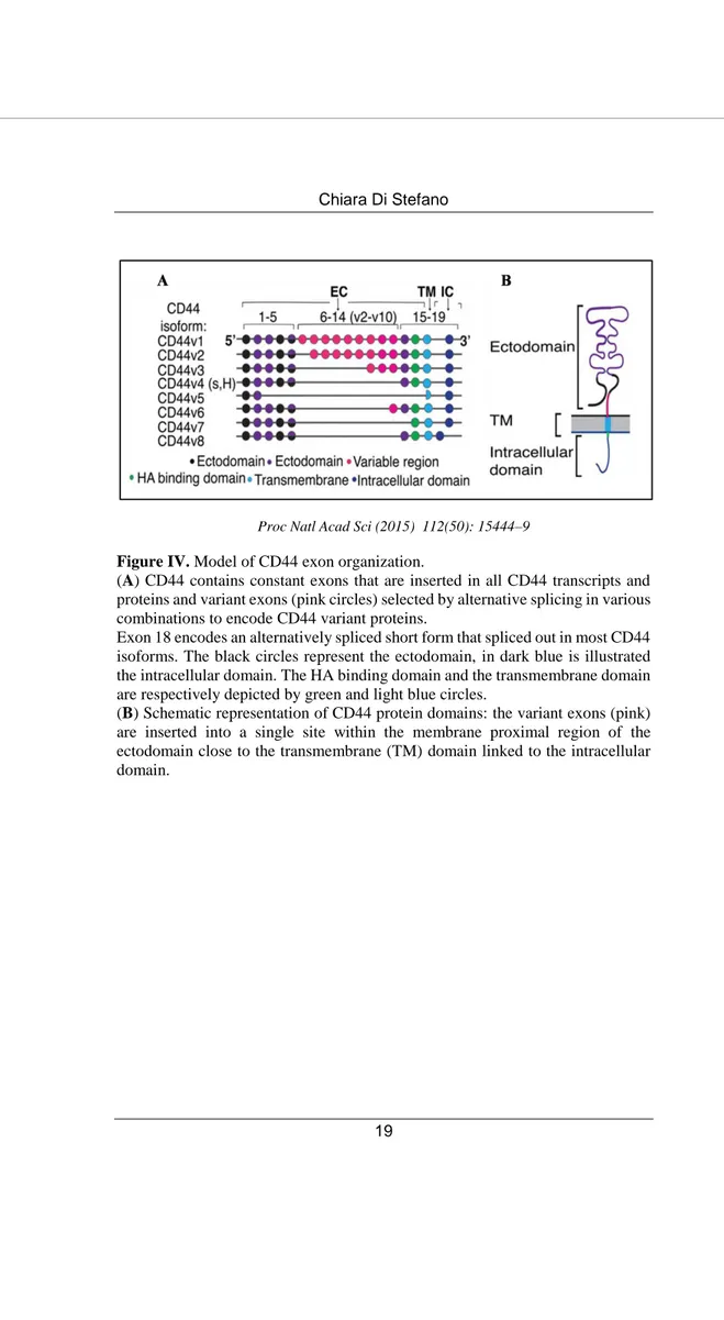
Proc Natl Acad Sci (2015) 112(50): 15444–9
Figure IV. Model of CD44 exon organization.
(A) CD44 contains constant exons that are inserted in all CD44 transcripts and proteins and variant exons (pink circles) selected by alternative splicing in various combinations to encode CD44 variant proteins.
Exon 18 encodes an alternatively spliced short form that spliced out in most CD44 isoforms. The black circles represent the ectodomain, in dark blue is illustrated the intracellular domain. The HA binding domain and the transmembrane domain are respectively depicted by green and light blue circles.
(B) Schematic representation of CD44 protein domains: the variant exons (pink) are inserted into a single site within the membrane proximal region of the ectodomain close to the transmembrane (TM) domain linked to the intracellular domain.
1.5 CD44 standard (CD44s) and CD44 variant (CD44v) isoforms as putative CSCs markers
Most human cancers, as well as prostate cancer, are characterized by dysregulated CD44 expression. PCa loses expression of CD44s (56), a marker of basal cells in benign epithelium, and overexpresses splice variant isoforms. CD44, especially CD44v isoforms have been identified as CSC markers and critical players in regulating tumor progression, metastases and treatment resistance in many cancers.
A significant reduction of CD44s expression in proportion to tumor grade was first described in 1996: CD44s resulted decreased in metastatic cancer compared to matched primary cancer (57). It was later found that patient-derived cancers had increased CD44 v7-v10 messenger RNA and knock-down of this variant isoform by RNAi significantly decreased invasion and migration (58).
Currently, Ni J. and colleagues have demonstrated high levels of CD44 v6 in primary prostate cancer tissues and lymph node metastases including cancer cells and surrounding stromal cells but not in normal prostate tissues. On the other hand, CD44s expression is negative in all primary prostate cancer tissues and lymph node metastases, while is strongly positive in all normal prostate tissues suggesting that CD44s may negatively regulate CaP progression while CD44 v6 may be involved in CaP progression and metastases. Finally, the authors demonstrated that CD44 v6 displays CSC-like properties and is closely associated with PCa stemness (59). CD44 v6 predicts poor prognosis and is a marker of constitutive and reprogrammed CSCs driving colon cancer metastases (60). CD44 v3 is a specific CSC marker of head and neck cancers (61;62). CD44 v8-v10 is a gastric CSC marker and its expression is low in normal tissues thus making it an ideal target to eliminate gastric CSCs (63). Moreover, CD44 v8-v10 contributes to ROS defense by promoting the sintesis of reduced glutathione (GSH). These findings reveal a role for CD44 v8-v10 in the protection of CSCs from high levels of
In addition to CD44 v8-v10 transmembrane expression, its enzymatic cleavage has also been investigated. The cleaved fragment, detected by presence of the soluble extracellular domain in ascites fluid of ovarian cancer patients, correlated with worse prognosis (65). However, the prognostic value of CD44s and CD44v isoforms seems to vary by cancer type and discrepancies or controversial results have been reported even within the same tumor type. The contradictory conclusions, regarding the correlation between CD44 expression and disease prognosis, may be caused by using different methodologies (66). Then commercially available CD44 assay kits containing well-characterized CD44s and CD44v antibodies with standardized protocols are crucial for comparison among different studies to come to general conclusions.
1.6 Toll-Like receptors: properties and functions in health and disease
Toll-Like receptors (TLRs) are a family of transmembrane proteins that recognize pathogen associated molecular patterns (PAMPS), molecules highly conserved in bacteria, viruses, fungi and parasites (67).
TLRs are expressed in epithelial and immune cells. Ligand activation of TLRs initiates the onset of inflammation (68) by either MyD88-dependent pathways, that culminate with the induction of inflammatory cytokines through MAPKs and NF-kB activation, or TRIF-dependent pathways, which are responsible for the induction of inflammatory cytokines as well as type I interferons by IRF-3 activation (69).
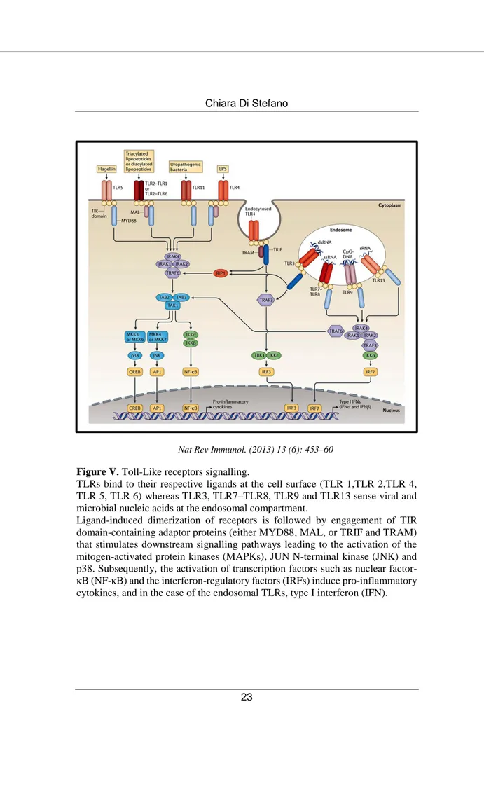
To date, 10 isoforms have been identified in humans: TLR 1, 2, 4, 5 and 6 are expressed at the cell surface membrane and recognize pathogens-derived lipids, lipopolysaccharides, peptidoglycan or proteins. In contrast, TLR 3, 7, 8 and 9 recognize (poly) nucleotides in endolysosomes (70) (Figure V).
Nat Rev Immunol. (2013) 13 (6): 453–60
Figure V. Toll-Like receptors signalling.
TLRs bind to their respective ligands at the cell surface (TLR 1,TLR 2,TLR 4, TLR 5, TLR 6) whereas TLR3, TLR7–TLR8, TLR9 and TLR13 sense viral and microbial nucleic acids at the endosomal compartment.
Ligand-induced dimerization of receptors is followed by engagement of TIR domain-containing adaptor proteins (either MYD88, MAL, or TRIF and TRAM) that stimulates downstream signalling pathways leading to the activation of the mitogen-activated protein kinases (MAPKs), JUN N-terminal kinase (JNK) and p38. Subsequently, the activation of transcription factors such as nuclear factor-κB (NF-factor-κB) and the interferon-regulatory factors (IRFs) induce pro-inflammatory cytokines, and in the case of the endosomal TLRs, type I interferon (IFN).
TLRs are the key sensors of the innate immunity, mediated by cytokine and chemokine secretion, and are critically involved in priming the adaptive immune response necessary for killing invading pathogens (71) thus a negative regulation of their signalling is crucial to prevent over-activation of the immune system resulting in chronic inflammatory diseases and autoimmune disorders (72).
Besides the canonical antimicrobial function of TLRs, it has been demonstrated that functional TLRs are also expressed on a wide variety of tumors playing a crucial role in tumor growth and progression. Their expression is deregulated in cancerous epithelial tissues (73).
The role of TLRs in cancer is controversial: it has been demonstrated both an anti-apoptotic effect (protumoral) and a pro-apoptotic function (antitumoral) for several TLRs.
The immune system has the potential to either promote tumor progression as well as mediate tumor destruction. Which of these conditions prevails depends on the balance between the protumor and antitumor mediators of both innate and adaptive immunity. Chronic inflammation is involved in polarizing immunity toward a tumor-promoting phenotype producing an immune suppressive environment that is permissive for carcinogenesis. Conversely, acute inflammation promotes a tumor-rejecting response (74). It was suggested that TLR gene polymorphisms might alter TLR signaling, thus affecting inflammation and prostate cancer risk. Sequence variants of TLR1, TLR6 and TLR10 were confirmed to increase prostate cancer risk (75) as well as variants of TLR4 (76). However, controversial results were also obtained (77).
1.7 The dual role of Toll-Like receptor 3 in cancer
Although conflicting reports have been published concerning the pro or antitumoral role of several TLRs, most literature data agree on an antitumor role for TLR3 (78;79). In fact, the TLR3 ligand Ampligen has been proposed as a potentially safe immune-adjuvant in cancer therapy (80). The activation of TLR3 by the specific agonist polyinosinic:polycytidylic acid [poly (I:C)], a synthetic dsRNA analogue, exerts an antitumoral effect by inducing apoptosis with higher efficiency in the androgen-dependent prostate tumor cell line LNCaP than in the androgen-independent cell line PC3 depending on differential degree of up-regulation of hypoxia inducible factor-1 (6). Furthermore, poly (I:C) stimulation recruits an antitumoral immune response.
Galli R. and coworkers have previously demonstrated that activation of TLR3 in PCa cell lines induces the secretion of cytokines and chemokines that can recruit and activate immune cells in the tumor site promoting their anticancer activity (81).
Different hypothetic models of action for TLR3-dependent anticancer mechanism have been proposed (82): an immune-mediated tumour growth suppression (83), a direct apoptotic effect on TLR3 expressing cancer cells (4;84-86) and an inhibition of tumor growth through both immune-mediated anticancer mechanisms and cancer cell apoptosis.
Although the majority of studies have reported that TLR3 activation leads to tumor suppression, it has also been described to promote tumor development by enhancing cancer cell proliferation and survival (87). Increased TLR3 expression in patients with breast cancer was correlated with poor prognosis (88). The same author, 1 year later, showed that 85% of the examined PCa cases were positive for TLR3 expression and that high levels of TLR3 were associated with biochemical recurrence (89). Furthermore, the outcomes of clinical trials using TLRs agonists are in general inconsistent (90). This extremely intricated context reflects our insufficient understanding of the direct role of TLR3 in cancer cells,
in particular cancer stem cells (CSCs), capable of initiating new tumors in vivo and interconvertible between a stem and non-stem-cell state.
CSC plasticity has been linked to inflammation which may select for, or induce, stem-like cell even if the underlying mechanisms remain poorly understood.
Although the stimulation of TLR3 with poly (I:C) induces the death of particular subpopulations of tumor cells blocking tumor growth initially, it promotes breast cancer cells toward a CSC phenotype through the proliferation of CSCs together with the induction of a CSC phenotype from non-CSCs. Inhibition of both β-catenin and NF-kB is an effective strategy to control the growth of human breast cancer induced by TLR3 activation (7). This study suggest that the therapeutic potential of a given TLR agonist should be carefully evaluated with consideration of its possible role in mediating CSC phenotypes and potentiating more robust cancer recurrences.
2. AIMS
In the past years, much progress has been made towards better understanding the development and progression of PCa as well as the factors which drive the development of androgen independence. However, the highly heterogeneous nature of prostate cancer poses many obstacles to therapy.
Several groups have proposed that NE cells may be resistant to hormonal therapy and therefore responsible for tumor recurrence following androgen ablation. In addition, the selective expression of the putative prostate cancer stem cell marker CD44 in NE tumor cells of PCa further supports the importance of such cells in therapy resistance and tumor recurrence and raises interesting questions about the relationship of the NE tumor cells to the elusive PCa stem cells. Our laboratory group has previously demonstrated that the TLR3 agonist poly (I:C) induces apoptosis in the androgen-dependent tumor cell line LNCaP negative for neuroendocrine markers and CD44, while the androgen-independent cell lines PC3 and DU-145 are nearly resistant to poly (I:C) treatment and share NE features associated with the expression of the stem/progenitor cell marker CD44(3). Thus, we called into question the existence of a subpopulation negative for neuroendocrine markers more prone to poly (I:C)-induced apoptosis. My PhD project aimed to evaluate whether the apoptosis resistance to poly (I:C) of the androgen-independent PCa cell lines can be correlated to CD44 expression. To test if poly (I:C) targets or selects CD44high population, the modulation of membrane-CD44 protein in PC3 cells after poly (I:C) treatment was assessed by flow cytometry. The unexpected enrichment, following poly (I:C) treatment, of a CD44-negative population very small in size besides a CD44high cell subpopulation selection, led us to investigate the biological properties of CD44-negative and CD44hi gh cell subpopulations, resistant to poly (I:C), and in parental PC3 and DU-145 cells by FACS sorting. Therefore, we aimed to determine whether the cell populations generated by CD44-negative and CD44high sorted
cells were functionally distinct, in particular if CD44-negative sorted cells could be considered “stem-like cells” giving rise to CD44high cells, described to be stem-like and more tumorigenic in
prostate cancer. To this purpose, we used in vitro gold standard functional method, the colony forming assay, to determine whether it enriches for a holoclone phenotype in the PC3 cells verifying the holoclone peculiar ability to regenerate the full spectrum of clonal heterogeneities. Moreover, we proposed to examine CD44 expression in different clone types from primary clonogenic assay and to perform secondary clonogenic assay of these clones in order to compare the clone forming ability of the clones generated by CD44-negative cells and the clones derived by CD44high cells. The
secondary clonogenic assay will be performed also on expanded two sorted subpopulations since they reflect the behavior of the single derived clones.
Finally, CD44v isoforms have been identified as CSC markers and critical players in regulating tumor progression metastases and treatment resistance in many cancers (64;65). Thus, we aimed to investigate if the cell populations generated by CD44-negative and CD44high sorted cells expressed different CD44 splice variant isoforms at the mRNA and protein level.
3. RESULTS
3.1 Poly (I:C) treatment selects highly expressing CD44 and CD44-negative subpopulations.
We have previously demonstrated that the androgen-independent cell line PC3 is largely resistant to poly (I:C) treatment (6). To establish whether such incomplete resistance can be possibly correlated to differences in CD44 expression, we treated PC3 cells with 25 µg/ml poly (I:C) for increasing times up to 4 days and analyzed the composition of the cell population by flow cytometry using an anti-CD44 antibody that recognizes both standard and all CD44 isoforms. We observed that poly (I:C) treatment resulted in an increased percentage of CD44-negative population (only,2-4% in control sample) up about 10-fold in 3 days treatment (Figure 1 A) and a decrease of CD44-positive cells, accompanied by a shift to a median fluorescence intensity (MFI) higher than the control (Figure 1 B and 1 C). The population lacking CD44 protein was very small in size : ~ 60% of these cells were < 7 µm, ~ 30% ranged from 7-15 µm and only 10% of CD44 negative cells were > 15 µm (data not shown).
Figure 1. Modulation of CD44 expression following poly (I:C) treatment.
CD44 expression was evaluated in PC3 cells after treatment with poly (I:C) for 2, 3 and 4 days. Poly (I:C) stimulation enriches for CD44-negative cell subpopulation (A) while reduces the percentage of CD44+ cells (B) at the indicated times. CD44 expression was calculated as percentage of total live cells. Each data point is the mean ± SEM of three independent experiments *p < 0.05; **p < 0.01, ***p < 0.001, Student’s paired t-test. (C) Representative flow cytometry overlay of gated live cells before (dotted histogram) and following 72 hours poly (I:C) treatment (black histogram) show an increase of CD44-negative cells (marker M1) and a decrease of CD44-positive population (marker M2). The grey histogram represents the isotype control APC-labeled IgG2B. Cells were gated using sytox blue stain to exclude dead cells
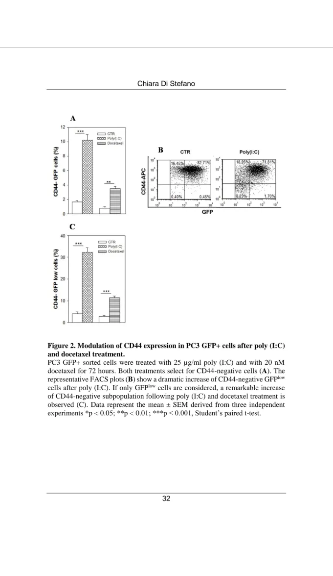
Most of the CD44-negative-enriched population resulting from poly (I:C) treatment is included in a size area generally comprising the debris in flow cytometry analysis based on forward scatter vs side scatter. Thus, to verify our data excluding debris, we used PC3 cells infected with TWEEN-EGFP lentiviral vector and sorted only the GFP+ cell population. We treated the PC3 GFP+ cells with poly (I:C) or with the standard therapeutic agent docetaxel for 72 hours and again flow cytometry analysis confirmed the high increment of CD44-negative population after poly (I:C) stimulation (Figure 2 A). The representative dot plot (Figure 2 B), highlights that the CD44-negative/ GFP+ population enriched by poly (I:C) treatment derives from the very small size GFPlow population cells (0.4% CD44 -GFPlow cells in CTR vs 8.2% in poly (I:C)-treated cells). In fact, by gating exclusively the GFPlow cells we observed a much higher percentage of CD44-negative cells after poly (I:C), and, at a lesser extent, docetaxel stimulation (Figure 2 C).
Figure 2. Modulation of CD44 expression in PC3 GFP+ cells after poly (I:C) and docetaxel treatment.
PC3 GFP+ sorted cells were treated with 25 µg/ml poly (I:C) and with 20 nM docetaxel for 72 hours. Both treatments select for CD44-negative cells (A). The representative FACS plots (B) show a dramatic increase of CD44-negative GFPlow cells after poly (I:C). If only GFPlow cells are considered, a remarkable increase of CD44-negative subpopulation following poly (I:C) and docetaxel treatment is observed (C). Data represent the mean ± SEM derived from three independent experiments *p < 0.05; **p < 0.01; ***p < 0.001, Student’s paired t-test.
3.2 Poly (I:C)-enriched negative cells give rise to CD44-positive cells
In order to explore the biological properties of the cell populations enriched after poly (I:C) treatment, single PC3 GFP+ cell suspensions were obtained and sorted by FACS into CD44high and CD44-negative cells. The purity of these cell populations was generally >95% as revealed by post-sort analysis (not shown). Surprisingly, 48 hours after sorting, CD44-negative GFP+ cells gave rise to a population expressing membrane-CD44 protein. Data in Figure 3 shows that 75% of the cells generated by CD44-negative sorted population become positive for CD44.
Figure 3. CD44 expression in CD44-negative and CD44high -sorted cells
resistant to poly (I:C) stimulation.
Percentage of CD44+ cells in CD44-negative GFP+ and CD44high GFP+ sorted PC3 populations, resistant to 72 hours poly (I:C) treatment, immediately after sorting (0 d) and 48 hours (2d) after culturing. n=3 mean ± S.E.M **p < 0.01, Student’s paired t-test.
3.3 CD44-negative cells sorted from parental PC3 and DU-145 bulk populations spontaneously convert to highly positive CD44 cells
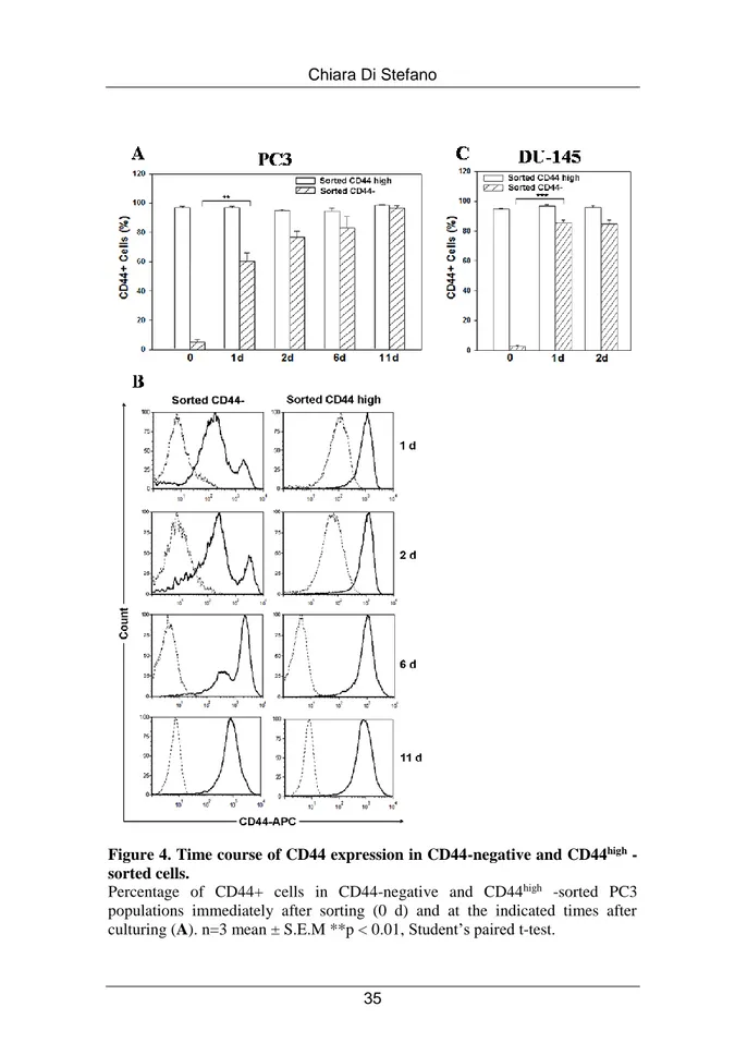
We next explored whether the minority CD44-negative population present in parental PC3 and DU-145 cell lines could also undergo spontaneous conversion. To this aim, we isolated pure (average, >95%) populations of CD44high and CD44-negative cells fractionated by FACS directly from PC3 and DU-145 cultures. Cells from these two PC3 subpopulations were seeded into 24-well plates immediately following cell sorting and monitored for CD44 expression over the subsequent 11 days. Purified CD44high cells remained a pure CD44high population, whereas CD44-negative cells gave rise to a CD44-positive cell population that progressively increased, restoring the initial CD44 expression profile in 11 days (Figure 4 A). CD44high cells represent ~ 4% of CD44-negative fraction collected immediately after sorting (0 d), whereas their percentage increases in culture to ~ 60% in 24 hours. The representative FACS analysis of CD44 expression in the time-course experiments represented in Figure 4 B shows that CD44-negative cells acquired CD44 expression on their surface in just 24 hours post-sorting and that this expression reached higher levels after 2, 6 and 11 days in vitro expansion. Of note, cell counts revealed that, in the first 24 hours post sorting, no cell proliferation occurred, since the number of cells plated remained constant. In addition, similar FACS experiments were performed on DU-145 cells to test whether the fast conversion from CD44-negative to CD44high cells was shared by another androgen-insensitive PCa cell
line. As shown in Figure 4 C, CD44-negative subpopulation sorted from DU-145 spontaneously converted into CD44high, even with higher efficiency than PC3 cells.
Figure 4. Time course of CD44 expression in CD44-negative and CD44high
-sorted cells.
Percentage of CD44+ cells in CD44-negative and CD44high -sorted PC3 populations immediately after sorting (0 d) and at the indicated times after culturing (A). n=3 mean ± S.E.M **p < 0.01, Student’s paired t-test.
(B) Representative histograms of CD44 expression at the indicated time points of culturing CD44-negative and CD44high cell populations. The dotted histogram represents the isotype control APC-labeled IgG2B, the black histogram indicates APC-labeled CD44 antibody staining.
(C) Percentage of CD44-positive cells in CD44-negative and CD44high -sorted DU-145 populations immediately after sorting (0 d), 24 and 48 hours after in vitro expansion. n=3 mean ± S.E.M ***p < 0.001, Student’s paired t-test.
3.4 Cells derived from CD44-negative subpopulation show higher clonogenic potential than cells derived from CD44high
population producing mainly holoclones
Our results obtained by sorting experiments suggest that CD44high
cells, described to be stem-like and more tumorigenic in prostate cancer (39), may arise from components of the CD44-negative population. Therefore, in order to investigate and compare the functional properties of CD44-negative and CD44high cells, we
evaluated their respective clonogenic potential in limiting dilution assays. We plated these two PC3-sorted subpopulations at 1 cell per well in 96-well plates, one day after plating we selected those wells that contained only 1 viable cell and then we followed the development of each individual clone over the following two weeks. Although CD44-negative and CD44high cells showed a similar
clonogenic potential in primary clonogenic assay (Figure 5A 0 d), 28 days after sorting CD44-negative-derived cells exhibited a significantly higher clonogenic potential than those derived from CD44high cell population (Figure 5 A). To investigate this delayed
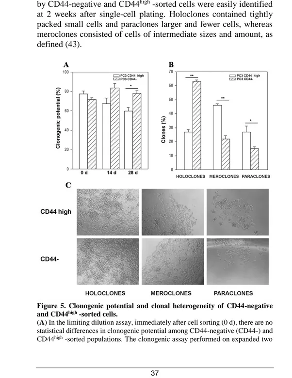
increase in clonogenic potential, we analyzed the clone types in primary clonogenic assay, since the formation of holoclones has been adopted as a surrogate stem cell assay, particularly in the study of prostate cancer (91). In primary clonogenic assay, 2 weeks after plating, 63% of clones generated by sorted CD44-negative cells developed into typical holoclones, whereas 22% were meroclones and 15% formed paraclones. Conversely, the clones derived by
5 C, the distinct morphologies of the three types of clones generated by CD44-negative and CD44high -sorted cells were easily identified at 2 weeks after single-cell plating. Holoclones contained tightly packed small cells and paraclones larger and fewer cells, whereas meroclones consisted of cells of intermediate sizes and amount, as defined (43).
Figure 5. Clonogenic potential and clonal heterogeneity of CD44-negative and CD44high -sorted cells.
(A) In the limiting dilution assay, immediately after cell sorting (0 d), there are no statistical differences in clonogenic potential among CD44-negative (CD44-) and CD44high -sorted populations. The clonogenic assay performed on expanded two
sorted subpopulations (cultured for 28 days) shows a significantly higher capability to form clones of CD44 negative-derived cultured cells than CD44high -derived cells (*p < 0.05). The CD44-negative-sorted cells produced mainly holoclones whereas CD44high -sorted population gave rise principally to meroclones (B). All the data were analyzed by Student’s paired t-test. n=3 mean ± S.E.M. The phase contrast representative images of the different types of clones generated by CD44-negative and CD44high -sorted cells are shown in (C).
3.5 Clones derived from CD44-negative cells show a greater
clonogenic potential than clones generated by sorted CD44high
population in secondary clonogenic assay.
We then wondered to examine CD44 expression in different clone types from primary clonogenic assay and to perform secondary clonogenic assay of these clones. To this aim, we randomly chose some clones derived from the primary clonogenic assay of sorted CD44-negative and CD44high PC3 subpopulations and evaluated
CD44 expression after 30 days in vitro expansion of 12 CD44high -derived clones (5 holoclones, 6 meroclones, and 1 paraclone) and 13 CD44-negative-derived clones (10 holoclones, 1 meroclone and 2 paraclones). All the clones became positive for CD44 expression as revealed by flow cytometric analysis (Figure 6 A). Furthermore, we verified the holoclone ability to regenerate the full spectrum of clonal heterogeneity selecting, in total, 7 holoclones and 3 meroclones derived by CD44high and by CD44-negative primary clonogenic assay. In line with published evidence (44), holoclones, but not meroclones, gave rise to all three types of clones in the secondary clonogenic assay (Figure 6 B). Moreover, when serially in vitro, 17 CD44high and CD44-negative derived total holoclones and 14 meroclones, the holoclones showed an higher long-term propagation ability than meroclones (Figure 6 C).
In order to compare the clone forming ability of the clones generated by CD44-negative cells and the clones derived by CD44high cells, we
CD44-enrichment in CD44-negative-derived clones in primary clonogenic assay, we observed a significantly higher clonogenic potential in the clones generated by cells derived from CD44-negative population than in those derived from sorted CD44high population (Figure 6 D).
Figure 6. CD44 expression, proliferative and self-renewing capacities of CD44-negative and CD44high -derived clones.
(A) Percentage of cells expressing CD44 protein in PC3 bulk population and in CD44-negative and CD44high derived clones 30 days after cell sorting.
(B) Clone classification, following secondary clonogenic assay, of CD44-negative and CD44high -derived primary holoclones (n=7) and meroclones (n=3). (C) Percentage of clone survival during 120 days follow-up period.
By 120 days, most holoclones proliferated robustly (88,2%) and only 2 holoclones aborted. 7 of the 14 meroclones (50%) were abolished and could not be further propagated.
(D) The clones generated by CD44-negative cells showed an higher clonogenic potential than CD44high -derived clones (**p < 0.01). The table indicates the number of wells that developed 1 clone starting from 1 single viable cell for CD44-negative and CD44high –derived clones (n=6 for each group). The data were analyzed by chi-square test.
3.6 CD44-negative-derived population specifically re-expresses CD44 v8-10 splice variant isoform
Our data indicate that the cellular population generated by CD44-negative-sorted cells after 2 days in culture was almost indistinguishable from the CD44high -derived cells in terms of CD44
expression (Figure 4). In addition, both the CD44high and CD44-negative-derived clones become positive for CD44 as revealed by using an anti-CD44 antibody that recognizes a common epitope to CD44 standard (CD44s) and all CD44 variant (CD44v) isoforms. However, the variant isoforms (CD44v) have been linked to subpopulations endowed with stem cell potential, to enhanced metastatic potential and a poor prognosis in several types of cancer (60;91).
Therefore, we aimed to investigate if the functionally distinct cell populations generated by CD44-negative and CD44high cells
expressed different CD44 splice variant isoforms at the mRNA and protein level. We first determined the expression of CD44 isoforms in PC3 cells by designing PCR primers in constant exons 5 and 15, flanking the variable region of the CD44 gene (Figure 7 A). Semiquantitative RT-PCR analysis from PC3 cells revealed the presence of three main products at a size of 396, 600 and 792-bp, whose sequences correspond to CD44s, CD44 v10 and CD44 v8-v10 isoforms, respectively. Subsequently, we performed RT- PCR from both PC3 and DU-145 CD44-negative and CD44high -derived cells 14 and 28 days after cell sorting and we found that CD44 transcript variant 3, containing variant exons v8–v10, was more highly expressed in the population generated by CD44-negative-sorted cells (data not shown). In order to evaluate the CD44 v8-v10 levels in the two sorted subpopulations, we performed splice isoform-specific quantitative RT-PCR (qRT-PCR) analysis by designing specific primers inside CD44 v8-v10 exons. Our results show a twenty-fold higher expression of CD44 v8-v10 mRNA in PC3 CD44-negative-derived cells compared to CD44high -derived population 21 days after sorting (Figure 7 B). We next used an
antibody specific to CD44 v8-v10 to prove the differential expression of this splice variant at the protein level. About 80% of CD44-negative derived cell population was positive for CD44 v8-v10 membrane protein, while only 2% of CD44high-derived cells
expressed this CD44 splice variant isoform (Figure 7 B).
An enriched expression of CD44 v8-v10 transcript was also observed in CD44-negative-derived DU-145 cells, although at less extent (Figure 7 C). Data obtained on membrane CD44 v8-v10 protein level mirrored that on CD44 v8-v10 mRNA (Figure 7 C).
Figure 7. CD44 v8-v10 expression in CD44-negative and CD44high -derived
(A) Total RNA isolated from PC3 cell line was subjected to RT-PCR analysis with primers targeted to exons 5 and 15 of human CD44 cDNA. Three main PCR products derived from CD44 v8-v10 (792 bp), CD44 v10 (600 bp) and CD44s (396 bp) are indicated.
(B) CD44 v8-v10 specific qRT-PCR analysis showed an higher expression of CD44 v8-v10 mRNA in PC3 cells derived from CD44-negative cell population compared to those derived from CD44high cell population 21 days after sorting. PC3 CD44-negative-derived cells express 82% and 77% of CD44 v8-v10 membrane protein 14 and 21 days after culturing, respectively.
(C) DU-145 CD44-negative-derived cell populations express higher levels of CD44 v8-v10 transcript and membrane protein than CD44high -derived cells 14 and 28 days after sorting. Data are shown as fold increase of CD44 v8-v10 positive cells in CD44-negative-derived cells (at the mRNA and protein level) compared to CD44high -derived cells (adjusted to 1). All the data were analyzed by Student’s paired t-test. n=3 mean ± S.E.M. *p < 0.05; **p < 0.01.
4. DISCUSSION
So far, the effect of poly (I:C) treatment has been evaluated on the overall cell population and the possible presence of sub-populations with different sensitivity to poly (I:C) has not yet been explored. Therefore, we have started investigating if apoptosis following poly (I:C) stimulation takes place only in specific sub-populations of androgen-independent prostate cancer cell lines first of all evaluating if the apoptosis resistance to poly (I:C) treatment can be linked to CD44 expression, given that CD44 has been proven to be a candidate marker for normal prostatic epithelium stem cells and prostate cancer stem cells (92).
Unexpectedly, our experimental evidence showed that poly (I:C) treatment enriched for a CD44-negative population very small in size besides a CD44 highly positive population. Moreover, an enrichment of CD44-negative cell subpopulation was observed also following docetaxel, the standard chemotherapeutical treatment of PCa. Mounting evidence suggests that CSCs may be enriched following anti-cancer therapeutics (93;94) that lead to the selection of even more aggressive cells, possibly because they hit mostly the bulk tumor mass without impacting the stem compartment thus producing only temporary success followed by a relapse.
Patralawa and coworkers obtained CD44high and CD44-negative tumor cell populations from multiple PCa cell cultures and xenograft such as PC3 cell line showing that CD44 high PCa cells are more proliferative, clonogenic, tumorigenic, and metastatic than the isogenic CD44-negative PCa cells (39). On the other hand, they also observed tumor development in CD44-negative PCa cells and the emergence of CD44-positiveclones from a pure CD44-negative cell population. The authors explained the tumorigenicity of the CD44-negative PCa cells as a possible contamination from CD44-positive cells or as the ability of a minor subset of cells, in the CD44-negative population, to develop into CD44-positive cells. Our results are partially in accordance with this latter explanation because we demonstrated that both CD44-negative-sorted cells, resistant to poly
(I:C), and CD44-negative cells from parental PC3 and DU-145 spontaneously converted to highly positive CD44 cells, known to be stem-like cells in PCa. Nevertheless, we sorted a CD44-negative population very small in size, usually excluded when gating cells on the basis of Forward Scatter / Side Scatter. In contrast, the negative population selected by Patralawa and colleagues represented the bottom 5% most dimly stained (CD44low), whereas for the positive ones, only the top 10% most brightly stained (CD44high) was considered. In fact the Authors observed CD44 expression in 100% of PC3 cells. Thus, we believe that both in PC3 and DU-145 cells there is an highly dynamic and fast-converting CD44-negative population that express CD44 protein as soon as 24 hours after cell sorting. This conversion has been observed in other systems. Qian H and colleagues showed that primary mesenchymal stem cells (MSCs) of bone marrow reside in the CD44-negative cell fraction that rapidly converts into a CD44 positive phenotype (95). Moreover, a cell population negative for CD44 expression with stem-cell like traits has been identified in head and neck squamous carcinoma as well (96).
In addition, evidence for tumor cell populations that can reversibly shift between stem-like and more committed cells, is emerging. It has been demonstrated that a subpopulation of basal-like human mammary epithelial CD44low cells spontaneously dedifferentiated into CD44high stem-like cells. Moreover, oncogenic transformation
enhanced the spontaneous conversion, so that non-stem cancer cells gave rise to CSC-like cells in vitro and in vivo (97). However, the Authors show that CD44low cells converted into the CD44high state at an highest rate displayed minimal interconversion ability (up to 10%), whereas we observed a dramatic high rate of conversion in the small size CD44-negative- sorted population. Furthermore, clonal analysis assay revealed that all the clones produced by CD44-negative-sorted cells become as positive for CD44 as the CD44high -sorted population. Chaffer and colleagues showed that the tumors arising from purified CD44low cells contained a small fraction of
purify CD44low cells but a population completely lacking
CD44-membrane protein. Lentiviral infection experiments, by using an EGFP expression vector, allow us to identify and sort a CD44-negative cell population characterized by small size.
Then, to determine whether the cell populations generated by CD44-negative and CD44high -sorted cells were functionally distinct, in
particular if CD44-negative-sorted cells could be considered “stem-like cells” giving rise to CD44high cells, we used the holoclone
formation assay, adopted as a surrogate stem cell assay, particularly in prostate cancer (98). This assay circumvent the isolation of CSCs by using cell surface markers that are often subjected to dynamic expression changes. We found that CD44-negative-derived cells showed an higher clonogenic potential than CD44high -derived cell
population producing mainly holoclones. The primary clones, generated by CD44-negative cells and subjected to secondary clonogenic assay, exhibited a greater clonogenic potential than the clones produced by CD44high -sorted cells as well. According to evidence reported in literature (44), holoclones expressed high levels of CD44, sustained in vitro long-term propagation and gave rise to all three types of clones regenerating the clonal heterogeneity in secondary clonogenic assay. Unlike Li H and colleagues, we did not observe differences in CD44 expression among holoclones, meroclones and paraclones, suggesting that this molecule is probably expressed in both cancer stem, progenitor and more differentiated cells. Our data suggest that CD44h igh -derived cell population contains a smaller proportion of self -renewing cells than CD44-negative-derived cells.
Although CD44 has been identified as CSC marker in several cancers (99), its ubiquitous expression in many cell types has limited its use in targeting CSCs. Furthermore, conflicting data implicate CD44 in both tumor suppression and progression (63;100;101), largely attributed to the expression of alternatively spliced variants. In this thesis, we show that the transcript of a CD44 variant containing variable exons 8, 9, and 10, called CD44 variant 3 or CD44 v8-v10, as well as CD44 v8-v10 protein were significantly
elevated in CD44-negative-derived cells compared with CD44high
-generated population. CD44 v8-v10 was first described in colon cancer and was found to be upregulated in primary and metastatic tumors but rarely expressed in normal mucosa of adults and to promote metastasis in colon (102) pancreatic and breast cancer cells (103;104). Moreover CD44 v8-v10 is a gastric CSC marker having a low expression in normal tissues thus it is considered an ideal target to eliminate gastric CSCs (63).
Notably, there is evidence that CD44 v8-v10 enhances the CSC-like characteristics also in prostate cancer. Stress-response protein RBM3 was shown to impair the splicing of CD44 v8-v10 and consequently increase the expression of its standard isoform attenuating the stem-like properties of prostate cancer cells. When cells experience stress, RBM3 is down-regulated with an increase of CD44 v8-v10 that enhances the CSC-like characteristics in prostate cancer cells (105).
CD44 v8-v10 may also be responsible for conferring resistance to therapeutic treatment stabilizing the cystine transporter xCT, thereby promoting the ability of cancer cells to defend themselves against reactive oxygen (64).
In conclusion, we have shown that CD44 v8-v10 is the major variant found in the stem-like CD44-negative-derived population.
Finally, our data could provide new insights about the role of TLR3 stimulation in tumors. Although the majority of studies by us and other groups have reported that TLR3 activation leads to tumor suppression, it has also been described to promote breast cancer cells toward a CSC phenotype through the proliferation of CSCs together with the induction of a CSC phenotype from non-CSCs (7).
Future work will elucidate if the higher clonogenic ability observed in CD44-negative-derived population may be specifically imputed to CD44 v8-v10 splice isoform and if may also be responsible to therapeutic treatment resistance. Given that many CD44v isoforms are preferentially expressed on cancer cells and involved in tumor progression, CD44v isoforms might be better CSC markers than
will open new field of investigation to find innovative targeted therapies against CSCs.
Finally, we know that the CD44 v8-v10 enriched population arise from a small in size cell population lacking CD44 expression. Thus, a specific characterization of CD44-negative stem-like cells will open new field of investigation to find innovative therapies that selectively target the putative stem-like subpopulation involved in therapy resistance and tumor relapse.
5. MATERIALS AND METHODS 5.1 Cell lines
PC3 (Lot: 61777391) and DU-145 (Lot: 59722255) cells were obtained from the American Type Culture Collection (ATCC, Manassas, VA) and cultured respectively in RPMI-1640 and Minimum Essential Eagle Medium (Sigma, Saint Louis, MO, USA) supplemented with 2 mM L-glutamine (Sigma), 200 U/ml penicillin–streptomycin (Sigma), sodium pyruvate 1 mM (Sigma), Hepes 10 mM (Sigma) and 10% fetal bovine serum (FBS) (Life Technologies-Gibco Eugene, OR, USA). They were mantained at 37°C in a humidified 5% CO2 incubator. For all the experiments,
prostate cancer cell lines were stimulated with poly (I:C) High Molecular Weight (InvivoGen, San Diego, CA) and docetaxel (Sigma) in 3% FBS medium.
5.2 Flow cytometry
Cells were detached with trypsin (0.25%)/ EDTA (Sigma), washed with PBS and incubated with APC-conjugated CD44 antibody (BD-PharMingen, San Diego, CA, USA) and isotype control APC-conjugated IgG2B (BD-PharMingen) in PBS BSA 1% (Sigma) for 30 minutes on ice prior to flow cytometric analysis. Sytox Blue Dead Cell Stain (Life Technologies, Eugene, OR, USA) was added to exclude dead cells. For CD44 v8-v10 staining 1x10⁵ cells were incubated with 3 µg/ml Anti-human CD44 v9 primary antibody (clone: RV3) (Cosmo Bio Co.Ltd, Tokyo, Japan) in PBS 0.2 % BSA for 45 minutes at 4°C. Cells were then washed twice in PBS BSA 0.1 % and incubated with the secondary antibody APC-labeled Goat anti-Rat IgG (H+L) (Invitrogen, Carlsbad, CA, USA) diluted 1:200 in PBS BSA 0.1 % for 30 minutes at 4 °C in the dark. Finally, the cells were washed two times in PBS BSA 0.1 % and resuspended in 300 µl PBS containing 0.5 µg/sample Sytox Blue Dead Cell Stain in 5 ml FACS tubes. Cells were assayed using aCyAn ADP flow
cytometer (Beckman Coulter, Brea, CA, USA) and data were analyzed employing FCS5 express Software (De Novo Software). 5.3 Fluorescence-activated cell sorting (FACS) analysis
For sorting experiments, the PC3 and DU-145 cells were stained with APC-conjugated CD44 antibody (BD-PharMingen, San Diego, CA, USA) and were sorted into RPMI medium whereas DU-145 cells into Minimum Essential Eagle Medium. Cell populations were analyzed post-sorting to ensure purity of sorting before progressing with additional experiments. Apoptotic cells were excluded during FACS by elimination of cells positive for staining with Fixable Viability Stain 780 (FSV780) (BD Biosciences, San Jose, CA, USA). FACS was performed with a FACSAria Cell Sorter (BD Biosciences, Tokyo, Japan).
5.4 Single-cell cloning by limiting dilution
CD44high and CD44-negative-sorted cells were resuspended in fresh medium to generate a single-cell suspension with a density of 10 cells/mL. Then, 100 μL single-cell suspension was dispensed into each well in a 96-well culture plate. After plating, we selected only the wells that contained 1 viable cell excluding the wells with no cells or with more than one cell. These single-cell wells were maintained in 10% FBS medium and were checked 14 days after cell sorting experiment to establish clonogenic potential. The resulting colonies were graded on their morphology and ability of cells from these colonies to proliferate and differentiate. When the colonies grew confluent, they were expanded in order to perform further experiments on the CD44-negative and CD44high -derived clones.