Angelo Balestrazzi, MD Ophthalmic Unit, Misericordia Hospital
Via Senese, IT–58100 Grosseto (Italy) E-Mail [email protected]
Case Report
Diagnosis, Clinical Trend, and
Treatment of Diffuse Lamellar
Keratitis after Femtosecond
Laser-Assisted in situ Keratomileusis:
A Case Report
Angelo Balestrazzia Alessandra Balestrazzib Maria Ilaria Giannicoc
Paolo Michielettob Emilio Balestrazzic
aOphthalmic Unit, Misericordia Hospital, Grosseto, Italy; bOphthalmic Hospital,
Rome, Italy; cBalestrazzi Associated Ophthalmological Practice, Rome, Italy
Keywords
Diffuse lamellar keratitis · Femtosecond laser · LASIK Abstract
We report a severe case of diffuse lamellar keratitis (DLK) following femtosecond laser-assisted in situ keratomileusis (femto-LASIK). A 25-year-old man was submitted to 150 kHz iFS® Intra-Lase-assisted LASIK in both eyes for compound myopic astigmatism. The day after surgery, clinical examination showed a diffuse whitish granular cell reaction particularly in the right eye. High-dose dexamethasone eyedrops with topical antibiotic and artificial tears were prescribed. Five days after surgery, a central corneal opacity with convergent striae was detected at bio-microscopy. The suspicion of DLK was confirmed. Additional therapy based on hyperosmolar ophthalmological solution, oral doxycycline, and topical 10% sodium citrate was prescribed. Treatment was continued and tapered for over 3 months. Improvement in corneal transpar-ency were obtained 2 weeks after the systemic therapy had been started. Uncorrected visual acuity improved from 20/32 to 20/20 at 1-year follow-up. DLK represents an infrequent com-plication after femto-LASIK. It should resolve without sequelae if promptly diagnosed and treated, without necessity of corneal flap lifting. © 2018 The Author(s)
Introduction
Femtosecond laser-assisted in situ keratomileusis (femto-LASIK) is considered a safe sur-gical procedure to correct low and medium myopia, hyperopia, and astigmatism. Diffuse la-mellar keratitis (DLK) is a relatively infrequent complication of LASIK [1]. It may occur after any surgical procedure in which a corneal lamellar incision has created an interface through the stromal tissue.
DLK is a self-perpetuating sterile inflammation of the cornea characterized by the appear-ance of a diffuse, multifocal, polymorphonuclear infiltration in the flap interface [1, 2]. Its ori-gins are unclear; early signs are usually evident since the first postoperative day, but in some cases by day 3–4, such as cloud-like white areas evident in the interface due to the stromal melting caused by the release of proteolytic enzymes from polymorphonuclear cells. Conse-quently, a reduction in visual acuity occurs.
Robert Maddox was the first surgeon who reported on this mysterious post-LASIK inflam-matory syndrome in 1996 [3]. The differential diagnosis includes infective keratitis and inclu-sions in the interface.
We report a case of severe DLK following femto-LASIK successfully treated without cor-neal flap lifting with 1 year of follow-up. To our knowledge, there are only few case reports or series of grade 4 DLK after femto-LASIK [4, 5], and no descriptions but one [6] of the clinical evolution of this syndrome with anterior segment optical coherence tomography (OCT) has been published. We suggest anterior segment OCT as a very effective method to follow the course and the severity of corneal stromal involvement.
Materials and Methods
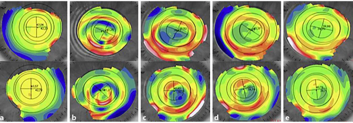
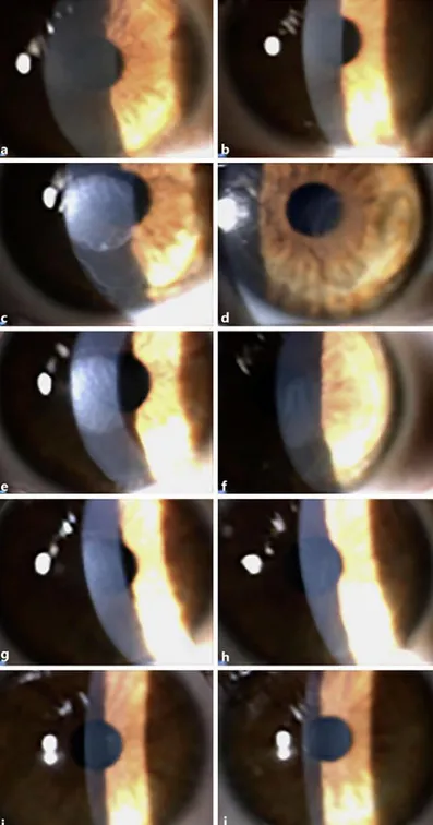
In February 2017, a 25-year-old man was referred to us for correction of his myopic astig-matism. Best corrected visual acuity was 20/20 in both eyes (cycloplegic refraction: –1.50 = –0.50 at 180° in the right eye and –1.75 = –0.25 at 180° in the left eye). Slit lamp biomicroscopy revealed normal features. Intraocular pressure by applanation tonometry was 17 mm Hg in both eyes. Topographic simulated keratometry (Sim K) readings at 3 mm were 42.23 D at 1°, 43.04 D at 91° in the right eye and 42.70 at 1°, 43.57 D at 91° in the left eye (CSO Corneal Topography MODI’02, Florence, Italy) (Fig. 1a). Central corneal thickness was 600 μm in the right eye and 610 μm in the left eye at OCT of the anterior segment (Optovue RTVue-100, USA). Femto-LASIK was performed the same day in both eyes with 150 kHz iFS® Femtosecond Laser (Abbott Medical Optics, AMO Manufacturing, USA). The corneal flap was 110 μm thick and 8.8 mm large. Laser treatment was completed using the Bausch & Lomb 217z100 Excimer Laser Platform (Rochester, NY, USA). The day after surgery, uncorrected visual acuity (UCVA) was 20/25 in the right eye and 20/20 in the left eye, but the patient complained of blurred vision in his left eye since a few hours after the procedure. At slit lamp examination a slightly diffuse whitish granular cell reaction was detected, more evident in the right eye. A diagnosis of early DLK was made (Fig. 2a, b).
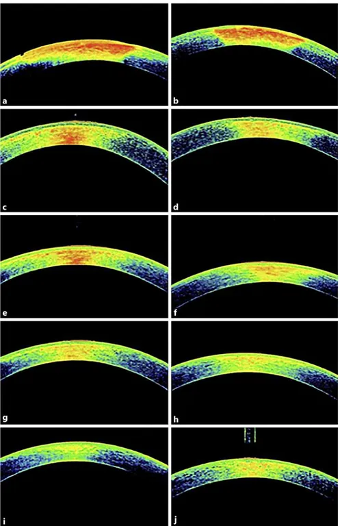
The patient was examined day by day. We prescribed topical dexamethasone eyedrops hourly for the first day and every 2 h from day 2, tobramycin eyedrops 3 times a day for the first week, and artificial tears. On day 5, slit lamp biomicroscopy showed a corneal opacity in both eyes, like a scar, more dense in the center, with corneal striae moving towards the infil-trate (Fig. 2c, d). UCVA had decreased to 20/32 in both eyes. OCT of the anterior segment showed a large hyperreflective area about 3 mm wide and 330 μm deep (Fig. 3a, b). Sim K
readings at 3 mm were 39.24 D at 97°, 38.63 D at 56° in the right eye and 37.86 D at 75°, 34.05 D at 10° in the left eye. Minimal corneal thickness was 413 μm in the right eye and 438 μm in the left eye. A diagnosis of severe DLK (grade 4) was made.
We started treatment with oral doxycycline twice a day for 2 weeks, then once a day for 1 month, and topical 10% sodium citrate 3 times a day. Dexamethasone eyedrops were pre-scribed 5 times a day for 1 month, 3 times a day for another month, and twice a day during the third month, with artificial tears.
Two days later, the infiltrate and the whitish granular cell reaction were slightly reduced. UCVA was 20/50 in both eyes. Two weeks after surgery, UCVA was 20/32, and central corneal thickness was 435 μm in the right eye and 489 μm in the left eye. We tapered topical therapy over 4 months and started treatment with topical hyperosmolar ophthalmological solution (sodium chloride 5%, sodium hyaluronate 0.15%) 3 times a day for 3 months.
One month after surgery, UCVA was 20/80 in both eyes, 20/20 with pinhole occluder; autorefractometry readings were +3.50 = –4.25 at 130° in the right eye and +4.50 = –5.75 at 25° in the left eye; the cornea was clearer (Fig. 2e, f). At the anterior segment, OCT stromal hyperreflectivity was less wide and deep (Fig. 3c, d). Corneal topography showed irregular astigmatism (Fig. 1b).
At 3 months, thin microstriae affected the flap in both eyes (Fig. 2g, h), and UCVA was >20/25 in both eyes. Sim K readings at 3 mm were 38.43 D at 156°, 39.52 D at 66° in the right eye and 39.52 D at 1°, 40.43 D at 91° in the left eye; the corneal topographic pattern was more regular and the corneal flattening area smaller (Fig. 1c). Anterior-segment OCT showed only few signs of hyperreflectivity (Fig. 3e, f).
At 6 months, UCVA was 20/25 in both eyes, OCT was almost normal (Fig. 3g, h), and Sim K readings at 3 mm were 39.69 D at 145°, 40.37 D at 55° in the right eye and 39.79 D at 11°, 41.02 D at 101° in the left eye (Fig. 1d). Central corneal thickness was 500 μm in the right eye and 450 μm in the left eye; the latter showed very thin vertical microstriae.
At 1 year, UCVA was 20/20 in both eyes, and OCT confirmed completely DLK regression (Fig. 3i, j). Topographic Sim K readings at 3 mm were 39.23 D at 167°, 39.99 D at 77° in the right eye and 39.71 D at 7°, 40.78 D at 97° in the left eye (Fig. 1e). Central corneal thickness was unchanged. Thin microstriae were still visible in the left eye (Fig. 2i, j).
Discussion
In 1998, Smith and Maloney [2] described an unusual noninfective inflammatory reaction of the interface early after LASIK in 13 cases of keratitis with whitish granular elements; the condition was called DLK. Several communications were presented on this topic in 1998, and the condition was named “sands of the Sahara” syndrome, alluding to the sand-like granular aspect of the infiltrate in the interface, nonspecific diffuse intralamellar keratitis and diffuse intralamellar keratitis [7].
The incidence of DLK reported in the literature varies between 0.2 and 1.8% [8]. It occurs after primary LASIK procedures, retreatments or lamellar procedures such as lamellar kera-toplasty and intracorneal rings segment implantation, and small incision lenticule extraction, as described recently [9–11]. In case of simultaneous bilateral surgery, DLK affects both eyes although corneal involvement should be different.
Classification of DLK is extremely important for an early diagnosis and effective treat-ment. There are various classifications reported for DLK. The most common classifications are those of Machat (1999) [8], Linebarger (2000) [12], and Azar (2001) [13]. Based on the
severity of the inflammation, DLK has been classified into four stages [13], from stage I with traces of white granular cells in the periphery of the flap, vision and refraction usually unaf-fected, to stage IV that is very rare (incidence: 1/5,000 cases), characterized by stromal melt-ing, scarrmelt-ing, and vision loss. At this stage, there is a peak in aggregation of inflammatory cells and release of collagenase by the neutrophils in the interface, which results in corneal melting and tissue loss [12–14].
Early diagnosis and appropriate treatment should resolve DLK without sequelae; the final visual outcomes are comparable to those of cases with an uneventful postoperative course, as suggested by Stulting et al. [14]. Once DLK has been diagnosed, the surgeon should follow DLK evolution with accurate biomicroscopy, autorefractometry, refractive examination, corneal topography, anterior segment OCT, and noncontact pachymetry.
The majority of surgeons agree that topical steroid-based therapy (0.2% dexamethasone, 1% prednisolone acetate, 1% rimexolone) and topical antibiotic therapy (fluoroquinolones) are the gold standard for the treatment of less severe and peripheral cases [15]. Once a more advanced stage has been identified (grade 2 Machat, stage 3 Linebarger, type II Azar), the flap can be lifted with careful irrigation of the stromal bed and of the internal face of the flap with balanced saline solution and additional washing with steroids and antibiotics, if necessary. This should be performed as soon as possible, on day 2 or 3, to interrupt the inflammatory response and to prevent corneal melting and permanent scarring [15]. However, lifting the flap can increase the incidence of striae [13], and evidence in the literature showed that treat-ment of severe DLK with topical and oral high-dose steroids produces excellent results with-out flap lifting and interface irrigation [4, 13, 14, 16].
It is also known that when the cornea is injured, corneal epithelial cells and other local cells immediately release a large number of mediators, including nitric oxide, interleukin 1, and matrix metalloproteinases (MMPs), to recruit inflammatory cells into the local injured cornea [4], so treating the corneal inflammatory response could prevent corneal melting even in the advanced stage of DLK.
Based on these data, we immediately started intensive topical therapy with dexameth-asone eyedrops in addition to prophylactic antibiotic treatment. On day 5, when slit lamp biomicroscopy showed worsening of DLK in both eyes, we started to use oral doxycycline and topical 10% sodium citrate for their anticollagenase activity, as suggested by Jarade et al. [4]. Oral doxycycline is a semisynthetic tetracycline that is well recognized for its therapeutic efficacy in treating MMP-mediated ocular surface diseases such as rosacea, recurrent epithe-lial erosions, and ocular burns. Doxycycline can promote wound healing by reducing inflam-mation and protease activity of MMPs, by inhibiting macrophage recruitment, and by decreas-ing vascular endothelial growth factor C, interleukin 1b, inducible nitric oxide synthase, and tumor necrosis factor-α expression. By chelating divalent actions, sodium citrate might inter-fere with polymorphonuclear cell diapedesis and complement activation; it has been reported to decrease the production of chemotactic factors and to inhibit the stimulation and release of enzymes from polymorphonuclear cells [4].
In our case, improvements in topographic pattern (Fig. 1), corneal thickness, anterior seg-ment OCT (Fig. 3), and corneal transparency were evident since the second treatseg-ment week and at 3 months (Fig. 2g, h), 6 months, and 1 year (Fig. 2i, j), respectively. OCT documented a reduction in corneal reflectivity from the periphery to the center and from the deeper stroma towards the surface during follow-up (Fig. 3). No corneal scarring or permanent vision loss were detected and, even if visual acuity was the same in both eyes, the patient reported a worse quality of vision in the left eye affected by residual thin microstriae.
In conclusion, DLK represents an infrequent complication after femto-LASIK. It should resolve without sequelae if promptly diagnosed and treated. High-dose topical steroids, hy-perosmolar ophthalmological solution, oral doxycycline, and 10% sodium citrate eyedrops could promote corneal healing, without necessity of corneal flap lifting.
Statement of Ethics
The study was carried out in accordance with the Declaration of Helsinki. Disclosure Statement
The authors have no conflict of interest or financial interest to disclose. References
1 Buratto L, Brint SF. Custom LASIK – surgical techniques and complications. Thorofare: Slack; 2003. p. 214–9, 242–6.
2 Smith RJ, Maloney RK. Diffuse lamellar keratitis. A new syndrome in lamellar refractive surgery.
Ophthalmology. 1998 Sep;105(9):1721–6.
3 Maddox R, Hatsis A. Shifting sands of the Sahara: interface inflammation following LASIK. In: Gimbel HV, Anderson Penno EE, editors. LASIK complications; Prevention and Management. Thorofare (NJ): Slack; 1999. pp. 30–6.
4 Jarade E, Slim E, Antoun J, Khzam RA. Treatment of grade IV diffuse lamellar keratitis with oral doxycycline and topical 10% sodium citrate. Can J Ophthalmol. 2016 Dec;51(6):e178–84.
5 Hoffman RS, Fine IH, Packer M. Incidence and outcomes of LASIK with diffuse lamellar keratitis treated with topical and oral corticosteroids. J Cataract Refract Surg. 2003 Mar;29(3):451–6.
6 Agarwal, Amar; Kumar et al. Thieme Verlagsgruppe, Stuttgart, New York, Delhi, Rio 2015. Essentials of OCT in Ocular Disease, Part 2. Anterior-Segment Optical Coherence Tomography.
7 Kaufman SC, Maitchouk DY, Chiou AG, Beuerman RW. Interface inflammation after laser in situ keratomileusis. Sands of the Sahara syndrome. J Cataract Refract Surg. 1998 Dec;24(12):1589–93. 8 Machat JJ. The art of LASIK. Thorofare: Slack; 1999. p. 393–6.
9 Zhao J, He L, Yao P, Shen Y, Zhou Z, Miao H, et al. Diffuse lamellar keratitis after small-incision lenticule extraction. J Cataract Refract Surg. 2015 Feb;41(2):400–7.
10 Tomita M, Sotoyama Y, Yukawa S, Nakamura T. Comparison of DLK incidence after laser in situ
keratomileusis associated with two femtosecond lasers: Femto LDV and IntraLase FS60. Clin Ophthalmol. 2013;7:1365–71.
11 Choe CH, Guss C, Musch DC, Niziol LM, Shtein RM. Incidence of diffuse lamellar keratitis after LASIK with 15 kHz, 30 kHz, and 60 kHz femtosecond laser flap creation. J Cataract Refract Surg. 2010 Nov;36(11):1912–8. 12 Linebarger EJ, Hardten DR, Lindstrom RL. Diffuse lamellar keratitis: diagnosis and management. J Cataract
Refract Surg. 2000 Jul;26(7):1072–7.
13 Johnson JD, Harissi-Dagher M, Pineda R, Yoo S, Azar DT. Diffuse lamellar keratitis: incidence, associations, outcomes, and a new classification system. J Cataract Refract Surg. 2001 Oct;27(10):1560–6.
14 Stulting RD, Randleman JB, Couser JM, Thompson KP. The epidemiology of diffuse lamellar keratitis. Cornea. 2004 Oct;23(7):680–8.
15 Steinert RF, Swami AU. Diffuse interface keratitis. Rev Ophthalmol 2001;8(suppl to Jan):46–52.
16 Michieletto P, Balestrazzi A, Balestrazzi A, Alegente M, Boccassini B. Stage 4 diffuse lamellar keratitis after laser in situ keratomileusis Clinical, topographical, and pachymetry resolution 5 years later. J Cataract Refract Surg. 2006 Feb;32(2):353–6.
Fig. 1. Corneal topography, tangential map, of the right eye (upper line, from left to right) and of the left eye (bottom line, from left to right) during follow-up. a Before surgery. b One month after surgery. c Three months after surgery. d Six months after surgery. e One year after surgery.
Fig. 2. Digital slit lamp images of the corneal surface of the right eye (left column) and of the left eye (right column) during follow-up. a, b One day after surgery. c, d Five days after surgery. e, f One month after surgery. g, h Three months after surgery, i, j One year after surgery.
Fig. 3. OCT of the anterior segment of the right eye (left column) and of the left eye (right column) during follow-up. a, b Five days after surgery. c, d One month after surgery. e, f Three months after surgery. g, h Six months after surgery. i, j One year after surgery.