Received: 28 May 2020 Revised: 26 December 2020 Accepted: 13 January 2021 DOI: 10.1002/jev2.12066
R E S E A R C H A R T I C L E
Proteome-minimized outer membrane vesicles from
Escherichia
coli as a generalized vaccine platform
Ilaria Zanella
Enrico König
Michele Tomasi
Assunta Gagliardi
Luca Frattini
Laura Fantappiè
Carmela Irene
Francesca Zerbini
Elena Caproni
Samine J. Isaac
Martina Grigolato
Riccardo Corbellari
Silvia Valensin
Ilaria Ferlenghi
Fabiola Giusti
Luca Bini
Yaqoub Ashhab
Alberto Grandi
,Guido Grandi
1Department of Cellular, Computational and Integrative Biology (CIBIO), Laboratory of Synthetic and Structural Vaccinology, University of Trento, Trento, Italy 2Toscana Life Sciences Foundation, Siena, Italy
3GlaxoSmithKline Vaccines, Siena, Italy
4Department of Life Sciences, Functional Proteomics Laboratories, University of Siena, Siena, Italy 5Palestine-Korea Biotechnology Center, Palestine Polytechnic University, Hebron, Palestine 6BiOMViS Srl, Siena, Italy
Correspondence
Prof. Guido Grandi, CIBIO, University of Trento, Via Sommarive, 9, 38123 Trento, Italy. Email:[email protected]
Current address
Francesca Zerbini, Medimmune/Astra Zeneca, Granta Park, CB21 6GP, Cambridge, UK. Ilaria Zanella and Enrico König contributed equally to this study.
Funding information
Advanced European Research Council, Grant/Award Number: OMVAC 340915; National Funding PRIN-2015, Grant/Award Number: CUPE62F16001630001
Abstract
Because of their potent adjuvanticity, ease of manipulation and simplicity of produc-tion Gram-negative Outer Membrane Vesicles OMVs have the potential to become a highly effective vaccine platform. However, some optimization is required, includ-ing the reduction of the number of endogenous proteins, the increase of the loadinclud-ing capacity with respect to heterologous antigens, the enhancement of productivity in terms of number of vesicles per culture volume. In this work we describe the use of Synthetic Biology to create Escherichia coli BL21(DE3)Δ60, a strain releasing OMVs (OMVsΔ60) deprived of 59 endogenous proteins. The strain produces large quantities of vesicles (> 40 mg/L under laboratory conditions), which can accommodate recom-binant proteins to a level ranging from 5% to 30% of total OMV proteins. Moreover, also thanks to the absence of immune responses toward the inactivated endogenous proteins, OMVsΔ60 decorated with heterologous antigens/epitopes elicit elevated antigens/epitopes-specific antibody titers and high frequencies of epitope-specific IFN-γ-producing CD8+ T cells. Altogether, we believe that E. coli BL21(DE3)Δ60 have the potential to become a workhorse factory for novel OMV-based vaccines.
K E Y W O R D S
cancer, infectious diseases, outer membrane vesicles (OMVs), synthetic biology, vaccines
INTRODUCTION
More than 60 years ago, it was accidentally discovered that Gram-negative bacteria release Outer Membrane Vesicles (OMVs) (Bishop & Work,1965; Chatterjee & Das,1967). As often happened in science, the discovery remained almost unnoticed for
This is an open access article under the terms of theCreative Commons AttributionLicense, which permits use, distribution and reproduction in any medium, provided the original work is properly cited.
© 2021 The Authors. Journal of Extracellular Vesicles published by Wiley Periodicals, LLC on behalf of the International Society for Extracellular Vesicles
J Extracell Vesicles. 2021;10:e12066. wileyonlinelibrary.com/journal/jev2 of
of ZANELLA et al.
several years and only recently, scientists became aware of the amazing aspects of the OMV biology and of their potential for biotechnological applications. OMVs are closed spheroid particles of heterogeneous size, 50–300 nm in diameter, generated through a ‘budding out’ of the bacterial outer membrane. They represent a distinct secretory pathway with a multitude of func-tions, including inter and intra species cell-to-cell cross-talk, biofilm formation, genetic transformation, defence against host immune responses, and toxin and virulence factor delivery to host cells (Ellis & Kuehn,2010; Kulp & Kuehn,2010).
OMVs are becoming an attractive vaccine platform for their excellent adjuvanticity (Chen et al.,2010; Ellis & Kuehn,2010), the possibility of decorating them with heterologous antigens (Gerritzen et al.,2017; Irene et al.,2019; Kesty & Kuehn,2004), and the simplicity of their production and purification process (Berlanda Scorza et al.,2012). Indeed, OMV-based vaccines have already reached the market (Ladhani et al.,2016; Serruto et al.,2012), while others are in advanced clinical phases (Gerke et al.,
2015; Rossi et al.,2016).
OMV-based vaccines can be classified in two categories. The first one includes vaccines constituted by OMVs directly purified from the pathogen of interest (Kaparakis-Liaskos & Ferrero,2015; Van Der Pol et al.,2015). The rationale of developing such vac-cines is that OMVs provide both the immune stimulatory molecules (adjuvants) and the protective antigens. The prototype of this category is Bexsero, the Meningococcal Group B vaccine, which, in addition to purified meningococcal antigens, is formulated with vesicles from the Neisseria meningitidis New Zealand strain (Parikh et al.,2016). The second category includes OMVs dec-orated with heterologous antigens and obtained from a properly engineered Gram-negative bacterial species (Kaparakis-Liaskos & Ferrero,2015; Van Der Pol et al.,2015). Such vaccines exploit the excellent adjuvanticity properties of OMVs and have the advantage to potentially target any kind of bacteria, viruses and cancer, provided that specific protective antigens are selected.
No vaccines belonging to the second category are currently available for humans. This is in part because for its full-blown development, the OMV platform still needs to be optimized. Such optimization should include the development of efficient strategies for OMV engineering, the modulation of the OMV adjuvanticity to avoid potential reactogenicity, and the elimination of unnecessary OMV endogenous proteins.
Here, we describe the application of Synthetic Biology for the construction of a novel E. coli strain, named E. coli BL21(DE3)Δ, releasing OMVs (OMVsΔ) deprived of 59 endogenous proteins. The rationale for creating such strain is threefold. First, we hypothesized that by eliminating OMV endogenous proteins we could increase the loading capacity of OMVs with respect to recombinant antigens. Second, we predicted that heterologous antigens expressed in proteome-minimized OMVs should be more immunogenic because of their increased amount in OMVs and of the absence of endogenous proteins that could dilute the antigen-specific immune responses. Third, in view of human applications, the availability of OMVs carrying a reduced number of irrelevant proteins should offer an important additional advantage. As predicted, we show that OMVsΔhave a number of important features, which ultimately lead to the elicitation of potent antibodies and T cell responses against heterologous antigens. Overall, our data indicate that E. coli BL21(DE3)Δ is a particularly useful strain for the development of effective vaccines against infectious diseases and cancer.
MATERIALS AND METHODS
.
Bioinformatics analysis and software
The E. coli BL21(DE3) proteome was analyzed to predict membrane-associated and periplasmic proteins. A meta-prediction pipeline that employs different subcellular localization prediction tools in three phases of positive and negative selections was used (Hisham & Ashhab,2018). The identified proteins were subsequently assessed for their biological function and classified (essential vs. non-essential) according to the ‘Keio collection’ (Baba et al.,2006), and EcoCyc E. coli database (Keseler et al.,2017), respectively. Oligonucleotides for guide DNAs, donor DNAs and primers were designed using Serial Cloner and Benchlink and BLAST was used for alignments (Altschul et al.,1990). Densitometry was performed with Image Studio Lite Version 5.2 (Li-Cor Biosciences, Lincoln, NE USA) and for 2-DE map analysis, ImageMaster 2D Platinum v.6.0 software was used (GE Healthcare, Chicago, IL USA).
.
Bacterial strains, culture conditions and plasmids
E. coli strains (BL21(DE3) derivatives, DH5α, TOP10) were routinely cultured in lysogeny broth (LB Miller; Merck, Darmstadt,
Germany) supplemented with 100 mg/L ampicillin (Carl Roth, Karlsruhe, Germany), 25 mg/L chloramphenicol (Merck, Darm-stadt, Germany), 50 mg/L kanamycin (Thermo Fisher Scientific, Waltham, MA USA) or a combination of them, where necessary. For recombinant protein expression in OMVs, cultures were supplemented with 0.1 mM isopropyl-β-D-thiogalactopyranoside (IPTG, Merck, Darmstadt, Germany) at OD600between 0.5 and 0.7 and OMVs were collected after 4 h from induction.
ZANELLA et al. of
then growth continued at 25◦C, pH 6.8 (±0.2), dO2 > 30%, 280–500 rpm. At OD6001.0, the culture was induced with 0.1 mM
IPTG together with a feed consisting in ampicillin (50 mg/L) or chloramphenicol (12.5 mg/L), glycerol (15 g/L), and MgSO4
(0.25 g/L).
Plasmids encoding FhuD2, SpAKKAA, Csa1, HlaH35L, FhuD2-mFAT1 and Nm-NHBA were already described (Fantappiè et al.,
2017; Grandi et al.,2018; Irene et al.,2019). For the construction of plasmids encoding ClfAY338A, FhuD2-Bp, FhuD2-OVA and
FhuD2-hFAT1, the PIPE-PCR method was used (Klock & Lesley,2009). Briefly, the coding sequences were chemically synthetized (Geneart, Thermo Fisher Scientific, Waltham, MA USA) and PCR amplified or several oligonucleotides were assembled stepwise (in case of Bp) using the primers reported in Table S3. In parallel, a pET21b+ derivatives carrying either the sequence encoding the leader peptide for secretion of E. coli Lpp (pET-Lpp plasmid) or Lpp-FhuD2 (for epitope fusions) (Irene et al.,2019) were linearized using the primer couples Lpp-R-plasmid/nohisflag and Lpp-FhuD2-R-plasmid/nohisflag, respectively (Table S3). The PCR products were digested with DpnI (New England Biolabs, Ipswich, MA USA), combined (2μL each) and used to transform
E. coli HK100 chemically competent cells. Positive clones identified by colony PCR using GoTaq Green Master Mix (Promega,
Madison, WI USA) and gene specific primer (Table S3) were subjected to sequence analysis (Eurofins Scientific, Luxemburg) before using the resultant plasmids (Table S3) for transformation of OMV producer strains.
.
Construction of
E. coli BL(DE)Δ60
Gene deletions were accomplished using our previously established CRISPR/Cas9-based genome editing protocol (Zerbini et al.,
2017). The preparation of the 90 pCRISPR-SacB-gDNA plasmids were done using double-stranded synthetic oligonucleotides (Merck, Darmstadt, Germany) encoding for the guide RNA reported in Table S4. Stepwise single gene mutagenesis was achieved by co-transformation of electrocompetent cells of the recipient strain (E. coli BL21(DE3)ΔompA_pCasRed, E. coli Δ_pCasRed, etc.) with the gene-specific pCRISPR-SacB-gDNA and its respective double-stranded donor DNA (ds-dDNA) (Table S5). Gene-specific primer pairs used for screening of colonies with GoTaq Green Master Mix (Promega, Madison, WI USA) are listed in Table S6. Genomic DNA was extracted from E. coli BL21(DE3)ΔompA and E. coli BL21(DE3)Δ using PowerSoil DNA Isolation Kit (Mo Bio Laboratories, Carlsbad, CA USA) and the genomes were fully sequenced. Briefly, libraries were prepared using the Nextera XT DNA Library Preparation Kit (Illumina, San Diego, CA USA), and library quality was assessed using the Caliper LabChip GX (High-Throughput Bioanalyzer) following manufacturers guidelines. Sequencing was performed on the HiSeq 2500 system (Illumina, San Diego, CA USA).
.
Expression and purification of recombinant proteins
FhuD2 and HlaH35LS. aureus proteins were purified as previously described (Irene et al.,2019). The genes encoding the E. coli
BL21 proteins Agp, CpoB, EfeO, FepA, GlpQ, LamB, MalE, MalM, OppA, Pal, YdcL and YncD were amplified from chromosomal DNA and cloned as His-TAG C-terminal fusions using linearized pET15+ plasmid using PIPE method (Table S3). Proteins were purified using Ni-NTA resin (Qiagen, Hilden, Germany) following standard purification procedures.
.
OMV purification and electrophoretic analysis
For assessment of OMV productivity of each mutant we used 50 ml culture volume, whereas OMVs for antigen expression analysis and batch preparation for immunization were purified from 200 ml and 1 L cultures, respectively. OMVs were collected from culture supernatants by filtration through a 0.22μm pore size filter with a syringe for small volumes up to 50 ml (Merck, Darmstadt, Germany) or with a filter system for larger volumes (Starlab, Hamburg, Germany). Small volumes (50 ml and 200 ml) were applied to high-speed centrifugation (174,900 x g for 2 h at 4◦C) and pellets containing OMVs were resuspended in PBS (Gibco brand, Thermo Fisher Scientific, Waltham, MA USA). Purification of OMVs from cultures in bioreactor was carried out using Tangential Flow Filtration (ÄKTA flux system; GE Healthcare, Chicago, IL USA) with a Hollow Fibre cartridge UFP-500-C-3MA (GE Healthcare, Chicago, IL USA). Protein content of OMVs was quantified using DC Protein Assay (Bio-Rad) and the quality of OMVs was monitored by SDS-PAGE loading OMVs (normalized byμg) on Any kD Criterion TGX Stain-Free Protein Gel (Bio-Rad Laboratories, Hercules, CA USA) stained using ProBlue Safe Stain (Giotto Biotech, Sesto Fiorentino, Italy). The detailed purification protocol has been submitted to EV-TRACK (ID EV200158).
2-DE was performed as previously reported (Fantappiè et al.,2017; Gagliardi et al.,2016). For analytical and MS-preparative gels, 250μg and 500 μg of protein, respectively, were diluted in 350 μL of denaturation buffer and 0.2% or 2% (v/v) IPG-buffer (pH 3–10; GE Healthcare, Chicago, IL USA). Analytical gels and MS-preparative gels were stained with ammoniacal silver nitrate and MS-compatible silver staining, respectively, before being scanned using the ImageScanner III (GE Healthcare, Chicago,
of ZANELLA et al.
IL USA). Image analysis was performed on 2-DE analytical gels using the ImageMaster 2D Platinum v.6.0 software (GE Healthcare, Chicago, IL USA). For each OMV preparation, OMVsΔompA and OMVsΔ, image analysis was carried out on three different spot maps. Qualitative differences between OMVsΔompAand OMVsΔ were considered significant only when absence/presence of protein spots was observed in all three corresponding gels.
.
Negative staining electron microscopy method
A volume of 5μl of OMV diluted at 80 ng/μl in PBS were loaded onto a copper 200-square mesh grid of carbon/formvar rendered hydrophilic by glow discharge using a Q150R S (Quorum, East Sussex, UK). The excess solution was blotted off after 30 s using Whatman filter Paper No.1. The grids were negatively stained with NanoW (Nanoprobes, Yaphank, NY USA) for 30 s, then blotted using Whatman filter Paper No.1 and finally let air dry. Micrographs were acquired using a Tecnai G2 Spirit Transmission Electron Microscope equipped with a CCD 2kx4k camera at a final magnification of 120000x.
.
Protein identification by mass spectrometry
Protein identification was carried out by peptide mass fingerprinting (PMF) using an Ultraflex III MALDI-TOF/TOF mass spectrometer (Bruker Daltonics, Billerica, MA USA), equipped with a 200 Hz smartbeam I laser, as already described (Fan-tappiè et al.,2017; Gagliardi et al.,2016). Mass spectra were acquired in reflector positive mode with a laser frequency set to 100 Hz. Flex Analysis software v.3.0 was used for analysis. Acquired spectra were internally calibrated using auto-proteolytic trypsin peptides (842.509 m/z and 2,211.105 m/z) and contaminants were removed from the obtained mass lists. PMF search-ing was carried out against entries for E. coli in the SwissProt database ussearch-ing Mascot software (Matrix Science Ltd., London, UK, www.matrixscience.com) with 100 ppm as peptide tolerance, monoisotopic peptide mass, allowing one missed cleavage by trypsin, carbamidomethylation of cysteine as fixed modification, and oxidation of methionine as variable modification. The parameter used to accept identifications was the default Mascot protein score greater than 56, corresponding to a 5% probability that the observed match was a random event (P< 0.05). The number of matched peptides and the extent of sequence coverage were also considered to confirm protein identification. Protein spots that emerged as significant qualitative differences along with the MS data for those that were successfully identified by MS are reported in Table S2.
.
Assessment of phospholipid content, particle size and dimension of OMVs
For lipid quantification, styrylic membrane dye FM4-64 (Thermo Fisher Scientific, Waltham, MA USA) at a final concentration of 3.3μg/ml was added to 125/250 ng of OMVs (protein content) resuspended in PBS in a black 96-well OptiPlate (Perkin Elmer, Waltham, MA USA). As standard, we used E. coli L-α-phosphatidylglycerol (Merck, Darmstadt, Germany) in a range between 5
μg and 0.15 μg. The reaction was incubated for 10 min at 37◦C and fluorescence (excitation at 485 nm, emission at 670 nm) was
measured with an Infinite M200PRO plate reader (Tecan, Männedorf, Switzerland).
Assessment of particle size and dimension on OMVs diluted in PBS to a concentration of 100 ng/ml was performed using a Nanosight NS300 (Malvern Instruments, Malvern, UK) with a 532 nm laser, camera level 15, slider shutter and gain of 1206 and 245, respectively. Three videos of 60 s were recorded and analyzed with the software NTA 3.4 Build 3.4.003.
.
Toll-Like-Receptor agonistic activity and IL- stimulation
OMV agonistic activity of human Toll Like Receptor 2 (hTLR2) and mouse Toll Like Receptor 4 (mTLR4) was measured using HEK-Blue™ hTLR2 or HEK-Blue™ mTLR4 Cells, respectively, and QUANTI-Blue (InvivoGen, San Diego, CA USA) as substrate as previously described (Irene et al.,2019). For the IL-6 assay, THP-1 human leukemic monocyte cells were cultured in RPMI 10% FBS and differentiated into macrophages by adding 10 ng/ml of phorbol 12-myristate 13-acetate (PMA) and incubating cells at 37◦C 5% CO2for 48 h. Then 4× 10⁵cells in a volume of 10μl/well of RPMI culture medium were aliquoted into a 96 well-plates
(Corning, Corning, NY USA). Different concentration of OMVs and purified LPS (control) were added to 90μl/well of RPMI in duplicate starting from an initial concentration of 10,000 ng/ml to a final concentration of 10 pg/ml with 10-fold serial dilutions. Cells and OMVs were incubated at 37◦C O/N. IL-6 release in supernatants was measured by Human IL-6 Uncoated ELISA™ Kit (Thermo Fisher Scientific, Waltham, MA USA). Corning Costar ELISA plates were coated with 100μL/well of anti-human IL-6 antibodies and incubated at 4◦C O/N. The day after 100μL/well of cell supernatants were transferred to the plate and incubated 2 h at room temperature (RT) and the assay was completed following the manufacturer’s protocol. Plates were read at 450 nm using a SpectraMax M2 Microplate reader (Molecular Devices, San José, CA USA).
ZANELLA et al. of
.
Animal experiments
All mice were purchased from Charles River Laboratories (Wilmington, MA USA). Animal experiments were carried out in accordance with experimental protocols reviewed and approved by the Animal Ethical Committees of University of Trento (Trento, Italy) and Toscana Life Sciences (Siena, Italy) and by the Italian Ministry of Health. Animal health was evaluated using a 0 (no signs of stress and suffering) to 6 (severe signs of stress and suffering) ‘pain score’ scale approved by the Animal Wel-fare Committees of University of Trento and Toscana Life Sciences in accordance with the ‘Working document on a severity assessment framework - National Competent Authorities for the implementation of Directive 2010/63/EU on the protection of animals used for scientific purposes’. The evaluation considers the recommendations of the Federation of European Laboratory Animal Science Associations (FELASA) (Baumans et al.,1994). This ‘pain score’ scale is based on objective parameters and are assigned as follows: score 0: normal hair, mucosa, physical activity and nourishment; score 1: ruffled hair coat (piloerection), lesions or dehydration; score 2: as in 1 and reduced physical activity (e.g., grooming, exploration); score 3: as in 2 and lethargy and loose< 10% body weight; score 4: as in 3 and signs of respiratory impairment (dyspnoea or tachypnea); score 5: as in 4 and loss of body weight between 10% and 20%, lordosis or kyphosis; score 6: as in 5 and body weight loss> 20% (animals have to be sacrificed). For antibody titers, female BALB/c mice (5 per group) were immunized intraperitoneally (i.p.) on days 0, 14 and 28 with 2μg of OMVs in the presence of Alum (2 mg/ml). Blood was collected by cardiac puncture from euthanized mice at day 35 and serum was obtained from blood through centrifugation at 2,000 rpm for 10 min. For T cell analysis, C57BL/6 mice were subcutaneously (s.c.) injected on days 0 and 7 with 10μg of OMVsΔ60+ 5 μg of either OVA or SV40 synthetic peptides. At day 12, spleens were collected and used to perform intracellular cytokine staining for subsequent analysis.
.
Analysis of OMV immunogenicity by western blot and ELISA
Purified recombinant E. coli proteins (1μg each) or total OMV proteins (10 μg) were loaded on Any kD Criterion TGX Stain-Free Protein Gel (Bio-Rad, Laboratories, Hercules, CA USA) and then transferred onto a nitrocellulose membrane using iBlot transfer stack (Thermo Fisher Scientific, Waltham, MA USA). Sera from mice immunized with OMVsΔompAand OMVsΔwere used at 1:1,000 dilutions and immunogenic proteins were detected with anti-mouse IgG-HRP antibodies (Agilent, Santa Clara, CA USA) at a dilution of 1:2,000 using ECL Select Western Blotting Detection Reagent (GE Healthcare, Chicago, IL USA).
Antigen specific antibody titers elicited by engineered OMVs were measured by ELISA as previously described (Irene et al.,
2019) using FhuD2, HlaH35L, Burkholderia pseudomallei flagellin epitope (Bp) as recombinant proteins and D8-FAT peptide
(IQVEATDKDLGPNGHVTYSIVTDTD; Genscript Biotech, Piscataway Township, NJ USA) (200 ng each).
.
HLA neutralization assay
HLA neutralization assay was performed as previously described (Irene et al.,2019).
.
Analysis of OVA and SV-specific T cells
Percentage of OVA- and SV40-specific T cells in immunized mice was performed as previously described (Grandi et al.,2018). Statistical significance was assessed using two-tailed Student’s t-test.
RESULTS
.
Selection of endogenous proteins to be eliminated from
E. coli-derived OMVs
With the final goal to create an E. coli derivative releasing ‘proteome-minimized’ OMVs, we first experimentally defined the pro-teome of OMVs from the hypervesiculating strain E. coli BL21(DE3)ΔompA by using two-dimensional gel electrophoresis (2-DE) coupled to mass spectrometry analysis (MS) as described in Fantappiè et al (Fantappiè et al.,2017). A total of 128 sequences were identified corresponding to 81 unique E. coli proteins. 2-DE-MS was coupled to bioinformatics analysis of the E. coli BL21(DE3) genome aimed at predicting all periplasmic and outer membrane proteins. We selected 504 proteins, which included all the 81 proteins identified by MS. Next, from these 504 proteins we excluded those ones that were classified as “indispensable” according to the Keio library (Baba et al.,2006), and we finally generated a priority list of 90 proteins, 63 of which identified by MS on 2D map, to be eliminated from our progenitor strain E. coli BL21(DE3)ΔompA (Table S1).
of ZANELLA et al.
F I G U R E Creation of E. coli BL21(DE3)Δ strain releasing proteome-minimized OMVs. (a) The selected 90 genes (+ ompA) encoding dispensable periplasmic and outer membrane proteins are reported above and below the arrow according to the order used for CRISPR/Cas9-guided inactivation. ‘D’ indicates the genes that were successfully inactivated and ‘ND’ those genes whose inactivation either failed or was obtained but resulted in an impaired growth and/or OMV production and therefore were excluded (written in ‘red’). For each mutant strain, the average (n= 3) OMV productivity (mg OMV (protein-based)/litre/OD600 of culture) is reported and compared to the productivity of the progenitor E. coli BL21(DE3)ΔompA strain (dotted line). Bar colours indicate genes encoding outer membrane proteins (magenta), lipoproteins (green), periplasmic proteins (blue). Error bars, mean± s.d. (b) Left pie chart. Classification of the 91 proteins (90 selected proteins+ OmpA) according to their predicted compartmentalization (colour code as in a). The total number of proteins belonging to each category is written inside each slice. Right pie chart. Same representation as on the left, in which each slice is subdivided to highlight the number of the corresponding genes that were deleted (D, solid slices) or not-deleted (ND, open slices). (c) The 90 selected genes+ ompA are grouped into eight categories according to the biological/physiological function of their protein products. Bars (colour code as in A) indicate the total number of genes (T) and the number of deleted (D) and non-deleted (ND) genes of each category
.
Construction of
E. coli BL(DE)Δ60 releasing proteome-minimized OMVs
We previously published a CRISPR/Cas9-based genome editing protocol, which allows the cumulative inactivation of any dis-pensable gene in E. coli at a pace of one mutation every two working days (Zerbini et al.,2017). By using this protocol, we attempted the stepwise inactivation of the 90 selected genes by deleting 30 nucleotides at the 5′ end of the coding region of each mature protein and by introducing an in-frame stop codon (Figure1a, Fig. S1). Before moving to the next gene editing cycle, growth kinetics and OMV productivity was assessed to exclude mutants with impaired growth and/or OMV productivity. At the end of this brute force approach, a mutant, named E. coli BL21(DE3)Δ, was generated, which carries, in addition to the mutation in the ompA gene present in the progenitor E. coli BL21(DE3)ΔompA strain, the inactivation of 57 out of 90 genes (63%). Of the remaining 33 genes, six could be deleted in the presence of the preceding mutations (written in red in Figure1a) but the mutants had impaired growth kinetics, while the inactivation of the other 27 genes was not compatible with the genetic background of the recipient strains. Most of the non-viable mutations were lipoproteins and periplasmic proteins (Figure1b) and included genes involved in cell envelope assembly (5 out of 9) and molecular chaperones activity (6 out of 7) (Figure1c). Interestingly, all mutants showed an OMV productivity higher than the E. coli BL21(DE3)ΔompA progenitor, with some muta-tions (mltA, bglX, emtA and mlaA) appearing particularly effective in promoting vesiculation (Figure1a). Finally, to reduce the
ZANELLA et al. of
F I G U R E Comparison of two-dimensional electrophoresis of OMVsΔompA and OMVsΔ - Silver stained 2-DE gels of OMVs from E. coli BL21(DE3)ΔompA (a) and E. coli BL21(DE3)Δ (b). Red circles visualize those protein spots visible in OMVsΔompAand not visible in OMVsΔ. All spots not present in OMVsΔwere removed from the OMVsΔompAgel, digested with trypsin and analyzed by mass spectrometry. Zoom areas (c) are taken as examples of qualitative differences between the two gels and report the ID of the MS-identified proteins (see Table S1 for annotations)
potential reactogenicity of LPS-associated OMVs, the genome editing process was completed with the addition of msbB and
pagP gene inactivation, which results in the synthesis of a penta-acylated Lipid A (Somerville et al.,1996; Vorachek-Warren et al.,
2002). The final strain, carrying a total of 60 gene deletions, was named E. coli BL21(DE3)Δ60.
The correctness of each gene inactivation was first verified by PCR and sequence analyses using primers spanning the mutated regions (Fig. S1) and subsequently by sequencing of the whole genome that has been deposited at GenBank under the accession JADXDS000000000. Moreover, the disappearance from the vesicular compartment of the proteins encoded by the deleted genes was followed by comparing the 2D map of OMVsΔand OMVsΔompA. A total of 98 protein spots were no longer visible in the 2-DE map of OMVsΔ(Figure2). Of these 98 spots, 75 were successfully identified in OMVsΔompA2-DE map by MS analysis, and they corresponded to 46 unique proteins, 43 of which encoded by the deleted genes (Table S2 and Fig. S2). The disappearance of three proteins not encoded by the deleted genes (Table S1) could be ascribed to the fact that their expression level, which was already low in OMVsΔompA, could be further reduced by the inactivation of other genes. As far as the remaining 16 out of the 59 inactivated proteins are concerned, two are MsbB and PagP and the other 14 belong to the group of proteins that were selected by bioinformatics analysis. From the 2D map analysis of OMVsΔthe disappearance of the 59 proteins did not correspond to a substantial increase of specific protein spots. The stained material that appeared at the centre of the 2D gel probably represents a technical artefact since it does not carry proteinaceous material, as judged by mass spectrometry analysis.
.
Properties of
E. coli BL(DE)Δ60 and OMVs
Δ60Compared to its progenitor E. coli BL21(DE3)ΔompA, E. coli BL21(DE3)Δ had similar growth kinetics, but released approxi-mately 3-folds more OMVs (Figure3b). OMVs from both strains were purified from the culture supernatants and their size and quality was analyzed by electron microscopy and light scattering using a Nanosight NS300 set with 532 nm laser. As shown in
of ZANELLA et al.
F I G U R E Properties of E. coli BL21(DE3)Δ and OMVsΔ60. (a) Morphology of OMVsΔompAand OMVsΔanalyzed by electron microscopy. (b) Comparison of growth kinetics and OMV productivity between E. coli BL(DE)Δ and E. coli BL(DE)ΔompA. E. coli BL21(DE3)Δ (▲) and E. coli BL21(DE3)ΔompA (♦) were grown in a bioreactor over a 10 h period and 50 ml cultures were collected at different time points and used for OMV purification and quantitation. OMVs productivity is given as mg/L (protein content). (c) Determination of OMV size - Purified OMVs were characterized using a Nanosight NS300. The size value is given as an average of two independent biological replicates, analyzed in duplicate. (d) Determination of particle number - OMVs were analyzed at a normalized concentration of 100 ng/ml and the number, determined using Nanosight NS300, represents the average value obtained from two OMV preparations and two independent analyses. (e) Determination of lipid content in OMVs - Styrylic membrane dye FM4-64 (Thermo Fisher Scientific, Waltham, MA USA) at a final concentration of 3.3μg/ml was added to 125 and 250 ng of OMVs. After 10 min at 37◦C fluorescence was measured (excitation at 485 nm, emission at 670 nm) and lipid content was estimated asμg/μg OMVs (protein based) using E. coli L-α-phosphatidylglycerol as standard. (f) Analysis of TLR agonistic activity of OMVs - HEK-Blue mTLR4 cells were incubated with different amounts of either OMVsΔompA(♦) or OMVsΔ(▲). After 17 h the levels of secreted alkaline phosphatase (SEAP) secreted upon mTLR4 stimulation by OMVs were determined by reading the absorbance of the culture supernatant at 655 nm in duplicate. (g) In vitro release of IL- by macrophages upon OMV stimulation - THP-1 human leukemic monocyte cells (4× 10⁵cells in 10μL/well) differentiated to macrophages by PMA were incubated with different concentrations of OMVsΔompA(♦) and OMVsΔ(▲) in a final volume of 100 μL/well. IL-6 release in the supernatants was measured by Human IL-6 Uncoated ELISA Kit (Thermo Fisher Scientific, Waltham, MA USA) in duplicate. (h) Body weight analysis of mice immunized with OMVsΔ- Mice (5 mice/group) were immunized three times at 2-week intervals with 10μg of OMVsΔ60/dose in 100μL PBS containing 2 mg/ml of Alum hydroxide. Immunization with 100μL PBS containing 2 mg/ml of Alum hydroxide was used as control. Body weight of each mouse was measured at 3–5 day intervals over a period of 30 days. Curves are reported as average body weight (n= 5) of mice immunized with OMVsΔ60(▲) or alum (○). Error bars in C-F and H: mean± s.d. Error bars in F and G: mean ± s.e.m.
ZANELLA et al. of
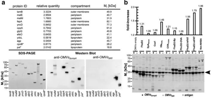
F I G U R E Immunogenicity of endogenous OMV proteins and expression of heterologous antigens in OMVs - (a) Ten proteins identified in OMVsΔompA by mass spectrometry were expressed as His-TAG fusions in E. coli BL21(DE3) and purified by IMAC. Purified proteins were analyzed by SDS-PAGE and Western Blot using sera from mice immunized with either OMVsΔompAor OMVsΔ60. As a control, two proteins (*) present in both OMVsΔompAand OMVsΔ60were also analyzed. Relative quantity refers to the number of pixel of the corresponding protein spot(s) in relation to the sum of pixel from all protein spots (= 100). (b) -E. coli BL21(DE3)ΔompA and E. coli BL21(DE3)Δ strains expressing different heterologous antigens as lipoproteins were grown in LB at 30◦C. At OD
600= 0.5, 0.1 mM IPTG was added and after 2 h OMVs were purified from culture supernatants by ultracentrifugation. Aliquots corresponding to 7.5μg of total OMV proteins were loaded on each lane. The amount of recombinant antigens expressed in the OMVs was estimated by densitometry analysis of each lane using the formula:μg recombinant protein = μg OMVs x (pixels recombinant protein)/(total lane pixels). The upper panel indicates the ratio between the amount of each recombinant protein expressed in OMVsΔ60and the amount of the same protein expressed in OMVsΔompA(see also Table S7). The arrowhead indicates the band corresponding to the most abundant protein (OmpF) in empty OMVs
Figure3a, the size and the morphology of the vesicles from both samples looked very similar by electron microscopy, with no visible contamination or presence of aggregates. The similarity in size and number of vesicles/μg of total proteins of OMVsΔompA and OMVsΔwas confirmed by Nanosight analysis (Figure3c, d), while the elimination of endogenous proteins (and probably the modification of the mla pathway) affected the phospholipid content, which was approximately twice as much in OMVsΔ with respect to OMVsΔompA(Figure3e). In line with the inactivation of msbB and pagP genes, OMVsΔhad a TLR4 agonistic activity and IL-6 stimulation capacity approximately one and three orders of magnitude lower than OMVsΔompA, respectively (Figure3f, g). These values appear to be in line with what reported for other OMV preparations for human use (Gerke et al.,
2015; Ladhani et al.,2016; Rossi et al.,2016) and the safety profile of OMVsΔwas further confirmed by the absence of body weight loss in immunized mice (Figure3h). Indeed, all OMVsΔimmunized mice experienced a ‘pain score’ 0/1, showing regu-lar or slightly ruffled hair coat, healthy mucous membranes, normal physical activity and normal consumption of food and water. Considering that the 59 inactivated proteins include a number of lipoproteins, we also compared the TLR2 agonistic activity of OMVsΔand OMVsΔompA. However, only a non-statistically significant decrease of TLR2 activation by OMVsΔwas observed (Fig. S3), suggesting that, in addition to lipoproteins, other OMV components can stimulate this innate immunity receptor.
One of the main motivations to create a strain releasing proteome-minimized OMVs was to reduce the immune responses toward OMV endogenous proteins and to enhance the immunogenicity of heterologous antigens. To test this, two pro-teins (the products of cpoB and pal genes) present in both OMVsΔ and OMVsΔompA and ten proteins only present in OMVsΔompA, were expressed in E. coli, purified and analyzed by Western Blot using sera from mice immunized with either OMVsΔor OMVsΔompA. While CpoB and Pal proteins were stained by both sera, five out of the ten proteins absent in OMVsΔ were recognized only by the sera from OMVsΔompA-immunized mice (Figure4a). Therefore, as expected, several OMVs endoge-nous proteins are immunogenic and their removal silenced the corresponding specific immune responses. To investigate whether the absence of some of OMV proteins could enhance the immune response toward the others, we separated the total proteins of OMVsΔompAand OMVsΔon SDS-PAGE and we analyzed them by Western Blot using sera from mice immunized with either OMVsΔompAor OMVsΔ. As shown in Fig. S4, the anti-OMVsΔompA serum recognized a higher number of protein species in OMVsΔompAthan in OMVsΔ. Moreover, no additional reactive proteins were observed in both OMVsΔompAand OMVsΔ using sera from mice immunized with OMVsΔ. Therefore, it appears that the poorly immunogenic proteins continue to remain so even if their relative abundance in OMVs would partially change as a result of the inactivation of others.
of ZANELLA et al.
Finally, we asked the question whether the inactivation of endogenous proteins could affect the stability of OMVs. To test this we incubated the OMV preparations at different temperatures and we followed their stability over a period of three months using SDS-PAGE. No appreciable difference in the protein pattern could be detected even if OMVs were conserved at 37◦C.
.
Engineering of OMVs
Δ60with heterologous proteins
We next investigated whether the removal of the OMV endogenous proteins could have affected the loading capacity of OMVs. We previously showed that foreign antigens and epitopes efficiently compartmentalize in OMVs when their coding sequences are either directly fused to a lipoprotein leader sequence (Irene et al.,2019) or hooked to the C-terminus of an OMV-associated lipoprotein (Grandi et al., 2017,2018). Therefore, we selected ten heterologous proteins and epitopes, we expressed them as lipidated antigens in both E. coli BL21(DE3)ΔompA and E. coli BL21(DE3)Δ and we purified the vesicles from each recombinant strain. Purified vesicles were analyzed by SDS-PAGE and the amount of recombinant protein in each vesicle preparation was determined by densitometry analysis (Figure4b). From these results, two main conclusions can be drawn. First, and in line with recently reported data (Irene et al.,2019), heterologous proteins efficiently compartmentalized in OMVs, where they could account for as much as 20%–30% of total OMVs proteins (Table S7). Second, the loading capacity of heterologous proteins was higher in OMVsΔthan OMVsΔompA, with an increase that could be as high as 90% (Figure4b).
Finally, we asked the question whether the expression of heterologous antigens could have affected the stability of OMVs. To test this, we analyzed the OMV protein pattern after storing FhuD2-hFAT1-OMVsΔompAand of FhuD2-hFAT1-OMVsΔat different temperatures (OMVsΔderived from E. coli BL21(DE3)Δ, a strain which differs from to E. coli BL21(DE3)Δ only for the absence of the msbB and pagP mutations. OMVsΔhave an identical proteome as OMVsΔ). Similarly to what observed with OMVsΔand OMVsΔompA, the stability of the engineered OMVs was not impaired even after three-month incubation at 37◦C.
.
OMVs
Δ60as vaccine platform
To test whether OMVsΔdecorated with heterologous antigens/epitopes could elicit antigen/epitope-specific immune responses, we selected four engineered OMVsΔloaded with lipidated FhuD2, HlaH35L, FhuD2-mFAT1 fusion and FhuD2-Bp fusion and
we used them to immunize (i.p.) groups of BALB/c mice. FhuD2, HlaH35Lare two S. aureus protective antigens (Irene et al.,
2019). FhuD2-mFAT1 is a C-terminal fusion carrying the murine homolog of a surface-exposed epitope of FAT1, a protein found overexpressed in colorectal cancer (Grandi et al.,2018; Pileri et al.,2016). Finally, FhuD2-Bp is a fusion carrying three copies of an immunogenic epitope (TRMQTQINGLNQGVSNAND) from Burkholderia pseudomallei flagellin separated by a short linker sequence (GS). After immunization, sera from animals of each group were pooled and antigen-specific antibody titers were measured by ELISA. High IgG titers were induced against all engineered antigens and in the case of the OMVsΔ60decorated with HlaH35L, FhuD2-mFAT1 and FhuD2-Bp the titers were approximately five to ten-fold higher than what obtained with
ani-mals immunized with engineered OMVs deriving from the progenitor E. coli BL21(DE3)ΔompA strain (Figure5a). The higher immunogenicity of engineered OMVsΔwas also confirmed by analyzing the ELISA titers of antigen-specific antibodies from single mouse sera. As shown in Fig.S6, sera from OMVsΔ–immunized mice had consistently higher levels of antigen-specific antibodies with respect to OMVsΔompA–immunized mice. Finally, in the case of HlaH35L, we also determined the inhibition of Hla
haemolytic activity of sera from mice immunized with engineered OMVs. Sera from mice immunized with HlaH35L-OMVsΔ
inhibited haemolysis at a three-fold higher dilution with respect to sera from HlaH35L-OMVsΔompA-immunized mice (Figure5b).
Moreover, the anti-HlaH35L-OMVsΔompAsera were not able to inhibit completely the Hla hemolytic activity, suggesting that the
two sera differ in both quantitative and qualitative terms.
To be broadly applicable, OMVs should elicit not only humoral immunity but also cell-mediated immunity. Therefore, we tested the capacity of OMVsΔto induce cytotoxic CD8+T cell responses. To this aim, 5μg of synthetic peptides corresponding to OVA and SV40 epitopes were absorbed to 10μg of both OMVsΔompAand OMVsΔand the OMV-peptide mixtures were used to immunize C57BL/6 mice. Animals were given two doses of vaccines, one week apart, and five days after the second immunization splenocytes were stimulated with the corresponding peptide and the frequency of IFN-γ-producing CD8+T cells was determined by flow cytometry. Both immunizations elicited epitope-specific T cells at a frequency ranging from 3% to 15% of total splenocytic CD8+T cells (Figure5c, d). In particular, OMVsΔ had a tendency to elicit higher frequencies of T cells even though, for a rigorous statistical analysis, a higher number of animals should be used.
DISCUSSION
In this work, we have described the creation of an E. coli derivative, E. coli BL21(DE3)Δ, in which 60 genes have been inactivated using a highly efficient CRISPR/Cas9-based genome editing approach. The strain features the release of OMVs deprived of
ZANELLA et al. of
F I G U R E Immunogenicity and functional immune responses in mice immunized with OMVsΔ60decorated with heterologous antigens/epitopes – (a) IgG titers in mice immunized with OMVs decorated with lipidated heterologous antigens. Groups of five female BALB/c mice were immunized i.p. 3 times at two-week intervals with 2μg of engineered OMVsΔompA(black bars) and OMVsΔ60(grey bars) in Alum. Sera were collected 7 days after the third immunization and IgG titers were measured in triplicate by ELISA, coating the plates (200 ng/well) with either the corresponding purified recombinant antigen (FhuD2 and HlaH35L) or the corresponding synthetic peptide (Bp and mFAT1). (b) Functional activity of anti-HLA antibodies elicit by HlaHL-OMVsΔ– Purified Hla was incubated with rabbit erythrocytes in the presence or absence of different dilutions of sera from mice immunized with HlaH35L-OMVsΔ60, HlaH35L
-OMVsΔompA, or purified recombinant HlaH35L(10μg) in Alum. Inhibition of Hla hemolytic activity is given as percentage of hemolytic activity obtained incubating the rabbit erythrocytes with water. Error bars: mean± s.d. (c and d) Analysis of epitope-specific CD+T cells elicited by immunization with OMVs absorbed with synthetic peptides corresponding to OVA and SV CD+T cell epitopes -C57BL/6 mice (5 animals/group) were immunized with 10μg of OMVsΔompAor OMVsΔ60‘absorbed’ to 5μg of either OVA peptide or SV40 peptide. After 5 days, splenocytes were collected from each animal and peptide-specific IFN-γ producing CD8+T cells were counted by flow cytometry, after stimulation with the specific peptide (OMVsΔompA♦ and OMVsΔ▲) or an irrelevant peptide (open symbols). Error bars: mean± s.e.m.
59 endogenous proteins and carrying a penta-acylated lipopolysaccharide. We also describe the main properties of the OMVs released by E. coli BL21(DE3)Δ and how the strain can be exploited for the development of effective OMV-based vaccines.
We believe that this work delivers a number of messages that can be of interest from technological, scientific and translational standpoints.
From a technical standpoint, we validated our CRISPR/Cas9-based gene editing approach (Zerbini et al.,2017), which demon-strated that sequential, multiple gene manipulations could be accumulated in the same strain at a pace of one mutation every other day. With the present work, once the 90 genes were selected, it took us approximately one month to produce the corresponding 90 pCRISPR-SacB-gDNA plasmids and double stranded donor DNAs, and we completed the whole process of sequential gene knockouts in roughly 1 year (approximately two mutations/working week). Considering that our protocol allows the efficient simultaneous introduction of at least two mutations at a time, strains carrying selective gene modifications could theoretically be accelerated to a pace of four mutations/week.
From a scientific standpoint, we believe that our work offers a collection of data, which might be useful to better understand the role of specific gene products and the complex network of protein-protein interactions occurring in the periplasmic and outer membrane compartments. As stated above, all genes selected for this study were classified as ‘dispensable’ (Baba et al.,2006) and could be singularly inactivated without affecting viability (Zerbini et al.,2017). The fact that some combinations of gene deletions appeared to be not compatible (or at least were not selected using the screening procedure adopted in our protocol) opens the question as to which relationship these genes have with the others to make their deletion incompatible with survival under the growth conditions used. Addressing this question could be of particular interest for those genes whose encoded proteins are still classified as ‘hypothetical and/or unknown’. If we look at the network connecting all selected 90 genes as it emerges from STRING database (Szklarczyk et al.,2019), it appears that most of the ‘non-permissive’ mutations fall in those genes/gene products that have some physical (i.e. transcriptional) or functional association with others (‘red nodes’ in Figure6). One possible explanation is that at least some of the genes exert important redundant functions. For instance, yncE, a gene with unknown function, is connected with a number of genes (fhuA, fepA, cirA, yncD, fecA) all involved in iron metabolism/transport. Therefore, the simultaneous deletion of all six genes could impair iron assimilation to a permissible level. Interestingly, five of the non-permissive mutations, mdoG, potD, kpsD, yifL and ppiA, include genes that seem to have no relationship with any others (‘yellow dots’ in Figure6). Such observation deserves further investigation.
Another interesting result from our study is that the removal of proteins did not substantially affect size and number of OMVs, as estimated after normalization to protein content (Figure3c, d) and by electron microscopy analysis (Figure3a). This could
of ZANELLA et al.
F I G U R E Physical and functional network among the 90 genes selected for gene knockout - The 90 genes selected for gene knockout (Figure1) were analyzed together with ompA using STRING database to visualize their possible physical (i.e. transcriptional) and functional association. Lines indicate which genes the database considers correlated according to different criteria and the number of connecting lines correlates with the number of association criteria. Red and yellow dots represent the genes whose inactivation failed or resulted in impaired growth and/or OMV productivity (see Discussion for more explanation). Asterisks indicate those proteins for which the STRING database has assigned different synonymous names with respect to those used in Figure1(see Table S1)
be explained assuming that OMVs carry a constant protein cargo and the loss of one protein species tends to be compensated with the accumulation of some others. This effect can be appreciated in Figure4b: the intensity of the abundant OmpF band is higher in OMVsΔthan OMVsΔompA(lanes 1 and 2) and the accumulation of heterologous proteins parallels the reduction of other bands, predominantly OmpF. While size and number of vesicles/μg of protein remain unchanged, phospholipid content is higher in OMVsΔ(Figure3e). This difference could be due to the structural modification of Lipid A between OMVsΔ and OMVsΔompA. The absence of an acyl moiety might either affect the phospholipid determination or be compensated by the additional transport of phospholipids in the external leaflet of the outer membrane. Moreover, phospholipid accumula-tion is physiologically regulated by an ABC transport system, comprising a number of genes involved in the maintenance of
ZANELLA et al. of
OM lipid asymmetry (mla pathway) (Malinverni & Silhavy,2009). It is likely that the inactivation of mlaA in E. coli BL21(DE3)Δ contributes to the increase of phospholipid content in OMVsΔ and to the hyper-vesiculating phenotype, similarly to what observed in Haemophilus influenzae, Vibrio cholerae and E. coli (Kulp et al.,2015; Roier et al.,2016).
From a translational standpoint, we believe that E. coli BL21(DE3)Δ can find concrete applications in vaccine development. As already pointed out, OMVs are becoming an attractive vaccine platform, but require optimization for a broad application. This study represents an important step forward in this regard. Essentially, E. coli BL21(DE3)Δ releases vesicles with several important properties. First, the strain produces> 40 mg/L of vesicles (protein based) under the laboratory conditions we used. Such yield, which can surely be further optimized in large-scale production processes, is nonetheless sufficient to guarantee the formulation of approximately 1,000 vaccine doses/L, assuming a 25–50μg OMVs/dose for human applications. Second, we have shown that the elimination of 59 OMV endogenous proteins has a beneficial effect on the level of expression and immunogenicity of heterologous antigens. Recombinant proteins can be incorporated into OMVs at a level consistently higher (up to 90%) com-pared to the progenitor strain. Moreover, in most of the cases we analysed, antigen-specific antibody titres elicited by engineered OMVsΔ60were superior to what obtained with OMVsΔompA. This could be a consequence of the higher antigen expression level or of the absence of the 59 endogenous proteins, which could otherwise interfere with, or dilute the antigen-specific immune responses. Indeed, we showed that many endogenous proteins are immunogenic in the mouse model and therefore their removal should be beneficial for an optimal immune response against heterologous antigens, as demonstrated for a number of antigen, including HlaH35L(Figure5a, b).
Immune responses elicited by OMVsΔare not restricted to antibodies. Our data show that when formulated with two CD8+ T cell synthetic epitopes elevated frequencies of epitope-specific CD8+T cells were induced in immunized mice, with frequen-cies ranging from 3% to 8% and 7% to 15% in the case of OVA and SV40 epitopes, respectively. In our hands, other adjuvants already used in humans, such as CpG and Hiltonol, did not promote such elevated epitope-specific CD8+T cell frequencies. Considering the importance of T cell responses in cancer immunotherapy, our data suggest that OMVsΔcan be exploited not only as ingredient for infectious disease vaccines but also for cancer vaccines.
Moreover, we showed that OMVsΔhave a safety profile similar if not superior to what reported for the OMV vaccines already in clinical use (Gerke et al.,2015; Rossi et al.,2016), an important feature in view of their future human applications.
Finally, the OMV stability appears to be particularly striking. Although stability studies using different OMV preparations are still in progress, prolonged conservation of our vesicles even at room temperature does not modify their overall protein profile, and this is true for both OMVsΔompAand OMVsΔas well as for the engineered OMVs analyzed so far. We are collecting 1-year stability data on different OMV preparations and the results so far available confirm the robustness of the preparations. High stability is a remarkable property of our OMVs also in view of their application in developing countries where it would be difficult to guarantee a cold chain for temperature-sensitive formulations.
Taking into account all advantageous characteristics presented here, we believe that E. coli BL21(DE3)Δ60 has the potential to become a workhorse factory for novel OMV-based vaccines.
A C K N O W L E D G E M E N T S
We thank Prof. M. Bolognesi (University of Milan, Italy), Prof. G. Colombo (University of Pavia, Italy) and Dr. L. Gourlay (Uni-versity of Milan, Italy), collaborators of the PRIN-2015 project, for providing Burkholderia pseudomallei epitope sequence and purified protein for immunogenicity studies. The authors wish to thank NGS Facility as well as Federica Armanini and Francesco Beghini (University of Trento, Italy) for library preparation, sequencing and data analysis. Furthermore, we gratefully thank the Department facilities for their technical support, in particular Sergio Robbiati (animal facility), Cristina Del Bianco (protein facility) and Isabella Pesce (cell analysis and separation facility). We are grateful to Alessandro Armini (University of Siena) for his assistance in the identification of protein spots.
Most of this work has been financially supported by the Advanced European Research Council grant OMVAC 340915 and partially supported by National Funding PRIN-2015 (CUPE62F16001630001) (both assigned to Guido Grandi).
AU T H O R C O N T R I B U T I O N S
Guido Grandi conceived and designed the study, analyzed data and wrote the manuscript; Yaqoub Ashhab coordinated the bioin-formatics analysis; Ilaria Zanella, Enrico König and Francesca Zerbini performed knockouts; Carmela Irene, Michele Tomasi, Riccardo Corbellari, Alberto Grandi: OMV engineering; Ilaria Zanella, Enrico König, Michele Tomasi, Luca Frattini, Laura Fan-tappiè, Carmela Irene, Riccardo Corbellari, Samine J. Isaac, Martina Grigolato, and Alberto Grandi: OMV production and purifi-cation; Enrico König, Luca Frattini, Michele Tomasi, Assunta Gagliardi and Alberto Grandi: biochemical and biophysical charac-terization of OMVs; Michele Tomasi, Ilaria Zanella, Enrico König and Alberto Grandi: OMV immunogenicity; Michele Tomasi, Laura Fantappiè, Silvia Valensin, Samine J. Isaac and Martina Grigolato: mouse models; Ilaria Zanella, Assunta Gagliardi and Elena Caproni: in vitro assays; Enrico König and Carmela Irene: recombinant protein purification; Assunta Gagliardi and Luca Bini:2-DE and MS analysis; Ilaria Ferlenghi and Fabiola Giusti electron microscopy analysis; Enrico König edited manuscript, Tables and Figures and all authors read, corrected and approved the final manuscript.
of ZANELLA et al.
C O N F L I C T S O F I N T E R E S T
Guido Grandi, Alberto Grandi, Enrico König, Ilaria Zanella, Laura Fantappiè and Carmela Irene are co-inventors of patents on OMVs; Alberto Grandi and Guido Grandi are involved in a biotech company interested in exploiting the OMV platform.
O R C I D
Enrico König https://orcid.org/0000-0002-1560-5948
R E F E R E N C E S
Altschul, S. F., Gish, W., Miller, W., Myers, E. W., & Lipman, D. J. (1990). Basic local alignment search tool. Journal of Molecular Biology , 403–410
Baba, T., Ara, T., Hasegawa, M., Takai, Y., Okumura, Y., Baba, M., Datsenko, K. A., Tomita, M., Wanner, B. L., & Mori, H. (2006). Construction of Escherichia coli K-12 in-frame, single-gene knockout mutants: The Keio collection. Molecular Systems Biology , 1–11
Baumans, V., Brain, P. F., Brugére, H., Clausing, P., Jeneskog, T., & Perretta, G. (1994). Pain and distress in laboratory rodents and lagomorphs. Laboratory Animals , 97–112
Berlanda Scorza, F., Colucci, A. M., Maggiore, L., Sanzone, S., Rossi, O., Ferlenghi, I., Pesce, I., Caboni, M., Norais, N., Di Cioccio, V., Saul, A., & Gerke, C. (2012). High yield production process for Shigella outer membrane particles. PLoS One , e35616.
Bishop, D. G., & Work, E. (1965). An extracellular glycolipid produced by Escherichia coli grown under lysine-limiting conditions. The Biochemical Journal , 567–576
Chatterjee, S. N., & Das, J. (1967). Electron microscopic observations on the excretion of cell-wall material by Vibrio cholerae. Journal of General Microbiology , 1–11
Chen, D. J., Osterrieder, N., Metzger, S. M., Buckles, E., Doody, A. M., Delisa, M. P., & Putnam, D. (2010). Delivery of foreign antigens by engineered outer membrane vesicle vaccines. Proceedings of the National Academy of Sciences of the United States of America , 3099–3104
Ellis, T. N., & Kuehn, M. J. (2010). Virulence and immunomodulatory roles of bacterial outer membrane vesicles. Microbiol. Molecular Biology Reviews , 81–94
Fantappiè, L., Irene, C., De Santis, M., Armini, A., Gagliardi, A., Tomasi, M., Parri, M., Cafardi, V., Bonomi, S., Ganfini, L., Zerbini, F., Zanella, I., Carnemolla, C., Bini, L., Grandi, A., & Grandi, G. (2017). Some Gram-negative lipoproteins keep their surface topology when transplanted from one species to another and deliver foreign polypeptides to the bacterial surface. Molecular & Cellular Proteomics : MCP , 1348–1364
Gagliardi, A., Lamboglia, E., Bianchi, L., Landi, C., Armini, A., Ciolfi, S., Bini, L., & Marri, L. (2016). Proteomics analysis of a long-term survival strain of Escherichia coli K-12 exhibiting a growth advantage in stationary-phase (GASP) phenotype. Proteomics , 963–972
Gerke, C., Colucci, A. M., Giannelli, C., Sanzone, S., Vitali, C. G., Sollai, L., Rossi, O., Martin, L. B., Auerbach, J., Di Cioccio, V., & Saul, A. (2015). Production of a Shigella sonnei vaccine based on generalized modules for membrane antigens (GMMA), 1790GAHB. PLoS One , e0134478.
Gerritzen, M. J.H., Martens, D. E., Wijffels, R. H., Van Der Pol, L., & Stork, M. (2017). Bioengineering bacterial outer membrane vesicles as vaccine platform. Biotechnology Advances , 565–574
Grandi, A., Fantappiè, L., Irene, C., Valensin, S., Tomasi, M., Stupia, S., Corbellari, R., Caproni, E., Zanella, I., Isaac, S J., Ganfini, L., Frattini, L., König, E., Gagliardi, A., Tavarini, S., Sammicheli, C., Parri, M., & Grandi, G. (2018). Vaccination with a FAT1-derived B cell epitope combined with tumor-specific B and T cell epitopes elicits additive protection in cancer mouse models. Frontiers in Oncology , 481
Grandi, A., Tomasi, M., Zanella, I., Ganfini, L., Caproni, E., Fantappiè, L., Irene, C., Frattini, L., Isaac, S. J., König, E., Zerbini, F., Tavarini, S., Sammicheli, C., Giusti, F., Ferlenghi, I., Parri, M., & Grandi, G. (2017). Synergistic protective activity of tumor-specific epitopes engineered in bacterial Outer Membrane Vesicles. Frontiers in Oncology , 1–12
Hisham, Y., & Ashhab, Y. (2018). Identification of cross-protective potential antigens against pathogenic Brucella spp. through combining pan-genome analysis with reverse vaccinology. Journal of Immunology Research , 1.
Irene, C., Fantappiè, L., Caproni, E., Zerbini, F., Anesi, A., Tomasi, M., Zanella, I., Stupia, S., Prete, S., Valensin, S., König, E., Frattini, L., Gagliardi, A., Isaac, S. J., Grandi, A., Guella, G., & Grandi, G. (2019). Bacterial outer membrane vesicles engineered with lipidated antigens as a platform for Staphylococcus aureus vaccine. Proceedings of the National Academy of Sciences of the United States of America , 21780–21788
Kaparakis-Liaskos, M., & Ferrero, R. L. (2015). Immune modulation by bacterial outer membrane vesicles. Nature Reviews. Immunology , 375–387
Keseler, I. M., Mackie, A., Santos-Zavaleta, A., Billington, R., Bonavides-Martínez, C., Caspi, R., Fulcher, C., Gama-Castro, S., Kothari, A., Krummenacker, M., Latendresse, M., Muñiz-Rascado, L., Ong, Q., Paley, S., Peralta-Gil, M., Subhraveti, P., Velázquez-Ramírez, D. A., Weaver, D., Collado-Vides, J., … Karp, P. D. (2017). The EcoCyc database: Reflecting new knowledge about Escherichia coli K-12. Nucleic Acids Research. , D543–D550
Kesty, N C., & Kuehn, M J. (2004). Incorporation of heterologous outer membrane and periplasmic proteins into Escherichia coli Outer Membrane Vesicles. The Journal of Biological Chemistry , 2069–2076
Klock, H. E., & Lesley, S. A. (2009). The Polymerase Incomplete Primer Extension (PIPE) method applied to high-throughput cloning and site-directed muta-genesis. Methods in Molecular Biology (Clifton, N.J.) , 91–103
Kulp, A. J., Sun, Bo, Ai, T., Manning, A. J., Orench-Rivera, N., Schmid, A. K., & Kuehn, M. J. (2015). Genome-wide assessment of outer membrane vesicle production in Escherichia coli. PLoS One , e0139200.
Kulp, A., & Kuehn, M J. (2010). Biological functions and biogenesis of secreted bacterial outer membrane vesicles. Annual Review of Microbiology , 163–184 Ladhani, S. N., Ramsay, M., Borrow, R., Riordan, A., Watson, J. M., & Pollard, A. J. (2016). Enter B and W: Two new meningococcal vaccine programmes launched.
Archives of Disease in Childhood , 91–95
Malinverni, J. C., & Silhavy, T. J. (2009). An ABC transport system that maintains lipid asymmetry in the Gram-negative outer membrane. Proceedings of the National Academy of Sciences of the United States of America , 8009–8014
Parikh, S. R., Andrews, N. J., Beebeejaun, K., Campbell, H., Ribeiro, S., Ward, C., White, J. M., Borrow, R., Ramsay, M. E., & Ladhani, S. N. (2016). Effectiveness and impact of a reduced infant schedule of 4CMenB vaccine against group B meningococcal disease in England: A national observational cohort study. Lancet , 2775–2782
Pileri, P., Campagnoli, S., Grandi, A., Parri, M., De Camilli, E., Song, C., Ganfini, L., Lacombe, A., Naldi, I., Sarmientos, P., Cinti, C., Jin, B., Grandi, G., Viale, G., Terracciano, L., & Grifantini, R. (2016). FAT1: A potential target for monoclonal antibody therapy in colon cancer. British Journal of Cancer , 40–51 Roier, S., Zingl, F. G., Cakar, F., Durakovic, S., Kohl, P., Eichmann, T. O., Klug, L., Gadermaier, B., Weinzerl, K., Prassl, R., Lass, A., Daum, G., Reidl, J., Feldman,
ZANELLA et al. of
Rossi, O., Caboni, M., Negrea, A., Necchi, F., Alfini, R., Micoli, F., Saul, A., Maclennan, C. A., Rondini, S., & Gerke, C. (2016). Toll-Like receptor activation by generalized modules for membrane antigens from Lipid A mutants of Salmonella enterica Serovars Typhimurium and Enteritidis. Clinical and Vaccine Immunology : CVI , 304–314
Serruto, D., Bottomley, M. J., Ram, S., Giuliani, M. M., & Rappuoli, R. (2012). The new multicomponent vaccine against meningococcal serogroup B, 4CMenB: Immunological, functional and structural characterization of the antigens. Vaccine , B87–B97.
Somerville, J. E., Cassiano, L., Bainbridge, B., Cunningham, M. D., & Darveau, R. P. (1996). A novel Escherichia coli lipid A mutant that produces an antiinflam-matory lipopolysaccharide. The Journal of Clinical Investigation , 359–365
Szklarczyk, D., Gable, A. L., Lyon, D., Junge, A., Wyder, S., Huerta-Cepas, J., Simonovic, M., Doncheva, N. T., Morris, J. H., Bork, P., Jensen, L. J., & Mering, C. V. (2019). STRING v11: Protein-protein association networks with increased coverage, supporting functional discovery in genome-wide experimental datasets. Nucleic Acids Research. , D607–D613
Van Der Pol, L., Stork, M., & Van Der Ley, P. (2015). Outer membrane vesicles as platform vaccine technology. Biotechnology Journal , 1689–1706
Vorachek-Warren, M. K., Ramirez, S., Cotter, R. J., & Raetz, C. R.H. (2002). A triple mutant of Escherichia coli lacking secondary acyl chains on lipid A. The Journal of Biological Chemistry , 14194–14205
Zerbini, F., Zanella, I., Fraccascia, D., König, E., Irene, C., Frattini, L F., Tomasi, M., Fantappiè, L., Ganfini, L., Caproni, E., Parri, M., Grandi, A., & Grandi, G. (2017). Large scale validation of an efficient CRISPR/Cas-based multi gene editing protocol in Escherichia coli. Microbial Cell Factories , 1–18
S U P P O R T I N G I N F O R M AT I O N
Additional supporting information may be found online in the Supporting Information section at the end of the article.
How to cite this article: Zanella I, König E, Tomasi M, et al. Proteome-minimized outer membrane vesicles from