Blood-based systems biology biomarkers
for next-generation clinical trials in
Alzheimer’s disease
Harald Hampel, MD, PhD; Andrea Vergallo, MD; Mohammad Afshar, MD, PhD;
Leyla Akman-Anderson, MD, PhD; Joaquín Arenas, PhD; Norbert Benda, MD;
Richard Batrla, MD; Karl Broich, MD; Filippo Caraci, MD, PhD; A. Claudio Cuello, MD;
Enzo Emanuele, MD, PhD; Marion Haberkamp, MD; Steven J. Kiddle, PhD;
Alejandro Lucía, MD, PhD; Mark Mapstone, PhD; Steven R. Verdooner, MD;
Janet Woodcock, MD; Simone Lista, PhD, for the Alzheimer Precision Medicine
Initiative (APMI)
Alzheimer’s disease (AD)—a complex disease showing multiple pathomechanistic alterations—is triggered by nonlinear dynamic interactions of genetic/epigenetic and environmental risk factors, which, ultimately, converge into a biologically heterogeneous disease. To tackle the burden of AD during early preclinical stages, accessible blood-based biomarkers are currently being developed. Specifically, next-generation clinical trials are expected to integrate positive and negative predictive blood-based biomarkers into study designs to evaluate, at the individual level, target druggability and potential drug resistance mechanisms. In this scenario, systems biology holds promise to accelerate validation and qualification for clinical trial contexts of use—including proof-of-mechanism, patient selection, assessment of treatment efficacy and safety rates, and prognostic evaluation. Albeit in their infancy, systems biology-based approaches are poised to identify relevant AD “signatures” through multifactorial and interindividual variability, allowing us to decipher disease pathophysiology and etiology. Hopefully, innovative biomarker-drug codevelopment strategies will be the road ahead towards effective disease-modifying drugs.
© 2019, AICH – Servier Group Dialogues Clin Neurosci. 2019;21(2):177-191. doi:10.31887/DCNS.2019.21.2/hhampel
Keywords: Alzheimer’s disease; systems biology; precision medicine; blood-based biomarker; context of use;
pathophysiology; clinical trial; predictive biomarker; biomarker-drug codevelopment
Alzheimer’s disease: systems biology and blood multiomics data
Recent years have witnessed an increasing understanding of the molecular and cellular underpinnings of Alzheimer’s disease (AD). Polygenic AD is a chronic neurodegenera-tive disease with an intrinsic genomic susceptibility and a complex and heterogeneous pathophysiology. This involves complex and intertwined pathophysiological cascades that, ultimately, induce axonal degeneration and deterioration of
synaptic integrity.1 AD is a multifactorial disease involving
genomic/epigenomic, interactome, and environmental factors. Next-generation molecular and high-throughput technologies hold promise to elucidate the mechanisms and networks underlying the complexity of AD. Consequently, comprehensive holistic and systems-level approaches are needed to characterize a complex multifactorial disease such as AD.2
The paradigm of systems biology aims: (i) to understand complex biological systems by integrating large multidi- Cop
yr ig ht © 2 01 9 A IC H – S er vi er G ro up. A ll r ig ht s r es er ve d. w w w .d ia lo gu es -c ns .o rg
mensional quantitative datasets; and (ii) to examine the relationships between components using computational modeling.3 The analysis of large and heterogeneous
data-sets poses a novel data analytical and modeling challenge, where the number of observations (n) is significantly smaller than the number of attributes (p).4 This issue is even more
prominent in systems biology using broad omic data. In this context, an unsupervised Formal Concept Analysis method, based on Galois lattices, may be used to extract all logical relations, and limit over-fitting issues.5 Systems biology is
currently regarded as an exploration tool for neurodegenera-tive diseases, including AD, that has the potential to discover new fundamental insights. While the current state of truly integrative systems-level semantic knowledge of preclinical AD is still in its infancy, when combined with valid and reli-able biomarker discovery and validation, Systems biology will be a cornerstone for precision medicine.6-8
Notably, since AD shows peripheral manifestations, it may serve as an ideal use-case for systems biology. Multiple systems are affected in AD, including systemic immune response and inflammation,9,10 renal and hepatic
clear-ance,11,12 lipid and glucose metabolism,13-15 and xenobiotics
from gut microbiome.16,17 Moreover, intercellular
commu-nication systems—including the glymphatic system18,19
and extracellular vesicles20,21—have been reported. The
study of peripheral blood—an easily accessible, informa-tion-rich matrix in which these complex systems can be interrogated—is expected to expand our knowledge of the systems biology of AD.22,23
There are a growing number of studies investigating single omics levels (genomic/epigenomic, transcriptomic, proteomic, and metabolomic) of preclinical AD and many of these studies tell a consistent story within individual omic levels. Here, we attempt to concisely summarize the general convergent findings at each level. Since the initial identifi-cation of the ε4 allele of the gene encoding apolipoprotein E (APOE ε4) as a risk gene variant for AD,24 numerous
large-scale genome-wide association studies (GWAS) and meta-analyses of GWAS have been performed.25-27
Moreover, recent studies using whole-exome sequencing,
Author affiliations: AXA Research Fund & Sorbonne University Chair, Paris, France; Sorbonne University, GRC n° 21, Alzheimer Precision Medicine (APM), AP-HP, Pitié-Salpêtrière Hospital, Paris, France; Brain & Spine Institute (ICM), INSERM U 1127, CNRS UMR 7225, Paris, France; Institute of Memory and Alzheimer's Disease (IM2A), Department of Neurology, Pitié-Salpêtrière Hospital, AP-HP, Paris, France (Harald Hampel, Andrea Vergallo, Simone Lista); Ariana Pharma, Paris, France (Mohammad Afshar); NeuroVision Imaging, Inc., Sacramento, California, USA (Leyla Akman-Anderson, Steven R. Verdooner); Research Institute of Hospi-tal 12 de Octubre (i+12), Madrid, Spain (Joaquín Arenas, Alejandro Lucía); Biostatistics and Special Pharmacokinetics Unit/Research Division, Federal Institute for Drugs and Medical Devices (BfArM), Bonn, Germany (Norbert Benda); Roche Diagnostics International, Rotkreuz, Switzerland (Richard Batrla); Head and Presi-dent, Federal Institute for Drugs and Medical Devices (BfArM), Bonn, Germany (Karl Broich); Department of Drug Sciences, University of Catania, Catania, Italy; IRCCS Associazione Oasi Maria S.S., Institute for Research on Mental Retardation and Brain Aging, Troina, Enna, Italy (Filippo Caraci); Department of Pharmacol-ogy and Therapeutics, McGill University, Montreal, QC, Canada; Department of NeurolPharmacol-ogy and Neurosurgery, McGill University, Montreal, Canada; Department of Anatomy and Cell Biology, McGill University, Montreal, Canada (A. Claudio Cuello); 2E Science, Robbio, Pavia, Italy (Enzo Emanuele); Neurology/Psychiatry/ Ophthalmology Unit, Federal Institute for Drugs and Medical Devices (BfArM), Bonn, Germany (Marion Haberkamp); MRC Biostatistics Unit, Cambridge Institute of Public Health, University of Cambridge, Cambridge, UK (Steven J. Kiddle); Universidad Europea de Madrid (Sports Science Department), Madrid, Spain (Ale-jandro Lucía); Department of Neurology, University of California Irvine School of Medicine, Irvine, California, USA (Mark Mapstone); Center for Drug Evaluation
and Research, US Food and Drug Administration, Silver Spring, Maryland, USA (Janet Woodcock). Address for correspondence: Professor Harald Hampel, MD,
PhD, Sorbonne University, GRC n° 21, Alzheimer Precision Medicine (APM), AP-HP, Pitié-Salpêtrière Hospital, 47 boulevard de l'hôpital, F-75013, Paris, France. (email: [email protected])
ABBREVIATIONS
Aβ: amyloid β
Aβ1-40: 40-amino acid-long amyloid β peptide Aβ1-42: 42-amino acid-long amyloid β peptide AD: Alzheimer disease
APMI: Alzheimer Precision Medicine Initiative BACE-1: β-site amyloid precursor protein cleaving
enzyme 1
COU: context-of-use CSF: cerebrospinal fluid18
18F-FDG-PET: 18F-fluorodeoxyglucose positron
emission tomography
MCI: mild cognitive impairment NFL: neurofilament light chain protein p-tau: phospho tau
t-tau: total tau
TRIPOD: Transparent Reporting of a multivariable
prediction model for Individual Prognosis Or Diagnosis
whole-genome sequencing, and targeted sequencing have led to the identification of rare variants in other novel late-onset AD genes.26 Besides the three identified causal gene
mutations (amyloid precursor protein [APP], presenilin 1 and 2 [PSEN1 and PSEN2]) for autosomal dominant early-onset AD, more than 40 susceptibility genes/
loci have been identified for late-onset AD.26
Most of these genes encode for functional pathways that are not obviously related to the primary amyloid β (Aβ) and tau prote-opathy. Accordingly, they are involved in orthogonal pathways such as immune response and inflammation (eg, ATP-binding cassette transporter A7 [ABCA7]), synaptic function (eg, Myc box-dependent-inter-acting protein 1 [BIN1]), and lipid metab-olism (eg, phospholipase D3 [PLD3]). This list will surely be enlarged in the future
as more genes/loci are discovered. For instance, a recent meta-analysis of AD on 9751 samples from Norway and the International Genomics of Alzheimer’s Project (IGAP) identified four novel risk loci: heparan sulfate-glucosamine 3-dulfotransferase 1 (HS3ST1), immunoglobulin heavy variable 1-68 pseudogene (IGHV1-68), USP6 N-terminal like (USP6NL)/enoyl-CoA hydratase domain containing 3 (ECHDC3), and benzodiazepine receptor associated protein 1 (BZRAP1-AS).28
Transcriptomics involves the measurement and study of the complete set of RNA transcripts, produced by the genome, and affecting protein expression and other cellular operations.29 Whole-transcriptome sequencing (RNAseq)
technology, using blood leukocytes, is a promising next- generation approach that has advantages for clinical trials. RNAseq is very efficient and has a broad range of detection.30 Transcript expression goes beyond genome
expression, reflecting state dependent demands on the organism, including long-noncoding RNAs (lncRNAs) and micro RNAs (miRNAs). Transcriptomic analysis can provide greater biological resolution viewed as coexpres-sion networks31 that may be more valid and reliable for
measuring perturbations of AD-relevant metabolic path-ways.32 To date, transcriptomic studies in AD revealed
alterations in lncRNA and miRNA expression reflecting aging and AD-related alterations in synaptic function,33
neurovascular coupling,34 immune response,35 and energy
metabolism and mitochondrial function.36
Proteins are the key functional molecules in biological systems providing structural, functional, and regulatory control of cells, tissues, organs, apparatuses, and systems. Proteins change in response to the demands of the organism in real time and, as a result, the proteome is dynamic. While early attempts at measuring the key prote-opathy of AD have been disappointing,37
recent attempts using newer technologies have been more successful.38 Accordingly,
numerous studies showed consistent alter-ations of immune,39 inflammatory,40
neuro-trophic,41 and vascular-related proteins that
may be promising candidates for proteomic blood biomarkers.
The metabolome consists of low-molecular weight compounds representing the end products of metabolism. With the advent of high-resolution mass spectrometry-based technologies over the past decade, numerous blood metabolomic AD markers have been reported, including those reflecting immune response,42 neurotransmitter biosynthesis43 lipid
and energy metabolism,44 and oxidative stress.45
While the major findings highlighted above suggest several compelling lines of biological dysfunction, it is clear that integration across the multiple levels of inquiry will yield much more information about the global processes involved in the preclinical AD state. Consequently, there is scien-tific rationale for creating multiomics panels which span multiple levels of systems biology to yield more reliable and informative properties. Although relevant approaches are emerging,46,47 the ultimate success of using systems-level
biomarkers in clinical trials will depend on the construc-tion of robust multiomic panels and their implementaconstruc-tion by translational scientists.48
Contexts of use for blood-based biomarkers
Blood (plasma/serum) is unquestionably the most appro-priate biological matrix for use in large exploratory studies. Given the benefits of blood-based biomarkers in terms of cost- and time-effectiveness, compared with the use of cerebrospinal fluid (CSF) or neuroimaging biomarkers, blood-based biomarkers can serve as the first step in a multistage approach similarly to the processes employed in other diseases (eg, cancer, cardiovascular disease, and
Blood-based
biomarkers may
help tackle
the burden of
Alzheimer disease
during early
preclinical stages
infectious disease). This multistep path can facilitate and optimize the early diagnosis of disease.49 The first steps
necessary for developing blood-based biomarkers for AD diagnosis should aim at establishing the specific contexts of use (COUs), “a statement that describes the manner and purpose of use for the biomarker in drug develop-ment”(Table I).50
Diagnostic
A diagnostic biomarker would help distinguish AD patients from individuals with normal cognitive aging. Ideally, it would differentiate AD and other dementias as well. Currently, this can be challenging without the use of lumbar puncture,51,52 expensive brain imaging,53 or post-mortem
histopathology.54 It would also have to complement the
diagnostic routines that are currently available in primary or in secondary health care. Each of these settings should be considered as two separate COUs due to differences in patients and routines. Diagnostic blood-based biomarkers for AD have been the most researched COU to date.50,55
Despite primary health care being by far the largest potential application area, most study populations are not representa-tive of this setting.49,50
Population screening
Screening for AD or for future risk of AD in the general population would have to meet the Wilson and Jungner
general screening criteria,56 requiring, for example, that:
(i) useful treatments exist; (ii) facilities for diagnosis and treatment exist; (iii) an agreed definition of on who to treat as a patient to avoid “diagnosis creep” exists57; (iv) the test
is accurate enough so that the cost of false positives does not outweigh the benefits of the test. Presently, none of these criteria are fully addressed.
Stratification into clinical trials
Biomarkers that have been or could be accurately measured in blood are already used to define eligibility for prevention trials. For example, in familial AD, the presence of a disease-causing mutation is an eligibility criteria for many trials.58,59
Genetic variants associated with late-onset AD increase the overall disease prevalence, but have poor disease pene-trance. Certain gene variants, however, including APOE
ε4, significantly increase the probability of developing AD;
they have been used as eligibility criteria for clinical trials (available at: http://clinicaltrials.gov/show/NCT02565511 and http://clinicaltrials.gov/show/NCT03131453).
The idea is that selecting patients at high risk of cognitive decline in prevention trials can greatly increase power to detect treatment efficacy.60 In the future, more complex and
accurate blood tests may be used for this purpose. Addition-ally, target treatments tailored to specific genetic variants or blood profiles should be pursued to develop stratified
SETTING(S) CONTEXT(S) OF USE
Drug research & development pipelines (biomarker-drug codevelopment programs)
Selection of trial participants
Assessment of drug mechanism of action (proof of mechanism) Dose optimization
Dose response monitoring
Efficacy maximization; minimization of toxicity and adverse events
Clinical management Screening subjects for existing AD pathophysiology (filtering the access to CSF and/or PET) Predicting the clinical onset in subjects proven positive for AD pathophysiology to facilitate/ optimize enrollment in clinical trials and access to future disease-modifying therapies Assessment of diagnosis, disease stage, and prognosis
Monitoring the disease progression
medicine approaches, either in prevention or dementia-stage trials, according to precision medicine. Current anti-Aβ trials require evidence of brain or CSF Aβ pathology as inclu-sion criteri for AD patients.61 Emerging blood biomarker
candidates showed promise for predicting the presence of Aβ pathology in the brain concordant with results shown by amyloid positron emission tomography (PET) inves-tigations.38 These tests might be used to identify patients
eligible for anti-Aβ trials, either directly or more likely as a prescreen. This approach is useful to target other aspects of AD pathophysiology, and, if biomarkers (and treatments) are being developed successfully, they could then serve to guide treatment decisions (ie, “biomarker-guided therapy” with “theragnostic” biomarkers).
Disease monitoring
Biological markers of reflecting core aspects of pathophys-iology and pathology, including Aβ peptides, tau protein, and regional brain volumes, are currently employed in clin-ical trials as secondary outcomes, since serial biomarker alterations can be standardized and precisely and objec-tively quantified across short time intervals compared with complex measures of cognitive decline.62 If blood
biomarkers were highly predictive of core pathophysiology and pathology, both cross-sectionally and longitudinally, they could also replace more established CSF and molecular neuroimaging biomarkers.
Treatment response monitoring
Disease monitoring biomarkers, accurately indicating the effect of therapy, should aid mapping of treatment response. In cases where they were required to evaluate safety and efficacy, they could be considered as companion diagnostic. The appealing notion that codevelopment of treatments and companion diagnostics will be required for successful AD trials needs to be further substantiated.
Recommendations
Across all potential COUs, 1039 studies have been performed to develop blood tests for AD and related pheno-types.50 Most investigations were academic discovery stage
studies, with no blood-based tests conclusively validated and qualified to any meaningful COU for AD. A growing number of promising candidates have been discovered and partly validated in independent studies and are going to be further tested,63 standardized, and qualified. It is not
surprising, however, that a number of promising candidate
tests have failed to replicate as well.64-67 For this reason,
standardized reporting using the “Transparent Reporting of a multivariable prediction models for Individual Prog-nosis Or DiagProg-nosis” (TRIPOD)68 and preanalytical variable
guidelines are recommended.69 Similarly, the release of all
negative findings, prediction model coefficients, study data, and analysis scripts is advocated to reduce barriers to future research and replication.70
Finally, researchers are encouraged to follow the checklist for developing a clinically useful blood-based biomarker.50
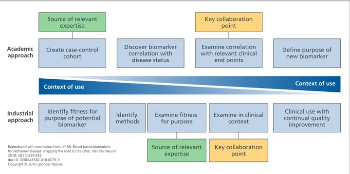
A key aspect of developing a blood test for AD is to decide on a COU and, then, to develop relevant evidence: this is designated as the “industrial approach.” However, most research in this area has followed the “academic approach,” that is, first developing a biomarker of disease status and then looking for other COUs (Figure 1). A problem with the latter approach is that the evidence needed to demonstrate utility in a COU might differ from that typically found in a case-control cohort (eg, the cohort might not be represen-tative of the COU [amyloid prevalence, disease stage]), the cohort might not have all the relevant measures, or the best biomarkers for each COU might differ.49,70
Positive and negative predictive biomarkers detecting target druggability and drug- resistance mechanisms
Current drug therapies for AD are considered transiently (“symptomatic”) biologically effective and could not substantiate clinically meaningful disease-modifying outcomes.71-73 The US Food and Drug Administration
(FDA) appealed for a new draft industrial guidance to develop AD drugs, including those for early-stage AD.74
Thus, there is an urgent medical need for drugs slowing the progression of the pathophysiological cascade causing synaptic dysfunction and neurodegeneration in AD.75,76
The identification of new molecular targets involved in AD pathophysiology represents the essential step to inves-tigate possible disease-modifying drugs counteracting the progression of the disease. Nevertheless, when considering pharmacological validation of new AD drug candidates, the need to develop newer transgenic models with more trans-lational value to the human condition should be considered. Effective drugs in currently available AD animal models do not necessarily translate as disease-modifying drugs in humans.77,78 The introduction of transgenic rodent models of
AD-like Aβ pathology represented a positive development in this direction.79,80 The repeated failures of several
poten-tial disease-modifying drugs in Phase III clinical trials led to question the right target and the Aβ hypothesis.81 However,
it should be considered that, frequently, AD patients recruited for clinical trials were already at a too advanced and potentially irreversible (decompensated) clinical stage of the disease (systems failure) and were not precisely selected using validated and qualified biomarkers.75
Never-theless, the case remains that late-stage therapies are likely bound to show only minimal effects or fail.82 Therefore,
early preclinical disease stage therapeutic trials focusing on Aβ, tau protein, and other disease-aggravating targets would probably have a better chance of delaying/halting AD-related pathophysiology.
According to this scenario, defining and validating appro-priate biomarkers is crucial for obtaining an early diagnosis of AD and assess the efficacy of disease-modifying drug treatments for AD,83,84 as stated by the FDA and by the
Euro-pean Medicines Agency (EMA).85 The recent revision of the
diagnostic criteria developed by the National Institute on Aging and the Alzheimer’s Association (NIA-AA) Working Group shifts the definition of the AD “construct” from symp-tomatic to biological and, specifically, introduces the use of biological markers—in the “A/T/N” classification scheme— as the new criteria reflecting AD pathophysiology.86 The
framework of the A/T/N scheme includes both a CSF and a neuroimaging biomarker in each of the three biomarker groups to identify the preclinical stage of AD and predict the following cognitive decline.86 In particular, “A” refers
to biomarkers of Aβ pathology, ie, the 42-amino acid-long Aβ peptide (Aβ1-42) in the CSF or amyloid PET); “T” refers
to biomarkers of tau pathology, ie, CSF phospho tau (p-tau) or tau PET; “N” refers to biomarkers of neurodegeneration or neuronal injury, ie, CSF total tau (t-tau), 18
F-fluorodeoxy-glucose [FDG]-PET (18F-FDG-PET), or structural magnetic
resonance imaging (MRI). These advanced unbiased biolog-ical criteria are required to better design clinbiolog-ical trials aimed at identifying disease-modifying compounds.86,87
Create case-control cohort Discover biomarker correlation with disease status Examine correlation with relevant clinical
end points Key collaboration point Key collaboration point Source of relevant expertise Source of relevant expertise Academic
approach Define purpose ofnew biomarker
Identify fitness for purpose of potential
biomarker
Industrial
approach methodsIdentify Examine fitness for purpose Examine in clinicalcontext
Clinical use with continual quality improvement
Context of use Context of use
Reproduced with permission from ref 50: Blood-based biomarkers for Alzheimer disease: mapping the road to the clinic. Nat Rev Neurol. 2018;14(11):639-652.
doi:10.1038/s41582-018-0079-7. Copyright © 2018 Springer Nature.
Figure 1. Potential collaboration points between academia and industry. Academic and industrial approaches to biomarker
development are inherently different, but combining these approaches could be extremely useful. Close collaboration between industry and academia would allow sharing of expertise in product testing, access to cohorts and clinical data, and sharing of ideas and theories with regard to clinical end points and context. By merging the two approaches, a method whereby the context of use is the primary focus throughout the process can be established. This model enabled synergistic development of a new biomarker between academics and industrial partners, sharing a wealth of experience.
When referring to both drug development and clinical trials, high costs, insufficient accessibility, and invasiveness of CSF biomarkers need to be critically assessed compared with blood-based biomarkers.5 Once validated in
indepen-dent large-scale cohorts, blood-based biomarkers will likely play a critical role to recognize—as early as in primary care settings—individuals with high risk of an early AD stage and to send these individuals to specialized centers where a confirmatory diagnosis can be done and the “A+/
T+/N+ biomarker profile can be established.86 The
useful-ness of blood-based biomarkers further increases when we consider the possibility that disease-modifying compounds, currently in Phase II/III clinical trials (eg, BAN2401 [Eisai], Aducanumab [Biogen] or Gantenerumab [Hoffmann-La Roche], or anti-tau therapies), might be approved in the coming future. In this scenario, blood-based biomarkers will increase the probability to get access to these treatments and will provide a fast and cost-effective rapid test to detect AD and, then, establish the eligibility of patients for inclusion into new clinical trials with new “potential” disease-mod-ifying drugs.
When considering the specific context in drug develop-ment, blood-based biomarkers should be validated and qualified for a specific COU, including assessment of mechanism of action (target engagement), dose optimiza-tion, efficacy maximizaoptimiza-tion, and monitoring of both drug response and safety.50,85 Once a blood-based biomarker is
validated for all or some of the above mentioned COUs, it can be implemented in clinical trials, designed to iden-tify disease-modifying agents, and combined with specific neuropsychological tests assessing both episodic memory and other relevant cognitive domains (ie, executive dysfunc-tion).76
Different blood-based biomarkers have been studied in the last 5 years, although preliminary evidence of validation is available only for the Aβ1-42/40-amino acid-long Aβ peptide
(Aβ1-40) ratio, the β-site amyloid precursor protein cleaving
enzyme 1 (BACE-1), t-tau, and p-tau.50
Recent studies using ultrasensitive analytical assays (Single Molecule Array [SiMoATM] platform) and fully automated
immunoassays showed that plasma Aβ1-42 concentrations
and, particularly, the Aβ1-42/Aβ1-40 ratio predict the risk
of progression to mild cognitive impairment (MCI) or dementia in cognitively normal individuals.88 Recent studies
showed that preconcentration of plasma Aβ peptides via immunoprecipitation substantially facilitated their immu-nological measurements.89 Plasma Aβ
1-42 concentrations
and Aβ1-42/Aβ1-40 ratio correlate with conventional and
validated AD biomarkers essential to detect an A+/T+/N+
biomarker profile,38,88,90,91 such as CSF Aβ
1-42
concentra-tions and brain Aβ deposition (as assessed by PET).88,90 The
inverse correlation found between plasma Aβ1-42 reduction
and brain Aβ deposition might be useful for future clin-ical trials using monoclonal antibodies directed against Aβ (ie, Aducanumab). Indeed, the decrease of plasma Aβ1-42
concentrations can predict Aβ positivity in subjective cognitive decline, MCI, and AD dementia.90 In this regard,
decreased plasma Aβ1-42 and increased nerve growth factor
precursor (proNGF) concentrations combined with inflam-matory biomarkers predict the worsening of “latent” AD pathophysiology and the subsequent cognitive decline in Down syndrome.93 The ability of Aβ
1-42/Aβ1-40 ratio to predict
cognitive decline might be useful not only for early diag-nosis, but also to monitor disease evolution differently from CSF Aβ1-42,which is stable over time and not useful for
predicting disease progression.85,94 This novel evidence,
when validated in long-term longitudinal (24 to 36 months) studies involving large cohorts, will be crucial to identify the specific COU of Aβ1-42/Aβ1-40 ratio as a novel biomarker to
assess the mechanism of action of disease-modifying drugs on the target Aβ (target engagement). We can hypothesize that a disease-modifying drug able to bind Aβ will prevent both plasma Aβ1-42 reduction and brain Aβ deposition as well
as related subsequent brain atrophy and cognitive decline. Another approach is to demonstrate target engagement of drugs affecting Aβ processing such as γ-secretase modula-tors or BACE-1 inhibimodula-tors.85 Plasma BACE-1 concentrations
are higher in MCI individuals who progressed to AD, over a 3-year follow-up, compared with stable MCI.95,96 Plasma
BACE-1 activity can predict disease progression; however, this novel COU needs to be validated in independent clinical trials.
When moving to the “tau scenario” of AD, recent highly sensitive immunoassays have been assessed for their potential in using plasma t-tau as a reliable blood-based biomarker for subject/patient selection (screening in primary setting) and AD prognosis.50,97-99 It is known that:
(i) plasma t-tau concentrations are significantly increased in AD patients compared with controls100; (ii)
blood-based p-tau is increased in AD patients and MCI individ-uals compared with controls101-103 and plasma p-tau
181 is a
more sensitive and specific predictor of elevated brain Aβ deposition than t-tau104; (iii) high baseline concentrations
of plasma t-tau in the Alzheimer’s Disease Neuroimaging Initiative (ADNI) cohort correlated with increased rates of atrophy (as assessed by MRI), hypometabolism (as assessed by 18F-FDG-PET), and the consequent cognitive decline98;
(iv) higher baseline concentrations of plasma t-tau in MCI individuals are associated with greater cognitive decline at 15 months not correlating with brain Aβ deposition.105
Whether the increased concentrations of plasma t-tau can be considered a specific biomarker for AD or just a prognostic marker for nonspecific cognitive decline is still debated.85
Recently, Chen and colleagues, using a highly sensitive detection platform combined with antibodies directed against the N-terminus of tau protein, found that N-termi-nal-detected tau (NT1) in plasma was able to discriminate between controls and AD or MCI-AD patients. Plasma NT1 did not predict disease progression, but it could be consid-ered a potential blood-based screening test for AD/AD-MCI useful to improve the selection of individuals eligible for clinical trials and to assess the clinical efficacy of tau-di-rected immunotherapies.106
Overall, the above considerations indicate a COU for blood-based biomarkers for better patient selection in clinical trials designed to assess the efficacy of investigative disease-mod-ifying drugs.50 This is a prospective scenario given the slow
evolution of cognitive decline (12 to 24 months) in early AD, as indicated by psychometric test data. As a conse-quence, well-validated blood-based biomarkers, integrated into a single panel, will help examine target druggability and resistance mechanisms, thus increasing the predictability of cognitive outcome changes in response to drug treatments. This innovation will enable to reduce costs and resources required by clinical trial pipelines.107
Regulatory viewpoint of biomarker-drug codevelopment towards individualized therapies for Alzheimer disease
CSF, as well as MRI and PET biomarkers, has been qual-ified by the EMA for the enrichment of study populations in pivotal clinical trials.108 However, these diagnostic
biomarkers are either considered invasive or expensive and there is a clear need for more practical, less invasive,
and less costly blood-based biomarkers.86,109 Recently,
plasma neurofilament light chain (NFL) protein emerged as a promising blood-based biomarker for neurodegener-ation in neurodegenerative diseases as well as plasma Aβ measures.38,91,110,111 Whereas these examples for candidate
blood-based biomarkers are considered promising, none of them can currently detect preclinical AD with reasonable diagnostic accuracy. Up to now, they have been mostly used as exploratory end points in clinical trials. However, their role as prescreening tools for selecting individuals (before more expensive and more invasive biomarkers are used) could be useful and, from a regulatory perspective, more qualification work in this direction is endorsed.112,113
Predictive biomarkers are used to identify treatment-re-sponsive patient subgroups. The usefulness of a biomarker to identify patients eligible to be treated with a new drug depends on the statistical interaction between biomarker and drug, ie, on the difference in the effect size between biomarker-positive and -negative patients. The biomarker is called predictive with respect to a given drug if this differ-ence is positive. Obviously, empirical demonstration that a biomarker is predictive in AD based on usual clinical data would require further corroboration in a well-powered clin-ical trial. Biomarker-drug codevelopment has been useful in recent years, especially in oncology, but remains a difficult task in AD due to the slow course of the disease and the lack of validated end points or surrogates, especially if early treatment at prodromal stages is contemplated.
Investigating pathway-based targeted drugs simultaneously in different pathologies in innovative study designs, such as recently proposed master protocols, could contribute to more efficient development. However, much effort is still required to explore and confirm reasonable predictive biomarkers based on clinical end points that are believed to predict the treatment effect. More sensitive clinical tools that can detect changes during early and preclinical stages of AD need to be developed.114,115
The difficulties in demonstrating the predictivity of a biomarker create regulatory challenges. If only biomark-er-positive patients are studied and there is inadequate external evidence for a differential treatment benefit, (for example, compelling mechanistic data or external control natural history information), the utility of the biomarker is unknown due to the lack of evidence in the nonselected
group. In such cases, studying both biomarker-positive and -negative patients will be necessary to obtain regulatory approval for the use of the drug only in a biomarker-iden-tified subgroup.
To foster investigation of targeted therapies using predictive biomarkers, regulators are prepared to discuss and advise on new, more sensitive end points as well as on statistical and pharmacometric modeling.
Industry viewpoint on the development of Alzheimer’s disease clinical trials focused on blood-based biomarkers
Neurodegenerative diseases require a better understanding of their pathophysiology to precisely address the relevant altered pathways with compounds targeting the relevant molecules involved.
This requires; (i) the identification of these pathophysiolog-ical pathways underlying clinpathophysiolog-ical phenotypes and breaking down one uniformly appearing clinical disease into disease subtypes characterized by biomarkers that are representative of the molecular phenotype. Such an approach has been successfully applied in oncology where biomarkers are used to differentiate subtypes of eg, breast cancer or lung cancer for better response rates.116 Furthermore, this may
require (ii) monitoring of the pathophysiological response during treatment to understand whether a patient’s disease is initially responding to a treatment and whether it continues and sustains response or (iii) monitoring whether an escape mechanism has been activated and the patient requires a different treatment approach.
In clinical trials for neurodegenerative diseases, this may require early identification of patients with an existing pathology but still absent of very discrete or unspecific symptoms, such as in AD, where biological indicators can precede the clinical symptoms by more than 10 to 15 years.117
In cases where a specific pathophysiological pathway defining a disease subgroup is targeted by a particular compound, a companion diagnostic approach may be required to test the status of biomarker(s) related to the molecular pathway(s) involved in order to provide safe and effective treatment options. Development of such
companion diagnostics often requires exceptional commu-nication and seamless collaboration between two companies (ie, diagnostics and drug companies) with distinct proficien-cies and disparate teams which, in practice, may present serious challenges during all stages of codevelopment from design and execution of clinical trials to market access and reimbursement, including Institutional Review Board over-sight, study management, monitoring and complex submis-sions to regulatory agencies. Even though a companion diagnostic approach allows for personalized treatments and may reduce the incidence of adverse effects and overall costs via avoidance of unnecessary and/or inefficient treatments, it should also be recognized that there are several disadvan-tages associated with a companion diagnostic approach as well. For instance, requirement of a specific diagnostic test to be performed before any treatment may be initiated, or requirement of continuous testing for monitoring purposes could actually add to the cost of individual patient care. In addition, lack of availability or limited accessibility of such a test for every individual potentially eligible for the drug may cause unwanted delays in access to the treatment. Similar to other in vitro diagnostic tests, companion diag-nostics must be accurate, reliable, and provide essential insights to be of clinical utility (Figure 2).
Implications and conclusions
Early detection and diagnosis of AD and other primary neurodegenerative diseases is a basis of timely and effective treatment. CSF biomarkers (CSF Aβ1-42 and tau
concentra-tions) and neuroimaging biomarkers (PET imaging of Aβ and tau aggregates, 18F-FDG-PET, and structural MRI) are
primarily used in academic expert clinical research centers and, therefore, not yet accessible as routine diagnostic tools in global primary care settings. There are international efforts to identify and validate innovative blood (plasma/ serum)-based biomarker candidates reflecting primary pathophysiological mechanisms associated with different neurodegenerative diseases, including AD.118
Blood-based biomarker candidates have the potential to be regularly analyzed both in primary care settings and in the community. Repeated (serial) blood sampling is accessible and practical even in elderly individuals and frail patients. There is (i) an ongoing dynamic process to identify and validate blood biomarkers for early detection, diagnosis, and prognosis of AD; and (ii) an increasing confidence that
blood-based tests for AD detection and diagnosis will be rapidly available, inexpensive, and easy to implement.118
Supporting the international Alzheimer Precision Medicine Initiative (APMI) (available at: https://www.apmiscience. com/) and its cohort program (APMI-CP),119 the
Blood-Based Biomarker Interest Group (BBBIG) has been created to provide global standards and best practices for the assess-ment of blood-based biomarkers. In a multistage diagnostic process, it is envisioned that blood-based biomarker tests would provide the screening entry point preceding further second-stage CSF analysis, MRI, and PET neuroimaging. Further profiling steps, based on genomic/epigenomic exploratory analyses, may be implemented as part of multi-model interventions targeted to specific biologically defined patient subgroups.118
There are many potential COUs for AD biomarkers including, but not limited to, identification of AD risk, risk for progression from MCI to AD, population screening,
stratification into clinical trials, disease monitoring, phar-macodynamics, monitoring of individual safety and toler-ability or treatment response monitoring.120 With regard to
the heterogeneous AD spectrum, blood-based biomarkers are particularly useful to specifically select individuals with Aβ pathology, using the Aβ1-42/Aβ1-40 ratio.121 The FDA
recently granted Breakthrough Device Designation for a brain amyloidosis blood test to screen for risk of AD.122 If
approved, it would be the first blood-based screening test to predict brain amyloid PET scan results in adults with memory complaints or dementia.
It is conceivable that other blood-based biomarkers will indicate and identify individuals with more acute and faster disease progression (eg, NFL or tau and, possibly, an inflammatory biomarker, such as YKL-40).123 In this
respect, the selection of individuals into a clinical trial will be enhanced using pathophysiological blood-based biomarkers identifying patients at risk for progression and
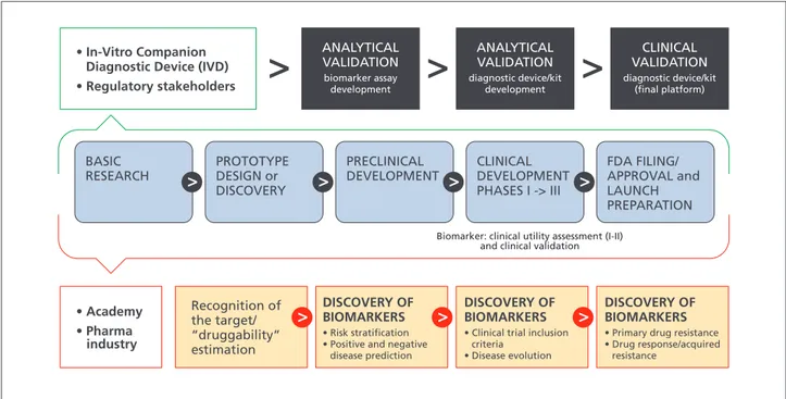
FDA FILING/ APPROVAL and LAUNCH PREPARATION CLINICAL DEVELOPMENT PHASES I -> III PRECLINICAL DEVELOPMENT CLINICAL VALIDATION diagnostic device/kit (final platform) ANALYTICAL VALIDATION diagnostic device/kit development ANALYTICAL VALIDATION biomarker assay development • In-Vitro Companion Diagnostic Device (IVD) • Regulatory stakeholders • Academy • Pharma industry Recognition of the target/ “druggability” estimation DISCOVERY OF BIOMARKERS • Risk stratification • Positive and negative disease prediction PROTOTYPE DESIGN or DISCOVERY BASIC RESEARCH
Biomarker: clinical utility assessment (I-II) and clinical validation
DISCOVERY OF BIOMARKERS • Clinical trial inclusion criteria
• Disease evolution
DISCOVERY OF BIOMARKERS • Primary drug resistance • Drug response/acquired resistance
Figure 2. Conceptual framework for biomarker-drug codevelopment strategies. Biomarkers should go through all the phases
of drug development and should be validated and qualified with regulatory guidance. Here, a “regulatory” setting for a single test that would be used together with a single drug in the clinical management of a patient is shown. The figure emphasizes key events for both the diagnostic test and drug regulation with coordination of the regulatory processes governing them, with the purpose of launching the products in parallel. Blue arrows, Drug development; orange rectangles, biomarker discovery, validation, and qualification.
decline. This approach will likely be applied to anti-tau ther-apies currently under development for the early AD, using NFL and tau protein, including different tau species and tau proteins phosphorylated at different phosphoepitopes. We envision the possibility to enter a novel era of next-gen-eration biomarker-guided targeted therapies for different neurodegenerative diseases, including AD. n
Disclosures/Acknowledgments: Contributors to the
Alzheimer Precision Medicine Initiative – Working Group (APMI–WG): Mohammad Afshar (Paris), Lisi Flores Aguilar (Montréal), Leyla Akman-Anderson (Sacramento), Joaquín Arenas (Madrid), Richard Batrla (Rotkreuz), Claudio Babiloni (Rome), Filippo Baldacci (Pisa), Norbert Benda (Bonn), Keith L. Black (Los Angeles), Arun L.W. Bokde (Dublin), Ubaldo Bonuccelli (Pisa), Karl Broich (Bonn), Francesco Cacciola (Siena), Filippo Caraci (Catania), Juan Castrillo† (Derio), Enrica Cavedo (Paris), Roberto Ceravolo (Pisa), Patrizia A. Chiesa (Paris), Jean-Christophe Corvol (Paris), Augusto Claudio Cuello (Montréal), Jeffrey L. Cummings (Las Vegas), Herman Depypere (Gent), Bruno Dubois (Paris), Andrea Duggento (Rome), Enzo Emanuele (Robbio), Valentina Escott-Price (Cardiff), Howard Federoff (Irvine), Maria Teresa Ferretti (Zürich), Massimo Fiandaca (Irvine), Richard A. Frank (Malvern), Francesco Garaci (Rome), Hugo Geerts (Berwyn), Filippo S. Giorgi (Pisa), Edward J. Goetzl (San Francisco), Manuela Graziani (Roma), Marion Haber-kamp (Bonn), Marie-Odile Habert (Paris), Harald Hampel (Paris), Karl Herholz (Manchester), Dimitrios Kapogiannis (Baltimore), Eric Karran (Cambridge), Steven J. Kiddle (Cambridge), Seung H. Kim (Seoul), Yosef Koronyo (Los Angeles), Maya Koronyo-Hamaoui (Los Angeles), Todd Langevin (Minneapolis-Saint Paul), Stéphane Lehéricy (Paris), Alejandro Lucía (Madrid), Simone Lista (Paris), Jean Lorenceau (Paris), Dalila Mango (Rome), Mark Mapstone (Irvine), Christian Neri (Paris), Robert Nisticò (Rome), Sid E. O’bryant (Fort Worth), Giovanni Palermo (Pisa), George Perry (San Antonio), Craig Ritchie (Edinburgh), Simone Rossi (Siena), Amira Saidi (Rome), Emiliano Santarnecchi (Siena), Lon S. Schneider (Los Angeles), Olaf Sporns (Bloomington), Nicola Toschi (Rome), Steven R. Verdooner (Sacramento), Andrea Vergallo (Paris), Nicolas Villain (Paris), Lindsay A. Welikovitch (Montréal), Janet Wood-cock (Silver Spring), Erfan Younesi (Esch-sur-Alzette). This research benefited from the support of the Program “PHOENIX” led by the Sorbonne University Foundation
and sponsored by la Fondation pour la Recherche sur
Alzheimer.
HH is supported by the AXA Research Fund, the
tion partenariale Sorbonne Université,” and the “Fonda-tion pour la Recherche sur Alzheimer,” Paris, France. The
research leading to these results has received funding from the program “Investissements d’avenir” ANR-10-IAIHU-06 (Agence Nationale de la Recherche-10-IA Agence Institut Hospitalo-Universitaire-6). SJK is supported by a MRC Career Development Award (MR/P021573/1). HH serves as Senior Associate Editor for the Journal Alzheimer’s & Dementia; he received lecture fees from Biogen and Roche, research grants from Pfizer, Avid, and MSD Avenir (paid to the institution), travel funding from Functional Neuro-modulation, Axovant, Eli Lilly, and company, Takeda and Zinfandel, GE-Healthcare, and Oryzon Genomics, consul-tancy fees from Qynapse, Jung Diagnostics, Cytox Ltd., Axovant, Anavex, Takeda, and Zinfandel, GE Healthcare, Oryzon Genomics, and Functional Neuromodulation, and participated in scientific advisory boards of Functional Neuromodulation, Axovant, Eisai, Eli Lilly and company, Cytox Ltd., GE Healthcare, Takeda, and Zinfandel, Oryzon Genomics and Roche Diagnostics. MA is employee and shareholder of Ariana Pharma. RB is an employee of Roche Diagnostics. SJK received an honorarium for serving on an advisory board of Roche Diagnostics. MM has patents pending to Georgetown University. The terms of this arrangement have been reviewed and approved by the University of California, Irvine, in accordance with its conflict of interest policies. SL received lecture honoraria from Roche.
AL, AV, LAA, NB, JA, KB, FC, ACC, EE, MH, SRV, and JW declare that they have no conflict of interest.
HH is coinventor in the following patents as a scientific expert and has received no royalties: In Vitro Multiparam-eter DMultiparam-etermination Method for The Diagnosis and Early Diagnosis of Neurodegenerative Disorders Patent Number: 8916388 • In Vitro Procedure for Diagnosis and Early Diagnosis of Neurodegenerative Diseases Patent Number: 8298784 • Neurodegenerative Markers for Psychiatric Conditions Publication Number: 20120196300 • In Vitro Multiparameter Determination Method for The Diagnosis and Early Diagnosis of Neurodegenerative Disorders Publication Number: 20100062463 • In Vitro Method for The Diagnosis and Early Diagnosis of Neurodegenerative Disorders Publication Number: 20100035286 • In Vitro
Procedure for Diagnosis and Early Diagnosis of Neuro-degenerative Diseases Publication Number: 20090263822 • In Vitro Method for The Diagnosis of Neurodegenera-tive Diseases Patent Number: 7547553 • CSF Diagnostic in Vitro Method for Diagnosis of Dementias and
Neuroin-flammatory Diseases Publication Number: 20080206797 • In Vitro Method for The Diagnosis of Neurodegenerative Diseases Publication Number: 20080199966 • Neurode-generative Markers for Psychiatric Conditions Publication Number: 20080131921
References
1. Hampel H, Lista S, Khachaturian ZS.
De-velopment of biomarkers to chart all Alzheimer’s disease stages: the royal road to cutting the thera-peutic Gordian Knot. Alzheimers Dement. 2012;8: 312-336.
2. Lista S, Khachaturian ZS, Rujescu D, Garaci
F, Dubois B, Hampel H. Application of systems theory in longitudinal studies on the origin and progression of Alzheimer’s disease. Methods Mol
Biol. 2016;1303:49-67.
3. Johnson CH, Ivanisevic J, Siuzdak G.
Metabo-lomics: beyond biomarkers and towards mecha-nisms. Nat Rev Mol Cell Biol. 2016;17(7):451-459.
4. Lee CH, Yoon HJ. Medical big data: promise
and challenges. Kidney Res Clin Pract. 2017;36(1): 3-11.
5. Jullian N, Jourdan N, Afshar M. Hypothesis
ge-neration for scientific discovery. Examples from the use of KEM®, a rule-based method for mul-ti-objective analysis and optimization. In: Kruse CG, Timmerman H. Towards Drugs of the Future:
Key Issues in Lead Finding and Lead Optimizati-on. Vol 9. Amsterdam, the Netherlands: IOS Press;
2008:75-80.
6. Hampel H, O’Bryant SE, Castrillo JI, et al.
Pre-cision medicine - the golden gate for detection, treatment and prevention of Alzheimer’s disease.
J Prev Alzheimers Dis. 2016;3(4):243-259.
7. Hampel H, O’Bryant SE, Durrleman S, et al.
A precision medicine initiative for Alzheimer’s disease: the road ahead to biomarker-guided in-tegrative disease modeling. Climacteric. 2017; 20(2):107-118.
8. Hampel H, Toschi N, Babiloni C, et al.
Revo-lution of Alzheimer precision neurology. Passage-way of systems biology and neurophysiology. J
Alzheimers Dis. 2018;64(suppl 1):S47-S105.
9. Hall JR, Wiechmann AR, Johnson LA, et al.
Biomarkers of vascular risk, systemic inflamma-tion, and microvascular pathology and neurop-sychiatric symptoms in Alzheimer’s disease. J
Alzheimer’s Dis. 2013;35(2):363-371.
10. Holmes C. Review: systemic inflammation and
Alzheimer’s disease. Neuropathol Appl Neurobiol. 2013;39(1):51-68.
11. Gronewold J, Klafki HW, Baldelli E, et al.
Fac-tors responsible for plasma beta-amyloid accumu-lation in chronic kidney disease. Mol Neurobiol. 2016;53(5):3136-3145.
12. Wang YR, Wang QH, Zhang T, et al.
Asso-ciations between hepatic functions and plasma
amyloid-beta levels-implications for the capacity of liver in peripheral amyloid-beta clearance. Mol
Neurobiol. 2017;54(3):2338-2344.
13. Proitsi P, Kim M, Whiley L, et al. Plasma
lipi-domics analysis finds long chain cholesteryl esters to be associated with Alzheimer’s disease. Transl
Psychiatry. 2015;5:e494.
14. Mapstone M, Cheema AK, Fiandaca MS,
et al. Plasma phospholipids identify antecedent memory impairment in older adults. Nat Med. 2014;20(4):415-418.
15. Arnold SE, Arvanitakis Z, Macauley-Rambach
SL, et al. Brain insulin resistance in type 2 diabetes and Alzheimer disease: concepts and conundrums.
Nat Rev Neurol. 2018;14(3):168-181.
16. Zhan X, Stamova B, Jin LW, DeCarli C,
Phin-ney B, Sharp FR. Gram-negative bacterial mole-cules associate with Alzheimer disease pathology.
Neurology. 2016;87(22):2324-2332.
17. Vogt NM, Kerby RL, Dill-McFarland KA, et
al. Gut microbiome alterations in Alzheimer’s di-sease. Sci Rep. 2017;7(1):13537.
18. Iliff JJ, Wang M, Liao Y, et al. A
paravascu-lar pathway facilitates CSF flow through the brain parenchyma and the clearance of interstiti-al solutes, including amyloid β. Sci Transl Med. 2012;4(147):147ra111.
19. Nedergaard M. Neuroscience. Garbage truck
of the brain. Science. 2013;340(6140):1529-1530.
20. Gámez-Valero A, Beyer K, Borràs FE.
Ex-tracellular vesicles, new actors in the search for biomarkers of dementias. Neurobiol Aging. 2019; 74:15-20.
21. Rajendran L, Honsho M, Zahn TR, et al.
Alz-heimer’s disease beta-amyloid peptides are relea-sed in association with exosomes. Proc Natl Acad
Sci U S A. 2006;103(30):11172-11177.
22. Henriksen K, O’Bryant SE, Hampel H, et
al. The future of blood-based biomarkers for Alzheimer’s disease. Alzheimers Dement. 2014; 10(1):115-131.
23. Rembach A, Ryan TM, Roberts BR, et al.
Pro-gress towards a consensus on biomarkers for Alz-heimer’s disease: a review of peripheral analytes.
Biomark Med. 2013;7(4):641-662.
24. Corder EH, Saunders AM, Strittmatter WJ, et
al. Gene dose of apolipoprotein E type 4 allele and the risk of Alzheimer’s disease in late onset fami-lies. Science. 1993;261(5123):921-923.
25. Pimenova AA, Raj T, Goate AM. Untangling
genetic risk for Alzheimer’s disease. Biol
Psychia-try. 2018;83(4):300-310.
26. Kamboh MI. A brief synopsis on the genetics
of Alzheimer’s disease. Curr Genet Med Rep. 2018;6(4):133-135.
27. Freudenberg-Hua Y, Li W, Davies P. The role
of genetics in advancing precision medicine for alzheimer’s disease-a narrative review. Front Med
(Lausanne). 2018;5:108.
28. Witoelar A, Rongve A, Almdahl IS.
Meta-ana-lysis of Alzheimer’s disease on 9,751 samples from Norway and IGAP study identifies four risk loci. Sci Rep. 2018;8(1):18088.
29. Lappalainen T, Sammeth M, Friedlander MR,
et al. Transcriptome and genome sequencing unco-vers functional variation in humans. Nature. 2013; 501(7468):506-511.
30. Sutherland GT, Janitz M, Kril JJ.
Understan-ding the pathogenesis of Alzheimer’s disease: will RNA-Seq realize the promise of transcriptomics?
J Neurochem. 2011;116(6):937-946.
31. Gaiteri C, Ding Y, French B, Tseng GC, Sibille
E. Beyond modules and hubs: the potential of gene coexpression networks for investigating molecu-lar mechanisms of complex brain disorders. Genes
Brain Behav. 2014;13(1):13-24.
32. Gaiteri C, Mostafavi S, Honey CJ, De Jager
PL, Bennett DA. Genetic variants in Alzheimer di-sease - molecular and brain network approaches.
Nat Rev Neurol. 2016;12(7):413-427.
33. Dillman AA, Majounie E, Ding J, et al.
Tran-scriptomic profiling of the human brain reveals that altered synaptic gene expression is associated with chronological aging. Sci Rep. 2017;7(1):16890.
34. Magistri M, Velmeshev D, Makhmutova M,
Faghihi MA. Transcriptomics profiling of Alzhei-mer’s disease reveal neurovascular defects, altered amyloid-beta homeostasis, and deregulated expres-sion of long noncoding RNAs. J Alzheimer’s Dis. 2015;48(3):647-665.
35. Verheijen J, Sleegers K. Understanding
Alz-heimer disease at the interface between genetics and transcriptomics. Trends Genet. 2018;34(6): 434-447.
36. Stempler S, Yizhak K, Ruppin E. Integrating
transcriptomics with metabolic modeling predicts biomarkers and drug targets for Alzheimer’s disea-se. PloS One. 2014;9(8):e105383.
37. Toledo JB, Shaw LM, Trojanowski JQ. Plasma
amyloid beta measurements - a desired but elusi-ve Alzheimer’s disease biomarker. Alzheimers Res
38. Nakamura A, Kaneko N, Villemagne VL,
et al. High performance plasma amyloid-β bio-markers for Alzheimer’s disease. Nature. 2018; 554(7691):249-254.
39. Hye A, Lynham S, Thambisetty M, et al.
Pro-teome-based plasma biomarkers for Alzheimer’s disease. Brain. 2006;129(Pt 11):3042-3050.
40. Dursun E, Gezen-Ak D, Hanağası H, et al. The
interleukin 1 alpha, interleukin 1 beta, interleukin 6 and alpha-2-macroglobulin serum levels in pa-tients with early or late onset Alzheimer’s disease, mild cognitive impairment or Parkinson’s disease.
J Neuroimmunol. 2015;283:50-57.
41. O’Bryant SE, Hobson VL, Hall JR, et al.
Se-rum brain-derived neurotrophic factor levels are specifically associated with memory performance among Alzheimer’s disease cases. Dement Geriatr
Cogn Disord. 2011;31(1):31-36.
42. Graham SF, Chevallier OP, Elliott CT, et al.
Untargeted metabolomic analysis of human plas-ma indicates differentially affected polyamine and L-arginine metabolism in mild cognitive impair-ment subjects converting to Alzheimer’s disease.
PLoS One. 2015;10(3):e0119452.
43. Varma VR, Oommen AM, Varma S, et al. Brain
and blood metabolite signatures of pathology and progression in Alzheimer disease: A targeted meta-bolomics study. PLoS Med. 2018;15(1):e1002482.
44. Whiley L, Sen A, Heaton J, et al. Evidence
of altered phosphatidylcholine metabolism in Alzheimer’s disease. Neurobiol Aging. 2014;35(2): 271-278.
45. Mapstone M, Lin F, Nalls MA, et al. What
suc-cess can teach us about failure: the plasma meta-bolome of older adults with superior memory and lessons for Alzheimer’s disease. Neurobiol Aging. 2017;51:148-155.
46. Rohart F, Gautier B, Singh A, Le Cao KA.
mi-xOmics: An R package for ‘omics feature selection and multiple data integration. PLoS Comput Biol. 2017;13(11):e1005752.
47. Singh A, Shannon CP, Gautier B, et al.
DIA-BLO: an integrative approach for identifying key molecular drivers from multi-omic assays.
Bio-informatics. 2019. doi:10.1093/bioinformatics/
bty1054.
48. Castrillo JI, Lista S, Hampel H, Ritchie CW.
Systems biology methods for Alzheimer’s disease research toward molecular signatures, subtypes, and stages and precision medicine: application in cohort studies and trials. Methods Mol Biol. 2018; 1750:31-66.
49. O’Bryant SE, Mielke MM, Rissman RA, et
al. Blood-based biomarkers in Alzheimer disease: Current state of the science and a novel collabo-rative paradigm for advancing from discovery to clinic. Alzheimer’s Dement. 2017;13(1):45-58.
50. Hampel H, O’Bryant SE, Molinuevo JL, et al.
Blood-based biomarkers for Alzheimer disease: mapping the road to the clinic. Nat Rev Neurol. 2018;14(11):639-652.
51. Hansson O, Seibyl J, Stomrud E, et al. CSF
biomarkers of Alzheimer’s disease concord with amyloid-β PET and predict clinical progression: A study of fully automated immunoassays in Bio-FINDER and ADNI cohorts. Alzheimers Dement. 2018;14(11):1470-1481.
52. Schindler SE, Gray JD, Gordon BA, et al.
Ce-rebrospinal fluid biomarkers measured by Elecsys assays compared to amyloid imaging. Alzheimers
Dement. 2018;14(11):1460-1469.
53. Amyloid PET AUDS diagnosis. But could CSF
do just as well? Available at: www.alzforum.org/ news/conference-coverage/amyloid-pet-aids-dia-gnosis-could-csf-do-just-well AlzForum. Accessed March 26, 2019.
54. Braak H, Braak E. Neuropathological staging
of Alzheimer-related changes. Acta Neuropathol. 1991;82(4):239-59.
55. Kiddle SJ, Sattlecker M, Proitsi P, et al.
Candi-date blood proteome markers of Alzheimer’s disea-se ondisea-set and progression: A systematic review and replication study. J Alzheimers Dis. 2014;38(3): 515-531.
56. Wilson JMG, Jungner G. Principles and
Prac-tice of Screening for Disease. Geneva: World
Health Organization; 1968.
57. Moynihan R. Caution! Diagnosis creep. Aust
Prescr. 2016;39(2):30-31.
58. Bateman RJ, Benzinger TL, Berry S, et al. The
DIAN-TU Next generation Alzheimer’s preven-tion trial: Adaptive design and disease progression model. Alzheimer’s Dement. 2017;13(1):8-19.
59. Reiman EM, Langbaum JBS, Fleisher AS,
et al. Alzheimer’s Prevention Initiative: a plan to accelerate the evaluation of presymptoma-tic treatments. J Alzheimers Dis. 2011;26(suppl 3):S321-S329.
60. Hu Y, Li L, Ehm MG, et al. The benefits of
using genetic information to design prevention trials. Am J Hum Genet. 2013;92(4):547-557.
61. Sperling RA, Rentz DM, Johnson KA, et al.
The A4 study: stopping AD before symptoms be-gin? Sci Transl Med. 2014;6(228):228fs13.
62. Cash DM, Rohrer JD, Ryan NS, Ourselin S,
Fox NC. Imaging endpoints for clinical trials in Alzheimer’s disease. Alzheimer’s Res Ther. 2014; 6(9):87.
63. Blood tests for amyloid step out at CTAD.
Available at: https://www.alzforum.org/news/ conference-coverage/blood-tests-amyloid-step-out-ctad AlzForum. Accessed March 26, 2019.
64. Ray S, Britschgi M, Herbert C, et al.
Clas-sification and prediction of clinical Alzheimer’s diagnosis based on plasma signaling proteins. Nat
Med. 2007;13(11):1359-1362.
65. Marksteiner J, Kemmler G, Weiss EM, et al.
Five out of 16 plasma signaling proteins are en-hanced in plasma of patients with mild cognitive impairment and Alzheimer’s disease. Neurobiol
Aging. 2011;32(3):539-540.
66. Casanova R, Varma S, Simpson B, et al. Blood
metabolite markers of preclinical Alzheimer’s disease in two longitudinally followed cohorts
of older individuals. Alzheimer’s Dement. 2016; 12(7):815-822.
67. Li D, Misialek JR, Boerwinkle E, et al.
Plas-ma phospholipids and prevalence of mild cog-nitive impairment and/or dementia in the ARIC Neurocognitive Study (ARIC-NCS). Alzheimer’s
Dement. 2016;3:73-82.
68. Collins GS, Reitsma JB, Altman DG, Moons
KGM; members of the TRIPOD group. Trans-parent Reporting of a Multivariable Prediction Model for Individual Prognosis or Diagnosis (TRI-POD): The TRIPOD Statement. Eur Urol. 2015; 67(6):1142-1151.
69. O’Bryant SE, Gupta V, Henriksen K, et al.
Guidelines for the standardization of preanalytic variables for blood-based biomarker studies in Alzheimer’s disease research. Alzheimer’s Dement. 2015;11(5):549-560.
70. Kiddle SJ, Voyle N, Dobson RJB. A blood test
for Alzheimer’s disease: progress, challenges, and recommendations. J Alzheimer’s Dis. 2018;64(sup-pl 1):S289-S297.
71. Goldman DP, Fillit H, Neumann P.
Accelera-ting Alzheimer’s disease drug innovations from the research pipeline to patients. Alzheimers Dement. 2018;14(6):833-836.
72. Salomone S, Caraci F, Leggio GM, Fedotova
J, Drago F. New pharmacological strategies for treatment of Alzheimer’s disease: focus on disea-se modifying drugs. Br J Clin Pharmacol. 2012; 73(4):504-517.
73. Winblad B, Amouyel P, Andrieu S, et al.
De-feating Alzheimer’s disease and other dementias: a priority for European science and society. Lancet
Neurol. 2016;15(5):455-532.
74. Tsukamoto K. Development of novel
phar-maceutical agents for Alzheimer’s disease: The impact of regulatory initiatives in Japan and the United States. Clin Ther. 2015;37(8):1652-1660.
75. Mullane K, Williams M. Alzheimer’s disease
(AD) therapeutics - 1: Repeated clinical failures continue to question the amyloid hypothesis of AD and the current understanding of AD causality.
Biochem Pharmacol. 2018;158:359-375.
76. Caraci F, Castellano S, Salomone S, Drago F,
Bosco P, Di Nuovo S. Searching for disease-modi-fying drugs in AD: can we combine neuropsycho-logical tools with bioneuropsycho-logical markers? CNS Neurol
Disord Drug Targets. 2014;13(1):173-86.
77. Jankowsky JL, Zheng H. Practical
considera-tions for choosing a mouse model of Alzheimer’s disease. Mol Neurodegener. 2017;12(1):89.
78. Puzzo D, Gulisano W, Palmeri A, Arancio O.
Rodent models for Alzheimer’s disease drug dis-covery. Expert Opin Drug Discov. 2015;10(7):703-711.
79. Do Carmo S, Cuello AC. Modeling
Alzhei-mer’s disease in transgenic rats. Mol
Neurode-gener. 2013;8:37.
80. Zimmer ER, Parent MJ, Cuello AC, Gauthier
S, Rosa-Neto P. MicroPET imaging and transge-nic models: a blueprint for Alzheimer’s disease