Università degli Studi di Ferrara
DOTTORATO DI RICERCA IN FARMACOLOGIA E
ONCOLOGIA MOLECOLARE
CICLO XXIV
COORDINATORE Prof. Antonio Cuneo
Nociceptin/orphanin FQ – NOP receptor system:
novel genetic and pharmacological tools
Settore Scientifico Disciplinare BIO/14
Dottorando Tutore
Dr. Stefano Molinari Dr. Girolamo Calo’
ABSTRACT
The neuropeptide nociceptin/orphanin FQ (N/OFQ) selectively binds and activates the N/OFQ peptide (NOP) receptor. In cells expressing the NOP receptor N/OFQ inhibits cAMP accumulation and Ca2+ conductance and stimulates K+ currents. Via these cellular mechanisms N/OFQ regulates several biological functions in the central nervous system (pain, locomotion, memory, emotional responses, food intake), as well as in the periphery (airways, cardiovascular, genitourinary and gastrointestinal systems). Several research tools including knockout mice and NOP selective agonists and antagonists have been developed in the past and used to investigate the role played by this peptidergic system in pathophysiology and to identify possible therapeutic indications of NOP receptor ligands. The aim of the present study was to make available to the scientific community novel genetic and pharmacological tools to speed up the process of target validation of the NOP receptor.
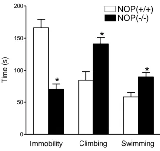
Knockout rats for the NOP receptor gene (NOP(-/-)) have been recently generated. These animals were used in the present study to investigate their emotional (open field, elevated plus maze, and forced swimming test), locomotor (drag and rotarod test), and nociceptive (plantar and formalin test) phenotype in comparison to NOP(+/+) littermates. The results were in line with previous findings obtained with selective NOP receptor antagonists in mice and rats and with mouse knockout studies and indicated that the blockage of N/OFQergic signalling elicits antidepressant and motor stimulant effects.
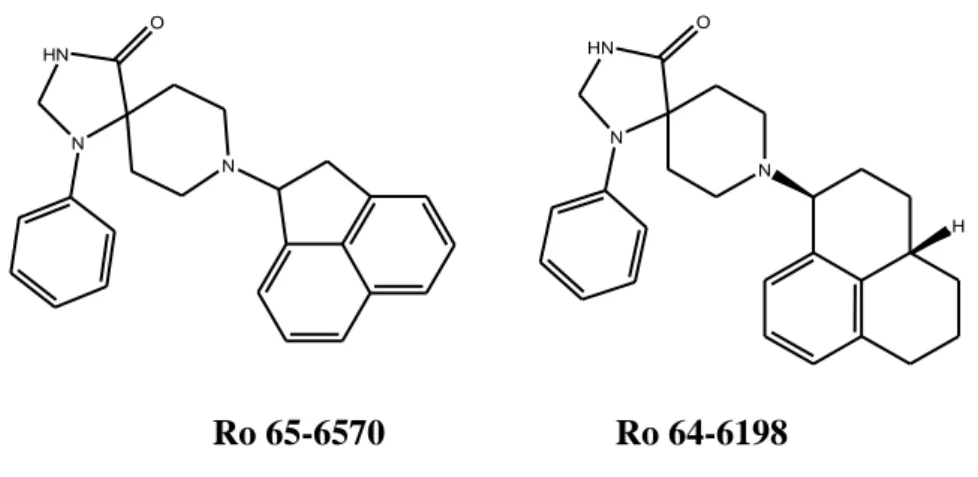
A detailed pharmacological characterization of novel NOP receptor non peptide ligands has been performed. The compound GF-4 displayed high affinity and potency at recombinant human NOP receptor associated with pure antagonist properties. This profile was confirmed in N/OFQ sensitive animal tissues. In vivo GF-4 elicited, similar to other NOP antagonists, beneficial effects in animal models of Parkinson disease. The NOP non-peptide agonists Ro 65-6570, SCH 221510 and compound 6d were characterized in vitro using a calcium mobilization assay and electrically stimulated mouse and rat vas deferens tissues. The results of these studies demonstrated that Ro 65-6570 and SCH-221510 behaved as full agonists showing however some level of NOP selectivity in rat, but not mouse, tissues. Compound 6d did not display NOP selectivity. Finally, mixed NOP/MOP receptor agonists were generated. [Dmt1]N/OFQ(1-13)NH2 was selected as the most potent compound. The mixed NOP/MOP full agonist activity and high affinity of [Dmt1 ]N/OFQ(1-13)NH2 was confirmed at human recombinant receptors in receptor and [
35
S]GTPS binding studies, at rat spinal cord receptors in [35S]GTPS binding experiments, and at guinea pig receptors inhibiting neurogenic contractions in the ileum. In vivo in the mouse tail withdrawal assay in mice [Dmt1]N/OFQ(1-13)NH2 was also able to elicit a robust antinociceptive effect being more potent than N/OFQ (by 30 fold) and morphine (by 3 fold). The antinociceptive properties of spinal [Dmt1]N/OFQ(1-13)NH2 were confirmed in non human primate studies. Collectively these results demonstrate that [Dmt1]N/OFQ(1-13)NH2 behaves as mixed NOP/MOP agonist and susbtantiate the suggestion that such mixed ligands are worthy of development as innovative spinal analgesics.
Summary 1
1. Introduction 3
1.1 Orphan GPCR and reverse pharmacology 3
1.2 NOP receptor 7
1.3 N/OFQ 12
1.4 N/OFQ and NOP receptor system: Cellular and Biological actions 18
1.4.1 pain 22 - Rodent studies 22 - monkeys studies 27 1.4.2 locomotor activity 28 1.4.3 anxiety 30 1.4.4 mood 33 1.4.5 food intake 35
1.4.6 reward and addiction 36
1.4.7 learning and memory 38
1.4.8 gastro intestinal system 39
1.4.9 airways 41 1.4.10 cardiovascular system 42 1.4.11 renal function 43 1.4.12 micturition reflex 44 1.4.13 immune system 45 1.5 Knockout animals 47 1.6 Pharmacology of NOP 49 2. Aims 66
3. Materials and methods 67
4. Results and discussion 88
4.1 Knockout studies
- phenotype of NOP(-/-) rats 88
4.2 Pharmacological studies
- NOP selective ligands 98
- NOP/MOP agonist 118
1. INTRODUCTION
1.1 Orphan G-protein coupled receptors and the reverse pharmacology approach
G-protein coupled receptors (GPCRs) are one of the largest family of proteins that are the main modulators of intercellular interactions and regulate a large variety of functions in the human body and in particularly in the central nervous system. There are numerous GPCRs in living organisms, but the function of many is still unknown. The human genome encompasses ~ 800 GPCRs, of which more than half are olfactory and/or taste GPCRs. They are targets of most of the primary messengers including the neurotransmitters, all the neuropeptides, the glycoprotein hormones, lipid mediators and other small molecules; thus have considerable pharmaceutical interest. Drugs that are acting on GPCRs are used to treat numerous disorders. In fact, more than 30% of the approximately 500 clinically used drugs, are modulators of GPCRs function, representing around 9% of global pharmaceutical sales, and making GPCRs the most successful of any target class in terms of drug discovery (Drews, 2000).
367 transmitter GPCRs have been identified within the human genome, the majority of these GPCRs have been identified on the basis of their sequence similarities, either by homology cloning or by bioinformatics analyses. Many of these receptors are still „orphans‟, i.e. its endogenous ligand is unknown.
The first step in the characterization of new orphan GPCRs is the search of the activating ligand. As the genomes of most studied model organism have now been sequenced, the process of discovery of GPCRs-ligand pairs has been reversed. In the past, neuropeptides have been traditionally identified either on the basis of their chemical characteristics (Tatemoto et al., 1980) or of their effects in particular assay systems (Erspamer et al., 1978). Although highly successful, these approaches had reached a stand still by the mid 80‟s.
Through DNA recombination techniques, it is now possible to transfect the sequence of an orphan receptor of which the function is not yet known, into an appropriate cellular expression
system. This leads to the use of orphan receptors as baits to isolate their natural ligands from mixtures of synthetic ligands, including known GPCR ligands, naturally occurring bioactive molecules of unknown function, and randomized compounds in high-throughput screening. This approach has been named “reverse pharmacology”. Thus, drug identification precedes the mechanistic understanding of mode of action of the drug candidate. The expression system provides the necessary trafficking and G-protein-signalling machinery to enable the successful identification of the activating ligand. By exposing the transfected cell to a tissue extract containing the natural ligand of the orphan receptor, a change in intracellular second messengers will be induced and will serve as a parameter to monitor orphan receptor ligand purification. Despite the logic of the theory, the process is not simple, since the physical nature of the ligand and the type of the second messenger response that it will generate, are unknown. However, structural features in an orphan GPCR will determine its relationship to known receptors and will help in evaluating the nature of the receptor‟s ligand and its activity. Indeed, an orphan receptor which is related, even to a low degree, to a particular receptor family has a higher probability of sharing a ligand of the same physical nature and a coupling to similar G proteins. Notably this strategy has already led to several significant discoveries. The orphan receptor strategy was first proven to be successful with the discovery of the neuropeptide Nociceptin/Orphanin FQ (N/OFQ), the subject of this thesis, as the endogenous ligand of the orphan GPCR Opioid Receptor-Like 1 (ORL-1) (Meunier et al., 1995; Reinscheid et al., 1995).
Figure 1.1. The orphan receptor strategy (Civelli et al., 2001). The orphan receptor strategy was developed
to identify the natural ligands of orphan G-protein-coupled receptors (GPCRs) with the aim of discovering novel transmitters (defined in the main text). This strategy involves: (1) expression of the cloned orphan GPCR in an heterologous cell line; (2) exposure of this transfected cell line to a tissue extract that is expected to contain the natural ligand; (3) recording of the change in second messenger response elicited by activation of the orphan GPCR; (4) fractionation of the tissue extract and isolation of a surrogate, the active component; (5) determination of the chemical structure of the active component and (6) chemical synthesis of the active component and demonstration that it exhibits identical activity to that of the purified ligand.
This first successful example of orphan receptor strategy was followed by the identification of other novel bioactive peptides such as: hypocretins and orexins, prolactin-releasing peptide, apelin, ghrelin, melanin-concentrating hormone, urotensin II, neuromedin U, metastin, neuropeptide B, neuropeptide W and neuropeptide S. Each of these discoveries was a landmark in its field (Civelli, 2005). The success in GPCRs deorphanization led to the approach being used by the pharmaceutical industry (Wise et al., 2004), which had mastered the high throughput screening (HTS) of thousands of ligands. This led to thousands of potential transmitters and unexpected ligands (also of non-peptide nature) being tested on dozens of orphan GPCRs and a revival of the reverse pharmacology approach. Table 1.1 summarizes the transmitters of peptide and non-peptide nature identified as ligands of orphan GPCRs after 1995 (Civelli, 2005).
Table 1.1. Transmitters identified as ligands of previously orphan GPCRs after N/OFQ; taken from (Civelli, 2005).
1.2 The NOP receptor
Pharmacological studies have defined at least three subtypes of opioid receptors, termed , and receptors, that are involved in the mediation of the numerous effects of opioid drugs, such as analgesia, respiratory depression, miosis, constipation, sensation of well being, tolerance and dependence.
The nomenclature for the opioid receptors remains controversial. A 1996 review and proposal for a novel nomenclature (Dhawan et al., 1996) based on guidelines from NC-IUPHAR has not been widely accepted by the research community. The 1996 proposal recommended replacement of the terms μ, δ, and κ with the terms OP3, OP1, and OP2, respectively. However, in the three years or more since the publication of this recommendation, almost all papers referring to opioid receptors have continued to use the well-established Greek symbol nomenclature. Since Greek nomenclature gaves many problems in manuscript preparation and particularly WEB searches, this was substituted with terminology more consistent with the overall guidelines of NC-IUPHAR that named the opioid receptors as: DOP, MOP, and KOP (Cox et al., 2000).
Molecular cloning of the DOP receptor (Evans et al., 1992; Kieffer et al., 1992) was soon followed by the cloning of the KOP and MOP receptors (Chen et al., 1993; Yasuda et al., 1993). Further attempts to clone additional opioid receptor types and/or subtypes, by hybridization screening at low stringency with opioid receptor cDNA probes, or using probes generated by selective amplification of genomic DNA with degenerate primers, led several laboratories to isolate a cDNA encoding a homologous protein with a high degree of sequence similarity to the opioid receptors (Bunzow et al., 1994; Chen et al., 1994; Fukuda et al., 1994; Lachowicz et al., 1995; Mollereau et al., 1994; Wang et al., 1994).
The novel clone, named Opioid Receptor Like-1 (ORL-1) receptor, displayed approximately 50 % identity with the traditional opioid receptors overall, with the transmembrane regions showing even higher homologies of up to 80 %. Despite the close homology with the other opioid receptors, opioid ligands displayed very low affinities towards ORL-1 receptor, thus it was considered an
orphan receptor. ORL-1 receptor is a typical GPCR with seven predicted transmembrane domains (Figure 1.2).
Figure 1.2. Schematic representation of ORL-1 receptor from (Topham et al., 1998)). TM helices are
numbered 1 to 7. E/IL: Extracellular/Intracellular Loop. Visible at the C-terminal of TM 6 is the Gln 286 (human receptor numbering) side chain.
ORL-1 was subsequently named NOP (Nociceptin/Orphanin FQ Peptide) receptor according to the IUPHAR nomenclature (Cox et al., 2000). The ORL-1 receptor was identified in different species and showed substantial sequence identities (>90%) between species variants, namely the human (Mollereau et al., 1994), rat (Wang et al., 1994; Fukuda et al., 1994; Bunzow et al., 1994; Lachowicz et al., 1995), mouse (Nishi et al., 1994) and pig (NOP (Osinski et al., 1999b)).
The human NOP receptor protein consists of 370 amino acids (Mollereau et al., 1994) and contains seven transmembrane (TM) domains. The N-terminal 44 amino acids contain 3 consensus sequences for N-linked glycosylation (Asn-X-Ser/Thr). There are also sites for potential phosphorylation by protein kinase A (in the third intracellular loop) and protein kinase C (in the second intracellular loop and the C-terminal).
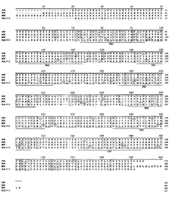
Figure 1.3. Alignment of the amino acid sequence of the rat NOP receptor (Hyp 8-1) with the amino acid
sequences of the rat brain DOP, MOP and KOP receptors. Sequences identical in at least 3 of 4 aligned sequences are boxed. Gaps in the alignment are indicated by a dash (-). Putative transmenbrane regions are underlined. Taken from Wick et al. (1994).
The NOP sequence has 57-58% amino acid (aa) identity to each of the rat MOP (Chen et al., 1994), DOP (Fukuda et al., 1993) and KOP (Minami et al., 1993). This percent identity is slightly lower than those obtained when the sequences of opioid receptors are compared to each other (62-67%).
The conservation among the four receptors is highest (70%) in the II, III, and VII transmembrane domains, and approximately 50% in the I, V and VI, but significantly lower (24%) in the IV. This high level of sequence conservation within the transmembrane domains lends weight to the view that the NOP receptor contains a TM binding pocket that is the structural equivalent of alkaloid binding pocket of the opioid receptors. Indeed, the NOP receptor has retained the ability, with low affinity, to bind and/or respond to opioid receptor ligands, agonist and/or antagonist such as etorphine and diprenorphine (Mollereau et al., 1994), buprenorphine (Wnendt et al., 1999), lofentanil (Butour et al., 1997), and naloxone benzoylhydrazone (Noda et al., 1998).
The NOP receptor gene, Oprl1, is located at the q13.2-13.3 region of the human chromosome 20 (Peluso et al., 1998) and has been mapped to the distal region of the mouse chromosome 2 (Nishi et al., 1994). In terms of intron-exon organization, the NOP receptor gene is nearly identical to that of the MOP, DOP, and KOP receptors, suggesting that the four genes have evolved from a common ancestor and hence belong to the same family (Meunier, 1997). Indeed, the NOP receptor appears to be evolutionary as old as the opioid receptors, since NOP receptor-like genomic sequences have been reported in teleost (Darlison et al., 1997), in cartilaginous fish (Li et al., 1996), in sturgeon (Danielson et al., 2001) and in zebra fish (Gonzalez-Nunez et al., 2003).
Although pharmacological studies have not firmly established the existence of NOP receptor subtypes (Calo et al., 2000b), NOP receptor heterogeneity is still an open question. NOP receptor heterogeneity may result from differential expression of NOP splice variants. So far, five splice variants of NOP mRNA have been isolated. One, identified in rat (Wang et al., 1994), encodes a NOP variant with an insertion (intron 5) in the second extracellular loop. The second splice variant, exhibiting an in frame deletion of 15 nucleotides at the 3‟ end of the TMD 1 coding region (Halford et al., 1995; Wick et al., 1994), does encode a functional receptor and has already been isolated from human tissue (Peluso et al., 1998). Further, insertions of exons 3 and 4 (Curro et al., 2001) after the first coding exon (exon 2) in rats result in three additional splice variants (Pan et al., 1998), which again encode truncated and not functional receptors.
The NOP receptor is widely expressed in the CNS, in particular in the forebrain (cortical areas, olfactory regions, limbic structures: hippocampus and amygdala, thalamus), throughout the brainstem (central periaqueductal gray, substantia nigra, several sensory and motor nuclei), and in both dorsal and ventral horns of the spinal cord (Mollereau et al., 2000; Neal et al., 1999a). The distribution patterns have suggested the involvement of the NOP receptor system in motor and balance control, reinforcement and reward, nociception, stress response, sexual behaviour, aggression and autonomic control of physiological processes (Neal et al., 1999a).
It is worthy of mention that NOP receptors co-express with MOP receptors in the dorsal horn of the spinal cord, the hippocampal formation and the caudate putamen (Anton et al., 1996; Letchworth et al., 2000) in the midbrain periaqueductal gray and the nucleus raphe magnus (Houtani et al., 2000). Distribution of NOP does not always overlap that of opioid receptors: these anatomical differences may provide a possible explanation for the different in vivo actions of N/OFQ and opioids (Ikeda et al., 1998; Monteillet-Agius et al., 1998; Sim et al., 1997).
The NOP receptor mRNA has also been identified in the peripheral nervous system and several other organs. It is expressed in peripheral ganglia and in the immune system. It has been detected in rat intestine, vas deferens, skeletal muscles and spleen (Wang et al., 1994) in porcine gastrointestinal tract and kidney (Osinski et al., 1999b), in several guinea pig ganglia (Fischer et al., 1998), also in rat retina and heart (Mollereau et al., 2000).
Peluso and colleagues (1998) were the first to describe the distribution of NOP receptor transcripts in man, in different brain regions by RT-PCR technique: the highest amplification was observed in cortical areas (the frontal and temporal cortex), in the hypothalamus, mamillary bodies, the substantia nigra, and thalamic nuclei. Transcripts have also been detected in limbic structures (the hippocampus and amygdala), brainstem (the ventral tegmental area, the locus coeruleus) and the pituitary gland. This distribution, which is similar to that of rodents, suggests the participation of the NOP receptor in numerous human physiological functions, such as emotive and cognitive processes, neuroendocrine and sensory regulation.
Berthele and colleagues (2003) studied the differential expression of NOP receptors in the human brain (cortex, basal ganglia, hippocampal area and cerebellum) by utilizing on-section ligand binding corroborated with mRNA detection on parallel sections of the same brain tissues. In general, [3H]-N/OFQ ligand binding and NOP receptor mRNA expression were widespread and indicative of a considerable high NOP receptor expression in these anatomical regions. [3H]-N/OFQ ligand binding and NOP mRNA expression studies showed that the highest amounts of NOP receptor were observed in the cerebellum, in the cortex (cingulate and prefrontal cortex), in the striatum (caudate nucleus and the putamen) and in the lamina II, followed by laminae III, V and VI, in the principal neurons of the dentate gyrus and in the hippocampal area (Berthele et al., 2003).
1.3 Nociceptin/orphanin FQ
In 1995 Meunier and Reinsheid simultaneously described N/OFQ as the endogenous ligand for ORL-1, now known as NOP. CHO cells expressing the orphan receptor were used to identify its endogenous ligand. Based on structural similarities with the known opioid receptors, both the chemical nature of the endogenous ligand (peptide) and the consequences of receptor activation (inhibition of cyclic AMP) were assumed to be similar to those of classical opioids. Consequently, cells were stimulated with forskolin to activate adenylyl cyclase and increase intracellular cAMP. As a Gi/o-coupled orphan receptor, endogenous agonists at this receptor will inhibit the formation of cAMP. Extracts from rat (Meunier et al., 1995) or pig (Reinscheid et al., 1995) brain were screened. Fractions that were able to inhibit the adenylyl cyclase activity were further fractionated through reverse-phase high-performance liquid chromatography. The purification and mass spectrometry analyses identified a heptadecapeptide (Figure 1.4), the sequence of which was determined. The synthetic peptide was shown to have high affinity (in the nanomolar range) and to strongly inhibit forskolin-induced accumulation of cAMP in CHO cells expressing the NOP receptor (EC50 about 1 nM), while showing no activity in non transfected cells (Meunier et al.,
1995). Moreover, when tested in vivo by intracerebroventricular (i.c.v.) injection in mice, the peptide induced hyperalgesia in the hot plate (Meunier et al., 1995) and tail flick tests (Reinscheid et al., 1995).
The group of Meunier termed the novel peptide nociceptin, based on apparent pro-nociceptive properties, while that of Reinscheid named it orphanin FQ, as ligand of an orphan receptor, whose first and last amino acids are Phe (F) and Gln (Q), respectively.
N/OFQ shares sequence homologies with the opioid peptide ligand dynorphin A (Figure 1.4); despite the structural similarities these peptides are functionally quite distinct. N/OFQ has no significant affinity for any of the opioid receptors (Reinscheid et al., 1998).
Figure 1.4. Structural similarities between dynorphin A and N/OFQ amino acid sequences (Guerrini et al.,
2000b).
The N-terminal tetrapeptide sequences (message domain) of the two peptides are very similar, with the only difference of the first amino acid residue (Phe in N/OFQ and Tyr in dynorphin A); the C-terminal parts (address domain) of the two molecules are both enriched in positively charged residues, such as arginine and lysine, even if distributed in different positions.
N/OFQ is a heptadecapeptide cleaved from the peptide precursor preproN/OFQ (ppN/OFQ). ppN/OFQ consists of a 181 amino acids in the rat, 176 amino acids in humans and 187 amino acids in the mouse (Mollereau et al., 1996; Nothacker et al., 1996) (Figure 1.5). The ppN/OFQ gene, that has been isolated from human, mouse and rat, is highly conserved in the three species.
Figure 1.5. Amino acid sequence for N/OFQ precursor, ppN/OFQ (Calo et al., 2000b).
Analysis of the nucleotide sequence of the ppN/OFQ gene revealed structural and organisational characteristics very similar to those of the opioid peptide precursors, in particular preproenkephalin and preprodynorphin, suggesting that these peptides derive from a common ancestor (Mollereau et al., 1996; Nothacker et al., 1996).
The ppN/OFQ gene is located on human chromosome 8 (8p21) (Mollereau et al., 1996). In the ppN/OFQ sequence there are several pairs of basic amino acids that present possible sites of cleavage for precursor maturation or for transcriptional regulation (Zaveri et al., 2000). Therefore, several biologically relevant peptides may derive from the N/OFQ precursor (Figure 1.5). Apparently two additional peptides are excised from the same precursor: N/OFQ2 and nocistatin (Okuda-Ashitaka et al., 1998). None of them bind to the NOP receptor (Mollereau et al., 1996; Nothacker et al., 1996), and until now specific receptors for them have not been identified. The peptide following the N/OFQ sequence, is a heptadecapeptide terminating with the couple FQ (N/OFQ2): it has been found to be biologically active, stimulating locomotor activity in mice (Florin et al., 1997) inducing antinociception both spinally and supraspinally (Rossi et al., 1998) and inhibiting gastrointestinal transit (Rossi et al., 1998).
The second peptide, named nocistatin (NST), has been reported to act as a functional antagonist of N/OFQ (Okuda-Ashitaka et al., 1998). In most studies, NST was found to be inactive per se, but was able to reverse several effects of N/OFQ, such as induction of allodynia after spinal administration in mice (Minami et al., 1998; Okuda-Ashitaka et al., 1998), inhibition of glutamate release from rat brain slices (Nicol et al., 1998), impairment of learning and memory in mice (Hiramatsu et al., 1999), stimulation of food intake in rats (Olszewski et al., 2000). Moreover, NST can, per se, cause antinociception after i.c.v. administration in the rat carrageenan test (Nakagawa et al., 1999) or after intratechal (i.t.) administration in the rat formalin test (Yamamoto et al., 2001). Interestingly, nocistatin or its C-terminal hexapeptide exerts anxiogenic-like effects in mice; in fact it has been reported that the C-terminal hexapeptide (the most conserved region among species), administered i.c.v., exerts clear anxiogenic-like effects in mice, in contrast to N/OFQ, that in the same experimental model, acts as an anxiolytic (Gavioli et al., 2002). Very recent findings demonstrated that the opposite effects of N/OFQ and NST on supraspinal pain modulation result from their opposing effects on the excitability of central amygdala nucleus-periaqueductal gray projection (CeA-PAG) neurons. Electrophysiological studies showed that N/OFQ hyperpolarized CeA-PAG projection neurons by enhancing an inwardly rectifying potassium conductance. In contrast, NST depolarized CeA-PAG neurons by causing the opening of TRPC cation channels via a Gαq/11-PLC-PKC pathway (Chen et al., 2009).
In vitro studies demonstrated that bovine nocistatin (bNST) inhibited the K+-induced [3
H]5-HT release from mouse cortical synaptosomes, displaying similar efficacy but lower potency than N/OFQ; this inhibitory effect was not prevented either by the NOP receptor antagonist UFP-101, or by the non-selective opioid receptor antagonist, naloxone. In contrast to N/OFQ, bNST reduced [3H]5-HT release from synaptosomes obtained from NOP receptor knockout mice (Fantin et al., 2007).
The localization of N/OFQ-immunoreactive fibres and terminals and/or the localization of the ppN/OFQ mRNA correspond reasonably well with the NOP receptor. Limbic areas highly
express N/OFQ, in particular the bed nucleus of the stria terminals, and the amygdala nuclei (Boom et al., 1999; Neal et al., 1999b). A matching pattern of N/OFQ and NOP receptor expression in the human and rodent central nervous system has been observed (Berthele et al., 2003; Peluso et al., 1998; Witta et al., 2004). As with the receptor, N/OFQ immunoreactivity and mRNA levels detected using in situ hybridization are closely correlated. N/OFQ is found in lateral septum, hypothalamus, ventral forebrain, claustrum, mammillary bodies, amygdala, hippocampus, thalamus, medial habenula, ventral tegmentum, substantia nigra, central gray, interpeduncular nucleus, locus coeruleus, raphe complex, solitary nucleus, nucleus ambiguous, caudal spinal trigeminal nucleus, and reticular formation, as well the ventral and dorsal horns of the spinal cord (Neal et al., 1999b). Recently N/OFQ was immunolocalized in rat lateral and medial olivocochlear efferents (Kho et al., 2006). Although N/OFQ and opioid peptides show a similar distribution, they are not colocalized in nociceptive centres such as the dorsal horn, the sensory trigeminal complex or the periaqueductal gray (Schulz et al., 1996). This N/OFQ distribution in the central nervous system suggests that the peptide is potentially involved in the regulation of a variety of brain functions, including emotional processing, learning and memory, locomotion, reward, pain transmission, and autonomic regulation of peripheral organs and systems.
In the periphery, mRNA of pp\N/OFQ was detected in rat ovary, in human spleen, lymphocytes, and fetal, but not adult kidney (Mollereau et al., 1996; Nothacker et al., 1996). Furthermore, it has been shown that, under physiological conditions, N/OFQ is present in the human plasma (~ 10 pg/ml) (Brooks et al., 1998). In several pathological conditions such as postpartum depression (Gu et al., 2003), Wilson‟s disease (Hantos et al., 2002), hepatocellular carcinoma (Horvath et al., 2004) and in acute and chronic pain states (Ko et al., 2002b), plasma levels of N/OFQ resulted increased. In contrast, lower N/OFQ plasma levels have been observed in patients suffering from fibromyalgia syndrome (Anderberg et al., 1998), cluster headache (Ertsey et
al., 2004) and migraine without aura (Ertsey et al., 2005). After all, a very recent findings indicate that N/OFQ plasma levels are increased in sepsis condition (Williams et al., 2008).
Little is known about the biosynthesis of N/OFQ, apart from the involvement of prohormone convertase 2 as demonstrated by studies performed in mice knockout for this enzyme (Allen et al., 2001). As far as N/OFQ metabolism is concerned, different studies demonstrated the involvement of aminopeptidase N (APN) that generates [desPhe1]N/OFQ a peptide lacking affinity for the NOP receptor. However different endopeptidases are also involved in N/OFQ metabolism. endopeptidase (EP) cleaves a variety of bonds to release inactive fragments, Figure 1.6 (Calo et al., 2000b), Endopeptidase 24.15 (EP 24.15) (Montiel et al., 1997) acts on the peptide bonds Ala7-Arg8, Ala11 -Arg12, Arg12-Lys13 and releases inactive compounds, where endopeptidase 24.11 (EP 24.11) acts on the cleavage site Lys13-Leu14 and plays a major role in the initial stage of N/OFQ metabolism in mouse spinal cord (Sakurada et al., 2002). C-terminal degradation also leads to a reduction in binding affinity of N/OFQ for NOP, loss of the 4 amino acids from the C-terminal tail as in N/OFQ(1-13) results in a 30-fold reduction in potency (Butour et al., 1997). However, amidation of C-terminus of N/OFQ(1-13) restores ligand affinity and potency, consequently N/OFQ(1-13)-NH2 is the shortest sequence retaining the full biological activity of the endogenous ligand (Guerrini et al., 1997).
Figure 1.6. N/OFQ metabolism by aminopeptidase N (APN) and endopeptidases (EP), from (Calo et al.,
2000b).
1.4 N/OFQ and NOP receptor system: Cellular and Biological actions
The cellular actions of the classical opioid receptors (MOP/KOP/DOP) and the NOP receptor have been shown to be pertussis toxin sensitive and therefore couple to inhibitory proteins i.e. G-proteins with Gi/o alpha subunits (Reinscheid et al., 1996). G-proteins are membrane bound/associated heterotrimeric proteins composed of α, β, γ subunits. There are four major classes of G proteins including Gi/Go, Gs, Gq.
Activation of NOP, similar to MOP, KOP, and DOP opioid receptors activation, leads to: i) closing of voltage sensitive calcium channels, ii) stimulation of potassium efflux leading to hyperpolarisation and iii) reduced cyclic adenosine monophosphate (cAMP) production via inhibition of adenylyl cyclase. Overall this results in reduced neuronal cell excitability leading to a
FGGFTGARKSARKLANQQ
F + GGFTGARKSARKLANQAPN
FGGFTGA + RKSARKLANQ FGGFTGARKSA + RKLANQ FGGFTGARKSAR + KLANQEP 24.15
FGGFTGARKSARK + LANQ FGGFTGARK + SARKEP
EP
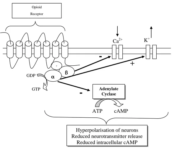
reduction in transmission of nerve impulses along with inhibition of neurotransmitter release (Figure 1.7) (Hawes et al., 2000).
Figure 1.7. Schematic representation of intracellular responses to NOP receptor activation.
N/OFQ inhibits forskolin-stimulated cellular cAMP production. Forskolin is used to directly activate adenylyl cyclase and consequently increase cAMP production. Elevating cAMP via other receptor driven systems (e.g. via the D1 dopamine receptor) is also inhibited by N/OFQ (Chan et al., 1998). Both the endogenous and recombinant NOP receptors are capable of inhibiting the formation of cAMP with remarkably consistent EC50 values in a range of cell systems.
K+ cAMP ATP
+
-
-
Ca2+ GTP GDP Adenylate Cyclase Hyperpolarisation of neurons Reduced neurotransmitter releaseReduced intracellular cAMP Opioid
The peptide also inhibits several types of voltage-gated Ca2+ channels: for example in human SH-SY5Y neuroblastoma cells it produces a partial inhibition of N-type Ca2+ conductance with an IC50 value of about 40 nM (Connor et al., 1996b), and in dissociated rat hippocampal neurones the peptide partially inhibits the three major types of Ca2+ channels, L, N and P/Q (Knoflach et al., 1996). The inhibition is no longer seen after β pertussis toxin treatment and cannot be prevented by high doses of naloxone. N/OFQ has been shown to mediate a pronounced inhibition of N-type calcium channels, whereas other calcium channel subtypes were not affected.
N/OFQ has also been reported to increase the inwardly rectifying K+ conductance in rat brain slices containing the dorsal raphe nucleus (Vaughan et al., 1996), the locus coeruleus (Connor et al., 1996a), and the periaqueductal grey (Vaughan et al., 1997), in hippocampal slices (Madamba et al., 1999) and cultured hippocampal neurones (Amano et al., 2000).
Collectively, these data are consistent with the hypothesis that N/OFQ acts primarily to reduce synaptic transmission and neuronal excitability in the nervous system (Meunier, 1997). In the CNS, studies using synaptosomes and brain slices revealed that N/OFQ inhibits the release of noradrenaline (NA), serotonine (5-HT), dopamine (DA), acetylcholine (ACh), γ-aminobutyric acid (GABA), and glutamate (Schlicker et al., 2000).
In the peripheral nervous system, studies showed the general modulatory effects (mostly inhibitory) of N/OFQ on neurotransmitter release from sympathetic, parasympathetic and nonadrenergic-noncholinergic sensory endings. In the respiratory, cardiovascular, genitourinary and gastrointestinal systems N/OFQ exerts inhibitory effects (Giuliani et al., 2000). Several isolated tissues from different species have been shown to be sensitive to N/OFQ. In particular, the electrically stimulated mouse and rat vas deferens and the guinea pig ileum have been described and used extensively in opioid receptor pharmacology: the guinea pig ileum, whose myenteric neuronal network contains mainly MOP receptors (Paton, 1957) has been shown to respond to N/OFQ (Calo et al., 1997; Calo et al., 1996; Zhang et al., 1997b); the mouse vas deferens whose nerve terminals contain mainly DOP receptors (Hughes et al., 1975) and the rat vas deferens, whose nerves contain
an uncharacterized opioid receptor (Lemaire et al., 1978), have also been reported to be N/OFQ sensitive preparations (Berzetei-Gurske et al., 1996; Calo et al., 1997; Calo et al., 1996; Nicholson et al., 1998; Zhang et al., 1997b). The twitch response in the three preparations is due to nerve activation and subsequent release of neurotransmitter since they are blocked by tetradotoxin. The release of NA from the sympathetic nerves is the major cause of the contractions of mouse and rat vas deferens, since they are blocked by the α-1 adrenoceptor antagonist prazosin. NOP receptors appear to be localized in sympathetic terminals since N/OFQ inhibits twitch evoked by electrical field stimulation, but does not modify contractions to exogenous NA (Calo et al., 1996). Similar results were obtained in the guinea pig ileum since N/OFQ inhibited atropine and tetradotoxin sensitive contractions derived from the release of ACh from cholinergic terminals of the myenteric plexus without affecting responses to exogenous ACh, thus demonstrating the prejunctional localization of the NOP receptor. In the three tissues the inhibitory effect of N/OFQ is not influenced by naloxone suggesting that classical opioid receptors are not targeted by the peptide.
A similar picture has been found regarding the inhibitory effects of N/OFQ on sensory fibres on the guinea pig bronchus (Fischer et al., 1998; Rizzi et al., 1999b), renal pelvis and heart (Giuliani et al., 1996; Giuliani et al., 1997b), and cholinergic contractions of human bronchus (Basso et al., 2005). The rat anococcygeus has also been described as a preparation in which N/OFQ produces a concentration-dependent inhibition of the adrenergic motor response to electrical field stimulation, but does not affect the response to exogenous NA. In addition, selective opioid ligands do not exert any effect on this preparation, suggesting that in this preparation the NOP receptor occurs without the co-presence of the classical opioid receptors (Ho et al., 2000). In all the preparations analysed above, the N/OFQ-NOP receptor system displays a prejunctional inhibitory function, as do classical opioid receptors.
Due to the widespread distribution of N/OFQ and NOP receptor, this peptidergic system is involved in a wide range of physiological responses with effects noted in the nervous system (central and peripheral), the cardiovascular system, the airways, the gastrointestinal tract, the
genitourinary and immune system. The role of this peptidergic system has been explored intensely with the pharmacological and biological tools available, such as i) antisense oligonucleotides targeting NOP receptor or ppN/OFQ gene, ii) antibodies directed against N/OFQ, iii) transgenic mice in which the receptor or the peptide precursor genes have been genetically eliminated, iv) selective and potent antagonists. Some of the more well-studied and noteworthy biological actions modulated by this system will be described below.
1.4.1 Pain regulation Rodent studies
Since the identification of N/OFQ there has been intense interest in the role of this peptide in pain processing. This is based on various factors, including the similarity of distribution of receptor and peptide to classical opioids within the defined pain pathway and the structural similarity to classical opioids. Application of N/OFQ has been shown to cause hyperalgesia, allodynia and analgesia. These conflicting findings are confounded by species and or strain differences in test animals, known to be fundamental in the supraspinal effects of nociception (Mogil et al., 1999). However, the route of administration and nociceptive paradigm under investigation are of paramount importance.
When administred supraspinally, N/OFQ was shown to increase pain sensitivity in mice and rats in the two initial studies of the functions of this peptide (hence the name nociceptin; (Meunier et al., 1995; Reinscheid et al., 1995)). However, the hyperalgesic effect of N/OFQ was only seen after intracerebroventricular (i.c.v.), but not after intrathecal (i.t.) administration. It has been demonstrated that most prominent role of N/OFQ in supraspinal pain modulation is a “functional opioid antagonism” directed against many different opioid receptor agonists (Mogil et al., 2001). Since behavioural testing in pain models, in particular i.c.v. injections, expose animals to acute stress, the apparent pronociceptive action seen in the initial studies may thus be interpreted as the
reversal of stress-induced antinociception rather than as a genuine pronociceptive or hyperalgesic effect (Mogil et al., 2001). I.c.v. injection of N/OFQ was stressful, resulting in a release of central endogenous opioid peptides with their effects subsequently reversed by the delivered dose of N/OFQ (Lambert, 2008). The suggestion for an anti-opioid role of N/OFQ has since been corroborated by results obtained in a variety of assays: indeed, it has been shown that N/OFQ counteracts the analgesic effect of the endogenous opioids (Tian et al., 1997a; Tian et al., 1997b) or that of exogenously applied morphine (Bertorelli et al., 1999; Calo et al., 1998b; Grisel et al., 1996; Zhu et al., 1997) or that of selective opioid receptor agonists (King et al., 1998). Worthy of mention is the fact that tolerance develops to the antiopioid effects of N/OFQ (Lutfy et al., 1999).
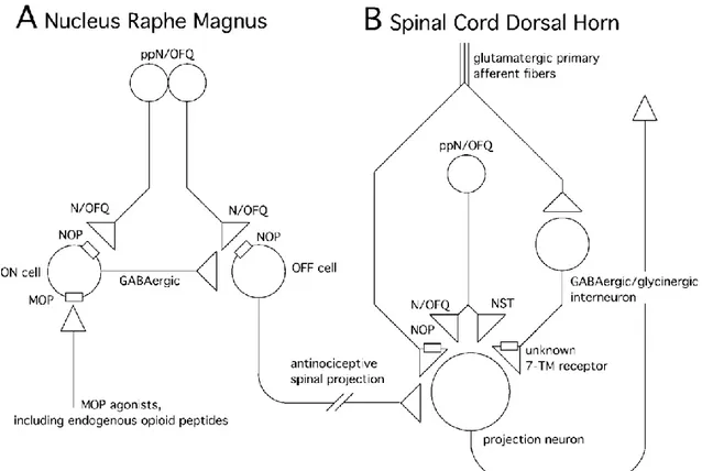
Since the NOP receptor and classical opioid receptors largely share the same transductional mechanisms, it is reasonable to speculate that their opposite effects on pain threshold are due to distinct localisations of N/OFQ and opioid peptides and their respective receptors on the neuronal networks involved in pain transmission at the supraspinal level. A cellular model explaining the antiopioidergic action of supraspinal N/OFQ focalizes on brain stem, in particular the nucleus raphe magnus of the RVM, the major site of supraspinal N/OFQ effects on pain processing. In this brain region, different types of neurons, so called ON and OFF cells, can be distinguished. ON cells fire immediately before a nociceptive reaction, while OFF cells are inhibited by the GABAergic ON cells and therefore silent at the same time. Activation of OFF cells induces spinal antinociception via descending antinociceptive tracts. MOP opioids inhibit ON cells and thereby cause a subsequent disinhibition of the antinociceptive OFF cells. By contrast, N/OFQ inhibits nearly all cell types in the RVM. Via a direct inhibition of OFF cells, N/OFQ counteracts the disinhibitory effects of MOP agonists on these cells and thereby reverses opioid-induced supraspinal analgesia. The same mechanism may also account for the apparent hyperalgesic effect of N/OFQ, providing a cellular basis for the reversal of stress-induced analgesia by N/OFQ. These studies demonstrate that the net effects of N/OFQ on nociception at supraspinal sites strongly depend on the activation state (resting versus sensitized) of pain controlling neuronal circuits (figure 1.8) (Zeilhofer and Calo, 2003).
The involvement of the NOP protein in the actions of N/OFQ on pain transmission has been investigated through the use of receptor antagonists, transgenic mice lacking the NOP receptor gene and with antisense oligonucletides. Indeed, the involvement of the NOP receptor in N/OFQ effects on nociception is supported by the following evidence: i) the pronociceptive action of N/OFQ is no longer present in NOP(-/-) mice (Nishi et al., 1997; Noda et al., 1998); ii) antisense oligonucleotides targeting the NOP receptor prevent the effect of N/OFQ (Tian et al., 1997b; Zhu et al., 1997); the pronociceptive effect of N/OFQ is reversed by NOP selective antagonists: [Nphe1]N/OFQ(1-13)-NH2 (Calo et al., 2000a; Di Giannuario et al., 2001; Rizzi et al., 2001b), UFP-101 (Calo et al., 2002), J-113397 (Ozaki et al., 2000; Yamamoto et al., 2001) and SB-612111 (Rizzi et al., 2007a; Zaratin et al., 2004).
Figure 1.8. Schematic drawing showing how N/OFQ modulates synaptic transmission in the nucleus raphe
magnus of the brain stem (A) and in the spinal cord dorsal horn (B). A, in the brain stem, MOP agonists including endogenous opioid peptides inhibit so-called secondary or ON cells. These cells are GABAergic and, in turn, inhibit descending antinociceptive OFF or primary cells. By inhibiting the ON cells, μ-opioids cause a disinhibition of OFF cells and elicit thereby antinociception. By contrast, N/OFQ inhibits both cell types at the same time. Under resting conditions, N/OFQ probably exerts no net effect on nociception. However, when MOP receptors on ON cells are activated N/OFQ can reverse this analgesia by inhibiting in addition OFF cells. B, in the spinal cord, N/OFQ selectively inhibits the release of glutamate and leaves the release of the inhibitory neurotransmitters glycine and GABA unaffected. Inhibition of glutamate release and the subsequent inhibition of nociceptive transmission through the spinal cord probably underlie the analgesic effect of nanomolar doses of spinally applied N/OFQ. NST by contrast reduces selectively the release of GABA and glycine via so far unknown 7-transmembrane receptor, but spares the release of glutamate.
Many lines of evidence indicate that the spinal cord is an equally important CNS area for nociceptive processing and its modulation by N/OFQ and classical opioids. Neurons and fibres networks containing ppN/OFQ mRNA and N/OFQ like immunoreactivity have been located in the dorsal spinal cord (Lee et al., 1997; Mamiya et al., 1998; Meis et al., 1998; Meunier et al., 1995), and endogenously released N/OFQ can be detected following electrical field stimulation of the spinal cord (Lai et al., 2000).
The role of N/OFQ in modulating pain threshold in the spinal cord is controversial. Although some studies reported that i.t. injection of N/OFQ produces hyperalgesia/allodynia (Hara et al., 1997; Inoue et al., 1999) others found no effect (Grisel et al., 1996; Reinscheid et al., 1995). Most of the studies, however demonstrated that i.t. N/OFQ induces an antinociceptive effect similar to that evoked by classical opioid receptor agonists (Candeletti et al., 2000b; Erb et al., 1997; Hao et al., 1998; Kamei et al., 1999; King et al., 1998; Nazzaro et al., 2007; Wang et al., 1999; Xu et al., 1996). While tolerance develops to the antinociceptive effect of i.t. N/OFQ upon repeated administration, there is no cross tolerance with morphine, suggesting that different receptors are involved in the actions of the two agents (Hao et al., 1997). Differences in animal species or even in strains, as well as in N/OFQ doses used, may account for the conflicting results reported with N/OFQ in the spinal cord. Worthy of mention is the work of Inoue et al. (1999) showing the dose response curve to N/OFQ is bell-shaped: very low doses of peptide (fmol range) cause hyperalgesia, while at higher doses (nmol range) N/OFQ is antinociceptive and blocks the scratching, biting and licking induced by i.t. substance P.
Collectively, the cellular mechanisms of the pronociceptive effects of N/OFQ in the spinal cord are still rather obscure, whereas inhibition of excitatory synaptic transmission presents as a clearly defined cellular mechanism underlying the spinal analgesia. The combination of opioid-like analgesia by N/OFQ at the level of the spinal cord with functional opioid antagonism at supraspinal sites, where most of the unwanted effects of classical opioids arise, has promoted the idea that NOP receptor agonists might be better tolerated spinally acting analgesics. It should however be noted that the only well studied non-peptide NOP receptor agonist Ro 64-6198 was anxiolytic, but not antinociceptive in acute pain rodent models (Jenck et al., 2000); Ro 64-6198 was recently reported to have an antiallodynic effect mediated by NOP receptors in a neuropathic pain model (Obara et al., 2005). Nevertheless, further studies with other NOP receptor agonists and in chronic pain models are desirable.
Nociceptive responses to acute noxious heat in NOP(-/-), ppN/OFQ(-/-) and double knockout mice were indistinguishable from those of NOP(+/+). However, NOP(-/-), ppN/OFQ(-/-) and double knockout mice showed markedly stronger nociceptive response during prolonged nociceptive stimulation (Depner et al., 2003). These results indicate that the N/OFQ system contributes significantly to endogenous pain control during prolonged nociceptive stimulation (e.g. formalin, zymosan A, writhing tests, SBL response to i.t. substance P), but does not affect acute (tail flick/immersion test) pain sensitivity (Depner et al., 2003).
Similar to what seen at the spinal level in the periphery both pro and antinociceptive effects were reported for N/OFQ. For instance intradermal administration of very low doses of N/OFQ stimulates the flexor reflex in mice. This effect involves stimulation of the release of substance P from peripheral nerve endings. However at higher doses N/OFQ prevented the facilitatory effect of substance P (Inoue et al., 1999). In addition several groups reported the ability of N/OFQ to inhibit neuropeptide release from peripheral sensory neuron terminals in different organs including the airways, heart, and renal pelvis (Giuliani et al., 2000; Giuliani et al., 1996; Lee et al., 2006).
Primate studies
As far as the role of N/OFQ in modulating pain in primate species is concerned, extremely interesting are the studies of Ko and colleagues, that demonstrated that in non human primates spinal administration of N/OFQ or synthetic NOP ligands i) does not elicit any effect at low doses, ii) in the nanomolar range of doses induces a robust antinociceptive action that is sensitive to NOP antagonists but not naltrexone, iii) in contrast to morphine, does not induce pruritus and iv) elicits a synergistic antinociceptive effect when given in association with morphine (Ko et al., 2006). Thus evidence coming from non human primates strongly suggests NOP receptor agonists as spinal analgesics. Interestingly, this same research group demonstrated that the NOP selective non peptide agonist Ro 64-6198 given systemically is able to induce dose dependent antinociceptive effects in non human primates while being inactive in rodents. In monkeys antinociceptive doses of alfentanil
(and in generally of opioids) are associated with respiratory depression, itch/scratching responses and reinforcing effects under self-administration procedures while in parallel experiments antinociceptive doses of Ro 64-6198 did not produced these side effects (Ko et al., 2009). Further studies are however needed to draw firm conclusions on the therapeutic potential of systemic NOP agonists as analgesics.
1.4.2 Modulation of locomotor activity
One of the seminal investigations of N/OFQ reported a dose-dependent decrease in locomotor activity (i.e., hypolocomotion) when the peptide was given supraspinally (Reinscheid et al., 1995). This effect was significant only after i.c.v. application of 10 nmol N/OFQ. This finding was later confirmed by other authors in mice (Nishi et al., 1997; Noble et al., 1997; Noda et al., 1998; Rizzi et al., 2001b).
Repeated daily N/OFQ injections result in rapid development of tolerance to this depressor effect on locomotion behaviour (Devine et al., 1996). The action of N/OFQ is insensitive to naloxone (Noble et al., 1997), while it is reversed by NalBzOH (Noda et al., 1998). The locomotor-inhibiting effects of N/OFQ seen in NOP(+/+) animals were not seen in NOP(-/-) mice, confirming the involvement of the NOP receptor in this effect (Nishi et al., 1997; Noda et al., 1998). However the spontaneous locomotor activity of NOP(-/-) mice is not different from that displayed by NOP(+/+) littermates which suggest that the N/OFQ-NOP receptor system does not play a tonic role in the physiological regulation of spontaneous locomotion. When microinjected directly into the hippocampus or ventromedial hypothalamus, but not the nucleus accumbens, high doses of N/OFQ (10–25 nmol) significantly decrease locomotor activity (Sandin et al., 1997).
N/OFQ has also been reported to stimulate locomotor activity and exploratory behaviour at very low doses (0.01-0.1 nmol) (Florin et al., 1997). This effect of N/OFQ has been related to the anxiolitic-like actions of the peptide (Jenck et al., 1997). Thus, N/OFQ shows a biphasic dose
response curve for locomotor activity: stimulation at low doses (0.01-0.1 nmol), inhibition at high doses (1-10 nmol).
It has been demonstrated that firing activity of dopaminergic cells of the substantia nigra, which express NOP receptors, is inhibited by microinjection of N/OFQ (Marti et al., 2004b). When microinjected into the substantia nigra, N/OFQ reduces dopamine release in the striatum and locomotor activity (Marti et al., 2004b). Conversely, the NOP receptor antagonists, UFP-101 and J-113397, injected into the substantia nigra, enhanced striatal dopamine release and facilitated motor performance (Marti et al., 2004b). These data confirm that improvement in locomotor activity is due to the enhanced striatal dopamine release caused by blockade of endogenous N/OFQ signalling. The inhibitory role played by endogenous N/OFQ on motor activity was additionally strengthened by the finding that mice lacking the NOP receptor gene outperformed wild-type mice on the exercise stimulated locomotion (rotarod test) (Marti et al., 2004b).Microinjection of UFP-101 into the substantia nigra also reversed akinesia in haloperidol-treated (Marti et al., 2004a) or 6-hydroxydopamine-hemilesioned rats (Marti et al., 2005). Enhancement of N/OFQ expression and release was observed in the latter parkinsonism model (Marti et al., 2005). Haloperidol-induced motor impairment and the dopaminergic neuronal toxicity induced by 1-methyl-4-phenyl-1,2,3,6-tetrahydropyridine, but not methamphetamine, were partially abolished in ppN/OFQ(-/-) mice (Brown et al., 2006; Marti et al., 2005). Increased locomotor activity was observed in NOP(-/-) mice (Marti et al., 2004b) and in rats treated with NOP (Blakley et al., 2004) or antisense-ppN/OFQ (Candeletti et al., 2000a). These studies suggest that endogenous N/OFQ might have a negative regulation in striatal dopamine levels and motor activity.
In the last few years, using a battery of behavioural tests, the group of Prof. Morari showed that NOP receptor antagonists such as J-113397 attenuated parkinsonian-like symptoms in 6-hydroxydopamine hemilesioned rats by reducing glutamate release in the SN whereas deletion of the NOP receptor gene conferred mice partial protection from haloperidol-induced motor depression (Marti et al., 2005), They subsequently showed that coadministration of the NOP
receptor antagonist J-113397 and L-DOPA to 6-hydroxydopamine hemilesioned rats produced an additive attenuation of parkinsonism: J-113397 and L-DOPA decreased thalamic GABA release and attenuated akinesia, their combination resulting in a more profound effect (Marti et al., 2007). Very recently, it has been demonstrated that Trap-101, a non-peptide NOP antagonist, changes motor activity in naive rats and mice and alleviates parkinsonism in 6-hydroxydopamine hemilesioned rats: Trap-101 stimulates motor activity at 10 mg/kg and inhibits it at 30 mg/kg (Marti et al., 2008); such dual action was observed in NOP(+/+) but not in NOP(-/-) mice suggesting a specific involvement of NOP receptors (Marti et al., 2008). Overall, these studies provide novel insights into the mechanisms underlying the antiparkinsonian action of NOP receptor antagonists that may be used alone or as an adjunct to L-DOPA in the therapy of Parkinson‟s disease. This indication has also been confirmed in non-human primate studies (Viaro et al., 2008; Visanji et al., 2008).
1.4.3 Anxiety
Different laboratories have reported anxiolytic-like effects in response to i.c.v administration of N/OFQ in rodents in several models of anxiety. The peptide and its receptor are found in a number of central nervous system loci involved in emotion and stress regulation, including the amygdala, septal region, locus coeruleus, and hypothalamus (Neal et al., 1999a; Neal et al., 1999b).
A number of standard behavioural assays reveal the ability of supraspinal N/OFQ to block fear and anxiety in both rats and mice (Jenck et al., 1997). Interestingly, these effects of N/OFQ were evident at relatively low doses (<1 nmol), which do not modify animal gross behaviour and are inactive with regard to other functions (i.e., nociception, food intake, etc.). Later, other laboratories confirmed the anxiolytic-like effects of the natural peptide N/OFQ in the elevated plus-maze test (Gavioli et al., 2002; Vitale et al., 2006), in the holeboard test (Kamei et al., 2004), and in the defence test battery (Griebel et al., 1999). Furthermore, at relatively low doses, several non-peptide agonists from Roche (Ro 65-6570 and Ro 64-6198) are generally reported as anxiolytic
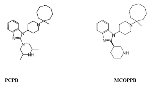
(Varty et al., 2005; Wichmann et al., 1999). After peripheral administration in the range of doses 0.1–3 mg/kg, Ro 64-6198 promoted anxiolytic-like effects in rats in the elevated plus-maze, fear-potentiated startle, and operant conflict tests (Jenck et al., 2000). This has been corroborated by the findings that ppN/OFQ deficient mice display an increased susceptibility to acute and repeated stress, as compared to their wild-type littermates (Koster et al., 1999). In a study from Schering-Plough the non-peptide agonist SCH 221510 was shown to be anxiolytic but with a reduced side-effect profile when compared with benzodiazepines (Varty et al., 2008). Pfizer has recently reported two new non-peptide NOP receptor agonists: PCPB (Hirao et al., 2008a) and MCOPPB (Hayashi et al., 2009; Hirao et al., 2008b); both compounds are orally active and showed anxiolytic-like effects in mice.
Importantly, very little information is present in the literature regarding the effects of selective NOP receptor antagonists on anxiety. It has been reported that the non-peptide molecule J-113397 at 10 mg/kg i.p. antagonized the anxiolytic-like effect of Ro 64-6198 in the conditioned lick suppression test in rats without having any effect in this assay per se (Varty et al., 2005). Although very few studies have been performed to date on this topic, the available evidence obtained with two chemically unrelated NOP antagonists, i.e., J-113397 and UFP-101 (Gavioli et al., 2006; Vitale et al., 2006), suggest that the acute blockade of NOP receptor does not modify the level of anxiety in rodents. In other words, these results suggest that N/OFQergic signalling does not tonically control anxiety-related behaviour.
Surprisingly, Fernandez et al. (2004) observed anxiogenic-like effects of N/OFQ given by i.c.v. injection in several anxiety-related procedures (i.e., open field, elevated plus maze, and dark-light preference) in rats. Explanations given by the authors are a difference in baseline stress between studies, and/or strain differences. Even though a control experiment suggested that the anxiogenic-like effects were, at least in part, independent of effects on locomotion (Fernandez et al., 2004), a more recent study by Vitale et al. (2006) may suggest the opposite. They replicated the anxiogenic-like effects of N/OFQ in the rat elevated plus-maze test, but observed anxiolytic-like
effects after 2 subsequent administrations of N/OFQ (Vitale et al., 2006). This change from anxiogenic- to anxiolytic-like effect of N/OFQ was accompanied by tolerance to the hypolocomotor effects of N/OFQ, suggesting that the anxiolytic-like effects of acute N/OFQ were masked by hypolocomotor effects.
So far the anxiolytic mechanisms of N/OFQ are not well understood. It is likely that N/OFQ effects on anxiety may depend on the ability of this peptidergic system to modulate endogenous 5-HTergic pathways since 5-HT is considered to play a pivotal role in the control of anxiety and fear (Millan, 2003). Moreover, recent findings suggested that the anxiolytic-like effects of N/OFQ might be mediated via activation of the GABA/benzodiazepine (Gavioli et al., 2008; Uchiyama et al., 2008). It has also been suggested that N/OFQ can act as a functional corticotrophin releasing factor (CRF) antagonist, since it is able to revert the hypophagia induced by either stress or the central administration of CRF (Ciccocioppo et al., 2001). Since CRF is a major mediator of stress and a potent anxiogenic agent, the functional relationships between the N/OFQ and CRF systems are worthy of further investigation aimed at clarifying the mechanisms by which N/OFQ exerts its anxiolytic-like effects.
Several lines of evidence also suggest that endogenous N/OFQ has an important role in anxiety and stress regulation. Enhanced anxiety was shown in ppN/OFQ(-/-) mice (Kest et al., 2001; Ouagazzal et al., 2003; Reinscheid et al., 2002) and antisense-NOP treated rats (Blakley et al., 2004), but not in NOP(-/-) mice (Mamiya et al., 1998). Gavioli et al. (2007) demonstrated that there are no clear differences between NOP(-/-) and NOP(+/+) mice in some classical models of anxiety (open-field, hole-board and marble-burying tests). In contrast, when subjected to other models of anxiety such as novelty-suppressed feeding behaviour and the elevated T-maze test, NOP(-/-) mice display lower anxiety-related behaviours compared to NOP(+/+) mice (Gavioli et
al., 2007).In the elevated plus-maze and light-dark box, NOP(-/-) mice displayed increased
Interestingly, the impact of ppN/OFQ deletion on the anxiety-like behaviours is more significant in group-housed, as compared with individual-housed mice, and male mice were more susceptible than females (Ouagazzal et al., 2003). In mice, differences in anxiety states are associated with differences in G protein coupling efficiency in the nucleus accumbens (but not in 12 other brain regions) (Le Maitre et al., 2006). A likely explanation of this finding is that the observed increase in coupling in non-anxious mice leads to increased N/OFQ-mediated transmission and thus protects from anxiety (Le Maitre et al., 2006).
However, the information that has been available to date has been too limited to propose a mechanistic interpretation of the anxiolytic-like effects of N/OFQ. Other studies aimed to the identification of the brain areas involved in this action are needed for a better understanding of N/OFQ role in this field.
1.4.4 Mood
Studies performed in rodents subjected to behavioural despair tests support a role of the N/OFQ-NOP receptor system in the modulation of mood behaviours.
NOP receptor antagonists, including [Nphe1]N/OFQ(1-13)-NH2, J-113397, UFP-101 and SB-612111 reduced immobility time in both the forced swim and tail suspension tests. N/OFQ (i.c.v.) alone did not affect immobility time (Gavioli et al., 2007; Gavioli et al., 2004; Redrobe et al., 2002).
In our laboratories, using a combined pharmacological and genetic approach, we demonstrated that blockade of N/OFQ-NOP receptor signalling in the brain produces antidepressant-like effects in the mouse and rat forced swimming test and in the mouse tail suspension test. I.c.v. injection of N/OFQ did not induce any behavioural modification in mice, but the co-administration of 1 nmol of N/OFQ reversed the antidepressant-like effect induced by the NOP receptor antagonists UFP-101 (Gavioli et al., 2003; Gavioli et al., 2004). In addition, N/OFQ (1 nmol) also reverted the effects induced by the non-peptide NOP receptor antagonist J-113397 in
the mouse forced swimming test (Gavioli et al., 2006). Moreover, it has been demonstrated that antidepressant-like effects elicited by the selective NOP receptor antagonist UFP-101 are probably due to the block of the inhibitory effects of endogenous N/OFQ on brain monoaminergic (in particular serotonergic) neurotransmission (Gavioli et al., 2004).
Whereas the immobility time in NOP(-/-) mice is less than that in NOP(+/+), the antidepressant-like effects of NOP receptor antagonists were not observed in NOP(-/-) mice (Gavioli et al., 2003), suggesting that endogenous N/OFQ plays a role in those depression-like behaviours. Treatment with UFP-101 (10 nmol) reduced immobility time in NOP(+/+) mice, while it was inactive in mice lacking the NOP receptor (Gavioli et al., 2003). Systemic administration of J-113397 (20 mg/kg) promoted a statistically significant reduction in immobility time in the forced swimming test in NOP(+/+), but not in NOP(-/-) animals. Additionally, SB-612111 (10 mg/kg, i.p.) reduced the immobility time in NOP(+/+) mice while being inactive in NOP(-/-) animals (Rizzi et al., 2007b).
Vitale and colleagues investigated the effect of UFP-101 in the chronic mild stress paradigm in rats; UFP-101 (10 nmol/rat, i.c.v. continuously infused by means of minipumps for 24 days) did not influence sucrose intake in non stressed animals, but reinstated the basal sucrose consumption in stressed animals, beginning from the second week of treatment as did fluoxetine (10 mg/kg, i.p.), used as reference drug (Vitale, 2008).
There is just one human study (Gu et al., 2003) in which plasma N/OFQ level was elevated in post-partum depressive women. This limited small study agrees with the notion that post-partum depression results from reduced 5-HT levels and that this is accompanied by elevated N/OFQ with the increase in N/OFQ possibly causing the fall in 5-HT (Lambert, 2008). Thus, NOP receptor antagonists may have the potential to be novel antidepressants.
1.4.5 Food intake
Soon after the isolation of N/OFQ, Pomonis and colleagues (1996) showed that supraspinal N/OFQ (1–10 nmol) increased food intake in satiated rats. N/OFQ effects are short-lasting, specific to food intake with neither water intake nor 1% sucrose intake affected, and accompanied by transient hypolocomotion (Polidori et al., 2000b).
N/OFQ hyperphagia can be blocked by antisense treatment to NOP mRNA (Leventhal et al., 1998)), competitive NOP antagonism (Polidori et al., 2000b) and functional antagonism by nocistatin (Olszewski et al., 2000). Surprisingly, naloxone/naltrexone pretreatment also blocks N/OFQ effects on food intake (Leventhal et al., 1998; Pomonis et al., 1996), although this is probably due to classical opioid receptors being involved in feeding control at a distal site or affecting motivational processes related to food intake. In addition, it was shown that the orexigenic action of 1 nmol of N/OFQ was prevented by SB-612111 (1 mg/kg) and no longer evident in NOP(-/-) animals, indicating that the orexigenic effects induced by N/OFQ are exclusively due to NOP receptor activation (Rizzi et al., 2007b).
The orexigenic action of N/OFQ is suggested to be attributed to both the inhibition of anorexigenic systems and the activation of orexigenic systems (Olszewski et al., 2004). N/OFQ has been found to inhibit pathways that promote termination of food intake in the hypothalamic satiety centers, such as oxytocinergic neurons in the paraventricular nucleus and neurons in the arcuate nucleus (Olszewski et al., 2004). Moreover, Ciccocioppo et al. (2004) found that N/OFQ, at doses without hyperphagic effects, inhibited stress-induced anorexia and that this anti-anorexic effect is due to the fact that N/OFQ acts as a functional antagonist of CRF at the bed nucleus of the stria terminalis (Ciccocioppo et al., 2004). [Nphe1]N/OFQ(1-13)-NH2 did not affect food consumption per se in satiated rats, but reduced that in food-deprived rats (Polidori et al., 2000b). UFP-101 also did not affect free feeding in the rat (Economidou et al., 2006b). This suggested that endogenous N/OFQ plays a role in orexigenic tone in response to food deprivation but not in normal feeding. On the contrary, (Rizzi et al., 2007a) showed that the antagonist SB-612111 (1 and 10 mg/kg, i.p.),
