DOTTORATO DI RICERCA IN
FARMACOLOGIA E ONCOLOGIA MOLECOLARE
CICLO XXVII
COORDINATORE Prof. Cuneo Antonio
Analysis of the anti-leukemic activity of
sodium dichloroacetate
Settore Scientifico Disciplinare
BIO/16
Dottorando Tutore Dott. Agnoletto Chiara Prof. Zauli Giorgio
Correlatore Prof.ssa Secchiero Paola
Table of contents
INTRODUCTION pg 1
B chronic lymphocytic leukemia pg 1
Prognostic markers in B-CLL pg 2
Current management of chronic lymphocytic leukemia pg 5
Metabolic transformation in cancer pg 8
Metabolism in chronic lymphocytic leukemia pg 10
Targeting aerobic glycolysis in chronic lymphocytic leukemia pg 11
The oncosuppressor TP53: role in metabolic transformation pg 12
Targeting p53 with Nutlin-3 as potential therapeutic option for B-CLL pg 16
OBJECTIVES pg 18
METHODS pg 19
Primary B-CLL patient samples and B leukemic cell lines pg 19
Culture treatments, assessment of cell viability, apoptosis, cell cycle profile and
Senescence pg 20
Assessment of mitochondrial alterations pg 20
Western blot analyses pg 21
Bi-dimensional gel electrophoresis (2-DE) and immunoblotting pg 21
Proteomic profiling pg 22
cDNA microarray and RT-PCR analyses pg 23
RNA analyses pg 24
Transfection experiments pg 24
RESULTS pg 26 DCA promotes cytotoxicity in primary B-CLL patient derived cells, but not in normal
peripheral blood cells pg 26
DCA activates the p53 pathway in p53wild-type B leukemic cell lines pg 29
DCA plus Nutlin-3 combination exhibits a potent synergistic anti-leukemic activity pg 32 Role of p21 induction in DCA and DCA+Nutlin-3 induced cytotoxicity in leukemic cells pg 34 DCA promotes comparable cytotoxicity in p53wild-type and p53mutated B-CLL patient cells pg 37
DCA effects on HL-60 apoptosis, cell cycle and mitochondrial energy metabolism
transcriptomic profile pg 42
DCA induces the transcription of p21 in both p53wild-type and p53mutated/null leukemic cells pg 45
DCA effects on HL-60 proteomic profile pg 46
Role of ILF3-p21 axis in DCA induced cytotoxicity pg 50
DISCUSSION pg 53
Conclusions pg 55
REFERENCES pg 56
Introduction
B chronic lymphocytic leukemiaB chronic lymphocytic leukemia (B-CLL) is the adult leukemia with the highest incidence in Western countries; more than 15.000 newly diagnosed cases and 4.500 deaths are currently estimated yearly. It is characterized by the clonal proliferation and accumulation of differentiated CD5+/CD23+ B lymphocytes in the peripheral blood and lymphoid organs [1,2]. B-CLL is an heterogeneous disorder in terms of progression, therapeutic response and outcome despite a common diagnostic immunophenotype (surface expression of CD19+, CD20+dim, CD5+, CD23+ and
sIgMdim) [2]. The leukemic transformation is initiated by specific genomic alterations, in particular
deletions on the long arm of chromosome 13 [del(13q14)]. Additional aberrations of the long arm of chromosome 11 [del(11q)], of the short arm of chromosome 17 [del(17p)] and trisomy of chromosome 12 seem to occur later in the course of the disease and predict a worse outcome [3-5].
The diagnosis of CLL is currently based on clinical analyses of lymphocyte morphology and the presence of >5x10⁹/l circulating clonal B cells with the specific immunophenotype persisting for
>3 months [1]. A recommended scoring system allocates one point each for the expression of weak surface membrane immunoglobulins, CD5, CD23 and absent or low expression of CD79b and FMC7. Using this system, 92% of B-CLL cases score 4 or 5, 6% score 3 and 2% score 1 or 2 [1]. Frequently at diagnosis patients present lymphadenopathy and systemic symptoms such as tiredness, night sweats and weight loss; signs of advanced stages of leukemia include impaired function of normal bone marrow elements, such as anemia and leucopenia, with higher susceptibility to infections [1]. 5-15% of B-CLL patients develop diffuse large B cell lymphoma (Richter syndrome) or, less frequently, Hodgkin lymphoma, either pre- or post-therapy. Clinical features suggestive of transformation include bulky (>5 cm) lymphoadenopathy, rapid nodal enlargement, extranodal disease, the development of B symptoms and marked elevation of lactate dehydrogenase (LDH). Moreover, autoimmune complications are common in B-CLL, occurring in 10-20% of patients and they target almost exclusively blood cells, in particular erythrocytes. The diagnosis of autoimmune
haemolytic anemia is based on the presence of an isolated reduction in haemoglobin with positive direct antiglobulin test, a rise in reticulocyte count, bilirubin and LDH and a reduction in serum haptoglobins [1]. Whereas the course of disease is indolent in some patients, it is steadily progressive in approximately half of patients, leading to substantial morbidity and mortality [2]. The variable clinical course of B-CLL is due, at least in part, to the disease's immunogenetic and molecular heterogeneity [2].
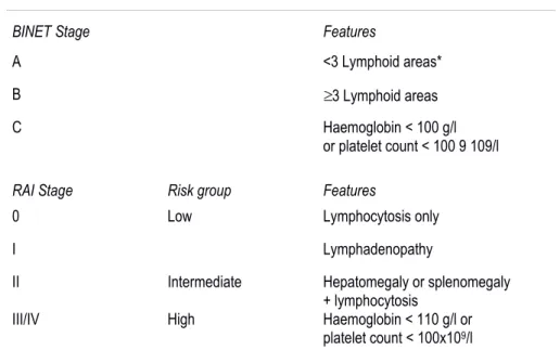
Numerous clinical and laboratory parameters have been developed to predict prognosis. Clinical staging systems of Binet and Rai still represent the most useful mean to determine prognosis and permit to divide patients into classes with different risk levels (Table 1) [1]. Commonly clinicians initiate therapeutic treatment only if patients reach advanced stages and develop a symptomatic disorder. However, while these staging systems are clinically useful, they can't predict long-term survival with high precision, especially for patients with early stage leukemia [6].
Table 1. Staging systems in CLL.
BINET Stage Features
A <3 Lymphoid areas* B 3 Lymphoid areas C Haemoglobin < 100 g/l
or platelet count < 100 9 109/l
RAI Stage Risk group Features
0 Low Lymphocytosis only I Lymphadenopathy
II Intermediate Hepatomegaly or splenomegaly + lymphocytosis
III/IV High Haemoglobin < 110 g/l or platelet count < 100x109/l
*The five lymphoid areas comprise: uni or bilateral cervical, axillary and inguinal lymphoid, hepatomegaly and splenomegaly.
Prognostic markers in B-CLL
The ability to predict a more aggressive disease course has improved with the use in clinical practice of tests for biologic markers (degree of somatic hypermutation in the variable region of the immunoglobulin heavy chain [IGHV] gene and expression of ZAP-70 [Z-associated protein 70] or CD38) and cytogenetic abnormalities [3,7-10]. High expression of both ZAP-70 or CD38 are associated with higher-risk CLL. While IGHV mutated CLL patients frequently show mild clinical features and high overall survival and progression-free survival, IGHV un-mutated CLL patients
suffer an aggressive form of the disease which may be refractory to treatment [11]. Moreover, although the somatic mutation status is highly associated with prognosis, recent data document that a specific heavy chain variable region genetic signature is associated with poor prognosis, independently of somatic mutational status [12].
Despite the relatively stable karyotype in CLL cells, genomic aberrations were recognized as important determinants of the clinical course of the disease [13]. In particular, chromosomal abnormalities represent strong independent predictors of prognosis in B-CLL. The most common is del(13q14.3), followed by del(11q22.3), trisomy 12, del(6q21-23) and del(17p13.1). As the sole chromosomal defect, del(13q14) is associated with a good prognosis [5,13,14]. In contrast, del(6q21-23), del(11q22.3), del(17p13.1), del(13q14.3) with other abnormalities, and trisomy 12 are associated with more rapid disease progression and reduced survival [13]. Finally, the deletions in chromosome 17p, containing the TP53 gene, and chromosome 11q, containing the ATM gene, represent the most important independent cytogenetic markers of poor prognosis [5].
Still, even with these advances, prediction of the disease course is not highly reliable, and the genomic events that dictate the initiation and heterogeneous evolution of B-CLL remained partially unknown [15,16]. Next-generation sequencing (NGS) studies have revealed previously unrecognized genetic lesions, uncovering recurrent somatic gene mutations that occur in B-CLL cells in parallel to the above-mentioned structural genomic aberrations [3-5,16,17]. These studies described about 1,000 somatic variants per patient, evidencing a marked genetic heterogeneity of the disease, with a relatively large number of genes recurrently mutated at low frequency and only a few genes mutated in more than 10% of the patients [5,10,18,19].
Among genes that are mutated at significant frequencies, TP53, ATM, MYD88 and NOTCH1 have been described previously in chronic lymphocytic leukemia [3,5,20-24]. TP53 mutations were detected in 15% of patients and most of them in CLL represent missense substitutions affecting the structural DNA-binding domain (DBD) of p53 protein (codons 175, 179, 248, 273, and 281) that is critical for its tumor-suppressor activity [5,21,22,25], with one mutation out of 105 cases in Quesada et al. [4] and 15 mutations out of 91 cases in Wang et al. [5]. The proportion of patients with a sole TP53 mutation [without del(17p)] vary from 3% to 4% among cohorts at diagnosis or before first therapy [21] to 12% in fludarabine-refractory CLL [26]. A prognostic impact of TP53 mutations in B-CLL has been suggested in a number of reports [21]. Patients with defective p53 have a significantly lower response rate to therapy and a short-progression-free survival [22]. Indeed, TP53 mutation and related alterations have been associated with disease progression and chemorefractoriness in B-CLL [27,28]. Additionally, although TP53 mutations in naïve B-B-CLL were usually considered a rare
(<5%) event [29,30], a recent study performed using the ultra-deep next generation sequencing technology demonstrated that very small TP53 mutated subclones (minor than 20%) are present in 9% of newly diagnosed B-CLL patients [31]. Of note, patients harboring small TP53 mutated subclones showed the same clinical phenotype and poor overall survival as patients carrying clonal TP53 lesions [31]. In addition, the percentage of TP53 mutations dramatically increases up to >30% after relapsed chemotherapy [32]. According to the revised National Cancer Institute (NCI) Working Group/International Workshop on Chronic Lymphocytic Leukemia guidelines for B-CLL, patients with del(17p) should be offered investigative clinical protocols or should be listed for allogeneic stem-cell transplantation [22].
9% of patients with B-CLL present mutations in ATM [5,20]. The ataxia telangiectasia mutated (ATM) protein kinase acts upstream of the p53 oncoprotein and coordinates an integrated cellular response to DNA double-strand breaks through phosphorylation of multiple target proteins. This results in the interruption of the cell cycle and either DNA repair or activation of apoptosis [20,33]. The ATM tumor suppressor gene is located within the minimally deleted region on chromosome 11q. Deletions of chromosome 11q were associated with a severe clinical phenotype and reduced overall survival in B-CLL. ATM mutations were associated with un-mutated IGVH genes and they provided independent prognostic information on multivariate analysis [20].
In addition, recurrent mutations in the myeloid differentiation primary response gene 88 (MYD88) were also identified in 10% of CLL patients [5]. This protein participates in the signalling pathways of interleukin-1 and Toll-like receptors during the immune response [34]. The missense mutations are localized within the interleukin-1 receptor–TLR domain [4,24], and they were documented to activate this proto-oncogene, promoting the duplication of tumour cells and protecting from apoptosis [24,35,36]. Patients with MYD88-mutated CLL were diagnosed at a younger age and with a more advanced clinical stage [3].
Finally, NOTCH1 mutations affect 5–10% of newly diagnosed B-CLL cases, with their prevalence increasing to 15–20% in progressive or relapsed patients [23]. NOTCH1 is constitutively expressed by B-CLL cells, where it increases cell survival and induces apoptosis resistance [37-39]. A recurrent mutation is due to the deletion of a CT dinucleotide (p.P2515Rfs*4), generating a truncated protein which is predicted to result in NOTCH1-impaired degradation and stabilizing effect on the NOTCH1 signaling pathway [3,16]. Indeed both the canonical and non-canonical NOTCH1 pathways are constitutively upregulated in NOTCH1-mutated patients [16]. NOTCH1-mutated patients had a more advanced clinical stage at diagnosis, more adverse biological features and an overall survival that was significantly shorter than those with wild-type NOTCH1 [3,10,23,38].
Interestingly, on a clinical perspective, among the novel genes that are recurrently mutated in CLL, some are preferentially altered in fludarabine refractoriness (FR) patients, namely TP53, NOTCH1, SF3B1 and BIRC3 [23,25,40,42]. FR remains an unsolved clinical problem associated with poor survival, involving 10% of patients after first-line treatment and virtually all patients after multiple lines of therapy [43,44]. Until recently, the molecular basis of CLL chemorefractoriness was incompletely characterized, and the only known genetic lesion with an established pathogenic role was inactivation of the TP53 gene occurring in 30% to 40% of refractory patients [26]. Interestingly, mutations of the splicing factor 3b, subunit 1 (SF3B1) gene have been identified in 15% of B-CLL patients [5] and they are recurrently associated with fludarabine-refractory CLL and poor clinical prognosis, with a frequency significantly greater than that observed in a consecutive CLL cohort sampled at diagnosis [40]. Therefore, SF3B1 mutated B-CLL require prompt treatment, independently of other established predictive markers [4,45].
In conclusion, somatic mutations affecting NOTCH1, SF3B1 and TP53 are improving our ability to predict the outcome of the disease from early stages [41,46], and several studies have been performed to clinically characterize the consequences of mutations affecting these genes on the prognosis of CLL. In a recent manuscript Landau et al. have shown that the presence of certain somatic mutations even in a small population of the tumor cells accurately predicts a high probability of relapse after treatment [17]. If this information is known at diagnosis, clinicians may choose to target those drivers early to prevent relapse [47].
Current management of chronic lymphocytic leukemia
The clinical course of patients with B chronic lymphocytic leukemia spans from an asymptomatic disease never requiring therapy to a rapidly progressive disorder requiring intensive treatment [2] The clinical heterogeneity of B-CLL and advanced age of a great number of subjects dictate that a diversified therapeutic approach, since no single treatment is applicable to all patients [1]. For more than 40 years, B-CLL has been treated with diverse chemotherapies [43]. Monotherapy with cytostatic alkylating agents, usually chlorambucil (CLB), has been used for several decades as initial, front-line therapy [48]; the principal limits of CLB are due to low or nonexistent complete remission (CR) rate and serious side effects after extended use, i.e. prolonged cytopenia, myelodysplasia and secondary acute leukemia [43,48,49]. Combinations of alkylating drugs with vinca alkaloids or anthracycline drugs did not improve outcomes [48]. Three purine analogs are
currently used in B-CLL, fludarabine, pentostatin, and cladribine (2-CdA). Fludarabine induced more partial remissions (PR) and more complete remissions (CR) (7%-40%) than other conventional chemotherapies, but did not improve overall survival (OS) when used as single agent [1,50].
A great collection of clinical data confirms that a combination of chemotherapies has proven more effective compared to monotherapy [49]. Since purine analogs and alkylating agents have different mechanisms of action and partially non-overlapping toxicity profiles, they were combined both in preclinical and clinical studies. The most thoroughly studied combination chemotherapy for B-CLL is fludarabine plus cyclophosphamide (FC) given as oral drugs or intravenous infusions [51]. Three randomized trials have demonstrated that FC chemotherapy improves clinical response, progression-free survival (PFS) and complete remission compared with fludarabine monotherapy [52-54]. An updated analysis of the CLL4 trial of the German CLL Study Group (GCLLSG) suggested that the front-line treatment of B-CLL patients with FC combination might improve the OS of the non-high-risk B-CLL patients [all patients without del(17p) or TP53 mutation] [49].
Chemo-immunotherapy was developed for the treatment of indolent and aggressive lymphoma. Rituximab, a chimeric monoclonal antibody directed against the CD20 antigen expressed on B-cells, became the standard drug for different B-cell lymphomas [42,55]. In B-CLL the expression of the CD20 antigen on leukemic cells is relatively low and standard-dose rituximab generates only poor response rates [43,56]. The results of phase 2 trials suggested that rituximab in combination with chemotherapy might have additive or synergistic effects in pretreated and treatment-naive patients [57,58]. In a study of 300 treatment-naïve patients, the combination of FC with rituximab resulted in an overall response rate of 95%, with 72% of patients achieving a complete response [43,57,58]. These response rates were among the highest reported so far for first-line treatments in patients with B-CLL [43]. Furthermore, this clinical study documented that patients with del(11q) presented complete remission after chemo-immunotherapy and patients with trisomy 12 and del(13q) greatly benefited from treatment. On the contrary, patients with del(17p), associated with a dysfunction of the p53 tumour suppressor and a very poor outcome, might not benefit substantially [43].
Immunotherapies for B-CLL treatment include other two drugs, which demonstrate therapeutic efficacy. Ofatumumab is a fully humanized antibody targeting the CD20 antigen, that induces an increased cell death due to greater complement-dependent cytotoxicity and similar antibody-dependent cellular-cytotoxicity activity compared with rituximab. Ofatumumab has shown some efficacy in patients with fludarabine and alemtuzumab refractory or bulky disease [59]. Alemtuzumab is a recombinant, fully humanized monoclonal antibody against the CD52 antigen.
Monotherapy with alemtuzumab had high response rates in patients with advanced B-CLL previously treated with alkylating agents and in patients nonresponsive or relapsed after second-line fludarabine therapy [49]. In addition, alemtuzumab has proven effective in patients with high-risk genetic markers such as deletions of chromosome 11 or 17 and TP53 mutations [60].
Currently numerous new compounds that target a relatively specific signaling defect or redirect the immune system against B-CLL cells are in clinical development [61,62]. These agents show clinical efficacy and may yield response rates above 50% even in relapsed and refractory CLL patients [43]. In particular, agents targeting B cell receptor (BCR) signaling have entered in phase 3 trials for B-CLL patients. Stimulation of BCR induces the activation of different tyrosine kinases, such as Bruton tyrosine kinase (BTK), spleen tyrosine kinase (Syk), ZAP-70, Src family kinases, and phosphatidyl inositol 3-kinase (PI3K), which stimulate malignant B-cell survival via activation of transcription factors such as NF-kB [61,63]. Idelalisib (CAL-101), an oral PI3Kp100 isoform– selective inhibitor, promotes apoptosis selectively in primary B-CLL cells, without reducing antibody-dependent cellular cytotoxicity [64]. In a phase 1 clinical trial in 54 heavily pretreated and high-risk B-CLL patients, idelalisib showed acceptable toxicity, positive pharmacodynamics effects, and favorable clinical activity. An overall response rate (ORR) of 56% was achieved, with 2 CRs and 28 PRs [65].
Ibrutinib (PCI-32765) is an orally active small molecule inhibiting BTK, which activates cell survival pathways such as NF-kB and MAPKs via Src family kinases. Inhibition of BTK might induce apoptosis in B-cell lymphomas and B-CLL cells [66]. Ibrutinib showed significant activity in patients with relapsed or refractory B-CLL [67]. Data from a phase 1b-2 multicenter study with single-agent ibrutinib on 85 patients with relapsed or refractory B-CLL or small lymphocytic lymphoma, the majority with high-risk disorder, were published recently [68].The ORR was 71%, the majority being PRs (68%). Interestingly, the response was independent of clinical and genomic risk factors, including advanced stage disease, the number of previous therapies and del(17p). This study illustrates that ibrutinib may soon become an additional treatment option for B-CLL patients with high-risk genetic lesions [68]. The above described novel therapies, targeting specific signaling proteins of B-CLL cells, present a moderate overall toxicity, without myelosuppression. However, CRs have not occurred frequently so far with any of these newer agents [43].
Patients who relapse and have not acquired a TP53 abnormality can be expected to respond to a further course of their initial therapy, although the PFS is usually shorter than after initial therapy and repeated courses often lead to drug resistance. However, re-treatment with the previous therapy is not recommended in patients whose initial treatment was sub-optimal or if a new treatment, shown
to be superior to the initial therapy, becomes available [1]. For patients with high risk B-CLL (TP53 defect and/or failing fludarabine combination therapy within 2 years), results of phase II and III studies using either FC, FCR or alemtuzumab, with or without high dose steroids, indicate that alemtuzumab plus pulsed methylprednisolone or dexamethasone should be regarded as the induction regimen of choice. However, this regimen is associated with a significant risk of infection and the duration of remission is relatively short. Allogeneic stem-cell transplantation provides the last opportunity of achieving long-term disease-free survival for patients with high-risk B-CLL, particularly those with TP53 defects [1,43].
Metabolic transformation in cancer
Over the past 25 years, the oncogene revolution has stimulated the research in cancer biology, revealing that the crucial phenotypes of tumour cells result from a host of mutational events that combine to alter multiple core signaling pathways [69,70]. High-throughput sequencing data suggest that the mutations leading to tumorigenesis are even more numerous and heterogeneous than previously thought [71]. Indeed, gene set enrichment analysis showed significant up-regulation of several core processes, known to participate also in cancer metabolic reprogramming, such as glycolysis/gluconeogenesis, the pentose phosphate pathway or oxidative phosphorylation [72,73]. Multiple key oncogenic signalling pathways converge to adapt tumour cell metabolism in order to support their growth and survival. Some of these metabolic alterations, that is rapid ATP generation, increased biosynthesis of macromolecules and tightened maintenance of cellular redox status, seem to be absolutely required for malignant transformation. Therefore, altered cellular metabolism represents one of the emerging hallmark of cancer [69,73,74].
The most evident and most studied metabolic phenotype observed in tumour cells is the Warburg effect, also termed aerobic glycolysis, which describes the shift from ATP generation through oxidative phosphorylation to ATP generation through glycolysis [75]. Indeed while normal tissues used mitochondrial oxidation to account for 90% of ATP production with glycolysis accounting for 10%, tumor cells used less of the highly efficient oxidative phosphorylation, producing 50% of the ATP from oxidation and 50% from glycolysis. This shift was thought to occur even though there is sufficient oxygen to support mitochondrial function [76,77]. The increase in glycolysis at the expense of mitochondrial energy production helps to support unbridled growth, provide precursors for the biosynthesis of macromolecules and protect cells from excessive and toxic levels of reactive
oxygen species (ROS) and oxidative stress [69]. This hypothesis greatly influenced the present perception of cancer metabolism, moving aerobic glycolysis into the mainstream of clinical oncology.
Figure 1. Molecular mechanisms driving the Warburg effect. Relative to normal cells (part a) the shift to aerobic glycolysis in tumour cells (part b) is driven by multiple oncogenic signalling pathways. PI3K activates AKT, which stimulates glycolysis by directly regulating glycolytic enzymes and by activating mTOR. The liver kinase B1 (LKB1) tumour suppressor, through AMP-activated protein kinase (AMPK) activation, opposes the glycolytic phenotype by inhibiting mTOR. mTOR induces the glycolytic phenotype by enhancing hypoxia-inducible factor 1 (HIF1) activity, which engages a hypoxia-adaptive transcriptional programme. HIF1 increases the expression of glucose transporters (GLUT), glycolytic enzymes and pyruvate dehydrogenase kinase, isozyme 1 (PDK1), which blocks the entry of pyruvate into the tricarboxylic acid (TCA) cycle. MYC cooperates with HIF in activating several genes that encode glycolytic proteins, but also increases mitochondrial metabolism. The tumour suppressor p53 opposes the glycolytic phenotype by suppressing glycolysis through TP53-induced glycolysis and apoptosis regulator (TIGAR), increasing mitochondrial metabolism via SCO2 and supporting expression of PTEN. OCT1 acts in an opposing manner to activate the transcription of genes that drive glycolysis and suppress oxidative phosphorylation. The switch to the pyruvate kinase M2 (PKM2) isoform affects glycolysis by slowing the pyruvate kinase reaction and diverting substrates into alternative biosynthetic and reduced nicotinamide adenine
dinucleotide phosphate. (NADPH)-generating pathways. MCT, monocarboxylate transporter; PDH, pyruvate dehydrogenase. The dashed lines indicate loss of p53 function. Image from Cairns et al,2011.
Recent advances in genomics and proteomics have provided insights into molecular mechanisms which underline the metabolic remodelling occurring in cancer cells [73,75]. Metabolic pathways in normal cells are tightly regulated and all parts of these networks can be highly perturbed in cancer cells [78]. Mutations in oncogenes and tumour suppressor genes cause alterations to multiple intracellular signaling pathways that affect tumour cell metabolism and reengineer it to allow enhanced survival and growth [69,71]. Consistently genetic analysis of tumors has since identified numerous oncogenes that, once activated, increase glycolysis and glutaminolysis, and, analogously, different tumor suppressor that, once lost, can shift energy production away from the mitochondria to a more glycolytic equilibrium [75,79]. Irrespective of the mechanisms involved, if certain tumors are reliant on glycolysis for bioenergetics, biosynthesis and invasive potential, therapeutic targeting of the underlying pathways may result in an anti-tumor activity (Figure 1) [79,80]. Indeed drugs that can selectively target the metabolic phenotype of the tumour are likely to at least delay tumour progression [74]. The resistance of tumours to both radiotherapy and chemotherapy can often be attributed to its aberrant metabolism. Therefore the reactivation of a more ‘normal’ metabolism could re-sensitize tumours to these agents. From a therapeutic perspective, knowledge of cancer cell metabolism will enable the identification of new drug targets and will facilitate the design of a new class of cancer therapeutics and diagnostic tools especially exciting [69]. The ultimate aim is to develop treatment strategies that slow tumour progression, improve the response to therapy and result in a positive clinical outcome [81]. Therapies that target tumour metabolism are currently being tested in pre-clinical and clinical studies [74].
Metabolism in chronic lymphocytic leukemia
Despite the high incidence of B chronic lymphocytic leukemia, metabolism of B-CLL cells remains a relatively unexplored field [82]. Although initially demonstrated in solid tumors, a recent study identified dysregulated glucose metabolism as a novel therapeutic target also in B-CLL [83]. In particular glycolytic enzymes appeared to be upregulated in a subset of CLL patients with either higher white blood cell counts and/or more aggressive clinical course. Additionally recent evidences documented that in leukemic cells mitochondrial uncoupling, i.e. the reduction in ATP synthesis in response to mitochondrial membrane potential, due to the overexpression of uncoupling protein UCP2, promotes glycolysis, increases the apoptotic threshold and induces chemo-resistance [84].
Results from few studies indicate that B-CLL cells might contribute to the so-called cancer-associated oxidative stress [85]. This metabolic condition results from accumulating reactive oxygen species (ROS) [86], compared with normal B-cell, due to cancer-related mitochondrial alterations, such as defective oxidative phosphorylation or an enhanced mitochondrial biogenesis [87]. Moreover, high levels of ROS correlated with the number of circulating CLL cells and have been detected in more aggressive B-CLL cells [88]. Mitochondria were identified as the key source for abundant ROS in B-CLL [76,82]. Escalated ROS levels promote genetic instability and development of drug resistance, determine alterations in cell-signaling [86] and altogether account for cancer cells’ aggressive behavior [82]. This is in line with studies demonstrating that B-cell stimulation promotes ROS production [89] and B-CLL cells partially resemble chronically activated B-cells [90]. Oxidative stress additionally attenuates immune responses by leading to dysfunctions and even apoptosis of NK- and T-cells, suggesting a role in tumor immune-escape [82]. Since cancer cells are continuously exposed to high levels of endogenous ROS, cells which adapt to high stress condition are preferentially selected. Consequently, an enhanced protective antioxidant capacity is reported in tumors, which in addition induce the development of drug resistance [91]. As a protective measure, the activation of the redox adaptive cascade (through increased expression of intracellular antioxidant catalase and the stress-responsive heme oxygenase-1, and reduced expression of mitochondrial manganese superoxide dismutase) would then additionally promote biogenesis of novel and functional mitochondria [82]. Thus increased mitochondrial ROS, adaptation to intrinsic oxidative stress and mitochondrial biogenesis are interconnected in B-CLL.
Interestingly, using agents that further aggravate mitochondrial ROS production and thereby overwhelm the cancer cells’ protective systems could selectively induce apoptosis in B-CLL cells [82]. Metabolic alterations can render malignant cells more susceptible to perturbations and therefore agents directed against the unique metabolic features of B-CLL cells might represent an opportunity to improve current therapeutic treatments and even to overcome drug resistance within B-CLL cells.
Targeting aerobic glycolysis in chronic lymphocytic leukemia
Several recent studies have confirmed that restricting glycolysis or diverting pyruvate into the mitochondria can significantly induce respiration in cancer cells [92]. The fate of pyruvate [either reduction in the cytosol by lactate dehydrogenase (LDH) or oxidation in the mitochondria by pyruvate dehydrogenase (PDH)] determines the direction of tumour metabolism. The inhibition of lactate dehydrogenase or the activation of pyruvate dehydrogenase can induce tumour cells to oxidize pyruvate in the TCA cycle and stimulate mitochondrial respiration [93-95]. Pyruvate dehydrogenase
represents the enzymatic step linking glycolysis to oxidative phosphorylation. One of four pyruvate dehydrogenase kinase (PDKs) and one of two pyruvate dehydrogenase phosphatases are associated with PDH and regulate the phosphorylation of the E1a subunit, dictating PDH activity. When phosphorylated, PDH is inactivated, resulting in channelling of pyruvate to lactate, whereas the non-phosphorylated form is active and converts pyruvate into mitochondrial acetyl-CoA [5]. Recent studies provided evidence that PDK1 expression is elevated in cancers, thereby attenuating mitochondrial function and respiration, and its activation may be a key regulatory switch contributing to the Warburg effect [5,69,70,77]. The inhibition of PDK is increasingly becoming seen as a valid target for cancer therapy [14,81]. Indeed ihibitors of PDK could potentially reverse some of the metabolic effects of tumorigenic signalling and several such candidates, including the mitochondria-targeting dichloroacetate molecule (DCA), are currently under evaluation for their therapeutic utility [71,74,75]. Indeed, preclinical studies confirm the relatively low toxicity of DCA and document its efficacy against numerous epithelial cancers (such as non-small cell lung, endometrial, prostate, breast and colorectal cancer), glioblastoma, as well as multiple myeloma [96-105]. DCA inhibits mitochondrial pyruvate dehydrogenase kinase (PDK) reverting the metabolic-electrical remodelling occurring in cancer cells; indeed it shifts the metabolism from glycolysis to glucose oxidation. In addition, DCA decreases the mitochondrial membrane potential, increases the reactive oxygen species (ROS) production and ultimately induces apoptosis and tumor growth arrest in mice models, without apparent toxicity [95]. DCA is an orally bioavailable, small molecule and currently being evaluated in phase I/II clinical trials for glioblastoma, gliomas and other solid tumors [106]. Interestingly, in solid tumor models it has also been reported that DCA may activate p53 [107]. In addition, a couple of recent studies proposed DCA alone or in association with bortezomib for the treatment of multiple myelomas [105,108], suggesting anti-cancer activity of DCA also in hematological disorders.
The oncosuppressor TP53: role in metabolic transformation
Although the tumour suppressor p53 is best known for its functions in mediating the cytotoxic response of most therapeutic molecules currently used in cancer therapy, p53 is also an important regulator of metabolism [69]. The role of p53 as a central component of the stress response machinery is well established, and numerous forms of stress - many of which are encountered during tumoral transformation - lead to the activation of p53 [109]. Virtually any stress signal can induce
p53, including hypoxia or the lack of nutrients and deregulated signalling through the nutrient-sensing pathways, namely the AKT–mTOR pathway [110]. Oxidative stress regulates p53 function by directly affecting its redox state and oxidation [111] and mitochondrial ROS represent an important component of the stress-induced activation of p53 [112]. Also, p53 is efficiently activated by ribosomal stress through the ability of several ribosomal proteins to bind and inactivate MDM2 [113]. Combinations of these abnormalities during tumour progression amplify the protective p53 response. However, increased glucose metabolism stimulated by the expression of the glucose transporter GLUT1 or hexokinase has been reported to suppress p53 activity [114], suggesting that high levels of glycolysis in cancer may help evade the tumour-suppressive effects of p53. The most obvious advantage of p53 activation under conditions of metabolic stress is a coordinated inhibition of cell proliferation and growth. However additional consequences of p53 activation are being uncovered, including an ability of p53 to promote the use of certain metabolic pathways and to increase cell survival.
Figure 2. Regulation of energy production by p53. Several functions of p53 reduce the flux through the glycolytic pathway and increase oxidative phosphorylation, thereby opposing the Warburg effect, in which cancer cells predominantly use glycolysis for energy production. However, there are also activities of p53, such as the activation of hexokinase and phosphoglycerate mutase (PGM), which could increase glycolysis under some circumstances. GLUT, glucose transporter; IKK, IκB kinase; NF-κB, nuclear factor-κB; SCO2, synthesis of cytochrome c oxidase 2; TCA, tricarboxylic acid. Image from Vousden and Ryan,2009.
Several studies documented that p53 exerts a role in the regulation of both glycolysis and oxidative phosphorylation (Figure 2) [78]. Different mechanisms have been described through which
p53 can slow glycolysis and therefore counteract its increase in cancer. p53 can inhibit the expression of the glucose transporters GLUT1 and GLUT4 and can decrease the levels of phosphoglycerate mutase (PGM), while increasing the expression of TP53-induced glycolysis and apoptosis regulator (TIGAR), which reduces the levels of the glycolytic activator fructose-2,6- bisphosphate [78,115]. Wild-type p53 also supports the expression of PTEN, which inhibits the PI3K pathway, thereby suppressing glycolysis [116] and hexokinase 2 (HK2), which converts glucose to glucose-6-phosphate (G6P) [117]. G6P then either enters glycolysis to produce ATP, or enters the pentose phosphate pathway. The effect of each of these actions is to impede flux through different steps of the glycolytic pathway. The restraint on glycolytic rate imposed by p53 is in accordance with its ability to help maintain mitochondria and drive oxidative phosphorylation [118]. These effects are the consequence of several p53-dependent functions, including the transcriptional activation of subunit I of cytochrome c oxidase [119]; the activation of expression of synthesis of cytochrome c oxidase 2 (SCO2), a key regulator which is required for the assembly of the cytochrome c oxidase complex of the electron transport chain [120]; and the induction of expression of the ribonucleotide reductase subunit p53R2 protein, that contributes to the maintenance of mitochondrial DNA [121]. Loss of either p53 or SCO2 expression results in a switch from cellular respiration to aerobic glycolysis, suggesting that inactivation of p53 in human cancers may directly contribute to the Warburg effect [75,122]. Indeed in tumors with non-functional p53 (majority of tumors), the mitochondrial electron-transport chain is compromised, and glycolysis is the more efficient mechanism for generation of ATP [80]. Interestingly, several of these p53 activities, such as the regulation of SCO2, seem to function under normal growth conditions in the absence of an acute stress signal, suggesting that p53 can help to maintain the aerobic respiration of most normal cells. However, the presence of p53-responsive elements in the promoters of phosphoglycerate mutase and hexokinase II suggests that p53 can promote at least some steps in glycolysis, resulting in increased survival signalling by helping to limit ROS production [78]. These different activities reflect context- or tissue-dependent differences in the metabolic functions of p53. Notwithstanding the role of p53 in enhancing oxidative phosphorylation, increased glycolysis in response to pharmacological inhibition of oxidative phosphorylation is reported to be dependent on p53 [123].
Our recent appreciation of the importance of basal levels of p53 in regulating metabolic pathways is mirrored by the importance of the antioxidant functions of low or uninduced levels of p53 [124]. ROS are heterogeneous in their properties and exert a plethora of downstream effects, depending on the concentrations at which they are present. Low levels of ROS are required for homeostatic signaling events, increasing cell proliferation and survival through the post-translational
modification of kinases and phosphatases [125]. At moderate levels, ROS induce the expression of stress-responsive genes such as hypoxia-inducible factor HIF-1Α, which in turn determine the expression of proteins providing pro-survival signals [69]. However, once levels of ROS become excessively high, they cause damage to macromolecules, induce the activation of protein kinase Cδ (PKCδ) and senescence [126], and/or cause permeabilization of the mitochondria, with the release of cytochrome c and apoptosis [127]. Cells counteract these effects of ROS by producing antioxidant molecules, such as reduced glutathione and thioredoxin [69]. There is currently a scientific consensus that cancer cells alter their metabolic pathways and regulatory mechanisms so that ROS and antioxidants are tightly controlled and maintained at higher levels than in normal cells, in such a way that the cell survives and the levels of ROS are reduced to moderate levels [128,129]. The mechanisms designed to counter the control of ROS abundance allow the cancer cell to avoid the detrimental effects of high levels of ROS, but also increase the chance that cell will experience additional ROS-mediated mutagenic events and stress responses that promote tumorigenesis.
p53 may promote oxidative stress while inducing apoptosis, but it also exerts an important role in reducing oxidative stress as a defense mechanism. Interestingly, through the p53 target gene cyclin-dependent kinase inhibitor 1A (CDKN1A, which encodes p21), p53 promotes the stabilization of the transcription factor NRF2 [130]. NRF2 is the master antioxidant transcription factor and upregulates the expression of several antioxidant and detoxifying molecules [130]. Loss of p53 in a cancer cell inactivates this redox maintenance mechanism: because p21 is not activated, NRF2 continues to be degraded, antioxidant proteins are not expressed and the redox balance is lost [130]. Regulation of the glycolytic pathway by p53 can also help to modulate oxidative stress by increasing flux through the pentose phosphate pathway. The activation of both TIGAR and hexose-6-phosphate dehydrogenase would promote the pentose phosphate pathway, with the generation of NADPH that enters in both anabolic and antioxidant pathways [78].
p53 can also exerts strong pro-oxidative effects. The ability of p53 to induce senescence and apoptosis is clearly related to the induction of oxidative stress, and even necrotic cell death is activated by the cooperation between ROS and p53-induced cathepsin Q expression [78]. Several p53-inducible proteins activated during the apoptotic response promote ROS production, including p53-induced gene 3 (PIG3), proline oxidase, Bax, PUMA and p66Shc [78]. To further increase this effect, p53 can also inhibit or modulate the expression of antioxidant genes such as superoxide dismutase 2 (SOD2), aldehyde dehydrogenase 4 (ALDH4) and glutathione peroxidase 1 (GPx1), which increases oxidative stress [78]. Less directly, the ability of p53 to drive oxidative phosphorylation through the activation of SCO2 will also promote the generation of ROS from the
mitochondria [120-122]. Depending on the cell type and the levels of ROS, the consequences of ROS regulation by p53 can include proliferation, migration, genotoxic damage, senescence and cell death. In cells in which both pro-oxidant and antioxidant responses are possible the difference lies in the extent and persistence of the damage-inducing stress. The regulation of ROS by p53 might be different under basal or low-stress conditions (in which p53 functions as an antioxidant) compared with high-damage and high-stress conditions (in which p53 contributes to cell death by increasing ROS levels) [131]. From a clinical point of view, it may be possible to exploit loss-of-function p53 mutations or other tumour suppressor genes by applying additional oxidative stress. In the absence of the redox maintenance pathway that is supported by these tumour suppressors, malignant cells might be selectively killed [86].
Numerous data indicate a role of wild-type p53 in averting metabolic transformation, although the possible role of mutant p53 is as yet largely unexplored. TP53 incurs point mutations in numerous cancers, with expression of mutant p53 proteins that have acquired activities which contribute to malignant progression independently of the loss of wild-type p53 function. A role for these mutant p53 proteins in promoting the metabolic transformation would be extremely interesting.
Targeting p53 with Nutlin-3 as potential therapeutic option for B-CLL
The cellular level of the protein p53 is regulated primarily by degradation via the ubiquitin-proteasome pathway, and the murine double minute 2 gene (MDM2) functions as a master regulator of p53 [132]. Vassilev et al. [133] reported the first potent and selective small molecule antagonist of MDM2, termed Nutlin-3. This is a cis-imidazoline analogue that binds to the p53 pocket on the surface of MDM2, activating the p53-pathway in vitro and in vivo. Notably, this interaction, while blocking MDM2-mediated inhibition of p53, does not interfere with p53 function and has little toxicity in animal models. In this context, Secchiero et al. demonstrated that Nutlin-3 is effective in inducing apoptosis in vitro in p53wild-type B-CLL samples independently of the negative prognostic markers
[134-136], as confirmed by other authors. These studies strengthened the concept that selective, non-genotoxic activation of p53 might represent an alternative to the current cytotoxic chemotherapy. Interestingly, Nutlins not only induced apoptotic death in leukemic cells, but also showed a synergistic effect when used in combination with the chemotherapeutic drugs commonly used for the treatment of hematological disorders. Furthermore Nutlins also exert non cell-autonomous biological activities, such as inhibition of vascular endothelial growth factor, stromal derived factor 1/CXCL12 and osteoprotegerin expression and/or release by primary fibroblast and endothelial cells. Moreover, Nutlins have a direct anti-angiogenetic and anti-osteoclastic activities. Thus Nutlins might have
therapeutic effects by two distinct mechanisms: a direct cytotoxic effect on leukemic cells and an indirect non-cell autonomous effect on tumor stromal and vascular cells, and this latter effect might be therapeutically relevant also for treatment of leukemia with 53 mutations [129]. Furthermore, it is noteworthy that, when used in association with other therapeutic compounds, Nutlin-3 showed synergistic cytotoxic activity also on p53mutated B-CLL cells [136,137].
Objectives
In spite of the recent therapeutic advances in the treatment of B-CLL patients, a major unsolved clinical problem is represented by the lack of effective treatments for B-CLL patients with more aggressive disease, carrying cytogenetic abnormalities affecting the p53-pathway activation (17p13.1 or 11q22.3 deletions), or characterized by recently identified somatic mutations in TP53, NOTCH1, SF3B1 and BIRC3 genes, who are less likely to respond to conventional treatments. Additionally, recurrent and/or relapsing disease remains a major concern. Taken together, these data underline the need to investigate new compounds for innovative anti-leukemic targeted therapies with less toxicity and increased efficacy, to be used alone or in combination with other therapeutics.
On these bases, the broad objective of my project was to preclinically assess the effects of the glycolytic inhibitor DCA in B-CLL in vitro. Particular attention has been directed to assess the potential therapeutic efficacy towards the B-CLL subtypes characterized by poor prognosis (i.e. p53 mutations). In order to achieve this aim, the experimental plan was structured in two phases:
i) we assessed the cytotoxic/cytostatic effects of DCA in a collection of primary p53wild-type
B-CLL samples obtained from patients and in normal peripheral blood mononuclear cells (PBMC), as well as a panel of human p53wild-type B leukemic cell lines (EHEB, JVM-2, JVM-3). DCA was used
either alone and in association with Nutlin-3. In addition, we have investigated the key intracellular molecular determinants mediating the activity of DCA, Nutlin-3 and Nutlin-3+DCA;
ii) we evaluated the cytotoxic/cytostatic effect of DCA in primary p53mutated B-CLL cells and in
human p53mutated/null leukemic cell lines (MAVER, MEC-1, MEC-2, HL-60). In order to dissect the
p53-independent molecular mechanisms of DCA cytotoxicity, a set of experiments based on proteomic and genomic approaches was performed using the p53null HL-60 leukemic cell line.
Methods
Primary B-CLL patient samples and B leukemic cell lines
For experiments with primary cells, peripheral blood samples were collected in heparin-coated tubes from B-CLL patients and healthy blood donors following informed consent, in accordance with the Declaration of Helsinki and in agreement with institutional guidelines (University-Hospital of Ferrara). The main clinical parameters of the B-CLL patients were abstracted from clinical records (Table 2). All patients had been without prior therapy at least for three weeks before blood collection. Peripheral blood mononuclear cells (PBMC) were isolated by gradient centrifugation with lymphocyte cell separation medium (Cedarlane Laboratories, Hornby, ON, CAN). T lymphocytes, NK lymphocytes, granulocytes and monocytes were negatively depleted from peripheral blood B-CLL with immunomagnetic microbeads (MACS microbeads, Miltenyi Biotech, Auburn, CA), with a purity >95% of resulting CD19+ population, as assessed by flow cytometry analysis.
To analyze the TP53 status, selected B-CLL patients’ DNA samples obtained from circulating CD19+ cells were sequenced on Ion Torrent Personal Genome Machine system using a
custom Ion AmpliSeq panel (Life Technologies, Carlsbad, CA) targeting the exonic regions and the exon-intron boundaries of TP53. Next generation sequencing data was annotated by a standard ANNOVAR pipeline supplied by COSMIC v68 database. All selected variants were validated by Sanger sequencing, first on genomic DNA and in case of splicing variants on cDNA. The potential pathogenicity of the identified mutations and their effects on p53 functionality was predicted by web tools (SIFT, http://sift.jcvi.org/; PolyPhen-2, http://genetics.bwh.harvard.edu/pph2/; MutationTaster,
http://www.mutationtaster.org/; HSF, http://www.umd.be/HSF; NNSplice,
http://www.fruitfly.org/seq_tools/splice.html) and by protein structural bioinformatics analysis (NCBI, http://www.ncbi.nlm.nih.gov/protein/; PDB, http://pdb.org; UniProt, http://www.uniprot.org; PFAM, http://pfam.sanger.ac.uk). The main clinical and molecular parameters of the selected B-CLL patients are reported in Table 4.
For the in vitro assays, B-CLL patients’ cells were cultured in RPMI-1640 medium containing 10% FBS, L-glutamine and Penicillin/streptomycin (all from Gibco, Grand Island, NY) and used
within the first 48 hours of cultures. The p53wild-type B leukemic cell lines EHEB, JVM- 2, JVM-3, the
p53mutated B lymphoblastoid leukemic cell lines MAVER, MEC-1 and MEC-2 and the p53null HL-60
leukemic cell line were all purchased from DSMZ (Deutsche Sammlung von Mikroorganismen und Zellkulturen GmbH, Braunschweig, Germany). The p53wild-type B leukemic cell lines, MAVER and
HL-60 cell lines were routinely cultured in RPMI-1640, whereas MEC-1 and MEC-2 were maintained in IMDM, all supplemented with 10% FBS, L-glutamine and Penicillin/streptomycin (all from Gibco).
Culture treatments, assessment of cell viability, apoptosis, cell cycle profile and senescence
For in vitro treatments with DCA (Sigma-Aldrich, St Louis, MO), used either alone or in combination with Nutlin-3 (Cayman Chemicals, Ann Arbor, MI), cells were seeded at a density of 1x10⁶ cells/ml and cultured under normoxic conditions. At different time points after treatment, cell
viability was examined by Trypan blue dye exclusion and MTT (3-(4,5-dimethilthiazol- 2yl)-2,5-diphenyl tetrazolium bromide) colorimetric assay (Roche Diagnostics Corporation, Indianapolis, IN) for data confirmation. Levels of apoptosis were quantified by Annexin V-FITC/ propidium iodide (PI) staining (Immunotech, Marseille, France) followed by analysis using a FACSCalibur flow cytometer (Becton-Dickinson, San Jose, CA). To avoid non-specific fluorescence from dead cells, live cells were gated tightly using forward and side scatter. The cell cycle profile was analyzed by flow cytometry after 5-bromodeoxyuridine (BrdU) incorporation. Briefly, leukemic cells were incubated with 50 μM BrdU (Sigma-Aldrich) at 37°C for 1 hour. The antibody anti-BrdU (BD Biosciences Pharmingen, San Diego, CA) was bound to BrdU incorporated into neosynthesized DNA and the complex was detected by FITC conjugated secondary antibody (Immunotech). Cells were then stained with PI (50 μg/mL) and analyzed by flow cytometry. Senescence was assessed under light microscope by senescence-associated β-galactosidase (SA-β-gal) staining, using a specific kit (Abcam, Cambridge, UK) and following the manufacturer’s instructions.
Assessment of mitochondrial alterations
Mitochondrial activity was evaluated staining cells with MitoTracker® GreenFM (Molecular Probes, Inc., Eugene, Oregon), which passively diffuses across the plasma membrane and accumulates in active mitochondria. Briefly, at different time points, cells were incubated with pre-warmed MitoTracker staining solution (obtained diluting MitoTracker® GreenFM in serum-free medium to a final concentration of 25 nM) for 30 minutes at 37°C. Cells were then washed with PBS (Gibco) and analyzed by flow cytometry.
The presence of morphological signs characteristic of mitochondrial alteration, such as mitochondrial fragmentation and cristae remodeling, were analyzed by transmission electron microscopy. Mitochondrial activity was evaluated by measuring the cellular ATP production. Briefly, ATP concentration was determined using the ATP Assay KIT (Abcam) in cell lysates of untreated and DCA treated cultures at different time points. Measurements were performed with the colorimetric method, following the manufacturer’s instructions. ATP contents were normalized per number of cells.
Western blot analyses
For Western blot analysis, cells were lysed and protein determination was performed by using the BCA Protein Assay (Thermo Scientific, Rockford, IL). Samples were supplemented with loading buffer (250 mM Tris pH 6.8, 2% SDS, 10% glycerin, 4% beta-mercaptoethanol, 1% bromophenol blue) and boiled for 2 minutes. Equal amounts of protein for each sample were migrated in SDS-polyacrylamide gels and blotted onto nitrocellulose filters. The following Abs were used: anti-p53 (DO-1), anti-MDM2 (SMP14), anti-p21 (C-19), anti- PUMAα/β (H-136), anti-PARP-1 (H-250) and Bax (2D2) purchased from Santa Cruz Biotechnology (Santa Cruz, CA); anti-Phospho-p53 (Ser15) and anti- anti-Phospho-p53 (Ser392) from Cell Signaling Technology (Danvers, MA); anti-tubulin from Sigma-Aldrich. After incubation with anti-mouse or anti-rabbit IgG horseradish peroxidase-conjugated secondary Abs (Sigma-Aldrich), specific reactions were revealed with the ECL Lightning detection kit (Perkin Elmer, Waltham, MA). Densitometry values for Western blot were estimated by the ImageQuant TL software (GE Healthcare, Buckinghamshire, UK) and were expressed as arbitrary units (a.u.). Multiple film exposures were used to verify the linearity of the samples analyzed and to avoid saturation of the film.
Bi-dimensional gel electrophoresis (2-DE) and immunoblotting
For bi-dimensional gel electrophoresis, protein extraction was performed by adding 150 μl of lysis buffer (5M urea, 2M tiourea, 2% CHAPS, 2% Zwittergent detergent; all from Calbiochem, San Diego, CA) with protease inhibitors (Complete, mini EDTA-free mixture, Roche Applied Science, Milan, Italy) to the cell pellets. Lysates were sonicated for 3 min, and 250 units of benzonase endonuclease (Novagen, San Diego, CA) were added to each sample and incubated for 40 minutes at room temperature on rotary shaker. Cell debris were removed by centrifugation at 13000 x g for 10 minutes, 15°C. The supernatants were aliquoted and stored at −80°C. Protein concentration was measured with a modified Bradford method (BioRad, Milan, Italy). Protein extracts (30 μg) from each
sample were diluted to a final volume of 125 μl in the rehydration solution (5M urea, 2M thiourea, 2% CHAPS, 2% Zwittergent, 100 mM DeStreak, 0.5% IPG buffer pH 3–10 linear; all from GE Healthcare) and then applied on immobilized pH 3–10 linear gradient strips, 7 cm (IPG strips, GE Healthcare). Briefly, IPG strips were hydrated on an IPGphor apparatus (GE Healthcare) for 16 h at 30 V/h and then focused for 26 h until 50,000 V-h. After the first-dimension run, proteins were reduced by LDS Sample Buffer (Life Technologies, Carlsbad, CA) containing 60 mM DTT (GE Healthcare). Proteins were alkylated by 100 mM iodoacetamide (Sigma-Aldrich). The strips were then embedded in 0.7% (w/v) agarose on the top of 1-mm-thick acrylamide precast gels at 10% (Life Technologies). After electrophoresis the proteins were transferred to nitrocellulose membrane by standard electroblotting and stained with MemCode™ Reversible protein stain kit (Fisher Scientific, Illkirch Cedex, France). After blocking with 5% dry milk in TBS-Tween20 for 1 h at room temperature, the membranes were probed with an anti-p53 (DO-1) antibody (Santa Cruz Biotechnology) and then exposed to the peroxidase-linked specie specific anti-mouse IgG (Sigma-Aldrich). The p53 positive proteins were visualized using chemiluminescence plus (ECL plus) Western blotting detection reagents (GE Healtcare).
Proteomic profiling
To perform one-dimensional gel electrophoresis (1-DE), for each experimental group triplicates of HL-60 cell protein extract (25 µg/replicate) were mixed with equal volume of Laemmli sample buffer and resolved on a NuPAGE® Novex® 4-12% Bis-Tris gel (Life Technologies). Each gel lane was manually cut with a sterile surgical blade into 24 bands of equal height (about 3 mm) and excised bands were crushed into small fragments, processed, submitted to in-gel trypsin digestion and peptide extractions. An aliquot of each digest (2 µl) was directly analyzed by liquid chromatography-tandem mass spectrometry (LC-MS/MS) using a LTQ Orbitrap XL™ (Thermo Scientific), interfaced with a 1200 series capillary pump (Agilent Technologies Inc., Santa Clara, CA). For protein identification, tandem mass spectra were extracted and the charge states deconvoluted by BioWorks version 3.3.1 (Thermo Scientific). For each sample, the MS/MS data from the 24 gel bands were merged and submitted as “mgf” file to the search engine Mascot (in-house version 2.2.06, Matrix Science, London, UK). Scaffold (version 3_00_02, Proteome Software Inc., Portland, ME) was used to validate MS/MS-based peptide and protein identifications. Mascot was set up to search both the forward and reversed sequences of the SwissProt_57.8 database (selected for Humans) assuming the digestion enzyme trypsin (1 missed cleavage allowed), mass tolerance of 1.00 Da and a parent ion tolerance of 2.0 ppm. Carbamidomethylation of cysteine was specified as a
fixed modification and deamidation of asparagine and oxidation of methionine were set as variable modifications in both search engines. Peptide identifications were accepted if they could be established at greater than 95.0%. Protein identifications were accepted if established at greater than 99.9% probability with at least 2 identified peptides. Identified proteins were quantified using spectral counts directly computed by Scaffold and the number of MS/MS spectra were grouped into “counts” associated to a specific protein. Spectral counts normalization was performed by Scaffold 8 version 3_00_02 (Proteome Software Inc.). Subsequently estimation of differential protein abundance was expressed as fold-change (ratio of the averaged spectral counts in the treated cells to the averaged spectral counts in the untreated cells). The normalized spectral count for each identified protein were submitted to SIMCA-P13 software package (Umetrics, Umea, Sweden) for multivariate data analysis (OPLS-DA). Variables that had significant contribution to discrimination between groups and a significant p-value (Mann-Whitney-Wilcoxon test, p<0.05), computed using JMP v6 software (SAS Institute, Inc., Cary, NC), were accepted as significantly modulated proteins upon treatment and submitted to network analysis. Functional interpretation of the differentially expressed proteins highlighted by proteomics was built using the MetaCore analytical suite version 5.3 (GeneGo, St. Joseph, MI).
cDNA microarray and RT-PCR analyses
Total RNA was extracted from cells using the Qiagen RNeasy Plus mini kit (Qiagen, Hilden, Germany) according to the supplier’s instructions. For the integrity measurement, 4 μl of the total RNA were analyzed on an Agilent Cary 60 UV-Vis Spectrophotometer (Agilent Technologies Inc). Microarray analyses was performed at the Qiagen SuperArray core facility (http://www.sabiosciences.com/customservice_pcrarray.php; SABiosciences, Qiagen, Hilden, Germany). To efficiently retro-transcribed mRNA, the Service Core uses the Qiagen RT² First Strand Kit (Qiagen); the cDNA was then hybridized with customized cDNA microarrays containing specific primers sets for 84 relevant apoptosis genes, cell cycle regulation genes or mitochondrial energy metabolism genes, plus five housekeeping genes. Analysis of gene expression was done essentially as described (available at http://www.sabiosciences.com/customservice_pcrarray.php). A set of controls present on each array enables data analysis using the ΔΔCT method of relative quantification, assessment of reverse transcription performance, genomic DNA contamination, and PCR performance. Alterations due to the treatment on the basal gene expression were determined as the ratio of relative gene expression compared with unstimulated cells and were considered significant when a > 2- or < -2 fold change of modulation was observed. The mitochondrial pathway
activity score was calculated using the bioinformatics tool provided by QIAGEN (available at http://www.sabiosciences.com/dataanalysis.php). Probability score >0,3 or <-0,3 indicates significant change in experimental sample as compared to control sample.
RNA analyses
Total RNA was extracted from cells using the QIAGEN RNeasy Plus mini kit (QIAGEN, Hilden, Germany) according to the supplier’s instructions. Once verified the quality of RNA preparation by agarose gel, total RNA was transcribed into cDNA, using the QuantiTect® Reverse Transcription kit (SABiosciences, QIAGEN). p21, MDM2, BAX, PUMA, TIGAR gene expression was analyzed using the SYBR Green-based real-time quantitative polymerase chain reaction (RT qPCR) detection method with SABiosciences RT2 Real- TimeTM Gene expression assays, which include
specific validated primer sets and PCR master mix (SABiosciences). All samples were run in triplicate using the real-time thermal analyzer Rotor-GeneTM 6000 (Corbett, Cambridge, UK). Expression values were normalized to the housekeeping gene POLR2A amplified in the same sample.
Transfection experiments
A pool of three specific small interfering RNA (siRNA) for each gene was resuspended in nuclease-free sterile water. Leukemic cells (2x10⁶ cells/0.1 ml) were mixed with either 1 μg of control
enhanced green fluorescence protein (EGFP) plasmid or 3 μg of small interfering RNA (siRNA) cocktails, transferred to a 2.0-mm electroporation cuvette and nucleofected with the nucleofector kit V (Lonza Cologne AG, Cologne, Germany) using the nucleofector device (Amaxa Nucleofector II apparatus, Lonza). After electroporation, cells were immediately transferred to a complete medium and cultured at 37°C until analysis. Transfection efficiency was monitored in each experiment by scoring the percentage of EGFP-positive cells by flow cytometry analysis. For specific gene knock-down, siRNAs were designed and manufactured by Ambion® (Life Technologies) according to the current guidelines for effective gene knock-down by this method. Negative control siRNA consisted of a 19 bp-scrambled sequence with 3’ dT overhangs (Ambion’s Silencer negative control siRNA). The Ambion’s Silencer negative control siRNA sequence has no significant homology to any known gene sequences from human and it has been previously tested for the lack of non-specific effects on gene expression.
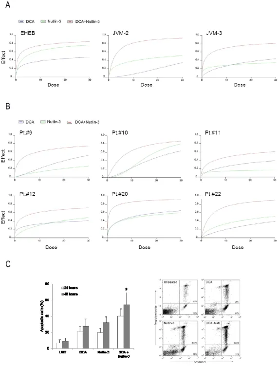
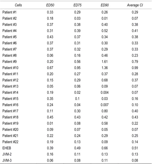
Statistical analysis and assessment of the effect of combination treatment
Descriptive statistics were calculated. For each set of experiments (ie, assays for cell cytotoxicity/functionality and RT-analysis), values were reported as means±standard deviation (SD). The results were analyzed by using the Mann-Whitney rank-sum test and statistical significance was defined as p<0.05. All statistical analyses were performed with SPSS Statistic 20 software (SPSS Inc., Chicago, IL). In order to investigate the effect of DCA+Nutlin-3 combination, leukemic cells were then treated with serial doses of DCA (range 3-30 mM) or Nutlin-3 (range 1-10 microM), individually or in combination using a constant ratio (DCA:Nutlin-3). Results were analyzed with the method of Chou and Talalay to determine whether combined treatment yields greater effects than expected from summation alone: a combination index (CI) of 1 indicates an additive effect, while a CI below 1 indicates synergism. For this purpose cell viability data were analyzed with the CalcuSyn software (Biosoft, Cambridge, UK) and reported either as CI values or as dose-effect curves directly drawn by the CalcuSyn software. Proteomics data were analyzed by both multivariate and univariate approaches. The normalized spectral counts for each identified proteins were submitted to SIMCA-P13 software package (Umetrics, Umea, Sweden) for multivariate data analysis. Variables were scaled using Pareto scaling and data were analyzed by orthogonal partial least-squares discriminant analysis (OPLS-DA). S-plots were calculated to visualize the relationship between covariance and correlation within the OPLS-DA results. Variables that showed significant contribution to discrimination between groups and a significant change in their expression (Mann-Whitney-Wilcoxon test, p<0.05), were accepted as significantly modulated proteins upon treatment and submitted to network analysis.
Results
DCA promotes cytotoxicity in primary B-CLL patient derived cells, but not in normal peripheral blood cells
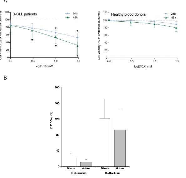
The first group of experiments was designed to investigate whether DCA promoted cytotoxicity in leukemic cells. For this purpose, the effect of DCA was comparatively analyzed on primary PBMC derived from B-CLL patients (n=22; Table 2) and from healthy blood donors (n=10). Treatment with DCA, used in the range of 1-30 mM for up to 48 hours, exhibited a dose- and time-dependent cytotoxicity, resulting in significant reduction of leukemic cell viability with respect to the untreated cultures, at concentrations ≥3 mM in B-CLL patient cell samples (Figure 3A). The IC50
(50% inhibition concentration) mean values (±SD) of cytotoxicity of DCA in B-CLL patient samples were 22±11 mM and 12±6 mM at 24 and 48 hours of treatment, respectively (Figure 3B). On the other hand, PBMC obtained from healthy donors were significantly less susceptible to DCA cytotoxicity as compared to primary B-CLL cells, with IC50 mean values (±SD) of cytotoxicity of
122±49 mM and 93±52 mM at 24 and 48 hours of treatment (Figure 3A-B), clearly showing that normal PBMC were completely resistant to DCA effects at concentrations ≤10 mM. Thus, in line with previous data obtained in solid tumor cell models [95-104] and multiple myeloma [105], we have demonstrated for the first time that DCA promoted a significant cytotoxicity also in primary B-CLL samples but not in normal PBMC.