Firmato
digitalmente da
FALASCHI
VALENTINA
C: IT
2
Summary
Introduction……….p.4 CHAPTER 1: Depression and pathophysiological mechanisms………p.8
1.1 Depressive episodes in mood disorders-implications of Monoaminergic
neurotransmission………. p.8 1.2 Other pathogenetic mechanisms of depression ……… p.18 1.3 Neurotrophins and BDNF……… p.23 1.3.1. BDNF in the CNS………. p.32 1.3.2. Peripheral and platelet BDNF……… p. 34 1.4. BDNF and the pathogenesis of depression……… p.41 1.5 Inflammation in depression and correlation with BDNF... p.45 1.6 Depression and metabolic status……… p.52 CHAPTER 2: Study Aims ……… p.59 CHAPTER 3: Materials and Methods
3.1 Subjects... p.63 3.2 Psychiatric evaluation... p.65 3.3 Determination of the hematological/hematochemical parameters and
circulating BDNF……… p.70
3.3.1 Chemicals, reagents and instruments……… p.70 3.3.2. Blood sampling, sample treatments and storage conditions……… p.71 3.3.3. Preparation of platelet soluble fractions for the analysis of intra-platelet
BDNF……….. p.73 3.3.4. Plasma and intra-platelet BDNF assay by sandwich ELISA ………p.75 3.3.5 Total protein determination by the Bradford’s method………. p.80 3.3.6. Uric Acid assay……….. p.83 3.4. Statistical analysis... p.84
3
CHAPTER 4: Results and Discussion
4.1 Clinical evaluation………...p.86 4.2 Evaluation of hematochemical parameters, pro-inflammatory indices and metabolism substrates in patients with Major Depressive Episode...p.92 4.3 Correlations and comparisons of hematochemical-inflammatory
parameters and clinical features of illness……….…….…………..p.95 4.4 Determination of PPP-BDNF and intra-platelet BDNF (ng/ml)...p.98 4.5 Correlations and comparisons of PPP-BDNF (ng/ml)
and PLT-BDNF(ng/mg) with hematochemical-inflammatory parameters and clinical features of illness………... p.103 5. Conclusions……….p.112. 6. References...p.115
4
Introduction
In these last two decades, the biochemical and molecular research of neuropsychiatric disorders has greatly expanded thanks to the attained technological and methodological advances that have become available to scientists and clinicians. Given the pathophysiological complexity of mental disorders, now also seen as real neuroendocrine and systemic dysfunctions, current research in the neuropsychiatric field is no longer focusing on a single substrate or neurotransmitter system but rather tends to integrate multiple parameters (Maes et al, 2009, Strawbridge et al, 2017). In the case of Mood Disorders, recent studies show how Major Depression can be considered as a systemic pathology, which involves primarily the brain, but also the whole organism, accordingly to a two-way communication model.
There is currently a large literature on the neuroanatomical, neurophysiological and neuroendrocrinological correlates of Major Depression, although no laboratory test has been sufficiently sensitive and specific as a diagnostic tool for the disorder so far. Numerous studies indicate, in addition to neurotransmitters and neuroendocrine markers, the involvement of the inflammatory response and, more generally, of the immune system, energy/redox metabolism and growth factors in depression, implying the presence of altered biochemical patterns/networks
5
rather than disturbances of single and separate parameters (Strawberidge et al, 2017).
The search of valuable biomarkers and correlates of depression has acquired an ever-increasing consent among neuropsychiatrists with the aim at differentiating clinical subtypes and improving patients’ clinical care. In fact, despite a high range of treatment options for major depression and the considerable therapeutic progress achieved, about one third of patients reach a full remission after initial treatment and the probability of non-response increases with the number of episodes experienced (Gaynes et al, 2009). This is probably due to the heterogeneity of the illness: not all patients show the same clinical presentation and, presumably, the same pathogenetic molecular substrates (Kauer-Sant’Anna et al, 2009). It is therefore necessary to ameliorate and even personalize treatments dedicated to this psychiatric illness, in order to possibly prevent its tendency to become chronic.
Numerous evidences have accumulated over time in favor of the presence of altered inflammation patterns in depression, related to an impaired activity of the neuroendocrine hypothalamus-pituitary-adrenal (HPA) axis and the system of neurotrophins, in particular of the Brain-Derived Neurotrophic Factor (BDNF). Under an integrated vision of the pathogenesis of depression, this illness would be defined by variable disturbances of these systems, known to reciprocally interact with the
6
functionality of the serotonin (5-HT) and other neurotransmitter signaling pathways. It is noteworthy that both the HPA responses and BDNF signalings work to maintain the homeostasis of metabolic processes during the occurrence of stressful events (Pitsillou et al, 2020, Levy et al, 2018). Specifically, an impaired expression of BDNF has been reported in some types of depressed patients, so that the administration of this neurotrophin has been thought as one of the most promising therapeutic strategies for the development of innovative antidepressant drugs, even if first data are not encouraging. Indeed, a main problem of this approach consists in the possible induction of a too rapid change of BDNF in the body that seems to increase the risk at developing tumors, due to the angiogenic properties of this neurotrophin. For this reason, some biological drugs capable of regulating the neurotrophin expression at the level of its gene transcription are currently under study (Radin et al, 2017). It is also important to understand how the neurotrophin system and BDNF interact with the inflammatory response in the pathogenesis of mood disorders. In support to the integrated hypothesis of depression, BDNF and its TrkB receptor, originally discovered in the brain, have been also detected in many peripheral tissues and in the bloodstream, suggesting their widespread, whole-body physiological role, linked to cell survival and differentiation (Serra-Millàs, 2016). Thus, the measurement of peripheral and circulating BDNF in depressed patients under different clinical conditions and
7
therapeutic approaches is still a challenging topic for the improvement of patients’ clinical care, even as a potential index of the risk or presence of specific psychiatric or somatic co-morbidities. Moreover, upgrading current knowledge on the regulatory mechanisms of gene/protein expression and post-translational/conformational modifications of BDNF, together the full understanding of its mechanisms of release or uptake in different cells and tissues, would help to explain the pathogenetic significance of an altered neurotrophic signaling within body districts. One of the first steps towards these goals consists, by consequence, in evaluating the circulating amount of BDNF and its correlation with patients’ clinical features (Hashimoto et al, 2010).
Taking into account these premises, the main objectives of this work were therefore:
1) to estimate the peripheral, bloodstream counterpart of BDNF in Major Depressive Episode through its measurement in plasma and platelet samples obtained from a well-defined group of patients;
2) to search possible correlations with patients’ clinical symptoms and results at routine laboratory tests, these last including the main blood metabolic, cellular and inflammation parameters.
8
CHAPTER
1:
Depression
and
pathophysiological
mechanisms
1.1 Depressive episodes in mood disorders-Implications of monoaminergic neurotransmission
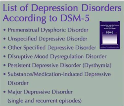
Mood Disorders include different conditions in which the most severe and common forms are Major Depressive Disorder (MDD) and Bipolar Disorders (BDs). Different depressive episodes can occur in the life of patients so a major depressive episode can be ascribed to a bipolar or a unipolar disorder. Furthermore, many definitions of types of major depression have been described in the years and then various specifiers in DSM-5, for more appropriate therapeutic strategies.
The Diagnostic and Statistical Manual of Mental Disorders (DSM) is one of the nosographic systems for mental or psychopathological disorders most used by psychiatrists, psychologists and physicians from all over the world, both in clinical practice and in research. Over the years, the manual, now in its 5-th edition, has been drafted taking into account the current development and results of psychological and psychiatric research in numerous fields, modifying and introducing new definitions of mental disorders.The first version dates back to 1952 (DSM-I) and was written by the American Psychiatric Association (APA), since then there have been further editions: in 1968 the DSM-II, in 1980 the DSM-III, in 1987 the
9
DSM-III-R (revised edition), in 1994 the DSM-IV, in 2000 the DSM-IV-TR (revised text) and in 2013 the DSM-5.
Unlike the DSM-IV-TR, in DSM-5 the chapter "Depressive disorders" has been separated from the chapter "Bipolar and related disorders".
The common features of Depressive Disorders (Figure 1) are the presence of a sad, empty or irritable mood, accompanied by somatic and cognitive changes that significantly affect the individual's ability to function. What differs among them are issues of duration, timing, or presumed etiology.
Figure 1: Classification of depressive disorders accordingly to the DSM-5. Major Depressive Disorder (MDD) is defined by single and recurrent episodes (DSM-5)
10
The DSM-5 describes “Major depressive Disorder” (MDD) as a complex disorder characterized by at least five symptoms present over a two-week period, at least one of which consists of depressed mood or loss of interest or pleasure. Symptoms may consist of weight loss or gain, insomnia or hypersomnia, psychomotor agitation or retardation, fatigue or lack of energy, feelings of worthlessness or guilt, impaired ability to think or concentrate, recurring thoughts of death, all causing a clinically significant distress and a relevant impairment in social, occupational, or other important areas of functioning (American Psychiatric Association, 2013). Major depressive disorder is a chronic and highly debilitating syndrome, one of the currently four main disorders affecting the world's population, with a lifetime probability of 10-30% in women and 7-15% in men (Kessler et al, 2003; Bromet et al, 2011; Hasin et al, 2018).
Furthermore, it is estimated that the prevalence of depressive episodes could continue to rise until they will become the second cause of illness among the World Health Organization (WHO) member states (Smith K. et al, 2014, Mathers and Locar 2006). In addition to provoking a significant decrease in social and physical functioning, MDD can lead to such discomfort to the point of causing a complete disability and/or even increasing the risk of suicide.
There is a high comorbidity with other neuropsychiatric disorders such as panic, obsessive-compulsive, eating and borderline-personality disorders,
11
substance abuse, but also with physical illnesses and pain (American Psychiatric Association 2013).
Major Depressive disorder has a variety of socio-economic consequences that include low education, job and emotional instability and poor job performance (Kessler and Bromet 2013).
Furthermore, this illness is tightly linked to a high rate of mortality, associated not only with suicide: depressed patients are more likely to develop coronary artery disease and type-2 diabetes (De Burgos-Lunar et al 2012, Mondal and Fatima 2018). Thus, depression continues to increase the global burden of disease and disability (Ahern et al, 2011).
The DSM-5 has described several specifiers that can be attributed to the single Major Depressive Episode with clinical and therapeutic implications. In fact, the specifiers for a depressive episode can be of different types: with anxiety, with mixed characteristics, melancholic, atypical or psychotic, with onset in the peri-partum and with a seasonal trend.
Depressive episodes can arise not only in the context of a diagnosis of unipolar depression, but also in patients with bipolar disorder, when, in the latter case, counter-polar (manic or hypomanic) episodes occur alternately with the depressive ones. According to DSM-5 in Bipolar I disorder, there are episodes that fully meet the criteria for a manic episode that can be preceded or followed by hypomanic or major depressive episodes
13
Therefore, Major Depression is a heterogeneous disorder both in the transverse that in the longitudinal course. Some authors have hypotized that unipolar and bipolar depression may represent distint nosological entities, possibly related to different biological substrates, but in years it has developed also a dimensional approach (mood spectrum model). So different authors and clinicians tend to identify Major Depressive Disorder (MDD) as a part of a continuum (bipolar spectrum), encompassing depressive and manic symptomatology of different severity levels along a longitudinal course (Akiskal et al, 1999).
Despite of the considerable impact of major depression on health, knowledge on the pathophysiology of this mental illness is very poor in respect to other common chronic diseases. Because of the complexity and heterogeneous nature of depressive episodes, current treatment outcome is suboptimal. From the most recent studies, depression is considered a multifactorial and multifaceted mental illness, related to variably combined genetic, biochemical, and non-genetic factors and triggers, such as stress, affective trauma, viral infection and abnormality in neurodevelopment which have more impact with the increased complexity of the pathogenesis of the disease (Chen, et al 2007, Ugo 2008). However, the precise pathogenic causes have not been defined yet.
The etiology of major depression can involve in fact both genetic and environmental factors (Lesch et al, 2004, Saveanu et al, 2012, Subbarao et
14
al, 2008). Environmental, lifestyle or transitional changes in the course of life, in relation to genetic and acquired vulnerabilities, define a variety of biological endophenotypes and clinical symptoms (Leuchter et al, 2014). After the discovery of the first antidepressants, (ADs), as monoamine oxidase inhibitors (MAOIs) and tricyclic ADs (TCA), the most relevant pathophysiological mechanism of MDD was considered the diminished monoamine neurotrasmission (Dell’Osso et al, 2016). The effectiveness of the first ADs consisted indeed in the rise of the extra-cellular levels of monoamines, in particular serotonin (5-hydroxytryptamine, 5HT) and norepinephrine (NE), through the blockade of their presynaptic catabolic enzymes MAO or respective re-uptake mechanism (membrane-bound transporters).
The classical monoamine theory, formulated in the ’60s, thus suggests that, at the origin of mood symptom presentations, stress or genetic or non-genetic vulnerabilities, lead to the reduction of biogenic monoamines as 5-HT, NE, and dopamine (DA) (Shilldkraut et al, 1965).
This theory was also formulated by the observation that reserpine, an antihypertension drug showing also antipsychotic properties, can provoke CNS monoamine depletion, precipitating depressive symptoms (Everett and Toman, 1959) (Figure 3).
15
Figure 3. Mechanism of action of antidepressant drug: selective serotonin re-uptake inhibitors (SSRIs), tricyclic (TCA) and selective noradrenaline reuptake inhibitors (SNRIs) (Van Rensburg and Reuter, 2019).
Tricyclics are a class of AD drugs so called due to their characteristic chemical structure formed by three condensed rings.
Although not all tricyclics share the same mechanism of action, they generally act through the inhibition of monoamine neurotransmitter reuptake and as antagonists of different receptors synaptic receptors,
16
particularly muscarinic cholinergic, serotonin and histamine receptors, causing various side effects. Toxicity occurs at approximately 10 times normal dosages (the therapeutic index is relatively low); these drugs are often fatal if taken in overdose, since they can cause ventricular arrhythmias, delirium and convulsions. Although they are still prescribed under special conditions, because they are known to be effective, in recent times their use has been largely replaced in the clinic by the most recent second-generation ADs (SGAs) as selective serotonin reuptake inhibitors (SSRIs) and serotonin and noradrenaline (SNRIs), which generally have fewer side effects (Feighner et al, 1999).
The importance of the monoamine hypothesis consisted in the introduction of the concept of “biochemical lesion” in depression (Leonard, 2000). Meanwhile, the monoamine hypothesis of depression has received much sustenance from neuroanatomical, brain imaging and functional studies conducted in animals and humans (Cummings, 1993; Arango et al, 2002). These investigations have shown a possible link between the onset of depression and brain areas as basal ganglia, mesencephalic nuclei, cortical and limbic districts (in particular, the amygdala and hippocampus), regions that have all been tightly associated with the control of mood tonus, emotion, psychomotor abilities, sleep, appetite and a variety of other stress-related functions. These regions have also shown high densities of monoamine biosynthesis enzymes (mesencephalic nuclei) and differential
17
expression patterns of monoamine receptor subtypes (Charney and Leger, 2010).
However, the monoamine hypothesis over time has been increasingly considered simplistic and incomplete, because it cannot fully explain complex clinical presentations (Dell'Osso et al, 2016, Catena dell'Osso et al, 2013), as well as the limited effectiveness of ADs that may arise in these and other cases.
Furthermore, currently available AD drugs are effective about 3 weeks after the beginning of the pharmacological treatment. Thus, if current ADs are surely golden treatments for the relief of severe depression, many pharmacological investigations are now more deeply considering their mechanisms of action in order to improve the AD efficacy. In particular, the complexity of monoamine receptors, their sensitization states or specific coupling to signal transduction pathways (Hamon and Blier, 2013), together the activity of some intracellular kinase pathways, are regarded as possible new targets to advance patients’ pharmacological care (Yuan et al, 2016). In the meantime, other SGAs have been developed and introduced into the clinical practice that have much improved the flexibility of the applied treatments in respect to specific symptom presentations. These relatively new ADs are the atypical ones, also named multifunctional ADs, such as venlafaxine, bupropion, mirtazapine, trazodone, nefazodone and agomelatine. Atypical/multifunctional compounds are characterized by
18
their ability at interacting with multiple recognition sites. Precisely, they can block the reuptake sites of 5-HT and/or NE, and/or behave as agonists, antagonists or partial agonists on specific 5-HT and NE receptor subtypes (5-HT1AR, 5-HT2R, alpha adrenergic receptors) (Stahl, 2009a; 2009b), as well as on receptors for neurotransmitters as glutamate, melatonin, dopamine, histamine or others (Hamon and Blier, 2013, Metts et al, 2019).
1.2. Other pathogenetic mechanisms of depression
Despite the previously reported advancements in the neurobiology and pharmacology of depression, the molecular bases of this invalidating illness, including the number of susceptible and resistant genes involved in its development, are still elusive. Moreover, drug resistance and relapses of episodes continue to be a clinical defy. Thus, the implication of other systems besides monoamines or other neurotransmitters and signal molecules, as melatonin and glutamate/GABA, has been postulated, being under intense investigation. Over years of research, additional metabolic and biochemical pathways have been thought to act in an orchestrated manner with neurotransmission for the pathogenesis of mood disorders. Among these, we mention tryptophan fates, cytokines, abnormalities of lipid/phospholipid metabolism, homocysteine or abnormalities of purinergic system and adenylate cyclase turnover (Raison et al, 2006; Folstein et al., 2007; Duman and Voleti, 2012; Danzer, 2017; Lener et al,
19
2017; Bartoli et al, 2020). In particular, some studies on ADs have shown that the prolonged exposure to these drugs can activate genes involved in the expression of trophic factors (Levy et al 2018). Furthermore, it has been demonstrated that ADs can influence the plastic properties of neurons associated with morphological changes and synapse re-modelling or dendritic spine formation (Cavez-Castillo et al, 2019, Cai et al 2015). Experimental studies also showed the phenomenon of “shrinking” on rats subjected to chronic stress: animals displayed a reduction in the density and volume of dendritic spines and synapse arborization concerning neurons of the nucleus accumbens, hippocampus and prefrontal cortex (Tang et al 2019). This condition, improved by ADs, was also observed in depressed patients and in people subjected to chronic stress (Sapolski et al, 2001).
These studies have led to the neurotrophic hypothesis of depression. Among neurotrophic factors that have attracted more attention in depression research there are the Brain-Derived Neurotrophic Factor (BDNF, also called abrineurin) and the neurotrophin-related Glial Cell-Derived Neurotrophic Factor (GDNF). Indeed, an ever-growing evidence has shown neurotrophic alterations in mood disorders, particularly in depression (Castren, 2014). Many studies have focused in particular on BDNF, a protein of the neurotrophic family, widely expressed in the human brain, which increases and maintain neuroplasticity favoring
20
neurogenesis, neurite arborization, synaptogenesis and synapse re-modelling (Colle et al, 2017). BDNF has been found mostly expressed in the hippocampus and cerebral cortex, the main brain areas that control cognition, mood and emotion (Mattson et al, 2008).
Current neurotrophic hypotheses show that depression is associated with a reduced expression and deregulation of neurotrophins accompanied by the aberrant neurogenesis of discrete brain regions (Duman et al, 2006). Accordingly, the disturbance of BDNF and GDNF expression/functionality affects the survival and plasticity of dopaminergic, GABA-ergic, glutamatergic, cholinergic and serotoninergic neurons in the central nervous system (CNS), disrupting monoamine neurotransmission (Martinowich et al 2008). The neurotrophic and monoamine hypotheses of depression are thus interlocked.
Beside the neurotrophic hypothesis of depression, other investigations have shown the involvement of the hyperactivity of the neuroendocrine stress response, as the hypothalamic-pituitary-adrenal axis (HPA), revealing how this dysfunction can be implicated in melancholy, psychotic characteristics and suicide risk. Concurrently, other cellular and molecular studies have shown the implication of inflammation and immune response in the pathogenesis of depression, together with the finding of the association of genetic variants of peripheral factors as pro-inflammatory cytokines in patients (Strawberidge et al, 2017).
21
It has been also hypothesized that HPA axis, for genetic and environmental reasons, is dysfunctional and hyperactive in depression due to the loss of the negative feedback exerted on the hypothalamus and pituitary gland by Glucocorticoid/Cortisol Receptors (GRs). Indeed, it has been extensively reported that a percentage of depressed patients have increased levels of cortisol in urine, plasma and saliva, with pituitary and adrenal glands increased in volume (Nemeroff and Vale, 2005; Pariante, 2006). The presence of HPA axis alterations, hypercortisolemia, desensitized GRs, the increase of circulating cytokines, the stimulation of specific leukocyte subpopulations and the alteration of flogosis indexes as well as blood cardiovascular risk factors, such as Erythrocyte Sedimentation Rate (ESR), C Reactive Protein (CRP) or platelet reactivity, could be part of the pathogenetic mechanisms occurring in the depressed patient. Receptors for mineralocorticoids or MRs are also investigated for their role in the HPA axis impairment present in depressed patients, suggesting that the loss of HPA feedback inhibition can occur at multiple levels in the depressive pathology (Pariante and Lightman, 2008). In summary, the monoamine dysfunction remains one of the main pieces of the puzzle that defines the pathogenesis of depression but more and more researchers suppose that it is not always the only determinant as was once believed. Essentially, we have here briefly introduced and pointed out those medical hypotheses that sustain the complexity of the neurobiology of depression and, more
22
broadly, of mood disorders. These severe psychiatric conditions presumably result from and/or delineate malfunctioning brain-body communication networks, variably involving the physiology and plasticity of some brain centers, stress-related neuroendocrine cascades and cellular and/or molecular effectors at the crossroad with the immune system, inflammation components as well as energy/redox metabolism. Neurotrophins and BDNF are considered main players in such an integrated vision of depression.
Before dealing in more depth with the involvement of BDNF in the pathogenetic mechanisms of depression, we will first present the main biochemical characteristics of this protein and its receptor.
24
human BDNF is an approximately 27 kDa homodimeric secretory protein (Radziejewski et al, 1992) encoded by the BDNF gene located on the chromosome position 11p14.1 (Silva de Olivera et al, 2019). Comparably to other neurotrophins, BDNF is characterized by a 55% homology of amino acid sequence with the other components of this protein family, including 6 cysteine residues involved in the intra-chain disulphide bridge formation (Isaacs, 1995). BDNF is a small protein, rich of basic amino acid residues, that can be separated both as a monomeric form (a polypeptide of about 120 aminoacidic residues) and as the physiologically active homodimer (Figure 4) or as a heterodimer, composed by a BDNF monomer and a monomer of another neurotrophin (eg. the BDNF/NT3 heterodimer) (Junbluth et al, 1993). The structural assembly of BDNF monomers, as for the other neurotrophins, is thought to be part of the complex BDNF signaling. The possible biological activity and physiological role of these neurotrophin heterodimers is under investigation, even at the pharmacological point of view (Dechant and Newman, 2013). From the comparison of the crystal structures of the different neurotrophins, as NGF (McDonald et al., 1991; 1993), monomers assume a typical arrangement of their -folds and disulphide bridges, the so-called cystine knot motif (Isaacs, 1995). Interestingly, neurotrophins share the cystine knot structure with other secretory proteins and growth
25
factors, as the multifunctional cytokine Transforming Growth Factor-2 (TGF-2) and platelet-derived growth factor (PDGF) (Isaacs, 1995). The cystine-knot protein conformation is known to contribute to the formation of the non-covalent dimeric form of neurotrophins and BDNF, presumably attributing a greater number of specific functional features to these molecules as well as to their interaction with specific receptors. It should be also mentioned that if BDNF shares structural similarities with the other neurotrophins, each neurotrophin is uniquely characterized by a specific pattern of charged (basic or acidic) residues exposed on its surface. Differences between the individual neurotrophins are particularly manifest within those loops extended from the -sheet core of the molecules that define the interaction with the specific receptor (Dechant and Neumann, 2013). These structural properties make neurotrophins endowed with a mix of hydrophilic and hydrophobic interactions together a limited capacity to diffuse and a tendency to stick to membranes, surfaces and other possible bonding sites (Dechant and Neumann, 2013). Beside its structural features, BDNF presents also a typical organization at the gene locus level. The human BDNF monomer is encoded by the BDNF gene, firstly described by the research group of Tonis Timmusk, Department of Gene Technology of the University of Tallinn (Estonia). The BDNF gene spans about 70 Kb and is composed of 11 exons and 9 promoters (Pruunsil
26
et al, 2007), being able to generate a variety of BDNF transcripts in relation to the cell type.
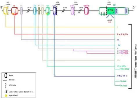
Figure 5. The human BDNF gene, its possible transcription initiation sites and the corresponding different transcripts (from Cattaneo et al., 2016).
Indeed, transcript variance is not only area-dependent, but it is also present within a same brain area, and even within neurons, as reported in cortical neurons (Kaneko et al, 2012; Hempstead, 2015). Moreover, the RNA messengers containing exons II, III, IV, V and VI are prevalent in the CNS level, while the others being rather transcribed in periphery (Pruunsild et al., 2007).
In Figure 5 is reported the BDNF gene organization and all the possible coding transcripts that can be produced by BDNF gene expression mechanisms. All transcripts are similar for their 3’ encoding region in exon IXd but are distinguishable by 5’ UTR sequences (untranslated region).
27
Substantially, the 3′exon encodes all or most of the protein, depending on the 5′exon used. The most recognized transcription mechanism consists in the conjugation of exon IX with another upstream exon (I-VIIIh) generating mature transcripts including only the downstream region of exon IX, or the IXd region. Exons I, VII and VIII display internal ATG codons used as possible translation initiation sites, forming a protein product with a longer N-terminal portion. Inside exons II, V and VI are also present splicing sites that produce transcripts similar for the coding region but differing for the 5’UTR size. Such a great number of exons, transcription initiation sites, different promoters and splicing sites imply the complex regulation activity of this gene locus. Moreover, the human BDNF gene is capable to produce natural antisense mRNA or non-coding anti-BDNF complementary to the sense BDNF transcript, a mechanism representing an additional post-transcriptional way to control BDNF protein expression (Pruunsild et al., 2007). These BDNF-anti-BDNF mRNA duplexes and these bidirectional transcription mechanisms play a main role in regulating protein expression in the brain (Pruunsild et al, 2007). The BDNF gene transcription is also regulated by CREB signaling, epigenetic mechanisms and microRNAs (Tao et al, 1998; Lubin et al, 2008; Caputo et al, 2011; Cattaneo et al, 2016; Khani-Habibabadi et al, 2019). At the translational level, the BDNF protein, as other secretory proteins, is firstly synthesized in the endoplasmic reticulum, leading to a precursor
29
Golgi network. The mature form (about 14KDa) is internalized into vesicles transported to the plasma membrane for exocytosis and secretion. For the regulated secretion, the signal peptide cleavage occurs later: pro-BDNF is internalized in granules that are transported from the Trans-Golgi to the plasma membrane. At this stage, proteolitic enzymes, as pro-protein convertases, transform pro-BDNF into the mature BDNF form (Cattaneo et al, 2016). The mature form then accumulates at the membrane level and BDNF is secreted only after specific stimuli (Lessmann et al., 2009). Otherwise, released pro-BDNF can be cleaved extracellularly by plasmin, matrix metalloproteinases and other factors. Vesicular secretion can involve both BDNF and mBDNF, with the amount of secreted mBDNF or pro-BDNF depending on the type and activity of convertase involved (Mowla et al, 2001). The mature form is the one recognized and bound by the TrKB receptor, a class of tyrosine kinase receptors involved in the regulation of CNS plasticity (Serra-Millàs, 2016). Neurotrophins and their receptors have a very high potential to contribute to the CNS and PNS repair after injury and damage (Meeker and Williams, 2015).
The membrane receptors for neurotrophins comprise three receptor tyrosine kinases belonging to the tropomyosin receptor kinases (Trk) family, which autophosphorylate after activation, and the p75 neurotrophin receptor, p75NTR. The p75NTR is a molecule that groups within the large family of homologs of death-domain containing receptors for the cytokine
31
residues transduces the signal to protein adapters coupled to intracellular cascades, as the Ras/ERK (extracellular signal regulated kinase), the PI3K/Akt (Phosphatidylinositol-3-OH kinase) and PLC-1 (phospholipase C) pathways (Kaplan and Miller, 1997; Pawson e Nash, 2000).
These signal transduction pathways are responsible of the biological function of each neurotrophin through the activation of transcription factors and the modulation of cell gene expression. The molecular relationship between Trk and p75 receptors is complex and incompletely understood, but it is known that the resulting cellular effects are highly ligand and cell specific. Under physiological conditions the BDNF binding to Trks or p75 receptors regulates cell survival. Indeed, p75 receptors have been found linked to both pro-apoptotic and anti-apoptotic features. Under physiological conditions, the anti-apoptotic signal deriving from the neurotrophin-TrK receptor complex antagonizes the pro-apoptotic action originating from the binding to the p75 receptors, synergistically acting with the p75 anti-apoptotic signal (Mazzoni et al., 1999; Aloyz et al., 1998; Maggirwar et al., 1998; Hamanoue et al., 1999). Under pathological conditions, the pro-apoptotic effect deriving from the interaction with p75 is not counteracted by the anti-apoptotic counterpart (pro/anti-apoptotic disequilibrium). The p75 neurotrophin receptor triggers signaling by non-covalent binding to intracellular molecules and the resulting cellular effects
32
of this signaling cascade are diverse. Moreover, if the p75 receptor function has not been still fully understood, it appears ever clearer that the biochemical and functional interactions between the Trk and p75 receptors belong to the fine-tuning of neurotrophin-receptor interactions determining distinct cellular responses and adaptation to different stimuli (Dechant and Neumann, 2013).
1.3.1 BDNF in the CNS
In the CNS, BDNF is the most widespread neurotrophic factor. It has been overall localized in neurons (Murer et al., 2001), but glial cells can express this neurotrophin under metabolic stress (Ceccatelli et al., 1991; Batchelor et al., 1999). Nakajima and coauthors (1998) have reported that glial BDNF is taken from the extracellular milieu through a truncated form of the TrkB receptor located in the plasma membrane that is capable to internalize BDNF without activate ensuing signal transduction pathways. The highest levels of CNS BDNF have been found in the neocortex, striatum, pro-encephalic nuclei, hippocampus, hypothalamus, brain stem and cerebellum (Murer et al., 2001). The expression pattern of BDNF mRNA is similar to that of the protein, even if the complexity of its transcriptional and translational mechanisms has brought to report the protein presence without appreciable amounts of its transcripts (Altar et al., 1997; Baquet et al., 2004). The neurotrophin BDNF has been linked to
33
both anterograde and retrograde transport in the CNS. The cortical-striatal network has been linked to the anterograde transport of BDNF from the cortex (II-III e IV-V layers) (Fusco et al., 2003; Baquet et al., 2004). The BDNF retrograde transport has been also observed (Altar et al., 1998), starting from dendrites and axons to the soma, a mechanism mediated by TrkB receptors present at the pre-synaptic level (Heerssen et al., 2004). The retrograde transport of BDNF seems to exert protective actions on BDNF-producing neurons by means of nuclear interactions linked to cell survival and counteraction against toxic stimuli (Lindholm et al., 1994; Heerssen et al., 2004).
BDNF central function has been linked to neuronal plasticity, learning, memory and Long-Term Potentiation (LTP) (Schinder et al., 2000; Schinder et al., 2002). The neurotrophin is also under investigation for its glial expression, as aforementioned, and therefore for its potential myelinization properties (Fletcher et al, 2018). The tight interaction observed between monoaminergic neurotransmission and BDNF in the brain is thought to correlate with a main role of this neurotrophin in neuropsychiatry disorders (Altar et al, 1998; Martinovich and Lu, 2008). The impaired BDNF signaling can be a vulnerability factor of these pathologies, by impacting neuroplasticity, inflammation patterns or hypothalamic–pituitary–adrenal axis action that are altered in psychiatric disorders (Cattaneo et al, 2016).
34
BDNF has been found also at the peripheral level. The physiology and distribution of peripheral BDNF is still under investigation, since many peripheral tissues have been found to produce BDNF and to express its receptor, implying that circulating levels of this neurotrophin cannot exclusively derive from the brain. At the same time, even if there is not a complete consensus on this assumption, neurotrophins can cross the blood-brain barrier (BBB), and the blood-brain and serum levels of these proteins have been found positively correlated to each other (Numakawa et al, 2010).
1.3.2 Peripheral and platelet BDNF
The BDNF and TrkB mRNAs are expressed in several non-neuronal tissues, including muscle, thymus, heart, liver, vascular smooth muscle cells, lung and spleen (Nakahashi et al, 2000). BDNF is also produced in monocytes, lymphocytes and eosinophils (Elding et al, 2004). The latter cells produce BDNF via the autocrine system and utilize it to evoke and extend the allergic reaction (Nockher et al, 2005). BDNF has been shown to play a pivotal role in the growth, survival and chemoresistance of tumor cells in various types of cancers, including Hodgkin lymphoma, myeloma, hepatocellular carcinoma and neuroblastoma (Yang et al, 2006). BDNF also mediates the survival and activation of endothelial cells through its interaction with TrkB (Kermani et al, 2005), suggesting its potential role in angiogenesis. Many non-neuronal cells, such as smooth muscle cells,
35
fibroblasts and astrocytes, may not express the molecular components of the regulated secretory pathway and therefore only secrete neurotrophins constitutively.
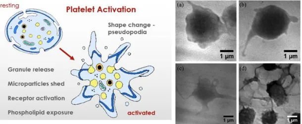
Furthermore, platelet BDNF represents probably the most relevant component of the peripheral neurotrophin: platelets are indeed the main storage source of BDNF secreted from all other tissues (Bus et al, 2011). Platelets are small un-nucleated blood cells with a size of approximately 3 μm deriving from megakaryocytes (MK) in the bone marrow, from which they are released into the bloodstream. Resting platelets have a rounded-discoid shape, while relevantly changing their morphology upon activation. They circulate for an average of 7-10 days and act to stop bleeding. Platelet surface proteins enable them to adhere to each other and to blood vessel walls. They possess several important organelles, microtubules, mitochondria, endoplasmic reticulum, the Golgi apparatus and 3 types of secretory granules: 1) α-granules - the most abundant; 2) dense granules; 3) lysosomes. Moreover, platelets also have an open canalicular system (OCS) connecting microtubules and the plasma membrane that is attracting a great attention for the physiology of these blood cells (Selvadurai and Hamilton, 2018).
36
Figure 8: Resting and activated platelets. Left panel: platelet rearrangements with activation (from memorang: the physiology of coagulation); right panel: the different grade of platelet shape changes during progressive activation (from Yang et al, 2014).
Under a variety of precise stimuli, as thrombin, ADP, arachidonic acid, serotonin and epinephrine, platelets can change their metabolism and shape, undergoing a more or less pronounced activation state, progressively becoming sticky and releasing factors from the different granules (Serra-Millàs, 2016) (Figure 8). The activation state of platelets induces not only deep morphological changes but also intense biochemical variations and rearrangements (Giannaccini et al, 2010). Platelets have been primarily related to coagulation, vascular repair functions and blood homeostasis, but there are more and more evidence indicating their active participation to inflammation and immune response (Serra-Millàs, 2016). Indeed, they can release vascular permeability factors and promote
37
chemotaxis of neutrophil granulocytes. Platelets release numerous inflammatory mediators that are not involved in hemostasis, as IL1, P-selectin, Platelet Factor 4 (PF4 or CXCL4) and MIP-1α (CCL3) (Thomas and Storey, 2015). Most of these mediators are stored in platelet granules, modifying, after release, leukocyte and endothelial-mediated responses, while activating bridges and networks between platelets, leukocytes and endothelium (Thomas and Storey, 2015). This suggests a role of platelets as active modulators of the interactions between monocytes, neutrophils, lymphocytes and the endothelium, in the framework of inflammation and both innate and adaptive immune responses (Semple et al, 2011). In should be also pointed out that platelets are under continuous investigation since years in the field of biological psychiatry, as they are considered a kind of “window to the brain”, containing enzymes, carrier proteins, neurotrophins, neurotransmitters and receptors also active in the CNS and synapses (Stahl, 1977; Da Prada et al, 1998; Leiter and Walker, 2019).
Ninety percent or more of blood BDNF is stored in platelets and their -granules (Fujimura et al, 2002). A close relationship has been indeed found between BDNF and platelets under physiological conditions. In addition, there is an approximately 100- to 200-fold estimated difference between the plasma and serum levels of BDNF because platelets release BDNF
38
during the clotting process (Lommatzsch et al, 2005). It has been demonstrated that the amount of BDNF in serum is nearly identical to that found in washed platelet lysates (Fujimura et al, 2002). Thus, the difference between the serum and plasma BDNF levels seems to reflect the amount of BDNF stored in circulating platelets, and the BDNF in platelets might serve as a reservoir for circulating BDNF. The BDNF in platelets may play a role during tissue trauma or nerve injury, releasing their contents into the circulation at the site of the injury (Radka et al, 1996). In the first studies, BDNF was not expressed in or produced from the megakaryocyte precursor cell of the mature platelets, in which protein synthesis is generally absent, but was sequestered from the circulation (Nakahashi et al, 2000). Recently, one study found that BDNF is present in the cytoplasm of platelets and in α-granules, suggesting that BDNF is either produced in platelets or passed down from MK (Tamura et al, 2011). A second study found that a megakaryocyte (MK) progenitor line, the MEG-01 cell line, produces BDNF upon thrombopoietin stimulation (Kaushansky et al, 2006), and the levels of BDNF in MEG-01 cells increased in a time-dependent manner (Tamura et al, 2012). This was the first report of the production of BDNF in a megakaryocyte cell line, a finding that led to the hypothesis of BDNF as a cell proliferation agent of the megakaryocyte lineage in vivo. It is also likely possible that there is a receptor for BDNF on the MEG-01 cell surface, but the TrkB receptor has
39
not been detected yet in MEG-01 cells or human platelets (Burnouf et al, 2012). Therefore, there should be an unidentified novel receptor in MKs or platelets. Some agonists, such as thrombin, collagen, Ca2+ and shear stress, could induce a rapid release of BDNF from platelets. Even with agonist stimulation, only approximately half of the BDNF in platelets is secreted, which suggests that platelets maintain a stable pool of BDNF as a buffer system (Bus et al, 2011). Anyway, there is still little knowledge about the relationship between BDNF and platelets: only a few studies have assessed issues such as the platelet activation mechanism that allows the release of BDNF and BDNF localization within platelets. The rate of BDNF release parallels the secretion of 5-HT from the dense granules and PF4 from the α-granules, although a greater proportion (90%) of the total 5-HT and PF4 are released compared to BDNF. Because only 40%-60% of the total content of platelet BDNF is released by maximal platelet activation, some authors postulated that platelets either have a non-releasable pool of BDNF, or that the released BDNF is sequestered by binding to a transporter or receptor on the platelet surface (Fujimura et al, 2002). Such binding could promote the internalization of BDNF by the platelets, as has been reported for 5-HT (Rendu et al, 2001) and for the BDNF astrocyte recycling (Alderson et al, 2000). The binding of BDNF to washed platelets was confirmed by FACS (cytofluorometric) analysis, microscopy as well as confocal microscopy, suggesting that platelets bind exogenous BDNF
40
(Fujimura et al, 2002). However, a recent study found two different locations of BDNF intra-platelet storage: the α-granules and the cytoplasm. Using immunoelectron microscopy, BDNF was clearly detected in the same fractions of P-selectin, a α-granule marker, and protein kinase C, a cytoplasmic marker (Tamura et al, 2011). BDNF is predominantly released from platelets through the activation of protease-activated receptor 1 (PAR1) during thrombin stimulation, along with the stimulation of vascular endothelial growth factor but not endostatin. Platelets stimulated with concentrations of the PAR1-activator peptide have shown a dose-response curve of BDNF release, exhibiting a two-phase pattern. The first phase is a drastic release phase occurring at a low-level of activation, which is completely inhibited by a pretreatment with Prostaglandin (PGE1), suggesting that this phase depends on calcium mobilization. The second phase is a mild release phase arising at a high-level of activation, which is not affected by the PGE1 pretreatment, suggesting that this step depends on the activation provoked by a PGE1-independent signal. BDNF response curve was found similar to that of PF4 (Tamura et al, 2011). There was no significant difference in BDNF release between the non-stimulated and the PAR4-AP-stimulated cells. PAR1 activation promotes the release of proangiogenic factors and these results support the action of BDNF as a proangiogenic factor (Italiano et al, 2009). Moreover, the α-granule BDNF component is released upon platelet activation, whereas the cytoplasmic
41
BDNF is not (Tamura et al, 2011). The maximum BDNF release is approximately 30%-40% with stimulation and the remaining 70% of BDNF is equivalent to that found in the cytoplasm, which is not released (Fujimura et al, 2002).
1.4 BDNF and the pathogenesis of depression
Many investigations have suggested BDNF as a main player in the pathophysiology of depression. Reduced BDNF gene expression and protein levels have been reported in many animal models of depression as well as in patients. The investigation of rodent models of depression, represented by exposition to a variety of stressful conditions and stimuli as social defeat, maternal deprivation or prenatal stress exposure, has shown that the induced depressive-like behavior is related to a reduced gene expression of BDNF mRNA in different brain regions, including the hippocampus and cortex, whereas AD drugs are able to upregulate BDNF expression in animal models (Cattaneo et al, 2016). These BDNF alterations have been linked to the reduction of the hippocampus size, a key region in the control of emotions in humans, providing the basis of the neurotrophin hypothesis of depression (Duman 2006). Results on animal models have been paralleled by investigations conducted in depressed patients, revealing the BDNF reduction both in post-mortem brains and in peripheral blood samples (Pandey et al, 2008; Sen et al, 2008).
42
Electroconvulsive therapy as well AD treatments have shown to increase BDNF in blood samples from patients suffering of severe depression (Polyakova et al, 2015; Cattaneo et al, 2016).
From preclinical studies a reduced BDNF activity, increased apoptosis and decreased hippocampal neurogenesis were found in association with depression and exposure to chronic mild stress (Filho et al, 2015). Besides, many other studies have appraised BDNF expression and amounts as possible peripheral biomarker of depression (Rana et al, 2020).
Some data show that depressed patients have lower BDNF plasma levels than controls suggesting that plasma BDNF may represent central BDNF and may be associated with clinical features of major depressive disorder (Polyakova et al, 2015; Klein et al, 2011; Dell'Osso et al, 2010).
Recent meta-analyzes have also shown that BDNF is more pronouncedly reduced in severe depression and increased during AD treatment even in the absence of clinical remission (Molendijk et al, 2014).
Some studies have shown low BDNF levels associated with more severe melancholic characteristics, psychomotor retardation or slowdown and insomnia (Alves et al, 2018; Monteiro et al, 2017).
However, if none of defined subsets of symptoms of depression were yet significantly associated with circulating peripheral BDNF, a negative correlation between BDNF (plasma or serum BDNF) and the number of
43
depressive episodes with melancholic features was found (Kotan et al 2012 Ceroleo 2019).
Low levels of BDNF have been observed in the brain of suicide subjects, depressed patients and stressed animals (Allen et al, 2015, Banerjee et al 2013). Negative environmental effects like psychological stress, chronic foot shocks and chronic social defeats also decrease BDNF levels in the hippocampus (Jiang et al, 2015). Additionally, as aforementioned, the administration of ADs increases BDNF levels, confirming a pathogenetic role of this protein in depression (Yoshikimura et a, 2010). Moreover, this hypothesis is corroborated by the finding that a high baseline BDNF level is associated to a positive response to ADs (Kurita et al, 2012).
Another hypothesis supports the fluctuation of peripheral BDNF levels in relation to different patterns of symptoms defining depressive episodes: for instance, an increase of circulating BDNF has been also associated with depression familiarity (Knorr et al, 2017) and mixed state episodes (Piccinni et al, 2015). According to DSM-5 in major depressive episodes the specifier with mixed characteristics is characterized by the presence of at least three manic/hypomanic symptoms present almost every day in most days of a depressive episode: high mood, expanded mood, hypertrophic self-esteem or grandiosity, increased talkativeness, flight of ideas, involvement in activities with the potential for harmful consequences, decreased need for sleep.
44
A recent report has shown the ability of the tricyclic AD imipramine to significantly increase the hippocampal BDNF levels in a rat model of depression induced by chronic unpredictable stress (Hazra et al, 2017). Yoshimura and co-workers revealed that a treatment with ADs or atypical antipsychotics at low doses can increase the plasma BDNF levels in patients with MDD or bipolar disorder (Yoshimura et al, 2010)
From these and many other investigations, peripheral BDNF was proposed as a potential diagnostic, prognostic, and therapeutic biomarker for mood disorders, particularly associated with disease severity and response to AD treatments (Dimitriadis et al, 2019). However, results are still controversial, non-conclusive and needing further investigation, and this protein is being actually rather considered a generic biomarker of depression (Poliakova et al, 2015).
It appears clear that the relationships between neuronal BDNF and its peripheral counterpart still require further investigation. In any case, the link between neuroendocrine systems, HPA axis and the immune/inflammatory response can be a key aspect to understand the regulation of BDNF release and storage into the bloodstream. This hypothesis deserves confirmation, in order to clarify if the BDNF platelet reservoir and BDNF extracellular levels can reflect, at different levels, the functionality of brain-periphery cross-talks actives in adaptation to stressors. Since these paths are supposed to be altered in depression,
45
peripheral BDNF variations in patients could be illness indices, allowing a more careful patients’ monitoring.
1.5 Inflammation in depression and correlation with BDNF
Several studies have showed that inflammation may interfere with the physiological 5-HT signaling, the neurotrophin synthesis, and the HPA axis functioning (Miller and Raison, 2016, Amodeo et al, 2018). Recent studies highlighted the possible role of proteins defined as "inflammasome", a cytosolic protein complex, usually generated in response to infection, but also to many other factors, and involved in a pro-inflammatory state, in oxidative stress and in the onset of depressive symptoms (Uint et al, 2019, Alcocer et al, 2014).
Recent studies have found increased levels of various pro-inflammatory markers in blood and cerebrospinal fluid (CSF) of depressed patients, including interleukin 1(IL-1), IL-6, interferon- (IFN), tumor necrosis factor alpha (TNF), CRP and neurotoxic factors such as superoxide anion and nitric oxide (NO) (Rosenblat JD et al, 2017, Muller et al, 2011).
Studies show that IL-6 and CRP appear frequently and reliably elevated in depression (Haapakoski, et al 2015). IL-8 has been reported elevated in subjects with severe depression, while the immunomodulatory 4 and IL-2 have decreased in line with symptom remission (Baune et al, IL-201IL-2).
46
TNF-alpha may only reduce with treatment in responders (Strawberidge et al, 2015). From these studies, it has been suggested that inflammatory responses appear aberrant in approximately one-third of patient with depression (Krishnadas et al, 2015, Raison et al, 2011). The inflammatory system, however is extremely complex and there are numerous biomarkers representing different aspects of its functionality and activation. Recently additional novel cytokines and chemokines have been found abnormally expressed in depression. Amongst these factors, we mention IL-1, IL-1, IL-7, IL-5, IL-16, IL-17 and IL-12p70.
It is worthy to mention that also traditional inflammation parameters as serum levels of C-Reactive Protein (CRP) have been related to decreased motivation and psychomotor retardation, but also to symptoms of anxiety in psychiatric patients (Miller, 2020). The presence of inflammation seems to reduce the response to ADs, as observed in a recent study in which almost half of resistant subjects, with failure response to conventional treatments, showed a CRP level >3 mg/L (with inflammatory status) (Arteaga-Henriquez et al, 2019). High CRP levels were correlated with a positive history of depression and treatment resistance, child abuse, or other comorbid medical diseases and metabolic syndromes (Arteaga-Henriquez et al, 2019). Even some polymorphisms of pro-inflammatory
47
genes, including genes encoding for IL-1, TNF, and CRP, have been linked to depression and treatment response (Lezheiko et al, 2018).
T-cells seem to be able to protect laboratory animals from stress and depression (Maes et al, 1999). By transferring T-cells to chronically stressed animals, an AD-taking phenotype was obtained. It was associated with the activity of pro-inflammatory cytokines released by T-cells in the meningeal space, particularly IL-4. Indeed, IL-4 levels were related to the stimulation of BDNF production by astrocytes and to the shift of microglia immune responses towards a neuroprotective M2 phenotype, together with an increase in hippocampal neurogenesis. Interestingly, regulatory T (T-reg) cells might also modulate inflammatory pathways and ensuring neuronal support during stress (Amodeo et al, 2018).
Moreover, according to the monoamine hypothesis of MDD, different factors can cause an alteration in monoamine activity, such as decreased plasma levels of the essential amino acid L-tryptophan (L-TRP). Low plasma L-TRP levels have been related to an increased production of IL-1β, TNF-α, and INF-γ, which would promote the degradation pathways of L-TRP named kynurenine shunt (Rosenblat et al, 2017).Other studies have shown that inflammatory patterns, impaired tryptophan metabolism and oxidative stress are related to HPA axis malfunctioning and altered cortisolemia in depression (Czarny et al, 2018). Neuroendocrine
48
dysfunctions in depressive patients can increase circulating cytokines, stimulate specific leukocyte populations and impair cardiovascular risk factors (as CRP) and platelet reactivity, possibly reflecting shared pathogenetic mechanisms between depression and inflammation (Figure 9). Moreover, many studies have shown that inflammation is involved in the pathophysiology of depression thanks to empirical evidence. First, inflammatory markers and HPA axis dysfunction are frequent in major depression, together the increase of medical conditions with predominant inflammation features, as multiple sclerosis or rheumatologic conditions including rheumatoid arthritis, chronic fatigue syndrome and fibromyalgia.
Figure 9: Hypothalamus-hypophysis-adrenal (HPA) axis and inflammation (modified from Thomson and Craighead, 2008; Dunn et al, 2007)
49
Then, the observation of depressogenic effects produced by cytokine immunotherapy such as interferon- in those subjects undergoing these treatments (Capuron et al, 2011, Howren et al, 2009, Krishnadas et al, 2012, Muthuramalingam et al, 2016).
It has also been shown that physical symptoms of depression are related to inflammation, in particular the "sickness behavior" (malaise, fatigue, loss of appetite, muscle and joint pain) effect of pro-inflammatory cytokines in the brain (Danzter et al, 2009) .
Studies on C reactive protein show that higher levels are associated with greater severity of symptoms, cognitive symptoms and suicidality, especially in women (Kölher-Forsberg et al, 2017a).
Furthermore, high levels of inflammation are associated with a subtype of depression, particularly atypical depression, which is also associated with the metabolic syndrome (Strawbridge et al, 2017).
Moreover, there is a correlation with BDNF. The effects of stress on BDNF mRNA expression in the hippocampus appears to be dependent on several factors: the type of stressor and its intensity, frequency, duration, number of exposures. Stress increases plasma corticosterone levels (Haque et al, 2013) and several studies reported an inverse correlation between corticosterone levels and BDNF expression in the hippocampus. Also, corticosterone administration has been reported to decrease BDNF levels in hippocampus
50
(Lee et al, 2014). If sleep deprivation, which is one of the symptoms of depression, appears to be associated with dysregulated levels of BDNF (Alzoubi et al, 2013), on the other hand training and ECT in depressed patients has been shown to increase or even restore BDNF levels.
Recent findings reported that BDNF is a key regulator in the neuroimmune axis regulation (Jin et al, 2019). Stress and its associated activation of inflammatory cytokines might have a negative effect on neurogenesis and neuroplasticity (Wohleb et al, 2016). The reduction in BDNF in hippocampus and cerebral cortex, caused by the administration of pro-inflammatory cytokines or the cytokine-inducer lipopolysaccharide (LPS) or INF-, was found to produce anxious and depressive symptoms (Fruhauf-Perez et al, 2018). Inflammation inhibits the BDNF/trkB expression, while pro-inflammatory cytokines influence the phosphorylation of the BDNF receptor (TrkB) interfering with BDNF signaling (Cortese et al, 2011).
In rat models, the injection of pro-inflammatory substances led to increased IL- 1, IL-6, and TNF- expression and decreased BDNF-mRNA expression, particularly in the hippocampus (Gibney et al, 2013). IL-1 is a pro-inflammatory cytokines that seems to influence hippocampal cytogenes and neurogenesis by interacting with its IL-1R1 receptor, by activating
NF-51
kB, and by stimulating the glucocorticoid secretion in response to environmentalstress (Pariante et al, 2017).
In humans, patients under treatment with IFN- showed reduced systemic BDNF levels in combination with increased levels of the cytokines IL-1 and IL-2 (Lotrich et al, 2013). Some studies suggested that an increase in IL-1 concentrations in depressed patients can be associated with a decrease in BDNF concentrations and also BDNF signaling pathways, reporting an altered BDNF-TrKB receptor efficiency, causing BDNF resistance despite its normal or high levels (Carlos et al, 2017).
This could explain the opposite results obtained on the relation between inflammation and neurotrophins. Additionally, it can explicate why in some cases of depression, despite drug treatment, there are high levels of BDNF. According to a recent study, even lipoproteins could decrease brain BDNF levels in the prefrontal regions and in the hippocampus, while increasing them in the nucleus accumbens (Kauppi et al, 2014).
Moreover, an increased brain microglia was observed in depressed patients. Microglia could regulate the release of BDNF and reduce the expression of BDNF and its affinity to the tyrosine receptor kinase B (TrkB) (Jin et al, 2019).
Conversely, BDNF can promote glia growth and proliferation (Zhang et al, 2017, Tu et al, 2017) thus contributing to the chronic inflammatory state of
52
the brain and neurotoxicity observed in several mood disorders (Muller et al, 2019).
A recent study (Jeenger at al, 2018) have shown that patients with a first depressive episode and recurrent depressive disorder had significant lower serum concentrations of BDNF together higher levels of IL-2 in comparison with the control group, while no significant difference was reported for CRP levels. CRP levels were found significantly higher in patients with 2 or more stressful life events.
However, despite several ongoing studies, there are still no reliable data on the correlation between inflammatory events and the expression of specific BDNF genes (Kauppi et al, 2014).
1.6 Depression and metabolic status
In the framework of the search of depression biomarkers, we cannot forget that obesity and metabolic syndrome are conditions often associated with inflammation and depressive disorders, especially major depression. Obesity and metabolic syndrome can increase the risk of major depression and vice-versa. The production of cytokines in adipose tissue (TNF- and IL-6) gives rise to a pro-inflammatory state in obesity (Luppino et al, Pan et al 2012)
The major biological markers associated with metabolic syndrome include leptin, adiponectin, ghrelin, triglycerides, high-density lipoprotin (HDL),
53
glucose, insulin and albumin. Associations of many of these markers with depression have been studied: for example, leptin and ghrelin can be reduced in depression compared to controls and may increase during AD treatment or in remission (Lu et al, 2007, Wittekind et al, 2015). Insulin resistance can be increased in depression (Kan et al, 2013).
The lipid profile, including HDL cholesterol, can be altered in many patients with depression, including those without comorbid physical illnesses (Liu et al, 2016).
In addition, associations between depression and hyperglycemia and hypoalbuminemia have also been reported (Lustman et al, 2000).
Some studies show how the increase in appetite in the context of a depressive episode is positively associated with BMI, waist circumference, metabolic markers, C reactive protein and TNF-. This feature is characteristic of a form of immune-metabolic depression, in particular the atypical depression (Lamers et al, 2017). Atypical depression according to DSM-5 involves the presence of mood reactivity plus at least two of the following symptoms: increased appetite or weight gain, hypersomnia, leaden paralysis and interpersonal rejection sensitivity. A study showed how women with atypical depression were more likely to have a higher fat mass than controls and, in geriatric depression, those with atypical forms had most metabolic dysregulation (Vogelzangs et al, 2014). Other studies
54
have found a low-grade inflammation profile in the atypical depression subtype only (Lames et al, 2013, 2016), revealing significantly higher levels of inflammatory markers, BMI, waist circumference and triglycerides and lower high-density lipid cholesterol (Lamers 2013). It is thus possible that the potential biological subtype of MDD, characterized by inflammation and metabolic syndrome patterns, may overlap with the clinical subtype of MDD defined by atypical depressive symptoms.
The implications of the prevalence of inflammation and metabolic syndrome can explain the high-risk rates of medical comorbidities and poor health outcomes in person with depression. These features would also outline poorer treatment outcomes, being indicative of specific biological subtypes of depression. A recent research has begun to identify potential novel treatment options for depression with elevated inflammation and suggest that infliximab, a TNF antagonist, may be efficacious in those patients showing elevated inflammation indexes (as CRP levels greater than 5.0 mg/L). Genetic transcription factors related to glucose and lipid metabolism were predictive of treatment response (Rethorst et al, 2014). Depression is associated with a significantly increased risk of developing type 2 diabetes and cardiovascular disease. These adverse effects have been mainly attributed to poor lifestyle behaviors including: increased caloric intake and reduced rates of physical exercise (Vallence et al, 2011). However, it should be evidenced that even when these factors have been