University of Siena – Department of Medical Biotechnologies
Doctorate in Genetics, Oncology and Clinical Medicine (GenOMeC)
XXXIII cycle (2017-2020)
Coordinator: Prof. Francesca Ariani
Modeling of cancer immune phenotype by new epigenetic drugs:
a strategy to improve efficacy of immunotherapy
Scientific disciplinary sector: MED/06 – Medical Oncology
Tutor
PhD Candidate
Prof. Michele Maio
Sara Cannito
Academic Year 2019/2020
Documento firmato da:MAIO MICHELE 20.11.2020 12:02:08 UTC
Digitally signed by CANNITO SARA
C=IT
Table of contents
RIASSUNTO ABSTRACT
1. INTRODUCTION ... 1
1.1. Epigenetic regulation in health and disease ... 1
1.1.1. Epigenetic control of the gene expression ... 1
1.1.2. The role of epigenetics in cancer initiation and progression ... 7
1.1.2.1. DNA methylation in cancer and DNMT inhibitors ... 8
1.1.2.2. Histone acetylation and inhibition of HDACs in cancer ... 11
1.1.2.3. Targeting the lysine methyltransferase EZH2 ... 14
1.2. Malignant Mesothelioma biology ... 17
1.2.1. Risk factors and pathogenesis ... 17
1.2.2. The genetic and epigenetic landscape of mesothelioma... 19
1.2.3. Epithelial-to-Mesenchymal Transition in mesothelioma ... 21
1.2.4. Standard treatments and novel immunotherapeutic strategies ... 23
2. AIM OF THE THESIS ... 27
3. MATHERIALS AND METHODS ... 28
3.1. Cell lines ... 28
3.2. Total RNA isolation ...28
3.3. Multiplexed gene expression analysis ... 29
3.4. Cell proliferation assay ... 30
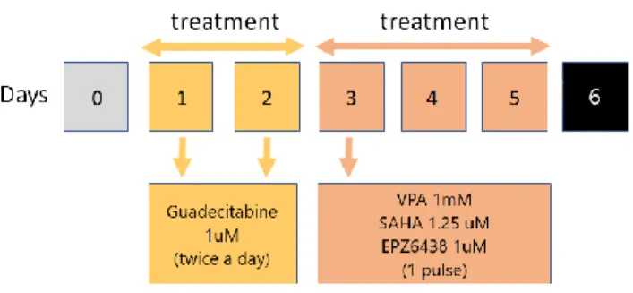
3.5. Compounds preparation and schedule of treatments ... 30
3.6. Flow cytometry analysis and antibodies ... 31
3.7. DNase I treatment of RNA samples and reverse transcription ... 32
3.8. Real-time polymerase chain reaction (RT-PCR) ... 32
3.9. Statistical analysis ... 33
4. RESULTS ... 34
4.1. nCounter gene expression panel analysis ... 34
4.2. Cytofluorimetric analysis of HLA class I antigens and ICAM-1 surface expression on MPM cell lines treated with combined epigenetic drugs ... 42
4.3. Molecular analysis of gene expression of CTA expression on MPM cell lines
treated with combined epigenetic drugs ... 43
4.4. Molecular analysis of gene expression of PD-L1 expression on MPM cell lines treated with combined epigenetic drugs ... 45
4.5. Molecular analysis of gene expression of NKG2DLs expression on MPM cell lines treated with combined epigenetic drugs ... 46
4.6. Molecular analysis of gene expression of EMT-regulating genes on MPM cell lines treated with combined epigenetic drugs ... 47
5. DISCUSSION ... 50
6. REFERENCES ... 55
RIASSUNTO
Il mesotelioma pleurico maligno (MPM) è un tumore molto aggressivo e rapidamente progressivo che si sviluppa a livello del mesotelio che compone la pleura; questa neoplasia può assumere diversi sottotipi istologici (epitelioide, bifasico e sarcomatoide), i quali sono strettamente correlati alla prognosi. Le modificazioni epigenetiche che avvengono nelle fasi di iniziazione e progressione del MPM possono svolgere un ruolo fondamentale nel regolare negativamente il crosstalk tra tumore e sistema immunitario, contribuendo a mantenere un microambiente tumorale immunosoppressivo. Conoscere più dettagliatamente il panorama epigenetico del MPM può contribuire a definire il razionale per nuove terapie antitumorali e porre le basi per studi di combinazione che prevedano l’utilizzo di farmaci epigenetici con farmaci immunoterapeutici.
Con il presente studio abbiamo voluto valutare, in un primo momento, le modificazioni nel profilo di espressione genica di 10 linee di MPM, di diverso istotipo, trattate con la guadecitabina, un agente demetilante il DNA di seconda generazione, tramite la piattaforma nCounter di Nanostring. I risultati ottenuti tramite Ingenuity Pathway Analysis (IPA) hanno mostrato che la guadecitabina era in grado di indurre l’attivazione dei geni coinvolti nel crosstalk tra cellule dendritiche e natural killer nel 50% delle linee cellulari di MPM indagate, accompagnata dall’attivazione di altre componenti coinvolte nella risposta immunitaria a infezioni e infiammazioni. I fattori trascrizionali “upstream” più frequentemente attivati appartenevano al pathway di segnalazione dell’interferon (IFN)-γ. Inoltre, è stata riscontrata l’up-regolazione (fold change medio (mFC) ≥ 1.5) di molecole immuno-relate, come NY-ESO-1 (mFC=13.16), MAGE-B2 (mFC=13.09), CD70 (mFC=5.27) e CTLA-4 (mFC=4.81). Abbiamo inoltre effettuato analisi istotipo-specifiche per esplorare le modificazioni molecolari indotte dalla guadecitabina nei 3 sottotipi di MPM. La guadecitabina ha indotto l’up-regolazione dell’espressione di marcatori del fenotipo epiteliale (es. CDH1, EPCAM e PECAM1), osservata ad alti livelli nelle linee cellulari sarcomatoidi; ciò è stato associato alla down-regolazione di molecole di origine mesenchimale (es. CDH2 e NCAM) e induttori della cascata metastatica (es. CDH11).
Successivamente abbiamo comparato gli effetti immunomodulatori della guadecitabina con quelli di altri farmaci epigenetici (gli inibitori delle iston acetiltransferasi (HDAC) VPA e SAHA o l’inibitore di EZH2 EPZ-6438) da soli o in combinazione con la guadecitabina in 5 linee cellulari di MPM (2 sarcomatoidi, 1 bifasica e 2 epitelioidi). Analisi citofluorimetriche e molecolari hanno rivelato che la guadecitabina up-regolava l’espressione delle molecole immuno-relate, quali HLA di classe I (mFC=1.59), ICAM-1 (mFC=3.27), PD-L1 (mFC=2.13), e NKG2DL (MICA mFC=1.88, MICB mFC=2.42, ULBP2 mFC=3.16), inducendo/up-regolando l’espressione dei Cancer Testit Antigens (CTA) NY-ESO-1, MAGE-A1 e MAGE-A3; il VPA up-regolava l’espressione degli antigeni di HLA di classe I (mFC=1.50), PD-L1 (mFC=2.76), NKG2DL (MICA mFC=1.69, MICB mFC=2.67, ULBP2 mFC=3.26) e quella dei CTA MAGE-A1 e MAGE-A3, rispettivamente in 2/5 e 3/5 linee cellulari di
MPM; il SAHA up-regolava l’espressione di MICA (mFC=1.57), MICB (mFC=4.05) e MAGE-A1 e MAGE-A3, rispettivamente in 2/5 e 4/5 linee cellulari; per contro, l’EPZ-6438 ha mostrato minime capacità immunomodulanti, inducendo solamente NY-ESO-1 e up-regolando l’espressione di PD-L1, MICB e ULBP2 in 1 linea cellulare ciascuno. Contrariamente ai risultati eterogenei ottenuti dai singoli farmaci, l’associazione di VPA, SAHA o EPZ-6438 alla guadecitabine ha rafforzato le capacità immunomodulanti di quest’ultima, influenzando l’espressione di tutte le molecole indagate. Specificatamente, le combinazioni di guadecitabine con VPA, SAHA o EPZ-6438 up-regolavano l’espressione degli antigeni HLA di classe I (mFC=2.21, 2.03, o 2.29 rispettivamente), di ICAM-1 (mFC=4.09, 4.63, o 5.33), di PD-L1 (mFC=6.95, 2.42, o 2.50), di MIC-A (mFC=3.48, 2.00, o 2.23), di MIC-B (mFC=6.80, 2.48, o 2.81) e di ULBP2 (mFC=13.45, 3.40, o 4.11). Infine, livelli di up-regolazione/induzione maggiori sono stati osservati per i CTA a seguito di tutti e 3 i trattamenti combinati rispetto alla guadecitabina in singolo. La modulazione delle caderine è stata influenzata dal sottotipo istologico di MPM: l’espressione di CDH1 è stata indotta dalla guadecitabina in singolo e dalla sua combinazione con VPA, SAHA e EPZ-6438 nelle 2 linee cellulari sarcomatoidi, costitutivamente negative per l’espressione del gene; l’espressione di CDH2 è stata up-regolata dal VPA e dal SAHA singoli in 1/5 linee cellulari e dalle combinazioni di guadecitabina con VPA o SAHA, rispettivamente in 3/5 o 1/5 linee cellulari di MPM; ciononostante, non è stata osservata alcuna up-regolazione del gene nelle 2 linee cellulari epiteliodi, costitutivamente negative per l’espressione di CDH2.
In conclusione, dalle analisi approfondite del pannello di espressione genica abbiamo confermato che la guadecitabina è in grado di up-regolare/indurre l’espressione di molecole immunitarie e immuno-relate cruciali per il crosstalk tra il tumore e il sistema immunitario; inoltre, abbiamo dimostrato che essa induce l’attivazione di geni correlati all’IFN, soprattutto nel fenotipo sarcomatoide, supportando l’ipotesi che i demetilanti possano aumentare la risposta immunitaria contro il MPM, potenzialmente anche del tipo istologico più aggressivo; la modulazione delle molecole di adesione tendente verso il fenotipo epitelioide suggerisce la possibilità di revertire la transizione epitelio-mesenchima, cruciale nel processo di metastatizzazione. Infine, combinando la guadecitabina con farmaci inibitori delle HDAC/EZH2 ha rafforzato la sua attività immunomodulante, fornendo il razionale per studi di associazione di farmaci epigenetici e agenti immunoterapici in modo da aumentare l’efficacia di questi ultimi nel trattamento del mesotelioma.
ABSTRACT
Malignant pleural mesothelioma (MPM) is a highly aggressive and rapidly progressive tumor that affect the mesothelium componing the pleura; it can acquire different histological subtypes (mainly epithelioid, biphasic, and sarcomatoid MPM), which are of prognostic significance. Epigenetic modifications occurring during MPM initiation and progression may play a relevant role in negatively regulating the crosstalk between the tumor and the immune system, as well as contributing to the highly immunosuppressive microenvironment. A better understanding of MPM epigenetics will contribute to refine antitumor strategies, laying the ground for epigenetic-based immunotherapy. The present study evaluated, in the first instance, changes in the gene expression fingerprint of 10 MPM cell lines of different phenotype treated with the second-generation DNA hypomethylating agent (DHA) guadecitabine, through the Nanostring Oncology panel with nCounter readout. Ingenuity pathway analysis results revealed that guadecitabine induced the activation of natural killer and dendritic cells signaling pathways in 50% of MPM cell lines, followed by the activation of other components involved in the immune system response to infections and inflammation. Besides, the most frequently activated upstream regulators belonging to the interferon (IFN)-γ signaling pathway. Also, the up- regulation (mean fold change (mFC) ≥ 1.5) of key immune-related molecules, such as the NY-ESO-1 (mFC=13.16), MAGE-B2 (mFC=13.09), CD70 (mFC=5.27), and CTLA-4 (mFC=4.81) was reported.
We also performed histological type-specific investigations to explore molecular changes induced by guadecitabine among the 3 histotypes. Guadecitabine induced the up-regulation of the expression of epithelial markers (e.g., CDH1, EPCAM, PECAM1), observed at higher levels in sarcomatoid cell lines; this was accompanied by the down-regulation of mesenchymal origin molecules (e.g., CDH2, NCAM), and inductor of metastatic signals (e.g., CDH11).
Secondly, the immunomodulatory effects of guadecitabine were compared to those of different epigenetic drugs (the histone deacetylase (HDAC) inhibitors VPA and SAHA, or the EZH2 EPZ-6438), alone or in combination with guadecitabine, in 5 MPM cell lines (two sarcomatoid, one biphasic, and two epithelioid). We performed cytofluorimetric and molecular qRT-PCR analyses and, in this regard, results showed that guadecitabine up-regulated the expression of immune-related molecules, such as HLA class I antigens (mFC=1.59), ICAM-1 (mFC=3.27), PD-L1 (mFC=2.13), and NKG2DLs (MIC-A mFC=1.88, MIC-B mFC=2.42, and ULBP2 mFC=3.16), and up-regulated/induced Cancer Testis Antigens (CTA: NY-ESO-1, MAGE-A1, and MAGE-A3) expression; VPA up-regulated the expression of HLA class I antigens (mFC=1.50), PD-L1 (mFC=2.76), NKG2DLs (MIC-A mFC=1.69, MIC-B mFC=2.67, and ULBP2 mFC=3.26), and the expression of CTA MAGE-A1 and MAGE-A3 in 2/5 and 3/5 MPM cell lines, respectively; SAHA up- regulated the expression of MICA (mFC=1.57), MICB (mFC=4.05), MAGE-A1 and MAGE-A3 in 2/5and 4/5 MPM
cell lines, respectively; conversely, EPZ-6438 induced minimal immunomodulatory effects, inducing only NY-ESO-1 and up-regulating PD-L1, MIC-B, and ULBP2 expression in 1 MPM cell line each. Despite the heterogeneous activities of single epigenetic drugs, the addition of both VPA, SAHA, and EPZ-6438 to guadecitabine strengthened the immunomodulatory effects of the latter, by affecting the expression of all investigated molecules. Specifically, guadecitabine plus VPA, SAHA, or EPZ-6438 upregulated the expression of HLA class I antigens mFC=2.21, 2.03, or 2.29; ICAM-1 mFC=4.09, 4.63, or 5.33; PD-L1 mFC=6.95, 2.42, or 2.50; MIC-A mFC=3.48, 2.00, or 2.23; MIC-B mFC=6.80, 2.48, or 2.81; ULBP2 mFC=13.45, 3.40, or 4.11, respectively. Lastly, higher levels of upregulated/induced CTA expression were observed after all 3 combination treatments versus guadecitabine alone. Cadherins modulation was MPM histotype-related: CDH1 expression was induced in the 2 constitutive-negative sarcomatoid MPM cell lines by guadecitabine alone or combined with VPA, SAHA, or EPZ-6438; CDH2 expression was upregulated by VPA or SAHA in 1/5 cell lines, and by guadecitabine plus VPA or SAHA in 3/5 or in 1/5 MPM cell lines, respectively; however, no induction of CDH2 have been reported in the constitutive negative epithelioid cell lines. Overall, from comprehensive gene expression panel analyses, we confirmed that guadecitabine induced/up-regulated the expression of immune and immune-related molecules, pivotal in the tumor- immune system crosstalk; also, we highlighted that guadecitabine-induced activation of IFN-related genes, especially in the sarcomatoid phenotype, supporting the hypothesis that DHA could increase the immune response against MPM, potentially also with sarcomatoid features; moreover, the modulation of adhesion molecules towards the epithelial type suggests the possibility to revert the epithelial-to-mesenchymal transition (EMT) event, crucial in the invasion-metastasis cascade. Also, combining guadecitabine with HDACi/EZH2i strengthened its immunomodulatory capabilities, laying the rationale for epigenetic drugs-based immunotherapies, to enhance efficacy of these strategy in the MPM clinic.
1. Introduction
1.1. Epigenetic regulation in health and disease
1.1.1. Epigenetic control of the gene expression
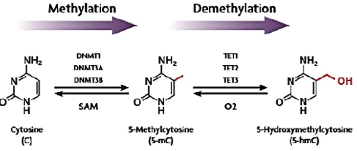
Epigenetic mechanisms refer to a series of potentially reversible and heritable changes in the chromatin structure and in the gene expression, which address the tissue-specific transcription and conserve cell identity, inducing changes in the phenotype, without altering the underlying nucleotide sequence (Esteller, 2008). Through interconnected biochemical modifications of DNA and histone/non-histone proteins, as well as by the activity of highly controlled remodellers/regulators, genes can be switched “on” or “off”, determining which proteins are transcribed at a specific time and in a particular cell type, in response to extracellular signals. The epigenetic landscape of complex organisms is set out during cell differentiation processes, playing a pivotal role in embryogenesis, but also involving a dynamical pattern of responses during critical stages such as pregnancy, the post-natal period and, to a lower extent, in later life. Epigenetic modifications are responsible, for example, for genomic imprinting processes, as well as for the inactivation of the X chromosome in female mammals, through events of initiation, spreading and maintenance of gene silencing (Hassler, 2012). In addition, epigenetic regulation plays as an interface between the genome and the environment, being characterized by plasticity in response to several factors, such as aging and environment/lifestyle. These mechanisms are potential source of missing heritability in complex traits. Theoretically, only germline heritable epigenetic events may contribute to the missing heritability, as opposed to the non-germline heritable components (inherited through mitosis or somatic epigenetic events). Identical twins, developed from a single fertilized egg, have the same genome, so any differences between twins are due to their epigenetics, not genetics. Since epigenetic modifications are involved in gene regulation during differentiation and homeostasis with the ability to integrate environmental stimuli, it is not surprising that abnormalities in these mechanisms have been linked to a wide range of diseases (Portela, 2010). In higher-order eukaryotes, epigenetic marks include principally DNA methylation, post-translational modifications (PTMs) of histone proteins, chromatin remodelling components, histone variant exchange, and non-coding RNAs. The most studied mechanism of epigenetic regulation is the DNA methylation, elicited by the addition of a methyl (-CH3) group to the 5′ carbon of cytosine to become 5-methylcytosines (Fig. 1). It affects especially small regions of DNA (<500 bp) enriched in cytosine-guanosine dinucleotides, known as CpG islands, owning a CG content greater than 55%. CpG-rich DNA is usually clustered around the promoter region of the gene and the methylation process can affect the transcriptional regulation of this gene. CpG sites
are methylated by the DNA methyltransferase (DNMT) enzymes, epigenetic “writers” that establish the mark, crucial for the normal development as evidenced by studies of embryology (Li, 1992; Egger, 2004). DNMTs are mainly grouped in enzymes performing de novo methylation, and those in charge to stably maintain the methylation patterns; all of them catalyse the reaction of methylation using the s-adenosyl-l-methionine (SAM) as the methyl groups donor. Currently, there are five known mammalian DNMTs and DNMT-like proteins: DNMT1, DNMT2, DNMT3A, DNMT3B, and DNMT3L (Robertson, 2002). The most abundant is the DNMT1, responsible for the maintenance of the methylation patterns on hemi-methylated replicating DNA within cell division, while de novo DNMTs, such as DNMT3A and DNMT3B, define the methylation fingerprint on unmethylated DNA (Morris, 2014). Methylation at CpG sites can be recognized by “readers” such as the methyl-CpG binding domain protein 1 (MBD1) and the methyl-CpG binding protein 2 (MeCP2), inducing repression. Besides classic CpG methylation, non-CpG methylation has been detected in embryonic stem cells at high levels (Pulverer, 2012). CpGs are hot spots for mutation, as 5-methylcytosine (5mC) can spontaneously undergo hydrolytic deamination to thymine (Smith, 2013), resulting in a mismatch with guanine opposite to the original 5mC. Conversely, methyl groups can be erased within active/passive demethylation pathways, followed by glycosylation and replacement with an unmethylated cytosine (Emran, 2019). Indeed, ten-eleven translocation (TET) cytosine oxygenases have been found to oxidase 5mC to 5-hydroxymethylcytosine (5hmC) (Fig. 1), present prevalently in neurons and embryonic stem cells, and further products. This methylation state has been defined as a demethylation intermediate, but some studies underline its role as an active epigenetic mark (Szulwach, 2011), associated with transcriptional activation, even though specific involvements in different contexts is yet to be elucidated (Branco, 2012).
Figure 1 – Regulation of DNA methylation/demethylation. Cytosine conversion in methylated cytosine, through DNMTs, and demethylation to 5-hydroxymethylcytosine by TET enzymes.
From Hong-Wei Y., J of Pharma Exp Ther, 2015.
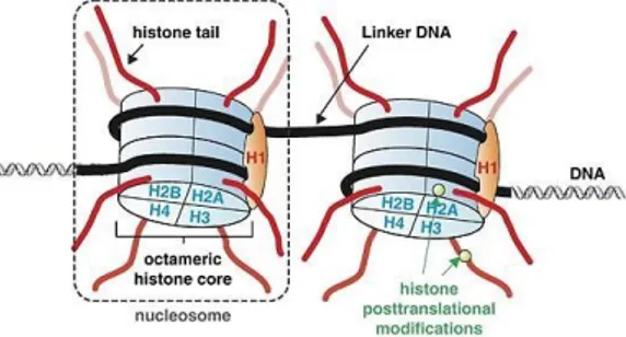
Another important mechanism of epigenetic regulation involves PTMs of histones. Histone proteins are key component of chromatin, which fundamental unit is the nucleosome. In eukaryotes, it consists of 147 base pairs of DNA wrapped around a histone octamer, the histone core, comprised of two copies for
each H2A, H2B, H3, and H4, while H1 acts as linker protein (Fig. 2).
Figure 2 - Schematic representation of the organization and packaging of elements of the nucleosome. From Füllgrabe, J., Oncogene, 2011.
Histones have got a specific structural and functional organization due to their role in the dynamic regulation of gene transcription, by controlling accessibility of nuclear transcription factors and RNA polymerase to regulatory DNA elements. Different amino acids of histone amino-terminal tails are subject to PTMs, including methylation, acetylation, phosphorylation, ubiquitylation, and sumoylation, even though several others have been reported (Kouzarides, 2007) (Fig. 3).
Figure 3 - Post-translational modifications on histone tails; in red are shown amino acids interested by acetylation, while in blue the ones potentially affected by methylation.
Lys: lysine. From Bottomley, M. J., EMBO Rep, 2004.
These covalent modifications can shape chromatin conformation in different ways: as a results of certain events, such as acetylation of lysine residues responsible for breaking down the positive charge of lysines, the chromatin structure is released facilitating the transcription; conversely, if the affinity between histones and DNA is enhanced, chromatin is condensed in form of heterochromatin, resulting in the inactivation of transcription, as observed for histone sumoylation (Shiio, 2004).
Acetylation and deacetylation are the most abundant and studied histone modifications, controlled by epigenetic writers, known as histone acetyltransferases (HATs) which lay the acetyl group, and erasers, called histone deacetylases (HDACs) which remove marks. The acetylation reaction consists in the transfer of an acetyl (CH₃CO–) group from the acetyl-CoA to the ε-amino group of the histone lysine. HATs can act on histone and non-histone protein and work through the HAT domains, who mediate acetylation, which are then recognized by bromodomains (“readers” of the mark) for the activation of
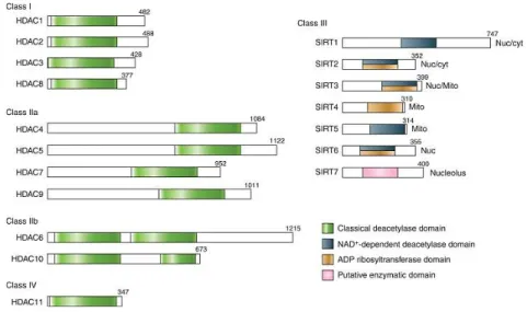
the signal. The acetylating enzymes comprise different families and are associated with cofactors that, making complexes, can direct both gene-specific and genome-wide acetylation (Bottomley, 2004). Some coactivator molecules, such as the CREB-binding protein (CBP) and its homologue p300, act as molecular switches that control gene transcription activation and both have intrinsic HAT activity (Chan, 2001). So, the structure of chromatin can be regulated within a cascade of intracellular signaling, involving cAMP, Ca2+, and ERK (Yuan, 2001). In contrast, HDACs, which remove the acetyl groups from lysine residues, confer to the latter a positive charge with the concomitant rearrangement of additional lysine PTMs, and the suppression of gene transcription (Seto, 2014). HDACs operate in complexes with corepressors and can act either on acetyl-lysine residues of histones tails or on non-histone proteins, such as the transcription factor p53 (Vaziri, 2001). The 18 HDACs known are classified in four classes, based on the homology with yeast enzyme counterparts (Fig. 4): class I enzymes (HDAC1, HDAC2, HDAC3, and HDAC8); class II HDACs (HDAC4, HDAC5, HDAC6, HDAC7, HDAC9, and HDAC10); class III HDACs (SIRT1, SIRT2, SIRT3, SIRT4, SIRT5, SIRT6, and SIRT7), known as Sirtuins; and class IV (HDAC11) which share only a weak homology with both class I and II HDACs. Class I, II, and IV are known as “classical” HDACs, sharing structural and functional homologies. They belong to the arginase/deacetylase superfamily, containing both the deacetylase and the arginase-like amidino hydrolase activity. The latter activity is explicated through a zinc finger-binding domain, required for the ion-dependent catalysis of the acetamide bond in acetylated lysine. Conversely, class III HDACs exert a NAD+-dependent mechanism of deacetylation through the NAD/FAD-binding domain, producing nicotinamide and the 2′-O-acetyl-ADP-ribose metabolite (Seto, 2014). Many of these HDAC substrates regulate proteins involved in different processes, such as cell-cell adhesion, cell-cell division, and apoptosis. In particular, HDAC-1,-2, and -3 repress genes involved cell-cell cycle regulation (Gui, 2004); HDAC8 regulates cell proliferation (Vannini, 2004); HDAC-4, -5, -7, and -9 recruit repressors on specific genomic regions, inhibiting gene transcription (Di Giorgio, 2016); HDAC6 controls cell migration, protein folding, mis-folded proteins degradation, cellular stress, immune synapse formation, and oncogenic tumorigenesis (Valenzuela-Fernández, 2008; Matthias, 2008; Lee, 2008). Given that histone PTMs modulates chromatin structure and gene expression, it is not surprising that abnormal events of acetylation are associated with multiple diseases, including cancer, interstitial fibrosis, autoimmune and inflammatory diseases, and metabolic disorders (Tang, 2013).
Figure 4 – Representation of different classes of HDACs. Enzymatic domains are shown in colours. From Seto, E.,Cold Spring Harb Perspect Biol, 2014.
Besides acetylation, methylation of histone tails is one of the major epigenetic PTMs, interesting arginine and, more commonly, lysine (K) residues. The effect of histone methylation depends on histone isoforms and lysine position at the level of N-terminal residue, as well as on the extent of methylation. Methylation of histone lysines is catalysed by lysine methyltransferase (KMT) and removed by lysine demethylase (KDM) enzymes. KDMs play a role both in repression (H3K4 demethylation) and activation (H3K9 demethylation) of transcription, and it has been reported their involvement in mental disorder development as well as cancer (Tahiliani, 2007; Kaniskan, 2018). Canonical lysine methylation sites are found on histone 3 at lysine 4 (H3K4), 9 (H3K9), 27 (H3K27), 36 (H3K36), or 79 (H3K79), and on histone H4 at lysine 20 (H4K20), regulating chromatin structure (Cao, 2015); also, other non-canonical sites have been described, but are much less characterized. Methylated sites on histones are recognized by chromodomains of proteins associated with chromatin remodelling, acting as readers of this specific mark. In this context, two of the most studied chromatin-modifying complexes include the evolutionary-conserved components of the Polycomb and Trithorax groups, operating as antagonists in the regulation of the development. Both groups are recruited directly on DNA motifs, through the recognition of Polycomb/Trithorax response elements (PRE/TRE), which, based on the recruited component, maintain a repressed/active transcriptional state, driving the epigenetic inheritance (Schuettengruber, 2017). Repression of gene expression is mediated by a Polycomb multiprotein system that include the Polycomb-Repressive Complex 1 (PRC1) and 2 (PRC2), and targets H3K27 and H3K9 methylation. The enhancer of zeste homolog (EZH) proteins, notably EZH1 and EZH2, are components of the catalytic subunit of the PRC2 machinery, mediating an independent silencing of gene expression through the H3K27me3 mark (Tan, 2014). In comparison to EZH2, EZH1 holds a low histone methyltransferase activity and its knockdown does not result in the global reduction of H3K27me2/3 levels (Margueron,
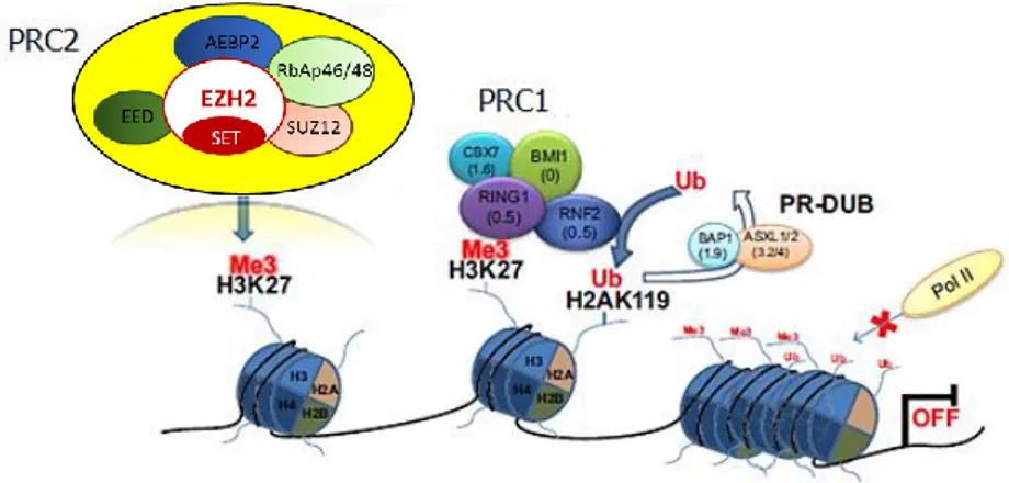
2008). The PRC2 machinery involve, overall, five subunits (EZH1/2, EED, SUZ12, RbAp46/48, AEBP2) which predominantly exert the methyltransferase activity through the C-terminal SET domain of EZH2, followed by a cascade of downstream events (Fig. 5). The complex is stabilised by non-catalytic subunits, which are also crucial for triggering the enzymatic function of EZH2 (Blackledge, 2015). Pathologic activation of the transcriptional repressor EZH2 is one of the most studied features observed in human cancers, and plays a key role in cell growth and differentiation (Bracken, 2003; Bryant, 2007; Qi, 2013), survival (Varambally, 2002), tumor invasion (Bracken, 2003; Bryant, 2007), and metastasis (Varambally, 2002; Mahmoud, 2016). In contrast to Polycomb multiprotein system, the Trithorax group proteins target H3K4 methylation, maintaining an active state of the gene expression.
Figure 5 - The PRC2 complex mediates, through the SET domain of EZH2, the tri-methylation of lysine 27 on H3 histones (H3K27me3); after that PRC1, mediating the mono-ubiquitination of lysine 119 on histone H2A (H2AK119ub) is recruited, additional epigenetic enzymes are
retrieved, compacting the chromatin structure. Edited from Marchese, I., Chromatin
Remodelling, 2012.
Overall, despite DNA methylation and histone PTMs are executed by different cellular machinery (all generally classified as writers, erasers, and readers), both are dynamically linked and act synergistically to regulate transcription. Therefore, it is now widely recognized that a significant interplay exists among these epigenetic modulation events. Histone modifications have been shown to induce DNA methylation, a process especially observed during the early development, regulating also the stability of DNMT enzymes (Cedar, 2009; Esteve, 2009). Once established, methylated CpGs are recognized by methyl-CpG-binding proteins, such as MeCP2, which form a complex with histone deacetylase and histone methyltransferases, acting as transcription co-repressive complexes (Feng, 2001; Cedar, 2009). Additionally, to explicate the local histone code, epigenetic rearrangements might be required: for example, if PRC2 is commissioned to methylate H3K27 but this lysine residue is acetylated, HDACs are recruited, making the amino-group of lysine side chain available for the PRC2-mediated methylation (Tan, 2014).
1.1.2. The role of epigenetics in cancer initiation and progression
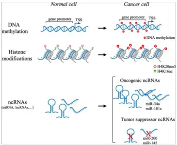
The epigenetic machinery is well known to be implicated in various physiological and pathological events, among the latter neurological disorders (Egger, 2004), asthma (Adcock, 2005), diabetes (Jones, 2007), autoimmunity (Javierre, 2010), and cancer (Jones, 2007). Since genetics alone does not provide an adequate explanation for the complexity of cancer, a comprehensive understanding of the role of epigenetics in cancer might unveil key mechanisms underlying its development and progression. In most cases, besides genetic alterations, the fine control of epigenetic mechanisms is lost; indeed, malignant cells harbour aberrations in DNA methylation and histone modifications, deregulation of non-coding RNAs (e.g., microRNA, long non-coding RNA), as well as alterations of numerous regulatory factors (Fig. 6). Genome sequencing revealed mutations in chromatin proteins in almost 50% of human cancers (You, 2012; Shen, 2013), and disruption of chromatin structure can induce inappropriate gene expression and genomic instability, resulting in the malignant transformation.
Being epigenetics modifications recognized as the key modulator of plasticity, their reversable nature makes them good therapeutic targets to potentially revert cancer-relevant processes by using specific inhibitors.
Figure 6 – Main epigenetic abnormalities in cancer. TSS: transcription start site; ncRNA: non-coding RNA; miRNA: microRNA; lncRNA: long non-coding RNA.
1.1.2.1. DNA methylation in cancer and DNMT inhibitors
A well characterized contribution of epigenetics to tumorigenesis concern DNA methylation disruption events, among which predominantly genome-wide DNA hypomethylation and focal hypermethylation, especially interesting CpG islands of tumor-suppressor gene (TSG) promoters (Esteller, 2008; Fernandez, 2012). One of the initial epigenetic abnormalities recognized to occur in tumors is the loss of DNA methylation, mostly affecting repeated DNA sequences, which constitute approximately half of the human genome. It has possibly evolved as a mechanism of defence against foreign DNA elements, including retrotransposons and viral pathogens (Richards, 2009). Different implications of hypomethylation in promoting tumorigenesis have been identified:
1) Chromosomal instability (CIN): a feature of most human cancers, particularly the solid ones. It is a type of genome instability, which ranges from single nucleotide changes to large-scale cytogenetic aberrations, able to alter different regulatory pathways, such as cell cycle control and DNA damages repair. Mutations in CIN genes increase the rate at which entire parts of chromosomes, or large parts of them, are lost or gained during cell division, or result in simple rather than complex chromosomal rearrangements. CIN is a cause of an imbalance in chromosome number (aneuploidy) and an enhanced rate of loss of heterozygosity, which is an important mechanism of inactivation of TSGs, such as for genes coding for p53, p21, or p19Arf (Lengauer, 1998). Nowadays, it is evident that more than 70% of common solid neoplasms are aneuploid and, in many instances, the onset of heterogeneous aneuploidy correlates with poor prognosis and aggressiveness of different tumors (Cimini, 2008; McGranahan, 2012); 2) Reactivation of transposable elements: hypomethylation of DNA in malignant cells can reactivate intragenomic DNA, such as the LINE (long interspersed nuclear element) and SINE (short interspersed nuclear element) sequences, as well as LTR (long terminal repeat) retrotransposons. These elements are normally hypermethylated and transcriptionally silenced in somatic cells, but they become demethylated to various degrees in cancer cells (Hoffmann, 2005). Higher levels of hypomethylation in LINE-1 was found in a wide number of tumors, compared to matched normal tissues, such as for urothelial carcinoma (Jurgens, 1996), hepatocellular carcinoma (Takai, 2000), melanoma (Sigalotti, 2011), and prostate cancer (Fiano, 2017). These demethylated transposons can be transcribed or translocated to other genomic regions, thereby disrupting the genome; 3) Loss of imprinting (LOI): it is a common epigenetic alteration observed in human cancers, involving loss of parental origin-specific expression of imprinted genes caused by defects of methylation, either in terms of activation of the normally silenced allele or silencing of the expressed one. For example, LOI of the insulin-like growth factor-2 (IGF-2), through the aberrant methylation of the maternal silent copy, represents a significant risk factor for colorectal
carcinoma (Feinberg, 2002); 4) Global hypomethylation: it’s largely secondary to hypomethylation of repeated DNA sequences, even though heterogeneous hypomethylation within gene-coding regions has been reported (Kaneda, 2004). The latter mainly leads to the enhancement of expression of proto-oncogenes and activation of cell proliferation. Hypomethylation of wide regions of the genome seems to be due, almost in part, to a reduction/deletion in DNMT1 enzyme, as well as mutations in TET genes (Gaudet, 2003; Zhang, 2020). From precancerous lesions to full-blown malignant tumors, the hypomethylation status of genomic DNA seems to be more prominent, being related to tumor progression (Hoffmann, 2005). A possible mechanism is that global hypomethylation also results in the hypomethylation and activation of cell motility and invasion genes, methylated in non-metastatic cells (Szyf, 2005). Therefore, DNA hypomethylation levels could be used as a biomarker of tumor aggressiveness (Fraga, 2004). However, Yamada et al. registered a dual function of global hypomethylation, able to sustain either tumor induction or inhibition, based on tumor site and stage (Yamada, 2005).
Hypomethylation has no proven relationship with aberrant hypermethylation in inducing cancer, since these processes are independent, targeting different programs at different stages of tumorigenesis (Ehrlich, 2002). The inactivation of TSGs through the hypermethylation of CpG island within their promoter region is a key element in cancer development. Esteller et al. hypothesize that the underlie mechanism involves DNMTs, which fail to recognize DNA repeat regions or intronic sequences to be methylated in a normal cell, resulting in the methylation of CpG islands that are normally not recognized by DNMTs (Esteller, 2002). It was discovered at the level of the RB human gene promoter, determining susceptibility to hereditary retinoblastoma; nowadays, it is considered a hallmark of cancer because it is found in every kind of human neoplasms, being responsible for the inactivation of genes dragged into cell cycle regulation, DNA repair, metabolism, cell-cell adhesion, apoptosis, angiogenesis, and invasion (Yánez, 2015). Several known TSGs are silenced via promoter methylation in different tumors, such as RB, p16INK4a, BRCA-1, VHL, E-cadherin, and MLH1 (Baylin, 2001). Both hypomethylation and hypermethylation processes are implicated in cancer metastasis development (Kong, 2015).
Since DNA methylation events are reversible, it is possible to reactivate TSGs, reprogramming the genome of tumor cells, inhibiting tumor proliferation and inducing cell death using inhibitors of DNMTs (DNMTi), also known as DNA hypomethylating agents (DHAs) (Gopisetty, 2006) (Table 1). DHAs are classified in two families: nucleoside and non-nucleoside analogues. Non-nucleoside inhibitors have low toxicity but exhibit limited hypomethylating activities. Nucleoside inhibitors, such as azacitidine (AZA) and decitabine (DAC), are, instead, characterized by the presence of a nitrogen atom in lieu of carbon 5-position of the pyrimidinic ring, linked to ribose/deoxyribose. After cellular uptake, they are metabolized
by different kinases for the incorporation into DNA (or mainly RNA for AZA) within the S phase of cell cycle. Both undergo the same reaction of normal cytosines leading to two major effects: i) a DNA damage response of apoptosis or, as documented for DAC, senescence (Navada, 2014); ii) the recognition by DNMT proteins which, once having bonded the modified cytidine, are complexed to the structure in an irreversible manner (Santi, 1984). As a result, DNMTs are physically depleted and, with concurrent cell divisions, demethylated DNA is accumulated (Fig. 7).
Figure 7 - Steps for the incorporation of azacitidine (5-aza) and decitabine (5-aza-dC) into DNA/RNA during cell cycle and main effects are reported. From Duchmann, M., Progress in
Hematology, 2019.
Both AZA and DAC received the Food and Drug Administration (FDA) endorsement for the treatment of myelodysplastic syndrome (MDS) and acute myeloid leukaemia (AML). Although AZA and DAC show basically similar mechanisms of action, they have been reported to exert distinct effects, showing different efficacy in clinical trials (Diesch, 2016). However, the latter have short half-life in the blood, due to rapid inactivation by cytidine deaminase (CDA). In order to prevent this, the second-generation DHA guadecitabine has been developed, consisting of a dinucleotide of DAC linked to deoxyguanosine, through a phosphodiester bond. The great advantage of this compound is that, thanks to its configuration, it is resistant to degradation by CDA, increasing in vivo exposure of DAC and demonstrating to be safe and well tolerated (Coral, 2013; Roboz, 2016; Jueliger, 2016). Guadecitabine is currently being evaluated in about 40 clinical trials worldwide, between whom phase III trials concerning untreated/previously treated patients with AML, MDS, and chronic myelomonocytic leukemia, as well as phase II studies for the treatment of solid tumors, including ovarian carcinoma, small/non-small cell
lung cancer (SCLC/NSCLC), melanoma, renal cell and hepatocellular carcinoma. Fazio et al. demonstrated that treatment of human melanoma cell lines with guadecitabine in vitro up-regulated/induced the expression of Melanoma Antigen Gene (MAGE) proteins of Cancer Testis Antigens (CTA) family (Fazio, 2018). The latter are antigens of the family of tumor-associated antigens, unexpressed or expressed at very low levels in normal tissues, except for placenta and testis, and able to positively influence immunogenicity of cancer cells and the immune recognition by cytotoxic T-lymphocytes (CTLs). Also, a strong up-regulation of the constitutive expression of HLA class I antigens and of the costimulatory molecule Intercellular Adhesion Molecule-1 (ICAM-1) was observed with both guadecitabine and DAC in melanoma and hematologic tumor cell lines in vitro (Fazio, 2018). These data have been extensively confirmed in vitro and in vivo over the years, highlighting the strong action of DHAs on augmenting immune responses with the up-regulation of innate and adaptive immunity-related molecules in tumors of different histotype (Covre, 2015; Jones, 2016; Nahas, 2019; Luker, 2020). Several studies also reported the DHA-mediated modulation of the expression of the Natural Killer Group 2D Ligands (NKG2DLs), whose bond with the immunoreceptor represent an activating and a costimulatory signal to boost natural killer-(NK)/T cell-mediated killing. The major histocompatibility complex class I chain-related A and B (MIC-A, -B), and the UL16 binding protein family (ULBP1-6) proteins are the stress-induced ligands, that are normally not expressed by healthy adult tissues but frequently found in tumor cells. However, tumor cells develop strategies to down-regulate NKG2DLs expression and avoid immune recognition, such as promoter hypermethylation. Baragaño Raneros et al. reported that AZA and DAC were able to induce demethylation and re-expression of NKG2DLs on the surface of AML cells, restoring the recognition of tumor cells by the immune system (Baragaño Raneros, 2015). The capability of DHA to induce/up-regulate NKG2DLs were also confirmed in different studies, as for glioma and melanoma cell lines, making them attractive targets for DHA-based strategies for solid tumors (Zhang, 2016; Fazio, 2018).
1.1.2.2. Histone acetylation and inhibition of HDACs in cancer
Histone acetylation has been shown to be frequently altered in many cancers, contributing to the epigenetic reprogramming. For example, loss of acetylation at lysine 16, together with the H4K20me3 repressive mark, has been reported to be a common epigenetic modification in human cancers, as well as low levels of H3K18ac observed in pancreatic, breast, prostate, and lung cancers, associated with poor prognosis (Fraga, 2005; Li, 2016). Also, increased expression of HDAC transcriptional repressors were reported in solid and hematologic malignancies: HDAC1 in prostate (Abbas, 2008) and gastric cancer
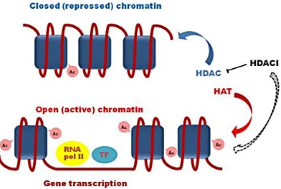
(Yu, 2019), HDAC-1 and -3 in ovarian cancer (Hayashi, 2010), HDAC-2 and -3 in colorectal carcinoma (Guan, 2000), HDAC6 and SIRT1 in AML (Bradbury, 2005), and HDAC8 in BRAF-mutated melanoma (Wilmott, 2015). In the majority of cases, high levels of HDACs are linked to advanced cancer and poor prognosis of patients, as for HDAC7 in pancreatic cancer (Ouaissi, 2014) or HDAC-1, -2, and -3 in gastric cancer (Sudo, 2011); some exception are reported, e.g., HDAC-3 and -8 in metastatic melanoma, whose overexpression is associated with improved survival (Wilmott, 2015). Underlie molecular mechanisms are not always well defined, but they can imply repression of TSGs (especially cell cycle regulators), down-regulation of microRNAs acting as tumor-suppressors, or alteration of oncogenic pathways (Li, 2016). HDAC inhibitors (HDACi), as a result of the inhibition of the zinc-containing enzymes, cause an accumulation of hyperacetylated histones, correlated with a relaxed chromatin conformation and transcription factors attraction, and enhance the expression of genes involved in various biological processes, including differentiation and cell death (Fig. 8). Indeed, HDACi are able to induce the down-regulation of anti-apoptotic genes, to activate the JAK/STAT signaling pathway, and the up-regulation of multiple pro-apoptotic proteins (e.g., Bim), such as the cyclin-dependent kinase inhibitor p21, which, together with the induction of oxidative DNA damage, contribute to cell cycle arrest (Grant, 2012).
Figure 8 – Representation of HDAC inhibitors (HDACi) mechanism of action. HDAC: histone deacetylase; HAT: histone acetyltransferase; Ac: acetylation; TF: transcription
factor. From Pasyukova, E.G., Mech Ageing Dev, 2017.
HDACi may act on different subclasses of HDACs, may have different biologic activity, acting at different concentration scales, based on the characteristics of the chemical compound. HDACi include a large range of drugs, which can be classified based on their chemical structure in: hydroxamic acids, benzamides, short chain fatty acids, and macrocycles, even though hybrid compounds are emerging. The majority of hydroxamic acids are classical HDACi, blocking all HDACs but not the NAD+-dependent class III enzymes, and work at very low concentrations, inducing differentiation and apoptosis of malignant cells (Richon, 2006). One of the earliest known hydroxamic acids is the natural compound vorinostat, also known as suberoylanilide hydroxamic acid or SAHA (Table 1), which has been the first
HDACi approved by FDA for the treatment, as single agent, of cutaneous T cell lymphoma (CTCL). Short chain fatty acids category includes only three compounds which inhibit class I and II HDACs, that tend to act at millimolar concentrations and are, in fact, less potent than hydroxamic acids, with a non-negligible toxicity. Valproic acid (VPA) (Table 1), approved for epilepsy and other neuropsychiatric disorders, belongs to the fatty acids-based inhibitors exerting its activities, among whom apoptosis, CTA induction, stimulation of T-cell recognition, induction of NKG2DLs and NK-mediated cytotoxicity, in a variety of tumors (Armeanu, 2005; Yamanegi, 2010; Makarevic, 2019). Due to the high concentrations required to achieve antitumor activity, many clinical trials are evaluating low-dose VPA efficacy within combinatorial approaches (Abaza, 2014; Suraweera, 2018). The most potent benzamide is the synthetic oral compound entinostat, a well-tolerated class I and IV HDAC inhibitor active at nanomolar concentrations. It induces inhibition of cell proliferation, terminal differentiation and apoptosis of different tumors, such as breast and non-small cell lung cancer with promising preclinical and clinical data (Ruiz, 2015; Connolly, 2017). Finally, macrocycles are the most potent HDACi, with pronounced selectivity for class I enzymes, whose component romidepsin has been approved by FDA for the treatment of CTCL in 2009. Different research groups have focalised their study on the properties of this class of HDACi, demonstrating their selectivity for HDAC-1,2, and -3, with significant anti-proliferative and immune-stimulating activities (Rajak, 2013).
Epigenetic drugs and their combinations have demonstrated to modulate the sensitivity of tumor cells to anticancer therapy. By far, the most relevant evidence is associated with the combination HDACi plus DHA, adding to the capability of inducing chromatin relaxation and apoptosis the restoration of TSGs (Grant, 2012). Numerous studies have supported this hypothesis. For example, the combination of AZA and entinostat in human lung adenocarcinoma cells induced a marked re-expression of pro-apoptotic genes, e.g., p16 and p21 cell cycle regulators, and reprogrammed the expression profile of different other pathways, including genes regulating DNA damage and tissue remodelling (Belinsky, 2011). Also, Tellez et al. showed that the combination of entinostat with guadecitabine induced a consistent reduction of the tumor burden in lung cancer mouse model, compared to single treatments, as well as the up-regulation/induction in the expression of p21, Bik, and more than 18 CTA in microarray studies (Tellez, 2014). Many questions, however, need to be resolved about the underlying mechanisms of action of drugs combinations. Nonetheless, many HDACi, such as SAHA, belinostat, VPA, panobinostat, and entinostat, are currently in clinical trials in combination with chemotherapeutic, radiation, and immunotherapeutic strategies, as well as with hormonal therapy, and inhibitors of topoisomerase, proteasome and tyrosin kinases (Suraweera, 2018). Indeed, Amnekar et al. demonstrated that the low-dose pre-treatment with HDACi (i.e., VPA and vorinostat) was able to sensitize gastric cancer cells to chemotherapeutic agents, increasing the amount of DNA-bound drug, enhancing also histone acetylation
and cell cycle arrest (Amnekar, 2020). Also, a phase II clinical trial for the treatment of NSCLC combining carboplatin and paclitaxel with vorinostat showed higher response rate, progression-free survival, and overall survival, compared to carboplatin and paclitaxel alone (ClinicalTrial.gov identifier: NCT01413750); however, a lower dose of vorinostat with carboplatin or paclitaxel is being evaluated in advanced solid tumor, in order to reduce toxicity (ClinicalTrial.gov identifier: NCT01281176). Finally, an interesting study has been recently published by Adeshakin et al. regarding the immunomodulatory properties of VPA. The study started from the observation, made by Xie et al., that VPA was able to attenuates immunosuppressive function of myeloid-derived suppressor cells (MDSC) either in vitro or
in vivo, and demonstrated synergistic antitumor efficacy when combined with blockade therapy of the
immune checkpoint molecule PD-L1 (Xie, 2018). This study demonstrated that VPA, inhibited MDSCs immunosuppressive functions through the down-regulation of IL10, IL6, and ARG1, and the up-regulation of inducible nitric oxide synthase (iNOS) and IL12 (Adeshakin, 2019).
1.1.2.3. Targeting the lysine methyltransferase EZH2
Several studies have highlighted the role of the transcriptional repressor EZH2 in cancer development and progression, reporting its hyperactivation in multiple tumors including, firstly, prostate cancer, but also melanoma, gastric, renal cell and breast carcinoma (Varambally, 2002; Gan, 2018). Besides its activity on histone methylation, EZH2 is also able to methylate non-histone proteins, as observed for cancer-relevant regulators of cell signaling and migration, such as STAT3, RORα, and talin (Gunawan, 2015; Rodriguez-Paredes, 2019). In addition, when EZH2 is phosphorylated in a PRC2-independent manner, it can also act as co-activator for transcription factors that promote tumor development and growth, such as β-catenin and ERα in breast cancer (Shi, 2007), the androgen receptor-associated complex in prostate cancer (Xu, 2013), and other genes of the Wnt/β-catenin pathway in cervical cancer, such as cyclin D and c-myc (Chen, 2016). However, either overexpression or loss-of-function mutations have been detected in the EZH2 gene in AML and MDS, suggesting its ambiguous role as both oncogene and TSG (Gan, 2018). Being EZH2 involved in various pathways of cancer regulation, from cell cycle, epithelial-to-mesenchymal transition (EMT), tumor immunity to drug resistance, it has become an interesting molecule to target using the different available/under investigation small molecules. Two major inhibitors have been reported (Tan, 2014): indirect inhibitors, such as DZNep, that interfere with the metabolism of SAM methyl donor thus inhibiting methylation reactions, and SAM-competitive inhibitors of EZH2, such as GSK126 (GSK2816126A) and EPZ-6438 (Tazemetostat); the latter has been approved by FDA on January 2020 for the treatment of adults and young patients (aged 16 years or over)
with metastatic or locally advanced epithelioid sarcoma ineligible for chirurgical resection, and on June 2020 in adults with relapsed/refractory follicular lymphoma bearing EZH2 mutations who have no alternative treatment options (Table 1).
Bracken et al. demonstrated, for the first time, that EZH2, highly expressed in primary human tumors, was restricted to growing cells and it is required for cell proliferation, acting as a downstream of the RB-E2F pathway (Bracken, 2003). In this regard, its role in cell proliferation has been extensively confirmed in different tumors, such as melanoma, breast and colorectal cancer (Zingg, 2015; Mahara, 2016; Yao, 2016). Indeed, Yao et al. demonstrated that in colorectal cancer cell lines the inhibition of EZH2, through DZNep or gene silencing, induced cell cycle arrest, inhibiting G1/S transition, and autophagy (Yao, 2016).
The overexpression of EZH2 has been also associated with aggressiveness and poor prognosis of many tumors, also related to the critical phenomenon of EMT (Tan, 2014). The latter refers to a highly plastic and reversable biological process in which non-motile, polarized epithelial cells undergo a series of biochemical alterations, becoming motile, non-polarized mesenchymal cells with high invasive potential. This process comprises a spectrum of intermediate states that involve alterations of several cell-cell adhesion molecules, such as adherens junctions, induced by EMT-activating transcription factors (e.g., Slug, Snail, Twist, and Zeb), but also modifications of cell-extracellular matrix (ECM) connections; in particular a switch from epithelial markers, such as E-cadherin or occludin, and mesenchymal markers, just like N-cadherin or vimentin, is reported, causing cells to lose anchor and to migrate, favouring the metastatic process (Lachat, 2019). Several epigenetic factors have been reported to be involved in the induction of EMT. For example, the E-cadherin-coding gene has been found to be one of the most frequently hypermethylated gene in malignant pleural mesothelioma (MPM) specimens (McLoughlin, 2017); also, HDAC-1 and -2 enzymes have been implicated in the E-cadherin promoter repression via the Zeb-1 transcriptional repressor (Aghdassi, 2012). More prominently, EZH2 has been strongly evidenced as a mediator of EMT through different mechanisms, such as the down-regulation of epithelial markers through the up-regulation of the transcription repressors Slug and Snail, and the repression of tissue inhibitor of metalloproteinases (TIMPs) with the concomitant invasion via metalloproteinases (MMPs)-mediated degradation of ECM (Cao, 2008; Yi, 2017). Recently, Stazi et al. showed that the inhibition of EZH2 in primary glioblastoma culture by two newly synthetized EH2i, not only induced cell cycle arrest and reduced inflammation, but also reverted EMT, up-regulating E-cadherin and down-regulating N-E-cadherin, dampening the aggressive phenotype (Stazi, 2019). Also, it was demonstrated that the inhibition of EZH2 by GSK126 could hinder cell migration and angiogenesis
EZH2 has been found to have a critical immune role, modulating T cells differentiation, NK activity, and T regulatory (Treg) cells functions, but also regulating notable immune cells within the tumor microenvironment (TME), such as dendritic cells and macrophages (Gan, 2018). Indeed, Wang et al. demonstrated that EZH2 was critical for tumor-infiltrating Treg immunosuppressive functions, and its pharmacologic inhibition not merely reprogrammed their activity, but also led to enhanced CD8+T cell response within the tumor, without autoimmune toxicity (Wang, 2018). Besides, it was found that EZH2-mediated H3K27me3 repressed the expression of Th1-type chemokines CXCL9 and CXCL10, key intermediaries of effector T cells trafficking within the TME (Nagarsheth, 2016). The same results had been obtained also by Peng et al. that also proved how combinatorial treatment with DHA and EZH2i in a mouse ovarian cancer model could restore CXCL9 and CXCL10 production by tumor cells, strongly reducing tumor growth and improving efficacy of immunotherapeutic agents. Also, a negative association between tumor EZH2 and DNMT1 expression levels and outcome of patients have been observed, highlighting the synergistic repressive activity of histone and DNA methylation events (Peng, 2015). Overall, the combination of EZH2 and DNMT inhibitors resulted in inducing consistent anti-neoplastic activity in different tumor histotypes, in terms of re-expression of several TSGs, inhibition of growth, induction of senescence and apoptosis, providing promising evidence for future ground-breaking explorations (Nascimiento, 2016; Momparler, 2017).
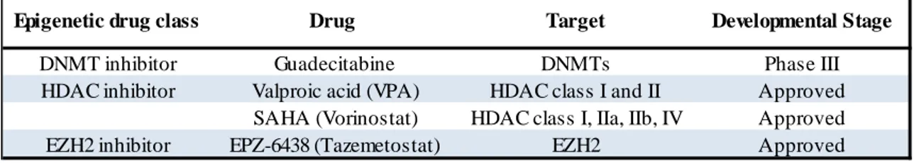
Table 1 – Investigated epigenetic drugs and targets.
Epigenetic drug class Drug Target Developmental Stage
DNMT inhibitor Guadecitabine DNMTs Phase III
HDAC inhibitor Valproic acid (VPA) HDAC class I and II Approved
SAHA (Vorinostat) HDAC class I, IIa, IIb, IV Approved
1.2. Malignant Mesothelioma biology
1.2.1. Risk factors and pathogenesis
Malignant mesothelioma is a neoplasm arising from mesothelial cells, cells of mesodermal origin, components of pleural and peritoneal cavities, of pericardium and vaginal tunic. The major interested site is the chest, with a percentage of MPM counting 70-80% of total cases (Delgermaa, 2011). Mesothelioma is a relatively low-frequency malignancy, causing the 4% of overall cancer mortality worldwide. However, it is highly aggressive (with a 5-year survival rate of about 5%) and highly resistant to therapies, resulting fatal within 24 months from the diagnosis (Fels Elliott, 2020; Gray, 2020). Its incidence began to increase from the second half of the XXth century, especially in industrialized countries, being closely related to the use of asbestos which is responsible for more than 80% of total cases of mesothelioma (Delgermaa, 2011; Micolucci, 2016); despite bans, the incidence is still growing. All types of asbestos have been associated with mesothelioma onset, having all being classified by the International Agency for Research on Cancer (IARC) as human carcinogens. Environmental exposure to either long or short fibers of erionite, a fibrous zeolite constituting volcanic rocks of rural areas, such as the Cappadocian region of Turkey, is likewise an important risk factor for mesothelioma (Baris, 1988). Other collectively recognized predisposing factors are high-dose ionising radiation (especially with the old radioactive contrast agent Thorotrast), simian virus 40 (SV40) infection, chest injuries, as well as genetic aberrations (Comin, 1997; Carbone, 2000; Micolucci, 2016). SV40, a DNA virus, appeared to be co-carcinogenic in humans after asbestos exposure, contributing to early-phase mesothelial cells transformation, impairing key cell-cycle regulators (Carbone, 2020). Nevertheless, the infection alone is not sufficient to cause human malignancy (Kroczynska, 2006). For the development of the disease, a long and variable latency period after cancerogenic fibers exposure (13-50 years or longer) is observed (Delgermaa, 2011). Since asbestos is responsible as proof of evidence of both benign and malignant cell transformation, ranging from asbestosis and pleural fibrosis to malignant tumors, studying lung responses toward it results pertinent to explore mesothelioma pathogenesis. Specific mechanisms induced by the inhalation of fibers are still poorly understood; anyway, they implicate direct mechanical injury, inflammation, oxidative stress, and DNA and chromosomal alterations. After the deposition in the airways, whose mechanism and site depend on the type of fiber, cytolytic and non-cytolytic injuries occur. The primary event is chronic inflammation, set up by alveolar macrophages. Long fibres, compared to the shortest, are difficultly phagocytized and hardly removed through the lymphatic system (Boulanger, 2014). The incomplete phagocytosis leads macrophages to produce reactive oxygen species (ROS) or reactive nitrogen species (RNS), promoting oxidative stress and interacting with cellular
components (Solbes, 2018). Moreover, protracted fibers persistence also causes the release of cytokines, such as interleukin-1β, transforming growth factor-β (TGF-β), and tumour necrosis factor-α (TNF-α) by alveolar macrophages, lung and pleural cells as well as high mobility group box 1 (HMGB1) protein by necrotic cells, leading to alterations of cellular signaling pathways and exacerbating the process (Carbone, 2012). Oxidative DNA damage, if not effectively resolved, is strongly mutagenic, triggering mutations, deletions, and genomic instability (Sage, 2018). In particular, TNF-α has been described to activate the nuclear factor-κB (NF-κB), a transcription factor regulating many genes involved in both cell proliferation and inflammatory pathways, such as c-myc, leading to the propagation of genetic aberrations and favouring malignant clones’ generation (Janssen, 1995; Carbone, 2012).
Mesothelioma can hardly be identified and distinguished from cancer metastasis (e.g., from lung, breast, ovarian, renal cell and colon carcinoma) and the diagnosis requires a multidisciplinary approach able to combine cytohistological aspects with clinico-radiological data. Indeed, the practical guidelines for the pathologic diagnosis of mesothelioma, formulated by the International Mesothelioma Interest Group, list a series of criteria to be considered, ranging from morphology to molecular markers, taking into account the context of the differential diagnosis (Husain, 2018). Overall, mesothelioma can be classified, from the histopathological point of view, into three main types: epithelioid, sarcomatoid and biphasic/mixed, between whom about 70-85% of all cases are represented by the epithelioid subtype, while only 10% are sarcomatoid. The most common epithelioid mesothelioma is characterized by clusters of polygonal cells with eosinophilic cytoplasm and a prominent nucleolus, in a context of fibrous stroma. Sarcomatoid type produces spindle-shape cells organized into bundles, with nuclear atypia and a various degree of mitosis, in an heterogenous stroma. The biphasic variant involves the combination of both previously described patterns. Interestingly, the histological subtypes strongly influence patients’ survival, thus defining a crucial prognostic factor (Alì, 2017). Indeed, the best outcome has been observed for the epithelioid variant, with a median overall survival (OS) of 20 months. Conversely, the sarcomatoid phenotype portends a particularly dismal prognosis, being the median OS of about 13 months. Finally, the biphasic subtype shows intermediate OS, depending on the prevalent histological component of the tumor (Yap, 2017). However, lack of subtype-specific markers hinders and delays the diagnosis. Clinical features involve non-specific signs and depend on the tumor site. The most common MPM generally exhibits debilitating symptoms, such as dyspnea, almost caused by pleural effusion, chest pain, fatigue, and weight loss. The early diagnosis is difficult but of pivotal importance, along with a proper surveillance programme of workers who, in the present or past, have been exposed to asbestos or other risk factors, in order to early recognise and control the pathology.
1.2.2. The genetic and epigenetic landscape of mesothelioma
A small proportion of cases of mesothelioma have been linked to germline mutations of the gene coding for the BRCA1-Associated Protein 1 (BAP1), leading to the rare “BAP1 cancer predisposition syndrome”, associated with MPM but also with uveal and cutaneous melanoma, clear-cell renal cell carcinoma, breast and other cancers (Carbone, 2013). Acquired somatic mutations of this gene have been also reported in almost 60% of patients with mesothelioma (Nasu, 2015). BAP1 is a nuclear deubiquitinating protein which contributes to double-strand DNA repair and gene expression control. Loss of BAP1 is one of the most frequent genetic features observed in mesothelioma, promoting genomic instability and altering gene transcriptional regulation (Sage, 2018). Other genes frequently mutated in mesothelioma are the neurofibromatosis type 2 gene (NF2), TP53, CDKN2A, and CDKN2B (Sage, 2018). The NF2 is a TSG and a membrane protein associated to cytoskeleton, which inhibit different oncogenic pathways, such as mTOR and Hippo; it was found altered in about 40% of mesotheliomas and loss-of-function mutations led to uncontrolled cell growth (Thurneysen, 2009). Some mutations, such as PIK3CA, STK11, and TP53, have been associated to the time to disease progression of MPM patients (Welch, 2017). Moreover, analysis across The Cancer Genome Atlas (TCGA) revealed that mesothelioma has a DNA damage repair footprint, significantly correlated to progression-free survival and OS (Knijnenburg, 2018). Besides single-gene mutations, different mutational signatures have been identified in mesothelioma. Indeed, Bueno et al. studied 216 human MPM and, interestingly, this signature was not significantly different between patients exposed or not exposed to asbestos, highlighting overall alterations in histone methylation, RNA elicase, Hippo, mTOR and TP53 signaling pathways (Bueno, 2016).
Although genetic aberrations of mesothelioma have been relatively well characterized, the epigenetic landscape of the disease still needs deeper investigations. Based on the analysis of asbestos-exposed MPM cell lines, the overexpression of DNMTs, such as DNMT1, DNMT3A, and DNMT3B have been identified (McLoughlin, 2017). Also, a significant correlation between asbestos exposure and DNA methylation at the gene loci of the metal-binding proteins MT1A and MT2A, involved in the regulation of transcription, has been described in MPM (Shivapurkar, 2004). Similarly, different epigenetic modifier enzymes were found overexpressed in MPM, for example the PRC2 components EZH2 and SUZ12, and the lysine demethylase KDM6A (McLoughlin, 2017; Cregan, 2017). The latter is a JmjC domain-containing protein that catalyzes the demethylation of di-/tri-methylated H3K27, playing an important role in inflammation and tumor progression (Liang, 2019). Besides, EZH2 activation appears to be a key pathogenetic mechanism involved in mesothelioma onset
dependent to BAP1, whose loss can induce either EZH2 over-expression or hypermethylation of PRC2 target genes (Welch, 2017). This is attributable to the interaction between PCR2 and BAP1, which opposes the suppressor function of the complex, acting as polycomb deubiquitinase which remove H2AK119ub mark (Fig. 5); the loss of the tumor suppressor BAP1, then, results in H3K27me3 accumulation (Yamagishi, 2017). EZH2 over-expression has been also demonstrated to significantly correlate with a poor survival of mesothelioma patients (McLoughlin, 2017). In addition, concomitant BAP1 loss and high EZH2 expression is of diagnostic utility, improving accurate malignant mesothelioma differentiation from benign proliferations (Shinozaki‐Ushiku, 2017). The BAP1 status in mesothelioma has also become of therapeutic utility, being its loss able to sensitize mesothelioma cells to EZH2 pharmacologic inhibition, opening the path to novel epigenetic-based approach for BAP1-mutant tumors (LaFave, 2015). Indeed, a phase II clinical trial with the EZH2 inhibitor EPZ-6438 is ongoing for the treatment of adult subjects with BAP1-deficient relapsed/refractory malignant mesothelioma (ClinicalTrial.gov identifier: NCT02860286). Sacco et
al. also showed the BAP1 regulated HDAC enzymes activity, altering sensitivity of mesothelioma
cancer cells to HDAC inhibitors (Sacco, 2015). Multiple studies demonstrated the effectiveness of HDACi-based epigenetic combinations in malignant mesothelioma. Anticancer effects of the combination of the HDACi SAHA or VPA plus the DNMTi DAC, compared to single-drug treatments, have been investigated in human MPM cells and in the corresponding murine model. Indeed, Leclercq et al. showed that the combination of DAC synergised with VPA/SAHA to induce cell death and CTA expression in vitro; also, DAC plus VPA led to a remarkable inhibition of tumor growth and promoted lymphocyte infiltration within the tumor in vivo (Leclercq, 2011). CTA up-regulation was also detected in MPM cell lines treated with guadecitabine, both at mRNA and at protein levels, correlated with the hypomethylation of MAGE-A1 and NY-ESO-1 CTA promoters; moreover, guadecitabine up-regulated HLA class I antigens and the immunostimulatory protein ICAM-1, resulting in an improved recognition of mesothelioma cells by CTLs, and highlighting DHAs utility to potentiate immunotherapeutic-based strategies (Coral, 2013). Finally, a relatively recent study of Bansaid et al. showed the immune check-point molecule PD-L1 was induced by the combination of DAC and different HDACi in MPM cell lines, suggesting that the epigenetic regulation of CTA and PD-L1 expression could be exploited using these epigenetic drugs combinations with anti-PD-L1 mAbs in malignant mesothelioma therapy (Bansaid, 2018).
1.2.3. Epithelial-to-Mesenchymal Transition in mesothelioma
As already hinted, EMT is a crucial plastic event through which epithelial cells gain a mesenchymal phenotype, remodulating the expression of critical cell-cell adhesion molecules, cell stiffness and polarity, acquiring ECM degradation capability and invasive potential. This multi-step process can occur either in physiological conditions, for example during development and organogenesis, or in wound healing, tumor development, and metastasis formation. During cancer progression, tumor cells might lose epithelial markers and acquire fibroblast-like morphology and characteristics, with the consequent leak of intercellular adhesion, facilitating migration (Cannito, 2010). Normally, the process ends with a mesenchymal-to-epithelial transition (MET) event, thanks to which metastatic cells from the primary tumor, once arrived at the distant homing site, become metastatic lesions leading to a secondary tumor growth (Thiery, 2009).
Common EMT markers include members of adherens and tight junctions, cytokeratins, cytoskeletal protein, and EMT-activating transcription factors. The most important epithelial marker in the context of MPM is the member of adherens junctions E-cadherin (encoded by the CDH1 gene), a calcium-dependent cell-cell adhesion protein, whose immunohistochemical expression has been correlated with MPM survival (Fassina, 2012). Different studies confirmed that the hypermethylation of CDH1 gene promoter occurred with a high incidence in MPM patients’ samples, with the consequent loss of its tumor suppressor role (Fischer, 2006). Interestingly, Fassina et al. showed that mesothelioma phenotypic subtypes corresponded to specific profiles of expression of EMT markers, investigated both by immunostaining and quantitative Real-Time PCR analyses. Indeed, epithelioid mesotheliomas were associated with the expression of epithelial marker proteins, such as E-cadherin, cytokeratin 5/6, and β-catenin; the sarcomatoid variant owned mesenchymal markers as vimentin, N-cadherin, S100A4, MMP-2 and -9, α-SMA, Zeb-1 and -MMP-2; finally, biphasic histotypes expressed β-catenin, N-cadherin, MMP-MMP-2 and -9, α-SMA, Zeb-2, but weak expression of vimentin, S100A4, and Zeb-1 (Fassina, 2012). The EMT-related features reflected the trans-differentiation steps of the tumor, resulting helpful in malignant mesothelioma histological subtyping (Fassina, 2012).
Given the evidence that a strong and continuous exposure to asbestos was found to induce chronic inflammation and that the HMGB1-mediated inflammation was associated with increased levels of EMT signaling pathways, the study of EMT in the malignant mesothelioma has become fundamental (Qi, 2013). Different factors are able to induce EMT in mesothelioma, among whom the aberrant expression of hepatocyte growth factor (HGF), the fibroblast growth factor-2 (FGF-2), TNF-α, periostin, mesothelin, and TGF-β (Qi, 2013; Chen, 2014; Farrell, 2014; Moustakas, 2012; Schramm, 2010; He,