Abstract:
This study assessed push-out strength, cement layer thickness, and interfacial nanoleakage of luted fiber posts fabricated with computer-aided design/computer-assisted manufacture (CAD/CAM) technology after use of 1 of 3 scanning techniques, namely, direct scanning of the post space (DS), scan-ning of a polyether impression of the post space (IS), and scanning of a plaster model of the post space (MS). Thirty premolars were randomly assigned to three groups corresponding to the scanning tech-nique. Posts were computer-designed and milled from experimental fiber-reinforced composite blocks. The mean (±SD) values for push-out strength and cement thickness were 17.1 ± 7.7 MPa and 162 ± 24 μm, respectively, for DS, 10.7 ± 4.6 MPa and 187 ± 50 μm for IS, and 12.0 ± 7.2 MPa and 258 ± 78 μm for MS specimens. Median (interquartile range) interfacial nanoleakage scores were 3 (2-4) for DS, 2.5 (2-4) for IS, and 3 (2-4) for MS. Post retention was better for fiber posts fabricated by DS technique than for those fabricated by IS and MS. Cement thickness did not differ between DS and IS specimens, but the cement layer was significantly thicker in the MS group than in the other two groups. Scanning technique did not affect sealing ability, as the three groups had compa-rable nanoleakage values.Keywords: CAD/CAM; scanning techniques; fiber post; post retention; nanoleakage; cement thickness.
Introduction
Functional and aesthetic rehabilitation of endodontically treated teeth with substantial loss of tooth structure often requires a post-retained fixed prosthetic restora-tion (1,2). Fiber-reinforced composite posts (i.e., fiber posts) are widely used because they have good aesthetics and elasticity close to that of dentin, which allows for uniform stress distribution within the root and decreases the risk of non-restorable root fracture (3,4). However, debonding of the post was the most frequent failure mode observed in fiber post-retained restorations (5).
The shape of the root canal and cement thickness around the post are important factors in the retention of fiber posts (6-8). A thicker cement layer around the post was reported to decrease bond strength between a fiber post and root dentin (8-12). Thus, insertion of a circular prefabricated fiber post into an oval-shaped root canal may be ill-advised, as using an undersized fiber post leads to excessive resin cement thickness (8,13). Bonding to apical thirds in such cases is even more challenging (13). Good post fitting and low cement thickness in oval-shaped canals can be obtained with a fine-grit oval tip combined with oval posts, particularly in the apical third (8). However, fracture resistance was not increased when oval posts were luted in oval post spaces (14). Custom fabrication of fiber posts has there-fore been proposed, as it may improve post adaptation to irregularly shaped, flared, or large-diameter root canals. A number of techniques have been used to custom-fabricate Journal of Oral Science, Vol. 60, No. 2, 262-268, 2018
Original
Effects of scanning technique on in vitro performance of
CAD/CAM-fabricated fiber posts
Nino Tsintsadze
1), Jelena Juloski
1,2), Michele Carrabba
1), Cecilia Goracci
1),
Alessandro Vichi
1), Simone Grandini
1), and Marco Ferrari
1)1)
Department of Medical Biotechnologies, University of Siena, Siena, Italy
2)Clinic for Paediatric and Preventive Dentistry, University of Belgrade, Belgrade, Serbia
(Received July 8, 2017; Accepted Sepember 8, 2017)
Correspondence to Dr. Jelena Juloski, Department of Medical Biotechnologies, University of Siena, Policlinico Le Scotte, Viale Bracci, Siena 53100, Italy
Fax: +39-577-233117 E-mail: [email protected] Color figures can be viewed in the online issue at J-STAGE. doi.org/10.2334/josnusd.17-0254
fiber posts, such as chair-side fabrication of posts with polyethylene fibers and composite resin (15), combined use of a fiber post and dual-curing resin cement to create an “anatomical post” (16), lateral compaction of several small fiber posts (17), and use of a fiber post customized to canal anatomy with a diamond-coated bur (18,19). However, these techniques are difficult to perform and time consuming.
Recent reports have suggested use of computer-aided design/computer-assisted manufacture (CAD/CAM)-fabricated post and core restorations, and several studies have investigated CAD/CAM-fabricated zirconia post and core techniques (20-24). However, it is nearly impos-sible to retrieve a zirconia post when fracture occurs, which leads to irreversible tooth failure (21). Therefore, Lee et al. suggested the combination of prefabricated fiber post and zirconia core, to avoid catastrophic failure caused by zirconia posts and achieve satisfactory aesthetic results with a zirconia core (23,24). Furthermore, fiber-reinforced composite blocks were used to CAD/ CAM-fabricate fiber posts for irregularly shaped or large root canals (25,26). Liu et al. (25) successfully restored a front tooth with a CAD/CAM-fabricated fiber post and core and used radiography to examine the results. Chen et al. (26) validated this technique by inserting a 1-piece glass fiber dowel-and-core fabricated with CAD/CAM technology in two patients. Additionally, two recent studies by Chen et al. tested von Misses stress level in dentin of extracted maxillary canines restored with CAD/ CAM glass fiber posts and lithium disilicate crowns (27,28). However, few in vitro or in vivo studies have investigated the performance of CAD/CAM-fabricated fiber posts inserted into oval-shaped root canals.
Digital images of the prepared tooth, which are required for CAD/CAM fabrication of a restoration, can be obtained by direct intraoral scanning, by scan-ning impressions, or by scanscan-ning stone models (25-29). Impressions and stone replicas can be digitized with high reliability (30). However, accuracy and precision are better for intraoral digital impression systems than for indirect digitization of polyether impressions or stone casts (31,32). Patient movement, the confined intraoral space, intraoral humidity, and saliva flow are patient-related factors that strongly influence scan quality and cause substantial variability in intraoral scans (32). However, no study has determined which of the three available procedures is best for scanning the post space and CAD/CAM fabrication of fiber posts.
The present study assessed the in vitro performance of CAD/CAM-fabricated fiber posts inserted into oval-shaped root canals. The posts were fabricated by using
three different procedures for digital data acquisition. The three null hypotheses tested were that there would be no statistically significant difference among these data acquisition techniques in 1) the push-out bond strength of the post, 2) the cement thickness surrounding the post, and 3) interfacial nanoleakage.
Materials and Methods
Specimen preparation
Thirty human single-rooted premolars extracted for orthodontic reasons were collected from patients after informed consent was obtained. The teeth were decoro-nated and endodontically treated with Reciproc rotary instruments (VDW, Munich, Germany) and warm vertical gutta-percha technique, namely, Beefill 2 in 1 (VDW) in combination with the resin root canal sealer AH Plus (Dentsply, Konstnaz, Germany). All roots were sealed with Fuji VII glass-ionomer cement (GC, Tokyo, Japan) and stored in water at 37°C. After 1 week, oval-shaped post space preparations 10 mm in depth were made by using size #6 Largo burs. The roots were randomly assigned to three groups (n = 10), in accordance with the digital data acquisition procedure used for CAD/CAM fabrication of the fiber posts, as detailed below.
Group 1: Direct scanning of the post space (DS).
The post space was covered with scan spray (VITA, Bad Säckingen, Germany) and scanned with an inEos scanner (Sirona, Bensheim, Germany). The digital 3-D model of the post was designed with inLab 3.88 software (Sirona), and the fiber post was milled from experimental fiber-reinforced composite blocks (RTD, St. Egrève, France) by using an inLab MC XL CAD/CAM milling unit (Sirona). Vinyl polyether silicone material (Black fit checker, GC) was used to control the fit of all fabricated fiber posts.
Group 2: Scanning an impression of the post space (impression scanning, IS). An impression of the post
space was made with polyether impression material (Parmadyne Garant 2:1, 3M ESPE, Neuss, Germany) by using stainless-steel impression trays (Asa Dental, Bozzano, Italy). The impression was covered with scan spray (VITA). The procedures for scanning the impres-sion and fabrication of the fiber post were identical to those used for Group 1. Vinyl polyether silicone material (Black fit checker, GC) was used to control the fit of all fabricated fiber posts.
Group 3: Scanning a stone model of the post space (model scanning, MS). An impression was made by using
the procedure described for Group 2. Then, a stone model was poured with type 4 stone. Scanning of the model and fabrication of the fiber post followed the protocol
described for Group 1. Vinyl polyether silicone material (Black fit checker, GC) was used to control the fit of all fabricated fiber posts.
In all groups, posts were cemented with Gradia Core dual-cure resin cement in combination with Gradia Core self-etching bond (GC). All materials were handled in accordance with the manufacturers’ instructions.
Thin-slice push-out test and cement thickness measurement
Twenty-four hours after cementation, 6 specimens per group were randomly selected for the thin-slice push-out test. All roots were sectioned with a cutting machine (Isomet; Buehler, Lake Bluff, NY, USA) with a diamond disc at slow speed (250 rpm) under water cooling. Six 1-mm-thick sections were obtained per root: two sections each from the coronal, middle, and apical thirds. Each section was marked with a permanent marker on its apical surface. Slice thickness was individually measured by using a digital caliper (Orteam srl, Milan, Italy) with an accuracy of 0.01 mm. Specimens were mounted on the jig, with the apical surface oriented upward to the punch-out rod. The thin-slice push-punch-out test was conducted by using a universal testing machine (Triax Digital 50, Controls, Milan, Italy) at a cross-head speed of 0.5 mm/ min until failure occurred. The load was applied at the center of the post, in the apicocoronal direction, to push each post toward the larger post space diameter. Load at failure was recorded in Newtons, and bond strength was calculated in MPa. Digital images of all specimens were taken with a digital microscope (Nikon Shuttle Pix, Nikon, Tokyo, Japan), and cement thickness around the post for each group was measured with Digimizer soft-ware (MedCalc, Mariakerke, Belgium).
Interfacial nanoleakage test
The remaining 4 roots per group were subjected to the interfacial nanoleakage test. Six 1-mm thin slices were obtained per root and covered with red nail polish. Only the dentin-cement-post interfaces were left exposed, and the specimens were immersed in silver nitrate solution (1 mL silver nitrate in 4 mL water) with a 0.22-nm filter (Carrigtwohill, County Cork, Ireland). All specimens were left in darkness for 24 h, then rinsed with water for 30 min and immersed in photo-developing solution (3 mL in 10 mL water, Kodak, Rochester, NY, USA) for 8 h. The slices were then rinsed with water for 30 min and polished. Polishing was performed by using 600- to 4,000-grit SiC papers in ascending order, until the root sections achieved a mirror-like appearance. To determine nanoleakage score, all specimens were observed under a
light microscope (Nikon SMZ645). Digital images were obtained with a digital microscope (Nikon Shuttle Pix). Nanoleakage was calculated as the percentage of black silver nitrate deposition along the post-cement-dentin interface, in accordance with the nanoleakage scoring system proposed by Saboia et al. (33) (0: no nanoleakage; 1: <25% of interface showing nanoleakage; 2: 25% to 50% of interface showing nanoleakage; 3: 50% to 75% of interface showing nanoleakage; 4: ˃75% of interface showing nanoleakage).
Statistical analysis
Push-out bond strength
Push-out strength data were normally distributed and thus assessed with two-way analysis of variance (ANOVA), with push-out strength as the dependent variable and scanning technique and root level as factors. The Tukey test for post-hoc comparisons was used after assessment of differences in push-out strength among the scanning techniques.
Cement thickness
Two-way ANOVA, with cement thickness as the depen-dent variable and scanning technique and root level as factors, was not possible because of the non-normal data distribution. Thus, the effects of the two factors on cement thickness were analyzed separately. Differences in cement thickness among scanning techniques were assessed with the Kruskal-Wallis ANOVA, followed by the Dunn multiple range test for post-hoc comparisons, as the data were not normally distributed. One-way ANOVA and the Tukey test for post-hoc comparisons were used to assess differences among root levels within DS group, as the data were normally distributed. For assessment of differences among root levels within the IS and MS groups, Kruskal-Wallis ANOVA and the Dunn multiple range test for post-hoc comparisons were used, as the data were not normally distributed.
Interfacial nanoleakage
Interfacial nanoleakage scores were analyzed with Kruskal-Wallis ANOVA and the Dunn multiple range test for post-hoc comparisons.
The level of significance was set at P < 0.05 in all analyses, and calculations were done with Sigma Plot 11 statistical software (Systat Software, Inc., San Jose, CA, USA).
Results
The mean (±SD) values for push-out bond strength were 17.1 ± 7.7 MPa for DS, 10.7 ± 4.6 MPa for IS, and 12.0
± 7.2 MPa for MS specimens. The mean (±SD) values for cement thickness were 162 ± 24 μm for DS, 187 ± 50 μm for IS, and 258 ± 78 μm for MS specimens. The median (interquartile range) values for nanoleakage score were 3 (2-4) for DS, 2.5 (2-4) for IS, and 3 (2-4) for MS specimens. Scanning technique was a significant factor in push-out bond strength, whereas post level was not (Two-way ANOVA). Posts fabricated after DS had the strongest retention (P < 0.001), and IS and MS group fiber posts showed comparable results (Table 1).
Furthermore, cement thickness significantly differed
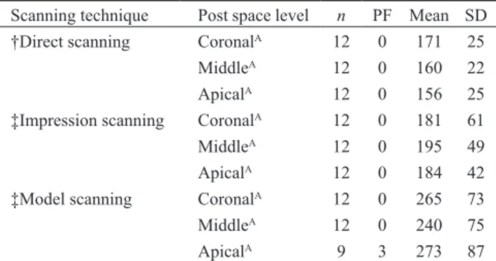
in relation to scanning technique. The cement layer was significantly thicker in the MS group than in the DS and IS groups (P < 0.001), which had comparable cement layer thicknesses (Table 2). Cement thickness did not significantly differ in relation to root level in any group (Table 3).
There was no significant difference in interfacial nanoleakage among the three groups (Table 4). Figure 1 shows representative digital images of nanoleakage in each group.
Discussion
Null hypothesis 1 was rejected, as push-out bond strength significantly differed among groups. Specifically, fiber posts fabricated after DS technique achieved higher retention than did those fabricated after the IS and MS techniques. Push-out bond strength did not significantly differ between the two indirect data acquisition proce-dures, i.e., the IS and MS proceproce-dures, in terms of post retention in oval-shaped root canals.
To our knowledge, no previous study has investigated direct scanning of the post space. Several studies used different indirect techniques for digital data acquisition of the post space (23-28,34). Lee et al. scanned stone models of the post space to custom-fabricate the fiber post and core restoration (23,24). In a study by Liu et al. (25), a wax pattern prepared from a stone model of the post space was digitized to design a 3-D model of the fiber post and core. Chen et al. (26) scanned vinyl-polysiloxane impressions taken from a die stone model to CAD/CAM-fabricate fiber posts. In a study by Bittner et al. (34), acrylic resin patterns were scanned to CAD/ CAM-fabricate a zirconia post and core. The scanning methods used were not specified in recent studies of CAD/CAM glass fiber posts by Chen et al. (27,28). In addition, no studies have examined the usefulness of procedures for digital acquisition of the post space.
When focusing on digitization of full arches and a
Table 1 Push-out bond strength (MPa)
Scanning technique* n PF Mean SD Post space level n PF Mean SD
Direct scanningA 36 0 17.1 7.7 Coronal 12 0 18.3 7.5
Middle 12 0 17.1 7.5
Apical 12 0 15.9 8.6
Impression scanningB 36 0 10.7 4.6 Coronal 12 0 10.5 4.9
Middle 12 0 10.4 3.6
Apical 0 0 11.1 5.4
Model scanningB 33 3 12.0 7.2 Coronal 12 0 11.2 5.7
Middle 12 0 11.4 6.8
Apical 9 3 13.3 8.8
n: number of specimens per group; PF: number of premature failures; SD: standard deviation. Asterisk (*) indicates a
significant factor in push-out bond strength. Different uppercase letters indicate statistically significant differences (Two-way analysis of variance, Tukey test, P < 0.001).
Table 2 Thickness of cement layer surrounding posts (µm)
Scanning technique n PF Mean SD Median 25% 75%
Direct scanningA 36 0 162 25 163 148 180
Impression scanningA 36 0 187 50 173 147 213
Model scanningB 33 3 259 78 236 187 314
n: number of specimens per group; PF: number of premature failures;
SD: standard deviation; 25%: 25th percentile (lower quartile); 75%: 75th percentile (upper quartile). Different uppercase letters indicate statistically significant differences (Kruskal-Wallis analysis of variance, Dunn multiple range test).
Table 3 Thickness of cement layer surrounding posts (µm),
by root canal level within each group
Scanning technique Post space level n PF Mean SD
†Direct scanning CoronalA 12 0 171 25
MiddleA 12 0 160 22
ApicalA 12 0 156 25
‡Impression scanning CoronalA 12 0 181 61
MiddleA 12 0 195 49
ApicalA 12 0 184 42
‡Model scanning CoronalA 12 0 265 73
MiddleA 12 0 240 75
ApicalA 9 3 273 87
n: number of specimens per group; PF: number of premature failures;
SD: standard deviation. Different uppercase letters indicate statistically significant differences within each group separately (†one-way analysis of variance, ‡Kruskal-Wallis analysis of variance).
single-unit crown or fixed dental prosthesis preparation, information on the accuracy and effectiveness of direct and indirect digital scanning methods varies. Direct digital scanning is regarded as more rational than scan-ning polyether impressions (31). Additionally, direct acquisition systems are believed to be less invasive, quicker, and more precise than indirect methods (35). Moreover, accuracy was significantly higher for direct digitalization than for indirect digitalization of impres-sions and gypsum casts (36). Syrek et al. (37) reported that marginal fit of single crowns was significantly better after direct data capturing than after digitization. Although these studies did not investigate procedures for scanning of post spaces, the present results are consistent with past findings regarding the accuracy of direct and indirect digitalization methods.
Previous studies suggest that the technical features of extraoral scanning result in better accuracy, as compared with intraoral digitization (32,38). Patient-related factors such as patient movement, the confined intraoral space, intraoral humidity, and saliva flow could cause deviations in intraoral scans (32). However, patient-related factors do not apply to the present direct scanning technique, as the specimens were not scanned in an intraoral environ-ment. However, even in clinical practice, when directly scanning post spaces, unlike scanning of crown/fixed dental prosthesis preparations, patient-related factors such as intraoral humidity and saliva flow can be
elimi-nated by using tooth-isolating systems.
Null hypothesis 2 was also rejected. The MS method was least accurate in measuring cement thickness around posts. The cement layer around posts was significantly thicker in the MS group than in the IS and DS groups, which had comparable results. This finding is consistent with the results of Güth et al. (36), who, although they did not examine posts, reported that results were more accurate for digitization of polyether impressions than for digitization of gypsum casts. Nevertheless, the literature is equivocal on this point. One study found no significant difference in precision between scanning of impressions and scanning of stone replicas of master dies prepared for crowns (30).
Null hypothesis 3 was accepted: interfacial nanole-akage did not significantly differ among the present groups. Teeth from all three groups showed considerable nanoleakage (interquartile range 2-4; Table 4, Fig. 1); thus, sealing ability in root canal dentin is a concern. In addition, this finding suggests that bond strength and cement thickness are not correlated with nanoleakage, as push-out bond strength and cement thickness signifi-cantly differed among groups. Previous studies showed no significant difference in interfacial nanoleakage in relation to the luting material used for bonding fiber posts to intraradicular dentin, despite differences in push-out strength (39). In addition, a previous study found no significant association between cement thickness and interfacial nanoleakage (40).
Regarding premature failure, debonding before testing or catastrophic fracture occurred during specimen preparation in the IS and MS groups (Tables 1-4). All premature failures were in the apical portion of the root, which indicates that post adhesion is most challenging in the deepest part of the root canal (13). Prematurely failed specimens could not be tested for push-out strength or nanoleakage expression and were thus excluded from the analysis. Reporting, but excluding, premature failures from statistical analysis is consistent with previously
Table 4 Descriptive statistics for nanoleakage scores
Scanning technique n PF Median 25% 75%
Direct scanningA 24 0 3.0 2.0 4.0
Impression scanningA 22 2 2.5 2.0 4.0
Model scanningA 19 5 3.0 2.0 4.0
n: number of specimens per group; PF: number of premature failures;
25%: 25th percentile (lower quartile); 75%: 75th percentile (upper quartile). Different uppercase letters indicate statistically significant differences (Kruskal-Wallis analysis of variance, Dunn multiple range test).
A B C
reported study designs (41,42).
The use of different experimental models, scanners, and accuracy measurement procedures might explain discrepancies regarding the efficacy of different digital data acquisition techniques. In this study, push-out bond strength, cement layer thickness, and interfacial nanoleakage were used to assess the accuracy of digital data acquisition techniques utilized for CAD/CAM fabrication of fiber-reinforced composite posts. To assess the accuracy of scanning procedures, Bittner et al. (34) compared marginal gap distance between the tooth and acrylic resin patterns and between the tooth and a definite zirconia post and core restoration. Other studies of the accuracy of different digitization procedures compared deviations between test and reference datasets by analyzing distances between Euclidean points (33) or on repeated measurements where the model rendered from the first scan serves as the control surface for consecu-tively acquired models (32). Currently, new reference scanners are used to test the precision and trueness of digital impressions (43). Therefore, additional laboratory and clinical studies are needed in order to test the effec-tiveness of the direct and indirect digital data acquisition techniques used to scan irregularly shaped root canals.
This study has limitations. It was not possible in an in
vitro study to account for patient-related factors. In
addi-tion, operator skill, experience, and knowledge might affect clinical results. However, CAD/CAM-fabricated fiber posts are attractive in clinical practice because they combine the advantages of traditional custom-made posts and prefabricated fiber posts. Such posts may better fit the post space and have a modulus of elasticity close to that of dentin—an important advantage of fiber-reinforced composite material. Nevertheless, CAD/CAM fabrica-tion of fiber posts requires a longer appointment, to scan the post space, the additional clinical step of cementation of the post, and a chair-side CAD/CAM device in the dental office. Therefore, fabrication of fiber posts can be regarded as an additional option for practitioners already working with a CAD/CAM chair-side system and for dental laboratories with a digital workflow.
Our results indicate that performance (in terms of post retention) is better for fiber posts fabricated after DS than for those fabricated after IS and MS. Cement layer thickness did not differ between DS and IS but was significantly higher in the MS group than in the other two groups. Scanning technique was not associated with sealing ability, as all the tested groups showed compa-rable nanoleakage.
Conflict of interest
All the authors deny any conflict of interest.
References
1. Ferrari M, Cagidiaco MC, Grandini S, De Sanctis M, Goracci C (2007) Post placement affects survival of endodontically treated premolars. J Dent Res 86, 729-734.
2. Zadik Y, Sandler V, Bechor R, Salehrabi R (2008) Analysis of factors related to extraction of endodontically treated teeth. Oral Surg Oral Med Oral Pathol Oral Radiol Endod 106, e31-35.
3. Martínez-Insua A, da Silva L, Rilo B, Santana U (1998) Comparison of the fracture resistances of pulpless teeth restored with a cast post and core or carbon-fiber post with a composite core. J Prosthet Dent 80, 527-532.
4. Torres-Sánchez C, Montoya-Salazar V, Córdoba P, Vélez C, Guzmán-Duran A, Gutierrez-Pérez JL et al. (2013) Fracture resistance of endodontically treated teeth restored with glass fiber reinforced posts and cast gold post and cores cemented with three cements. J Prosthet Dent 110, 127-133.
5. Cagidiaco MC, Radovic I, Simonetti M, Tay F, Ferrari M (2007) Clinical performance of fiber post restorations in endodontically treated teeth: 2-year results. Int J Prosthodont 20, 293-298.
6. Ferrari M, Vichi A, Mannocci F, Mason PN (2000) Retro-spective study of clinical performance of fiber posts. Am J Dent 13, 9B-13B.
7. Peters OA (2004) Current challenges and concepts in the preparation of root canal systems: a review. J Endod 30, 559-567.
8. Coniglio I, Garcia-Godoy F, Magni E, Carvalho CA, Ferrari M (2009) Resin cement thickness in oval-shaped canals: oval vs. circular fiber posts in combination with different tips/ drills for post space preparation. Am J Dent 22, 290-294. 9. Egilmez F, Ergun G, Cekic-Nagas I, Vallittu PK, Lassila LV
(2013) Influence of cement thickness on the bond strength of tooth-colored posts to root dentin after thermal cycling. Acta Odontol Scand 71, 175-182.
10. Mirmohammadi H, Gerges E, Salameh Z, Wesselink PR (2013) Effect of post diameter and cement thickness on bond strength of fiber posts. Quintessence Int 44, 801-810. 11. Özcan E, Çetin AR, Tunçdemir AR, Ülker M (2013) The
effect of luting cement thicknesses on the push-out bond strength of the fiber posts. Acta Odontol Scand 71, 703-709. 12. Gomes GM, Rezende EC, Gomes OM, Gomes JC, Loguercio
AD, Reis A (2014) Influence of the resin cement thickness on bond strength and gap formation of fiber posts bonded to root dentin. J Adhes Dent 16, 71-78.
13. Dietschi D, Duc O, Krejci I, Sadan A (2008) Biomechanical considerations for the restoration of endodontically treated teeth: a systematic review of the literature, Part II (Evaluation of fatigue behavior, interfaces, and in vivo studies). Quintes-sence Int 39, 117-129.
14. Krastl G, Lorch H, Zitzmann NU, Addison O, Dietrich T, Weiger R (2014) Do oval posts improve fracture resistance of
teeth with oval root canals? Dent Traumatol 30, 232-235. 15. Singh A, Logani A, Shah N (2012) An ex vivo comparative
study on the retention of custom and prefabricated posts. J Conserv Dent 15, 183-186.
16. Grandini S, Sapio S, Simonetti M (2003) Use of anatomic post and core for reconstructing an endodontically treated tooth: a case report. J Adhes Dent 5, 243-247.
17. Porciani PF, Coniglio I, Magni E, Grandini S (2008) Fiber post fitting to canal anatomy: a review of the morphology and shape of root canal system. Int Dent SA 10, 52-58.
18. Grande NM, Butti A, Plotino G, Somma F (2006) Adapting fiber-reinforced composite root canal posts for use in noncir-cular-shaped canals. Pract Proced Aesthet Dent 18, 593-599. 19. Plotino G, Grande NM, Pameijer CH, Somma F (2008)
Influence of surface remodeling using burs on the macro and micro surface morphology of anatomically formed fiber posts. Int Endod J 41, 345-355.
20. Awad MA, Marghalani TY (2007) Fabrication of a custom-made ceramic post and core using CAD-CAM technology. J Prosthet Dent 98, 161-162.
21. Ozkurt Z, Işeri U, Kazazoğlu E (2010) Zirconia ceramic post systems: a literature review and a case report. Dent Mater J 29, 233-245.
22. Marghalani TY, Hamed MT, Awad MA, Naguib GH, Elragi AF (2012) Three-dimensional finite element analysis of custom-made ceramic dowel made using CAD/CAM tech-nology. J Prosthodont 21, 440-450.
23. Lee JH, Sohn DS, Lee CH (2014) Fabricating a fiber-rein-forced post and zirconia core with CAD/CAM technology. J Prosthet Dent 112, 683-685.
24. Lee JH (2014) Accelerated techniques for a post and core and a crown restoration with intraoral digital scanners and CAD/ CAM and rapid prototyping. J Prosthet Dent 112, 1024-1029. 25. Liu P, Deng XL, Wang XZ (2010) Use of a CAD/CAM fabri-cated glass fiber post and core to restore fractured anterior teeth: a clinical report. J Prosthet Dent 103, 330-333. 26. Chen Z, Li Y, Deng X, Wang X (2014) A novel
computer-aided method to fabricate a custom one-piece glass fiber dowel-and-core based on digitized impression and crown preparation data. J Prosthodont 23, 276-283.
27. Chen A, Feng X, Zhang Y, Liu R, Shao L (2015) Finite element analysis to study the effects of using CAD/CAM glass-fiber post system in a severely damaged anterior tooth. Biomed Mater Eng 26, S519-525.
28. Chen A, Feng X, Zhang Y, Liu R, Shao L (2015) Finite element analysis of stress distribution in four different endodontic post systems in a model canine. Biomed Mater Eng 26, S629-635.
29. Boldt F, Weinzierl C, Hertrich K, Hirschfelder U (2009)
Comparison of the spatial landmark scatter of various 3D digitalization methods. J Orofac Orthop 70, 247-263. 30. Persson AS, Oden A, Andersson M, Sandborgh-Englund G
(2009) Digitization of simulated clinical dental impressions: virtual three-dimensional analysis of exactness. Dent Mater 25, 929-936.
31. Kurbad A (2011) Impression-free production techniques. Int J Comput Dent 14, 59-66.
32. Flügge TV, Schlager S, Nelson K, Nahles S, Metzger MC (2013) Precision of intraoral digital dental impressions with iTero and extraoral digitization with the iTero and a model scanner. Am J Orthod Dentofacial Orthop 144, 471-478. 33. Saboia VP, Nato F, Mazzoni A, Orsini G, Putignano A,
Giannini M et al. (2008) Adhesion of two-step etch-and-rinse adhesive on collagen-depleted dentin. J Adhes Dent 10, 419-422.
34. Bittner N, Hill T, Randi A (2010) Evaluation of a one-piece milled zirconia post and core with different post-and-core systems: an in vitro study. J Prosthet Dent 103, 369-379. 35. Galhano GÁ, Pellizzer EP, Mazaro JV (2012) Optical
impres-sion systems for CAD-CAM restorations. J Craniofac Surg 23, e575-579.
36. Güth JF, Keul C, Stimmelmayr M, Beuer F, Edelhoff D (2013) Accuracy of digital models obtained by direct and indirect data capturing. Clin Oral Investig 17, 1201-1208. 37. Syrek A, Reich G, Ranftl D, Klein C, Cerny B, Brodesser J
(2010) Clinical evaluation of all-ceramic crowns fabricated from intraoral digital impressions based on the principle of active wavefront sampling. J Dent 38, 553-559.
38. Luthardt RG, Kühmstedt P, Walter MH (2003) A new method for the computer-aided evaluation of three-dimensional changes in gypsum materials. Dent Mater 19, 19-24.
39. Marchesi G, Mazzoni A, Turco G, Cadenaro M, Ferrari M, Di Lenarda R et al. (2013) Aging affects the adhesive interface of posts luted with self-adhesive cements: a 1-year study. J Adhes Dent 15, 173-180.
40. Cantoro A, Goracci C, Vichi A, Mazzoni A, Fadda GM, Ferrari M (2011) Retentive strength and sealing ability of new self-adhesive resin cements in fiber post luting. Dent Mater 27, e197-204.
41. Nikolaenko SA, Lohbauer U, Roggendorf M, Petschelt A, Dasch W, Frankenberger R (2004) Influence of c-factor and layering technique on microtensile bond strength to dentin. Dent Mater 20, 579-585.
42. Roulet JF, Van Meerbeek B (2007) Statistics: a nuisance, a tool, or a must? J Adhes Dent 9, 287-288.
43. Ender A, Mehl A (2014) Accuracy in dental medicine, a new way to measure trueness and precision. J Vis Exp 29, 51374.