Adenomatous polyposis coli (APC)-induced apoptosis of HT29 colorectal
cancer cells depends on mitochondrial oxidative metabolism
☆
,
☆☆
Maricarmen Cristofaro
a,b,1, Annalisa Contursi
b,e,1, Simona D'Amore
a,b, Nicola Martelli
b,
Ada Fiorenza Spaziante
d, Antonio Moschetta
a,c,⁎
, Gaetano Villani
d,⁎⁎
aInstitute for Research, Hospitalization, and Scientific Care, Istituto Oncologico Giovanni Paolo II, 70124 Bari, Italy b
Laboratory of Lipid Metabolism and Cancer, Fondazione Mario Negri Sud, Santa Maria Imbaro, Chieti, Italy
c
Clinica Medica Cesare Frugoni, Interdisciplinary Department of Medicine, University of Bari Aldo Moro, 70124 Bari, Italy
d
Department of Basic Medical Sciences, Neurosciences and Sense Organs, University of Bari Aldo Moro, 70124 Bari, Italy
eUnit of Molecular Pathology and Genomics, Center for Sciences on the Ageing CeSI, G. D'Annunzio University of Chieti-Pescara, Italy
a b s t r a c t
a r t i c l e i n f o
Article history: Received 28 January 2015
Received in revised form 21 April 2015 Accepted 15 May 2015
Available online 22 May 2015 Keywords:
Mitochondria Respiratory chain
Reactive oxygen species (ROS) Wnt signaling
Apoptosis Colorectal cancer
Adenomatous polyposis coli (APC) is a tumor suppressor involved in the Wnt signaling, the primary driving force of the intestinal epithelium homeostasis. Alterations of components of the Wnt pathway, and in most cases mu-tations of APC, have been reported to promote colorectal cancer (CRC). During differentiation the enterocytes mi-grate from the crypt to the tip of the villus where they undergo apoptosis thus ensuring the continual renewal of the intestinal mucosa. The differentiation process is characterized by an activation gradient of the Wnt signaling pathway accompanied by a metabolic switch from glycolysis to mitochondrial oxidative phosphorylation along the crypt–villus axis. In the present work, we study the relationship between the expression of wild type APC pro-tein and mitochondrial oxidative metabolism in HT29 colorectal cancer cells, originally carrying endogenous in-active APC alleles. By generating mtDNA-depleted (rho0) APC-inducible HT29 cells, we demonstrate for thefirst time that the APC-dependent apoptosis requires the production of reactive oxygen species (ROS) by the mito-chondrial respiratory chain. The possible role of mitochondria as putative target in the prevention and/or therapy of colorectal cancer is herein discussed.
© 2015 Elsevier B.V. All rights reserved.
1. Introduction
Wnt signaling is the main pathway involved in the regulation of the intestinal epithelium homeostasis and differentiation along the crypt-villus axis[1]. This pathway includes the Adenomatous polyposis coli (APC) tumor suppressor gene product involved in the scaffolding of a so-called“destruction complex”, that destabilizes and controls the turn-over ofβ-catenin[2].
Under physiological conditions, intestinal epithelial cells migrate from the bottom of the crypt to the top of the villus where they differen-tiate from progenitor to mature enterocytes and lose their proliferative activity[3]. In this context, if the pathway is active,β-catenin translo-cates into the nucleus where it binds the LEF/TCF transcription factors and induces the expression of genes involved in the cellular prolifera-tion, such as c-Myc[4]and cyclin D1[5]. In the absence of Wnt ligands, β-catenin is phosphorylated by the destruction complex and targeted for ubiquitine-dependent proteasomal degradation[6]. Indeed, a gradi-ent of activation of the Wnt pathway occurs along the crypts with the progenitor cells located at the bottom showing the highest proliferative activity in response to the accumulation of nuclearβ-catenin[1].
Aberrant Wnt pathway activation is considered thefirst event in in-testinal tumorigenesis, with other mutations being required to facilitate
Abbreviations: APC, adenomatous polyposis coli; CDX2, caudal-related homeobox 2; COX, cytochrome c oxidase; CRC, colorectal cancer; DNP, 2,4-dinitrophenol; FAP, familial ad-enomatous polyposis; HT29 APCi, HT29 APC inducible; HT29βGal, HT29 β galactosidase; IRS1, insulin receptor substrate 1; KLF4, Kruppel-like factor 4; mt-COII, mitochondrial cytochrome c oxidase II; NAC, N-acetyl-L-cysteine; OXPHOS, oxidative phosphorylation; ROS, reactive oxygen species; TMPD, N,N,N′,N′-tetramethyl-p-phenylenediamine dihydrochloride; Tfam, mitochondrial transcription factor A; ZnCl2, zinc chloride.
☆ Potential conflicts of interest: The authors disclose no potential conflicts of interests. ☆☆ The work was funded by Italian Association for Cancer Research (AIRC, Milan, Italy, IG 14732 to A.M.); Italian Ministry of University and Education (Finanziamenti per la Ricerca di Base IDEAS RBID08C9N7 to A.M.; Programma Operativo Nazionale PON01_01958 to A.M.; PRIN 2010FHH32M_002 to A.M.); Italian Ministry of Health (GR-2008-1143546 and GR-2010-2314703 to A.M.); NR-Net Marie Curie ITN_606806 to A.M.; Apulian Region—Italy (POR Strategic Projects, CIP PS_101 to G.P.) and University of Bari, Italy (ORBA 08WEZJ, 07X7Q1, 06BXVC, IDEA GRBA0802SJ to A.M.).
⁎ Correspondence to: A. Moschetta, Clinica Medica Cesare Frugoni, Interdisciplinary Department of Medicine, University of Bari Aldo Moro, Piazza Giulio Cesare 11, 70124 Bari, Italy. Tel.: +39 0805593262; fax: +39 0805555388.
⁎⁎ Correspondence to: G. Villani, Department of Basic Medical Sciences, Neurosciences and Sense Organs, University of Bari Aldo Moro, Piazza Giulio Cesare 11, 70124 Bari, Italy. Tel.: +39 0805548534; fax: +39 0805548538.
E-mail addresses:[email protected](A. Moschetta),gaetano.villani@ uniba.it(G. Villani).
1
These authors contributed equally to this work.
http://dx.doi.org/10.1016/j.bbadis.2015.05.009
0925-4439/© 2015 Elsevier B.V. All rights reserved.
Contents lists available atScienceDirect
Biochimica et Biophysica Acta
the progression from adenomas to malignant, invasive and metastatic cancers[7]. Usually, germinal mutations in the APC gene underlie the development of large numbers of colon polyps, or adenomas, in Familial Adenomatous Polyposis (FAP) patients. Approximately 85% of all spo-radic and hereditary colorectal tumors is characterized by the loss of APC. APC is considered a classic tumor suppressor gene as both alleles must be inactivated for loss of tumor suppressing activity[7]. For CRC and FAP patients, the molecular mechanisms underlying the lack of APC protein can be a second truncating mutation or an allelic loss of the second allele, representing the limiting step for tumor initiation
[8]. Moreover, expression of the full length APC gene, in HT29 human co-lorectal cancer cells containing endogenous inactive APC alleles, pro-motes apoptosis[9].
The loss of proliferative activity and the differentiation process of the intestinal epithelium along the crypt-villus axis is also associated with a metabolic gradient, with crypt producing ATP primarily through glycol-ysis and differentiated cells depending on mitochondrial oxidative phosphorylation (OXPHOS). As a result of the increased demand in mito-chondrial activity[10], the differentiated epithelial cells at the apex of the
villus undergo apoptosis[11], mostly via mitochondrial ROS production
[12,13], thus guaranteeing the continual renewal of the intestinal mucosa. Mitochondria are the powerhouses of aerobic eukaryotic cells. They contain their own genome[14](~16.6 kb) encoding 22 tRNAs, 2 rRNAs and 13 protein subunits[15]belonging to the OXPHOS machinery. As a by-product of the electronflux through the respiratory chain, mito-chondria produce the majority of cellular ROS[16]. Besides their central role in cellular bioenergetics, mitochondria are also involved in the complex scenario of programmed cell death[17,18]. In fact, the release of the electron carrier protein cytochrome c from the intermembrane space to the cytoplasm is one of the primary activating steps of the ap-optotic caspase cascade[19,20].
The possible interaction between APC and mitochondria in relation to the apoptotic process has been previously investigated[9,21,22], yet nothing is known of their mechanistic relationship. In order to understand the functional link between mitochondrial oxidative metab-olism and Wnt signaling in intestine cell fate, we have generated HT29 APC inducible cells depleted of their mitochondrial DNA (rho0 cells), and thus lacking a functional respiratory chain and OXPHOS
Fig. 1. Expression of the wild type APC restores the Wnt signaling and promotes apoptosis in HT29 cells. Expression of wild type APC orβ-Gal gene was induced by ZnCl2(120μM) and cells
were harvested and analyzed at the indicated time points. *pb 0.05; **p b 0.01 for ZnCl2-treated cells vs. vehicle (EtOH)-treated controls. (A) Expression levels of APC, KLF4, c-Myc and
Cyclin D1 genes were analyzed after 24 h by RTqPCR. Values represent mean ± SEM for two independent experiments performed in triplicate. GAPDH was used as reference gene. (B) The percentage of the sub-G1 phase cells was measured byflow cytometry. Values shown represent mean ± SEM for two independent experiments performed in duplicate. 1720 M. Cristofaro et al. / Biochimica et Biophysica Acta 1852 (2015) 1719–1728
activity[23]. In this cell model, we show that the induced expression of the full length APC protein fails to stimulate apoptosis uncovering, for thefirst time, that this effect requires mitochondrial respiratory ROS production. Our results provide further insight on the pivotal role of mi-tochondria in intestinal cell differentiation and suggest the possibility to consider mitochondria as a possible target for the prevention and/or the treatment of colorectal cancer.
2. Methods
2.1. Cell line and culture conditions
APC-inducible (HT29 APCi) andβ galactosidase-inducible (HT29 βGal) HT29 cell lines were kindly provided by Dr Bert Vogelstein[9]. The cells were cultivated in McCoy's 5A media (Gibco, Life Technolo-gies) supplemented with 10% fetal bovine serum (FBS; Gibco, Life
Technologies), 1% penicillin/streptomycin (Life Technologies), and 0.6 mg/ml hygromicin B (Life Technologies) at 37 °C in a humidified at-mosphere of 5% CO2. The expression of the inducible genes was initiated
with 120μM zinc chloride (ZnCl2; Sigma-Aldrich).
The mtDNA-depletedρ0 cell lines were generated by exposure to ethidium bromide (EtBr; Promega)[23]. Briefly, HT29APCi and HT29 βGal cells were cultured for 40 days in McCoy's 5A media supplemented with 10% FBS, 0.2μg/ml of EtBr, 100 μg/ml pyruvate and 50 μg/ml uridine, without antibiotics. After assessment of the absence of mtDNA, HT29ρ0, HT29 APCiρ0 and HT29 βGal ρ0 cells were cultured in McCoy's 5A media supplemented with 10% FBS, 1% penicillin/streptomycin, 0.6 mg/ ml hygromicin, 100μg/ml pyruvate, and 50 μg/ml uridine.
For treatment with the antioxidant N-Acetyl-L-Cysteine (NAC; Sigma-Aldrich), NAC was dissolved in serum-free culture medium and added to afinal concentration of 20 mM. HT29 APCi, HT29 βGal, HT29 APCi ρ0and
HT29βGal ρ0cells were seeded in 6-well plates (Falcon) at a density of
Fig. 2. APC expression increases mitochondrial respiratoryfluxes in HT29 cells. Expression of wild type APC or β-Gal gene was induced by ZnCl2(120μM) and cells were harvested and
analyzed at the indicated time points. *pb 0.05; **p b 0.01 for ZnCl2-treated cells vs. vehicle (EtOH)-treated controls. Basal and DNP-uncoupled endogenous respiration rates are expressed
0.5 × 10⁶cells per well, pretreated with NAC for 2 h in serum-free
medi-um, and then induced with 120μM ZnCl2in normal growth medium.
2.2. Measurement of mtDNA by quantitative Real-Time PCR (RTqPCR) analysis
After APC induction, mtDNA was quantified by RTqPCR. RTqPCR primers were designed using Primer Express software. Briefly, cells were harvested and total DNA was extracted using the DNeasy Tissue Kit (Qiagen) according to the manufacturer's instruction. 60 ng of total DNA was used in realtime PCR analysis performed in 96 well optical reaction plates using the ABI 7500HT machine (Applied Biosystem). The PCR was performed with primers for the mitochondrial encoded cytochrome c ox-idase II (mt-COII) gene. PCR assays were conducted in triplicate wells for each sample. Baseline values of amplification plots were set automatically and threshold values were kept constant to obtain normalized cycle times and linear regression data. The following reaction mixture per well was used: 10μl Power Syber Green (Applied Biosystem), 2.4 μl of primers at thefinal concentration of 150 nM, 4.6 μl sterile water, and 3 μl total DNA (60 ng). For all experiments the following PCR conditions were
used: denaturation at 95 °C for 10 min, followed by 40 cycles at 95 °C for 15 sec then at 60 °C for 60 s. Quantitative normalization of mtDNA in each sample was performed using actin as internal control.
2.3. Detection of mtDNA depletion by multiplex PCR analysis
Total DNA was isolated from the indicated cell lines using the DNeasy Blood & Tissue Kit (Qiagen) according to the manufacturer's in-struction, and used as a template in a multiplex PCR test. The genomic DNAs isolated from HT29APCi, HT29βGal and HT29ρ0 cell lines were used as positive and negative controls, respectively. The PCR was carried out with primers for the mitochondrial cytochrome b gene (forward-5′ tacaaccacgaccaatgatatgaaa 3′ and reverse-5′ aggttttcatcatctccggtttacaag 3′) and the nuclear actin gene (forward-5′ tgactgactcctcatgaagatc 3′ and reverse-5′ ccgtcaggcagctcgtagctct 3′) using the ABI 7500HT machine (Applied Biosystem, Life Technologies). The annealing temperature was 55 °C for 30 cycles with an elongation time of 45 s.
2.4. RNA extraction
Total RNA was isolated by QIAzol reagent (Qiagen) following the manufacture's instruction. To avoid possible DNA contaminations, RNA was treated with DNAase-1 (Ambion, Life Technologies). cDNA was synthesized reverse-transcribing 4μg of total RNA in a total volume of 100μl using the High Capacity DNA Archive Kit (Applied Biosystem, Life Technologies) following the manufacturer's instruction.
2.5. Quantitative Real-Time PCR (RTqPCR)
RTqPCR primers were designed using the Primer Express software. PCR assays were performed in 96 well optical reaction plates using the ABI 7500HT machine (Applied Biosystem, Life Technologies). PCR assays were conducted in triplicate wells for each sample. Baseline values of am-plification plots were set automatically and threshold values were kept constant to obtain normalized cycle times and linear regression data. The following reaction mixture per well was used: 10μl Power Sybr Green Kit (Applied Biosystem, Life Technologies), 2.4μl of primers at the concentration of 150 nM, 4.6μl RNAse free water, and 3 μl cDNA (60 ng). For all experiments the following PCR conditions were used: 95 °C for 10 min, followed by 40 cycles at 95 °C for 15 sec then at 60 °C for 60 sec. Quantitative normalization of cDNA in each sample was per-formed using glyceraldehyde-3-phosphate dehydrogenase (GAPDH) as an internal control. Relative quantification was performed using the ΔΔCTmethod. Validated primers for RTqPCR are available upon request.
2.6. Flow cytometry for analysis of cell viability
Attached andfloating cells were harvested and suspended in phos-phate buffered saline (PBS; Gibco, Life Technologies). Therefore, cells were stained with 50μg/ml propidium iodide (PI; Sigma-Aldrich), 0.5μg/ml RNase (Sigma-Aldrich) and 1% Nonidet P-40 (Sigma-Aldrich) and incubated in the dark at room temperature for 1 h. Sub-G1 phase cells were detected using the FACSVantageflow cytometer (BD Biosci-ence) and analyzed by the Cell Quest-PRO software (BD BiosciBiosci-ence). Human lymphocytes were used as a control.
2.7. Measurements of ROS in HT29APCi and HT29 APCiρ0cells
The intracellular ROS levels were determined using the cell-permeant CM-H2DCFDA [5-(and-6)-chloromethyl-2′,7′-dichlorodihydrofluorescein
diacetate]fluorescent probe (Molecular Probes, Life Technologies). Cells were dissociated with trypsin (Gibco, Life Technologies), washed, and then incubated in PBS and 4μM CM-H2DCFDA. As negative control, cells
were incubated in PBS and vehicle (DMSO). As positive control, cells were incubated with PBS, 4μM CM-H2DCFDA and 8 mM H2O2. After
incu-bation, in the dark, for 20 min at 37 °C, cells were washed with PBS, and
Fig. 3. APC induction increases expression of cyt c oxidase subunit II gene (mt-COII) in HT29 cells. (A) Determination of the relative mtDNA level in HT29 APCi and in HT29 β-Gal cells upon ZnCl2-induced APC expression. Cells were analyzed for relative mt-COII
levels by real-time qPCR, and actin was used as a nuclear reference gene. (B) mt-COII mRNA levels were measured in HT29 APCi and in HT29β-Gal cells by real-time qPCR. GAPDH was used as housekeeping gene. Values shown represent mean ± SEM for two in-dependent experiments performed in triplicate. *pb 0.05; **p b 0.01 for ZnCl2-treated cells
vs. vehicle (EtOH)-treated controls.
then thefluorescence emission of the cell suspensions in PBS was assessed byflow cytometry.
2.8. Assessment of mitochondrial respiratory rates in intact cells
The respiratory activity of intact cells was measured polarographi-cally with a Clark-type oxygen electrode in a water-jacketed chamber (Hansatech Instruments), magnetically stirred at 37 °C, essentially as previously described[24]. Briefly, exponentially growing cells were col-lected by trypsinization and centrifugation, and then transferred into the polarographic chamber at 4–6 × 10⁶cells/ml in TD buffer [0.137 M
NaCl, 5 mM KCl, 0.7 mM Na2HPO4, 25 mM Tris–HCl (pH 7.4)], previously
air-equilibrated at 37 °C. The respiration rates by endogenous substrates were evaluated directly (basal endogenous respiration), after addition of 2,4-dinitrophenol (DNP) to afinal concentration of 60 μM (uncoupled maximal endogenous respiration) and after addition of sodium
ascorbate (10 mM) + N,N,N′,N′-tetramethyl-p-phenylenediamine
dihydrochloride (TMPD; 0.4 mM) as artificial membrane-permeant electron donors to cytochrome c oxidase (COX), in the presence of the upstream respiratory chain inhibitor antimycin A (40 nM). Due to the biphasic response of the cellular respiration to DNP, a DNP titration was carried out in HT29 APCi cells to choose the optimal DNP concentra-tion as the minimal one resulting in the maximal stimulaconcentra-tion of the res-piration rate.
2.9. Statistical analysis
All results are expressed as mean ± SEM. Data distribution and gene expression statistical analysis were performed using NCSS statistical and power analysis software 2007 (Kaysville, Utah, USA). Comparisons of two groups were performed using a Student's t test followed by Mann– Whitney U test where appropriate. A pb 0.05, p b 0.01 were considered significant.
3. Results
3.1. APC expression restores Wnt signaling and promotes apoptosis in HT29 APCi cells.
HT29 human colorectal cancer cells produce two carboxyl-terminal truncated APC proteins of approximately 100 kDa and 200 kDa, respec-tively. This cell line was stable transfected with vectors for ZnCl2
-inducible[25,26]expression of wild type APC (HT29 APCi) andβ-Gal (HT29β-Gal) genes, respectively[9,12]. As previously reported[9], the expression of the full length APC restored the physiological regula-tion of the Wnt pathway in HT29 APCi cells as revealed by RTqPCR anal-ysis of the KLF4 and c-Myc marker gene expression (Fig. 1A). In fact, the induction of APC by ZnCl2caused an increase in the mRNA level of KLF4,
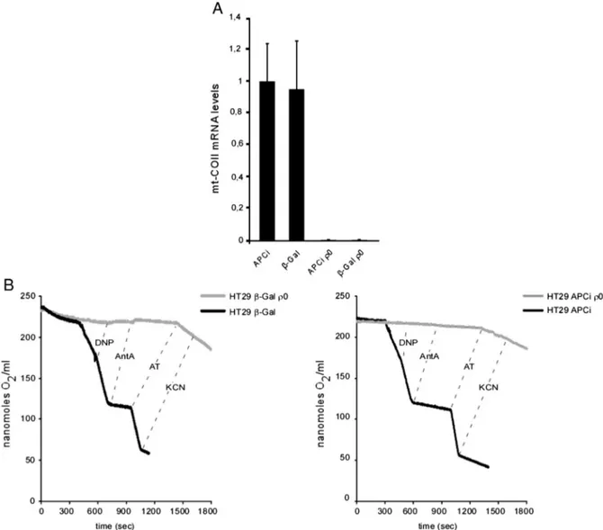
Fig. 4. Characterization of mtDNA-depleted (ρ0) HT29 APCi and HT29 β-GAL cells. (A) Expression of mitochondrial cytochrome c oxidase II gene before and after mt-DNA depletion. (B) Representative oxygraphic tracings of respiratoryfluxes in intact cells. DNP, 2,4-dinitrophenol (60 μM); Ant A, antimycin A (40 nM); AT, ascorbate (10 mM) plus N,N,N′,N′-tetramethyl-p-phenylenediamine (0.4 mM); KCN, potassium cyanide (2 mM).
an indirect target of APC activated through Caudal Type Homeobox transcription factor 2 (CDX2)[27], a significant decrease in the expres-sion of c-Myc, aβ-catenin target gene[4]and cyclin D1, a target for the β-catenin/LEF-1 complex[28]as compared to vehicle (EtOH)-treated cells. As expected, the expression levels of the same genes were un-changed in HT29β-Gal control cells.
To confirm the ability of the cells to undergo apoptosis upon APC in-duction, we measured the sub-G1 phase cell population byflow cyto-metric analysis. As shown inFig. 1B, after 48 h of APC induction by ZnCl2a significant increase in the sub-G1 cell content could be observed
in APCi, but not inβ-Gal cells when compared to mock-treated controls. The relative increase in sub-G1 cells reached its maximum (about 50%) after 96 h, but was still significantly present at 120 h from ZnCl2
exposure.
3.2. Induction of APC increases mitochondrial respiratoryfluxes in HT29 cells
Along the crypt–villus axis, intestinal cells show increasing levels of the APC protein, as well as a higher respiratory capacity measured via COX activity[12]. To identify a direct link between APC induction and
mitochondrial functions, we measured the respiratory capacities by en-dogenous substrates in HT29 APCi and HT29β-Gal intact cells (Fig. 2). A significant enhancement of both the basal and the maximal uncoupled endogenous respiratoryfluxes was detected upon ZnCl2-induced APC
expression in APCi cells, but not inβ-Gal cells, as compared with their control cells, respectively. These data, while confirming that the expres-sion of the full length APC protein restores a normal Wnt signaling path-way and promotes apoptosis, also suggest that is associated with an increase in mitochondrial respiration.
3.3. APC expression induces mitochondrial transcription
To investigate whether the induction of APC could result in an in-crease in mtDNA content, the total genomic DNAs of the HT29 APCi and HT29β-Gal cells, were used as templates in a RTqPCR to amplify the mitochondrial cytochrome c oxidase II gene (mt-COII). As shown inFig. 3A, levels of mtDNA remain unchanged upon ZnCl2-induced
APC expression in APCi cells and inβ-Gal cells. On the other hand, in order to check whether APC induction could influence mtDNA gene ex-pression, the same RTqPCR analysis of mt-COII gene, was carried out on total RNA from the same cells. We found that the expression of mt-COII
Fig. 5. Effects of the induction of wild type APC expression in HT29 APCiρ0 cells. (A) Expression levels of APC, KLF4, c-Myc and Cyclin D1 genes. Cells were harvested 24 h after ZnCl2
ex-posure and relative mRNA levels were quantified by RTqPCR on RNA samples using GAPDH as reference gene. Values shown represent mean ± SEM for two independent determinations performed in triplicate (*pb 0.05; **p b 0.01). (B) Time course analysis of apoptotic cell population after induction of wild type APC expression. Expression of wild type APC or β-Gal gene was induced by ZnCl2(120μM) and cells were harvested and analyzed by flow cytometry at the indicated time points. Values shown represent mean ± SEM for two independent
deter-minations performed in duplicate. (*pb 0.05; **p b 0.01).
gene results increased upon ZnCl2-induced APC expression in APCi cells,
but not inβ-Gal cells. These data point to an activated mitochondrial transcription due to inducible expression of APC (Fig. 3B).
3.4. Mitochondrial DNA-depletion (Rho0) in HT29 APCi and HT29β-Gal cells
To confirm that the increase in mitochondrial respiratory activity could represent the functional link between the restored APC func-tion and the subsequent stimulafunc-tion of the apoptotic process, we generated mtDNA-depleted HT29 APCiρ0and HT29β-Gal ρ0cell
lines by ethidium bromide treatment[23]. The complete removal of mtDNA was verified by the absence of any amplification product of the mtDNA cytochrome b gene (cyt b) (Fig. S1) in both HT29 APCi ρ0
and HT29β-Gal ρ0
cells. In addition, in the same cells, we verified the lack of mt-COII mRNA (Fig. 4A), further confirming the mtDNA depletion. To characterize theρ0state at a functional level, we
measured mitochondrial respiratoryfluxes. As expected, contrary to the parental counterparts, no antimycin-sensitive respiration was detected in the intactρ0cells. Similarly, no COX-specific activity
was present in bothρ0cell lines as revealed by the absence of
KCN-sensitive oxygen consumption elicited by the artificial membrane-permeant cytochrome c reductant ascorbate + TMPD (Fig. 4B).
We then checked if APC induction by ZnCl2would restore a normal
Wnt pathway in HT29 APCiρ0cells. As shown inFig. 5A, the ZnCl 2
-induced changes of the expression levels of APC, KLF4, c-Myc and cyclin D1 marker genes were maintained (see alsoFig. 1B) in HT29 APCiρ0
cells, thus demonstrating that the absence of mtDNA does not interfere with the normal regulation of the transcriptional program dependent on the APC/β-catenin pathway.
Finally, we determined whether the proapoptotic effect of the APC expression was also present in HT29 APCiρ0
cell line (Fig. 5B). Indeed, it has been previously demonstrated thatρ0cells can undergo apoptosis
[29]and that mtDNA depleted mitochondria can mediate the cell death
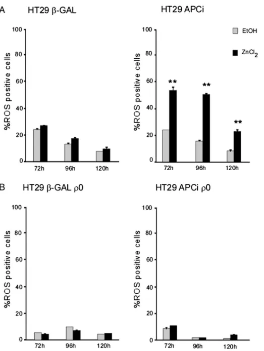
Fig. 6. Mitochondrial respiratory chain is needed for the APC-induced increase in ROS production. Expression of wild type APC orβ-Gal gene was induced by ZnCl2(120μM) and cells were
harvested and analyzed at the indicated time points. *pb 0.05; **p b 0.01 for ZnCl2-treated cells vs. vehicle (EtOH)-treated controls. The intracellular ROS levels were determined using the
cell-permeantfluorescent probe CM-H2DCFDA, and the percentage of ROS positive cells was quantified by flow cytometry. Values shown represent mean ± SEM for two independent
process by releasing cytochrome c[30]. In particular, the occurrence of the cell death process in response to different apoptotic stimuli has been studied in a variety of cancer cell lines[31,32]. Interestingly, no dif-ference was found in the time course analysis of the subG1 phase cell content when comparing ZnCl2-treated APCiρ0cells to their
mock-treated controls or toβ-Gal ρ0cells, respectively. These results suggest
a possible link between the APC-dependent apoptosis, and the presence of a functional mitochondrial respiratory chain.
3.5. APC-induced apoptosis requires mitochondrial respiratory ROS. We have previously demonstrated that one of the main apoptosis-driving factors in colorectal cancer cells is represented by the mitochon-drial respiratory chain-dependent accumulation of ROS[12]. To test the
hypothesis that the mitochondrial oxidative burst could have been the missing link between the restored Wnt pathway and apoptosis in APCi ρ0cells, we measured ROS accumulation upon APC induction in theρ0
cell lines and their parental counterparts. As shown inFig. 6, a signi fi-cant increase in the percentage of ROS-positive cells could be observed only in HT29 APCi cells, when comparing ZnCl2-treated cells to the
mock-treated controls or their correspondingρ0lines, respectively. It
is worth noting that the basal content of ROS positive cells was always lower inρ0
cells as compared to their parental cell lines, thus pointing to the mitochondrial respiratory chain as the main producer of ROS under our experimental conditions.
Finally, pre-treatment of the cells with the anti-oxidant N-acetyl-L-cysteine (NAC) almost completely abolished the increase in apo-ptotic cells in APCi cells upon ZnCl2-induced APC expression (Fig. 7
Fig. 7. Loss of APC-dependent apoptosis in NAC-treated HT29 cells. Expression of wild type APC orβ-Gal gene was induced by ZnCl2(120μM) and cells were harvested and analyzed at the
indicated time points. *pb 0.05; **p b 0.01 for ZnCl2-treated cells vs. vehicle (EtOH)-treated controls. Apoptotic cells were quantified as percentage of sub-G1 phase cells by flow cytometry
as previously described. ZnCl2-induction of APC expression was initiated after a 2 h pre-incubation with the antioxidant NAC (20 mM). Values represent mean ± SEM for two independent
determinations performed in duplicate. (*pb 0.05; **p b 0.01).
cfr.Fig. 1B), thus confirming the direct functional relationship be-tween the mitochondrial oxidative burst and the proapoptotic action of APC.
4. Discussion
Mitochondria, in addition to their main task of producing aerobic en-ergy through the OXPHOS apparatus, are increasingly shown to be in-volved in a variety of metabolic and signaling pathways of eukaryotic cells. We have recently demonstrated that mitochondria play a pivotal role in intestinal cell fate, not only by regulating the metabolic switch during cellular differentiation, but also by modulating the abundance of ROS production as the main driving signal for the apoptotic renewal of intestinal epithelial cells[12,33].
In this paper, we identify a functional link between mitochondria and Wnt signaling, i.e., the major signal transduction pathway involved in intestinal cells homeostasis and development along the crypt–villus axis. By generating a mtDNA-depleted APCi HT29 cell line, we clearly demonstrate that a functional respiratory chain is required for the proapoptotic effect associated with the expression of the full length APC protein, hence, with the re-establishment of a normal Wnt pathway, in colorectal cancer HT29 cells. Indeed, a direct interaction be-tween the caspase cleaved amino-terminus of APC and the mitochondri-al protein hTID-1 has been previously proposed to enhance sensitivity to apoptosis in colorectal cancer cells[22].
On the other hand, our results are apparently in contrast with previ-ous reports of an increased Wnt signaling as a potent activator of mito-chondrial biogenesis and respiratory capacity, as well as, of ROS production, via induction of the insulin receptor substrate-1 (IRS1) and c-Myc gene expression in C2C12 muscle cells and primary mouse embryonicfibroblasts[34]. In fact, the transcription factor c-Myc, known to be an important activator of mitochondrial biogenesis and function[35,36], is negatively regulated upon APC induction in HT29 colorectal cancer cells (Fig. 1B), thus pointing to a striking tissue-specificity regulation of the Wnt/IRS1/c-Myc pathway, in relation to mi-tochondrial activity. Interestingly, in line with ourfindings, IRS1 and c-Myc have been shown to be overexpressed in colorectal cancer as com-pared with paired normal mucosa samples[37]. Therefore, mitochon-dria seem to be differently regulated in the intestine, where OXPHOS and respiratory ROS-dependent apoptosis are required for epithelial cell differentiation and self-renewal, respectively, along the crypt to vil-lus axis[12].
Nevertheless, the role of mitochondrial ROS in intestinal tumorigen-esis remains controversial. A tumorigenic role for mitochondrial ROS has been, in fact, previously proposed, on the basis of an increase in in-testinal tumor initiation and growth in an APCMin/+Tfam+/−mouse, by a
mechanism independent of the canonical Wnt pathway[38]. In this an-imal model, however, ROS would have been involved in the process as major producers of mtDNA damage, rather than as a mediator of the ap-optotic cell death. Indeed, oxidative damage to biomolecules is caused by an unbalance between ROS production and scavenging and/or by a defective antioxidant repairing machinery. As well, ROS can act as sig-naling molecules[39,40]so that afine tuning of redox biology must be maintained in order to preserve physiological tissue homeostasis.
In conclusion, our study highlights the relationship between APC protein and mitochondrial functions in HT29 colorectal cancer cells. In the presence of wild-type APC, the negative regulation of the Wnt sig-naling reduces cell survival by promoting apoptosis through the mito-chondrial respiratory chain production of ROS. This intriguing scenario adds further insights into the role of mitochondria in intestinal epitheli-al cell fate and suggests the possibility to target the functionepitheli-al link be-tween Wnt/APC pathway and mitochondrial oxidative metabolism for the therapy and/or prevention of colorectal cancer.
Supplementary data to this article can be found online athttp://dx. doi.org/10.1016/j.bbadis.2015.05.009.
Transparency document
TheTransparencydocumentassociated with this article can be found in the online version.
Acknowledgments
HT29 APCi and HT29β-Gal cell lines were kindly provided by Drs. K. W. Kinzler and B. Vogelstein (Ludwig Center for Cancer Genetics and Therapeutics and Howard Hughes Medical Institutions, Johns Hopkins University School of Medicine, Baltimore, Maryland, USA). We grateful-ly acknowledge Drs. Elena Bellafante, Annalisa Morgano, Michele Vacca, Roberta Le Donne and the rest of the members of our group for the critical feedback and thoughtful discussions.
References
[1] A. Gregorieff, H. Clevers, Wnt signaling in the intestinal epithelium: from endoderm to cancer, Genes Dev. 19 (2005) 877–890.
[2] H. Clevers, Wnt breakers in colon cancer, Cancer Cell 5 (2004) 5–6.
[3] W. de Lau, N. Barker, H. Clevers, WNT signaling in the normal intestine and colorec-tal cancer, Front. Biosci. 12 (2007) 471–491.
[4] T.C. He, A.B. Sparks, C. Rago, H. Hermeking, L. Zawel, L.T. da Costa, P.J. Morin, B. Vogelstein, K.W. Kinzler, Identification of c-MYC as a target of the APC pathway, Sci-ence 281 (1998) 1509–1512.
[5] O. Tetsu, F. McCormick, Beta-catenin regulates expression of cyclin D1 in colon car-cinoma cells, Nature 398 (1999) 422–426.
[6] H. Aberle, A. Bauer, J. Stappert, A. Kispert, R. Kemler, beta-Catenin is a target for the ubiquitin–proteasome pathway, EMBO J. 16 (1997) 3797–3804.
[7] V. Korinek, N. Barker, P.J. Morin, W.D. van, W.R. de, K.W. Kinzler, B. Vogelstein, H. Clevers, Constitutive transcriptional activation by a beta-catenin–Tcf complex in APC−/− colon carcinoma, Science 275 (1997) 1784–1787.
[8] R. Fodde, R. Smits, H. Clevers, APC, signal transduction and genetic instability in co-lorectal cancer, Nat. Rev. Cancer 1 (2001) 55–67.
[9] P.J. Morin, B. Vogelstein, K.W. Kinzler, Apoptosis and APC in colorectal tumorigene-sis, Proc. Natl. Acad. Sci. U. S. A. 93 (1996) 7950–7954.
[10] B.J. Jeynes, G.G. Altmann, A region of mitochondrial division in the epithelium of the small intestine of the rat, Anat. Rec. 182 (1975) 289–296.
[11] H. Clevers, Wnt/beta-catenin signaling in development and disease, Cell 127 (2006) 469–480.
[12]I. D'Errico, L. Salvatore, S. Murzilli, S.G. Lo, D. Latorre, N. Martelli, A.V. Egorova, R. Polishuck, K. Madeyski-Bengtson, C. Lelliott, A.J. Vidal-Puig, P. Seibel, G. Villani, A. Moschetta, Peroxisome proliferator-activated receptor-gamma coactivator 1-alpha (PGC1alpha) is a metabolic regulator of intestinal epithelial cell fate, Proc. Natl. Acad. Sci. U. S. A. 108 (2011) 6603–6608.
[13]A. Turan, A. Mahmood, The profile of antioxidant systems and lipid peroxidation across the crypt–villus axis in rat intestine, Dig. Dis. Sci. 52 (2007) 1840–1844.
[14] D.D. Newmeyer, S. Ferguson-Miller, Mitochondria: releasing power for life and unleashing the machineries of death, Cell 112 (2003) 481–490.
[15] G. Attardi, G. Schatz, Biogenesis of mitochondria, Annu. Rev. Cell Biol. 4 (1988) 289–333.
[16] M.P. Murphy, How mitochondria produce reactive oxygen species, Biochem. J. 417 (2009) 1–13.
[17] D.R. Green, Apoptotic pathways: ten minutes to dead, Cell 121 (2005) 671–674.
[18]D.R. Green, G. Kroemer, The pathophysiology of mitochondrial cell death, Science 305 (2004) 626–629.
[19] X. Liu, C.N. Kim, J. Yang, R. Jemmerson, X. Wang, Induction of apoptotic program in cell-free extracts: requirement for dATP and cytochrome c, Cell 86 (1996) 147–157.
[20] R.M. Kluck, E. Bossy-Wetzel, D.R. Green, D.D. Newmeyer, The release of cytochrome c from mitochondria: a primary site for Bcl-2 regulation of apoptosis, Science 275 (1997) 1132–1136.
[21]M. Brocardo, Y. Lei, A. Tighe, S.S. Taylor, M.T. Mok, B.R. Henderson, Mitochondrial targeting of adenomatous polyposis coli protein is stimulated by truncating cancer mutations: regulation of Bcl-2 and implications for cell survival, J. Biol. Chem. 283 (2008) 5950–5959.
[22] J. Qian, E.M. Perchiniak, K. Sun, J. Groden, The mitochondrial protein hTID-1 partners with the caspase-cleaved adenomatous polyposis cell tumor suppressor to facilitate apoptosis, Gastroenterology 138 (2010) 1418–1428.
[23] M.P. King, G. Attardi, Isolation of human cell lines lacking mitochondrial DNA, Methods Enzymol. 264 (1996) 304–313.
[24] G. Villani, G. Attardi, In vivo control of respiration by cytochrome c oxidase in wild-type and mitochondrial DNA mutation-carrying human cells, Proc. Natl. Acad. Sci. U. S. A. 94 (1997) 1166–1171.
[25] S.S. Makarov, C. Jonat, S. Haskill, Hyperinducible human metallothionein promoter with a low level basal activity, Nucleic Acids Res. 22 (1994) 1504–1505.
[26] L. Poljak, C. Seum, T. Mattioni, U.K. Laemmli, SARs stimulate but do not confer posi-tion independent gene expression, Nucleic Acids Res. 22 (1994) 4386–4394.
[27]D.T. Dang, C.S. Mahatan, L.H. Dang, I.A. Agboola, V.W. Yang, Expression of the gut-enriched Kruppel-like factor (Kruppel-like factor 4) gene in the human colon cancer cell line RKO is dependent on CDX2, Oncogene 20 (2001) 4884–4890.
[28]M. Shtutman, J. Zhurinsky, I. Simcha, C. Albanese, M. D'Amico, R. Pestell, A. Ben-Ze'ev, The cyclin D1 gene is a target of the beta-catenin/LEF-1 pathway, Proc. Natl. Acad. Sci. U. S. A. 96 (1999) 5522–5527.
[29] M.D. Jacobson, J.F. Burne, M.P. King, T. Miyashita, J.C. Reed, M.C. Raff, Bcl-2 blocks ap-optosis in cells lacking mitochondrial DNA, Nature 361 (1993) 365–369.
[30]S. Jiang, J. Cai, D.C. Wallace, D.P. Jones, Cytochrome c-mediated apoptosis in cells lacking mitochondrial DNA. Signaling pathway involving release and caspase 3 acti-vation is conserved, J. Biol. Chem. 274 (1999) 29905–29911.
[31]S.M. Cardoso, A.C. Rego, N. Penacho, C.R. Oliveira, Apoptotic cell death induced by hydrogen peroxide in NT2 parental and mitochondrial DNA depleted cells, Neurochem. Int. 45 (2004) 693–698.
[32] P. Marchetti, S.A. Susin, D. Decaudin, S. Gamen, M. Castedo, T. Hirsch, N. Zamzami, J. Naval, A. Senik, G. Kroemer, Apoptosis-associated derangement of mitochondrial function in cells lacking mitochondrial DNA, Cancer Res. 56 (1996) 2033–2038.
[33] E. Bellafante, A. Morgano, L. Salvatore, S. Murzilli, T.G. Di, A. D'Orazio, D. Latorre, G. Villani, A. Moschetta, PGC-1beta promotes enterocyte lifespan and tumorigenesis in the intestine, Proc. Natl. Acad. Sci. U. S. A. 111 (2014) E4523–E4531.
[34] J.C. Yoon, A. Ng, B.H. Kim, A. Bianco, R.J. Xavier, S.J. Elledge, Wnt signaling regulates mitochondrial physiology and insulin sensitivity, Genes Dev. 24 (2010) 1507–1518.
[35] F. Li, Y. Wang, K.I. Zeller, J.J. Potter, D.R. Wonsey, K.A. O'Donnell, J.W. Kim, J.T. Yustein, L.A. Lee, C.V. Dang, Myc stimulates nuclearly encoded mitochondrial genes and mitochondrial biogenesis, Mol. Cell. Biol. 25 (2005) 6225–6234.
[36]F. Morrish, D. Hockenbery, MYC and mitochondrial biogenesis, Cold Spring Harb Perspect. Med. 4 (2014).
[37]D.L. Esposito, F. Aru, R. Lattanzio, A. Morgano, M. Abbondanza, R. Malekzadeh, F. Bishehsari, R. Valanzano, A. Russo, M. Piantelli, A. Moschetta, L.V. Lotti, R. Mariani-Costantini, The insulin receptor substrate 1 (IRS1) in intestinal epithelial differenti-ation and in colorectal cancer, PLoS One 7 (2012) e36190.
[38] D.K. Woo, P.D. Green, J.H. Santos, A.D. D'Souza, Z. Walther, W.D. Martin, B.E. Christian, N.S. Chandel, G.S. Shadel, Mitochondrial genome instability and ROS en-hance intestinal tumorigenesis in APC(Min/+) mice, Am. J. Pathol. 180 (2012) 24–31.
[39] R.B. Hamanaka, N.S. Chandel, Mitochondrial reactive oxygen species regulate cellu-lar signaling and dictate biological outcomes, Trends Biochem. Sci. 35 (2010) 505–513.
[40] K.M. Holmstrom, T. Finkel, Cellular mechanisms and physiological consequences of redox-dependent signalling, Nat. Rev. Mol. Cell Biol. 15 (2014) 411–421.