Endothelial Cell Activation In Vitro and Angiogenesis
In Vivo
Chiara Urbinati, Stefania Mitola, Elena Tanghetti, Chandra Kumar, Johannes Waltenberger,
Domenico Ribatti, Marco Presta, Marco Rusnati
Objective—The transactivating factor (Tat) of HIV-1 binds to␣v3integrin present on endothelial cells contributing to
neovascularization. Here, we investigated the biological consequences of Tat/␣v3interaction and the antagonist effect
of an Arg-Gly-Asp (RGD)-based peptidomimetic.
Methods and Results—Binding of Tat to endothelial␣v3triggers focal adhesion kinase and nuclear factor-B activation,
leading to endothelial cell proliferation, membrane ruffling, and motility in vitro and neovascularization in vivo. The RGD-peptidomimetic SCH221153 inhibits Tat/␣v3 interaction in a solid phase binding assay and endothelial cell
adhesion to immobilized Tat with a potency higher than that of RGD-containing peptides. Accordingly, SCH221153 inhibits Tat/␣v3-dependent focal adhesion kinase and nuclear factor-B activation, proliferation, membrane ruffling,
and motility in endothelial cells. Finally, SCH221153 inhibits the angiogenic response triggered by Tat in the chick-embryo chorioallantoic membrane without affecting physiological vascularization. SCH221153 exerts these inhibitory effects without affecting the interaction of Tat with endothelial heparan sulfate proteoglycans or with the vascular endothelial growth factor receptor-2/kinase domain– containing receptor. In all the assays the negative control SCH216687 was ineffective.
Conclusion—These data provide new insights on the mechanism of endothelial cell activation by Tat and point to RGD peptidomimetics as prototypes for the development of novel Tat antagonists. (Arterioscler Thromb Vasc Biol.
2005;25:2315-2320.)
Key Words: HIV-1 䡲 Tat 䡲 endothelium 䡲 integrin 䡲 angiogenesis
T
at protein, the main transactivating factor of HIV-1,1isreleased by HIV-1–infected cells2 targeting different
types of uninfected cells and causing a variety of biological effects related to distinct AIDS-associated pathologies. In AIDS patients, Tat contributes to tumorigenesis,2 to the
pathogenesis of dementia,3to the immune system suppression
(by interfering with the function of different cells of immu-nity),2and to heart disease and atherosclerosis.4
The involvement of endothelial cells (ECs) in Tat-dependent pathologies is manifold.5By inducing
neovascu-larization, Tat contributes to tumor progression and Kaposi sarcoma (KS) development.2Also, Tat increases endothelial
permeability and alters the expression of endothelial leuko-cyte receptors, leading to the extravasation of HIV-1⫹ monocytes and HIV-1 dissemination. These effects, in turn, contribute to the pathogenesis of lymphomas, AIDS-dementia,5 cardiovascular diseases, and atherosclerosis.6 A
decrease in endothelium-dependent vasorelaxation and endo-thelial nitric oxide synthase expression may also play a role in endothelial dysfunctions mediated by Tat.7
On these bases, Tat is considered a main target for the development of anti-AIDS therapies, and different anti-Tat strategies have been described, including gene therapies, vaccines, and Tat antagonists.8
Tat accumulates in the extracellular matrix as an immobilized protein,9 and substrate-immobilized Tat interacts with ␣
v3
integrin of ECs promoting their adhesion and triggering a complex signal transduction pathway that leads to activation of Tat-adherent ECs.10Accordingly,␣
v3interaction is required for
the chemotactic and mitogenic activity of Tat in vitro and for its angiogenic activity in vivo.11The Arg-Gly-Asp (RGD)
tripep-tide present in position 78 to 80 in Tat protein is required for ␣v3 interaction and EC adhesion.11 Accordingly,
RGD-containing peptides inhibit cell adhesion to immobilized Tat.12
Original received July 13, 2005; final version accepted August 15, 2005.
From the Chair of General Pathology and Immunology, Department of Biomedical Sciences and Biotechnology (C.U., S.M., E.T., M.P., M.R.), University of Brescia, Italy; the Department of Tumor Biology (C.K.), Schering-Plough Research Institute, Kenilworth, New Jersey; the Department of Cardiology (J.W.), University Hospital Maastricht, The Netherlands; and the Institute of Human Anatomy, Histology, and Embryology (D.R.), University of Bari, Italy.
Correspondence to Marco Rusnati, General Pathology and Immunology, Department of Biomedical Sciences and Biotechnology, Viale Europa 11, 25123 Brescia, Italy. E-mail [email protected]
© 2005 American Heart Association, Inc.
Arterioscler Thromb Vasc Biol. is available at http://www.atvbaha.org DOI: 10.1161/01.ATV.0000186182.14908.7b
Nonpeptidic analogs (peptidomimetics) of the RGD motif endowed with integrin-antagonist capacity block different pathological processes including angiogenesis13and
endothe-lial dysfunction that may lead to atherogenesis.4 Here, we
demonstrate that the RGD-peptidomimetic SCH221153, a selective␣v3 antagonist,13blocks␣v3/Tat interaction with
high potency and specificity, thus preventing Tat-mediated EC activation in vitro and angiogenesis in vivo.
Methods
ReagentsThe RGD-peptidomimetic SCH221153 and its control analog SCH21668713were from Schering-Plough Research Institute. Linear GRGDSPK and GRADSPK peptides were from Neosystem Labo-ratoire (Strasbourg). Cyclo(-Arg-Gly-Asp-D-Phe-Val) peptide (cRG-DFV) and cyclo(-Arg-Ala-Asp-D-Phe-Val) peptide (cRA(cRG-DFV) were from Bachem (Beidendorf). Synthetic biotinylated Tat was from Tecnogen. Bovine fibronectin (FN) was from Sigma (St. Louis, MO). The goat anti-rabbit biotinylated antibodies and Alexa Fluor 488 streptavidin were from Molecular Probes (Eugene, OR). Anti-3 antiserum was provided by G. Tarone, University of Turin, Italy. Anti-␣v3monoclonal LM609 antibody was from Chemicon Inter-national. Human integrin␣v3,glutathione-S-transferase (GST)-Tat, and GST-Tat fused to the green fluorescent protein were purified as described.14,15The GST moiety does not interfere with the transac-tivating, angiogenic, and cell-adhesive activities of Tat10 (and references therein).
Cell-Free␣v3Integrin/GST-Tat Interaction and
Cell Adhesion Assay
The assays were performed as described.14,16
Cell Cultures
Transformed fetal-bovine aortic endothelial GM7373 cells (obtained from the National Institute of General Medical Sciences [Institute for Medical Research, Camden, NJ]) were cultured in Eagle minimal essential medium containing 10% fetal calf serum (FCS) (Gibco, Grand Island, NY), vitamins, essential and nonessential amino acids. Human umbilical vein ECs (HUVECs; obtained from Biowhittaker, Walkersville, MA) were cultured in endothelial growth medium-2 medium (Biowhittaker). Porcine aortic ECs transfected with vascular endothelial growth factor receptor-2/kinase– domain containing re-ceptor (KDR) (KDR/PAECs)16were cultured in Ham F12 medium (Gibco) with 10% FCS and 2% glutamine. HL3T1 cells are derived from HeLa cells and contain integrated copies of the pL3CAT plasmid in which the chloramphenicol acetyltransferase (CAT) bacterial gene is driven by HIV-1 long terminal repeat.15They were cultured in Dulbecco modified Eagle medium containing 10% FCS (Gibco).
FAK and NF-B Activation Assays
GM7373 cells in 24-well culture tissue plates were made quiescent by 24-hour serum starvation and then incubated at 37°C for 30 minutes with GST-Tat (3 nmol/L) in the absence or in the presence of the peptidomimetics (0.3 mol/L). For focal adhesion kinase (FAK) activation assay, cells were lysed by a 5 minutes incubation at 90°C in reducing SDS-PAGE sample buffer and analyzed by Western blotting using an anti–phospho-Tyr397-FAK antibody (Bio-Source International). The autoradiography was digitized, and the integrated densities of the bands corresponding to phosphorylated FAK were evaluated with the Image Pro-Plus analysis system (Media Cybernetics). Data were expressed as fold increase of FAK phosphorylation in respect to untreated control cells. For nuclear factor-B (NF-B) activation assay, the amount of activated NF-B p50 subunit in cell extracts was determined using the TransAM NF-B p50 Assay Kit (Active Motif).
Tat Internalization and HIV-1 Long Terminal Repeat Transactivation Assays
The assays were performed as described.17For further details, see online Methods at http://atvb.ahajournals.org.
KDR-Binding and Phosphorylation Assays
KDR-binding assay was performed as described.16 For further details, see online Methods at http://atvb.ahajournals.org. For KDR phosphorylation assay, subconfluent cultures of KDR/PAECs were made quiescent by 16-hour serum starvation. Then, cells were preincubated for 30 minutes at 37°C with medium containing SCH221153 or SCH216687 (30 nmol/L) and then incubated for 15 minutes with GST-Tat (1.2 nmol/L). Cells were washed and lysed in Hepes buffer pH 8, 150 mmol/L NaCl, 1% Triton X-100, protease, and phosphatase inhibitors (70 nmol/L pepstatin, 100 nmol/L leu-peptin, 1.5mol/L aprotinin, and 1 mmol/L sodium orthovanadate). Aliquots (25g) of protein were separated by SDS-PAGE (7%) and probed with anti–phospho-tyrosine antibody (Santa Cruz Biotechnology).
Evaluation of the Mitogenic Activity of Immobilized GST-Tat
GM7373 cells were allowed to adhere onto 96-well plates coated with different proteins. Then, cells were washed with 2.0 mmol/L EDTA/PBS, trypsinized, and counted in a Burker chamber (T0) or incubated for 72 hours in fresh medium containing 0.4% FCS in the absence or in the presence of the peptidomimetics before counting. EC-Monolayer Wound Healing and
Membrane-Ruffle Formation Assay
GM7373 cell monolayers adherent to 3.5 cm-polystyrene nontissue culture plates coated with different substrata were wounded with a rubber policeman and incubated at 37°C with medium containing 0.4% FCS in the absence or in the presence of the peptidomimetics. Then, the cells at the edge of the wound were photographed, and those showing membrane ruffles were counted under an inverted microscope (Olympus 1⫻51) connected to a Camedia C-4040 digital camera (Olympus Biosystem). For wound-healing assay, wounded monolayers were incubated for 48 hours at 37°C and photographed under the inverted microscope. The extent of wound repair was evaluated by measuring the area of the wound by computerized image analysis using the Image Pro-Plus analysis system. In some experiments, wounded monolayers of GM7373 cells adherent to GST-Tat were fixed in 3% paraformaldehyde, 2% sucrose, perme-abilized with 0.2% Triton-X100, and saturated with 3% BSA (all in PBS). Then cells were incubated for 1 hour at room temperature with anti–phospho-Tyr397-FAK antibody (8 nmol/L plus 3% BSA in PBS), washed and incubated for 45 minutes with goat anti-rabbit biotinyl-ated antibodies (1:500 in PBS), then washed and incubbiotinyl-ated for 30 minutes with Alexa Fluor 488 streptavidin. Cells were photographed under an Axioplan 2 microscope equipped for epifluorescence (Carl Zeiss).
Chick-Embryo Chorioallantoic Membrane Assay The assay was performed as described.16
Statistical Analysis
Results are expressed as mean⫾SEM of 3 to 6 separate experiments. Student t test was used for statistical analysis.
Results
Effect of the RGD-Peptidomimetic SCH221153 on Tat/␣v3Interaction
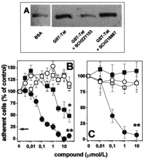
As shown in Figure 1A,␣v3binds to immobilized GST-Tat.
The interaction is specific because ␣v3 does not bind to
immobilized BSA nor to GST protein.10 Preincubation of
with GST-Tat whereas preincubation with the negative con-trol SCH216687 is ineffective (Figure 1A). It should be pointed out that recent observations using BIACORE tech-nology have shown that Tat/␣v3interaction occurs with an
affinity similar to that of different physiological␣v3ligands
(dissociation constant (Kd) being equal to 32 nM, 27 nM, and
64 nM for Tat, fibrinogen, and vitronectin, respectively).10
These data underline the efficacy of SCH221153 as an inhibitor of Tat/␣v3interaction.
GM7373 cells adhere to GST-Tat– coated plastic in a ␣v3-dependent manner, as demonstrated by the inhibition
exerted by anti-␣v3monoclonal antibody LM609, here used
as a positive control (Figure 1B). Accordingly, SCH221153 inhibits GM7373 cell adhesion to immobilized GST-Tat in a dose-dependent manner, whereas the negative control SCH216687 exerts only a limited inhibition with a potency ⬇200⫻ lower than that of SCH221153 (Figure 1B). In keeping with its selective ␣v3-antagonist activity,
SCH221153 does not affect GM7373 cell adhesion to the ␣51 ligand FN (Figure 1B). SCH221153 inhibits also the
adhesion of HUVECs to GST-Tat without affecting their adhesion to FN (Figure 1C). Again, SCH216687 was inef-fective in preventing HUVECs adhesion to immobilized Tat, further supporting the specificity of the Tat-antagonist effect of SCH221153.
SCH221153 inhibits GM7373 cell adhesion to immobi-lized GST-Tat with a potency that is 200⫻ and 25⫻ higher than that showed by the linear peptide GRGDSPK and by the cyclic peptide cRGDFV, respectively. Also, SCH221153 is 170⫻ more potent than its negative control SCH216687, whereas GRGDSPK and cRGDFV peptides are only 4 to 6⫻ more potent than their corresponding negative controls (see Table I, available online at http://atvb.ahajournals.org).
Tat binds also the vascular endothelial growth factor receptor-2/KDR18and heparan sulfate proteoglycans (HSPGs).19KDR/
PAECs and HL3T1 cells were used to study the effect of SCH221153 on Tat interaction with KDR and HSPGs, respec-tively.16 At variance with suramin (here used as a positive
control),16SCH221153 does not affect the binding of
biotinyl-ated Tat nor Tat-dependent KDR autophosphorylation in KDR-overexpressing PAECs (KDR/PAECs) (see Figure I, available online at http://atvb.ahajournals.org). Also, at variance with the HSPG-antagonist heparin,17SCH221153 affects neither
HSPG-dependent GST-Tat– green fluorescent protein internalization nor Tat-dependent HIV-long terminal repeat-transactivation in HL3T1 cells (see Figure II, available online at http://atvb. ahajournals.org). Nevertheless, SCH221153 retains the capacity to inhibit the adhesion of both KDR/PAECs and HL3T1 cells to immobilized GST-Tat without affecting their interaction with FN (see Figures I and II).
In conclusion, SCH221153 specifically inhibits the inter-action of Tat with␣v3without affecting its interaction with
KDR or HSPG receptors.
Effect of SCH221153 on Signal Transduction Triggered by Tat/␣v3Interaction
In ECs, integrin engagement by physiological ligands acti-vates FAK and NF-B that, in turn, mediate EC migration and proliferation in vitro and neovascularization in vivo.20On
the other hand, Tat triggers FAK21and NF-B22activation in
different cell types. Accordingly, free GST-Tat induces FAK phosphorylation and NF-B activation in GM7373 cells (Figure 2). Again, these responses are inhibited by SCH221153 but not by SCH216687, demonstrating that␣v3
engagement is required for the activation of these 2 second messengers by Tat in ECs.
Effect of SCH221153 on␣v3/Tat-Dependent
Endothelial Cell Activation In Vitro
Integrin-dependent EC adhesion and signal transduction can be considered the first steps of the process that, through EC migration and proliferation, leads to neovascularization.23
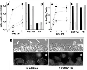
Accordingly, immobilized GST-Tat induces a significant increase in the proliferation rate of adherent GM7373 cells when compared with FN- or Poly-L-Lysin–adherent cells (Figure 3A). SCH221153 inhibits cell proliferation of GST-Tat–adherent cells whereas SCH216687 is ineffective. Also, SCH221153 has no effect on the limited proliferation ob-served in FN-adherent cells (Figure 3B). It must be pointed out that both compounds were added to cells only after their adhesion and spreading onto the substrate had occurred; this prevented cell rounding or complete detachment of ECs.10
Cell-membrane ruffling precedes the migration of EC body24 and is considered a morphological phenotype of
Figure 1. Effect of SCH221153 on Tat/␣v3interaction and EC adhesion. A,␣v3was incubated onto plastic-coated BSA or GST-Tat in the absence or in the presence of SCH221153 or SCH216687. Then, plastic-bound proteins were extracted and analyzed by Western blotting with anti-3antibodies. B, GM7373 cells were allowed to adhere to plastic-immobilized GST-Tat (black symbols) or FN (white symbols) in the presence of SCH221153 (circles) or SCH216687 (squares). Then, the number of adherent cells was evaluated and expressed as per-centage of cells adherent to GST-Tat or to FN in the absence of competitors. Arrow points to the value of cell adhesion to immo-bilized GST-Tat in the presence of anti–␣v3-neutralizing mono-clonal antibody LM609 (700 nmol/L). C, HUVECs were allowed to adhere to GST-Tat or FN in the absence or in the presence of SCH221153 or of SCH216687 (symbols as in B). Then, the number of adherent cells was evaluated. *P⬍0.001, **P⬍10⫺4, Student t test.
motile cells.25Wounded monolayers of GM7373 cells
adher-ent to GST-Tat rapidly form membrane ruffles at their leading edge. Cell-membrane ruffling is observed also in FN-adherent cells and, to a much lesser extent, in PL-adherent cells (Figure 3C). SCH221153 hampers membrane ruffling in GST-Tat–adherent cells (Figure 3D). Specificity of the effect is shown by the inability of SCH216687 to affect this process and by the lack of effect of SCH221153 on membrane ruffling in FN-adherent GM7373 cells (Figure 3D). Also, SCH221153 prevents the localization of phosphor-ylated FAK at the leading edge of migrating cells after wounding of GST-Tat–adherent monolayer (Figure 3E).
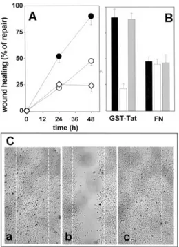
The ability of immobilized GST-Tat to stimulate prolifer-ation and motility in adherent GM7373 cells results in an increase in the capacity of a mechanically wounded cell monolayer to cover the denuded area when compared with FN- or PL-adherent cells (Figure 4). SCH221153, added to cell cultures after wounding, inhibits the capacity of GST-Tat–adherent cells to heal the wound, with no effect on the slower repair observed in FN-adherent cells (Figure 4B and 4C). Under the same experimental conditions SCH216687 does not alter wound repair in GST-Tat– or FN-adherent cells (Figure 4B).
Effect of SCH221153 on
Tat-Mediated Neovascularization
As shown in Figure 5, chick-embryo chorioallantoic mem-branes (CAM) implanted with gelatin sponges that were
loaded with GST-Tat show numerous allantoic vessels con-verging toward the implant. SCH221153 causes a significant inhibition of this activity, whereas SCH216687 is ineffective. Both compounds do not alter the unstimulated physiological vascularization of the chick-embryo CAM. No vascular reactions were detectable around the sponge in the specimens treated with vehicle alone.
Discussion
␣v3/Tat interaction plays an important role in the activation
of ECs that, in turn, contributes to the arise of AIDS-associated pathologies.26 On the other hand, nonpeptidic
analogs of the cell adhesion motif RGD endowed with integrin-antagonist capacity have been successfully used to block different angiogenesis-related processes.13 Here, we
demonstrate that the RGD-peptidomimetic compound SCH221153 inhibits Tat/␣v3interaction and the consequent
EC activation in vitro and neovascularization in vivo. Tat can be present as a free molecule or associated with the extracellular matrix.9Consistently,␣
v3is expressed both at
the basal and luminal aspects of endothelium.27 Here, we
observed that free Tat triggers an␣v3-dependent activation of
FAK and NF-B in ECs and that immobilized Tat induces EC adhesion, proliferation, and the acquisition of a motile phe-Figure 2. Effect of SCH221153 on signal transduction triggered
by Tat/␣v3interaction. Serum-starved GM7373 cells were treated with or without GST-Tat (3 nmol/L), SCH221153, or SCH216687 (both at 0.3 mmol/L). Then, FAK phosphorylation (A) or NF-B activation (B) were evaluated. In A, a indicates phosphorylated protein; t, total protein. *P⬍0.001, Student t test.
Figure 3. Effect of SCH221153 on Tat-dependent proliferation,
membrane ruffling, and FAK phosphorylation. GM7373 cells adherent to immobilized GST-Tat (F), FN (E), or PL (〫) were incubated for the indicated period of time (A). Alternatively, GST-Tat– or FN-adherent GM7373 cells were incubated for 72 hours in the absence (black bars) or in the presence of SCH221153 (white bars) or of SCH216687 (gray bars) (0.3mol/L) (B). Then, cells were counted. Data are expressed as cell population doublings. Monolayers of GM7373 cells adherent to the different proteins (symbols as in A) were wounded and incubated for the indicated periods of time (C). Alternatively, wounded monolayers of GM7373 cells adherent to GST-Tat or to FN were incubated for 3 hours in the absence or in the presence of peptidomimetics (symbols and doses as in B) (D). Then, membrane ruffling was evaluated. E, Photographs of the edge of a wounded monolayer of GST-Tat–adherent GM7373 cells in the absence (no addition) or in the presence of SCH221153. a and b, phase contrast photographs (200⫻). Arrows point to the most prominent ruffles. c and d, cells immu-nostained with anti–phospho-Tyr397-FAK antibody and photo-graphed under an epifluorescence microscope (630⫻).
notype; this contributes to the faster rate of healing of a Tat-adherent wounded EC monolayer. Notably, SCH221153 retains its Tat-antagonist capacity when the transactivating factor is presented to ECs either in its free or substratum-im-mobilized form.
Several experimental evidences point to the specificity of the Tat/␣v3 antagonist activity of SCH221153: (1) the
control compound SCH216687 was poorly effective in all the assays; (2) SCH221153 inhibits new blood vessel formation triggered by Tat in the chick-embryo CAM without affecting basal vascularization; (3) SCH221153 inhibits EC adhesion, membrane ruffling and wound healing in cells adherent to immobilized Tat but not immobilized FN. These observations are in keeping with the high specificity of SCH221153 for ␣v3when compared with different integrin receptors.13
In KDR/PAECs, SCH221153 does not affect the interac-tion of Tat with KDR. Relevant to this point, the occupancy and activation of KDR play a pivotal role in mediating the angiogenic activity of Tat.18 The observation that
SCH221153 inhibits Tat-mediated angiogenesis in the chick-embryo CAM without affecting Tat/KDR interaction and autophosphorylation suggests that the binding of Tat to both ␣v3and KDR receptors are required to stimulate
neovascu-larization, the single interaction with either 1 of the 2 receptors being insufficient to support a full angiogenic
response. This is in keeping with the existing cross-talk between integrins and tyrosine kinase receptors in neovascu-larization triggered by angiogenic growth factors.28
Accordingly, both␣v329and KDR30mediate the
biologi-cal effects of Tat in KS, pointing to the possibility of multitarget therapies aimed at blocking the 2 receptors simultaneously. Relevant to this point, a humanized anti-␣v3
antibody selectively targets human KS in nude mice,31and
the KDR inhibitor SU5416 exerts therapeutic benefits in patients with AIDS-associated KS.32It is also worth noting
that Tat synergizes with fibroblast growth factor-2 in the pathogenesis of KS.33 The observation that SCH221153
inhibits also ␣v3/fibroblast growth factor-2 interaction and
consequent EC activation13suggests that RDG
peptidomimet-ics may exert multitarget effects in KS.
Besides angiogenesis, the implication of ECs in AIDS-associated pathologies is manifold. Thus, RGD peptidomi-metics may inhibit also Tat-induced endothelial permeability and related dissemination of HIV-1 and HIV-1⫹ monocytes, possibly preventing the rise of lymphomas and of AIDS-dementia. Also, they may prevent Tat-dependent vasorelax-ation and endothelial nitric oxide synthase downregulvasorelax-ation, with possible benefits for AIDS-associated cardiovascular diseases. Related to this point, smooth muscle cells, well known effectors of atherogenesis, adhere to immobilized Tat via␣v3,34suggesting that RGD peptidomimetics may exert
Tat-antagonist activity also on this cell type.
A number of integrin antagonists have been developed, including cyclic RGD-containing peptides and anti-␣v3
monoclonal antibodies. Here, we found that the Tat-antago-nist potency of SCH221153 is higher than that of cyclic and linear RGD-containing peptides. Also, small-molecular weight RGD mimetics with oral bioavailability, like SCH221153, may have a number of advantages over peptide-based or antibody-peptide-based approaches. Indeed, nonpeptide
Figure 5. Effect of SCH221153 on Tat-induced angiogenesis in the
chick-embryo CAM. A, Chick-embryo CAMs were implanted with gelatin sponges adsorbed with vehicle or GST-Tat (12 pmoles) in the absence or in the presence of SCH221153 and SCH216687 (7 nmoles). At day 12 the angiogenic response was scored. B, Chick-embryo CAMs implanted with gelatin sponges containing GST-Tat in the absence (no addition) or in the presence of SCH221153 and SCH216687 (original magnification 5⫻).
Figure 4. Effect of SCH221153 on Tat-dependent repair of a
mechanically wounded EC monolayer. A, GM7373 cells adher-ent to GST-Tat (F), FN (E), or PL (〫) were wounded and incu-bated for the indicated periods of time. B, Wounded monolayers of GM7373 cells adherent to GST-Tat or to FN were incubated for 48 hours in the absence (black bars) or in the presence of SCH221153 (white bars) or of SCH216687 (gray bars) (0.3mol/ L). Then, the area of the wound was evaluated. Data are expressed as percentage of repair in respect to the denuded area measured at the beginning of the experiment. C, Photo-graphs (50⫻) of wounded monolayers of Tat-adherent GM7373 cells after 48 hours in the absence (no addition) or in the pres-ence of SCH221153 or of SCH216687. White dotted lines mark the edge of the wound at the beginning of the experiment.
RGD-analogs can be designed or modified to increase their oral bioavailability35 and their transepithelial transport.36
Taken together, our data suggest the possibility to use RGD peptidomimetics for the treatment of␣v3-dependent
pathol-ogies associated with AIDS.
Acknowledgments
This work was partially supported by grants from Istituto Superiore di Sanita (AIDS Project), from Associazione Italiana per la Ricerca sul Cancro (AIRC), from Ministero dell’Istruzione, Universı`ta` e Ricerca (MIUR) (Cofin 2003), and fondi locali per la rı`cerca (to M.R.), from AIRC, MIUR (Centro di Eccellenza “IDET”, Firb 2001, Cofin 2004), Fondazione Berlucchi, and ISS (Oncotechnological Program) (to M.P.), and from the European Commission (grant QLRT-2001-01955 “VEGF strategies”) (to J.W.).
References
1. Gatignol A, Jeang KT. Tat as a transcriptional activator and a potential therapeutic target for HIV-1. Adv Pharmacol. 2000;48:209 –227. 2. Noonan D, Albini A. From the outside in: extracellular activities of HIV
Tat. Adv Pharmacol. 2000;48:229 –250.
3. Dewhurst S, Gelbard HA, Fine SM. Neuropathogenesis of AIDS. Mol
Med Today. 1996;2:16 –23.
4. Krishnaswamy G, Chi DS, Kelley JL, Sarubbi F, Smith JK, Peiris A. The cardiovascular and metabolic complications of HIV infection. Cardiol
Rev. 2000;8:260 –268.
5. Rusnati M, Presta M. HIV-1 Tat protein and endothelium: from pro-tein/cell interaction to AIDS-associated pathologies. Angiogenesis. 2002; 5:141–151.
6. Matzen K, Dirkx AE, oude Egbrink MG, Speth C, Gotte M, Ascherl G, Grimm T, Griffioen AW, Sturzl M. HIV-1 Tat increases the adhesion of monocytes and T-cells to the endothelium in vitro and in vivo: impli-cations for AIDS-associated vasculopathy. Virus Res. 2004;104:145–155. 7. Paladugu R, Fu W, Conklin BS, Lin PH, Lumsden AB, Yao Q, Chen C. HIV Tat protein causes endothelial dysfunction in porcine coronary arteries. J Vasc Surg. 2003;38:549 –555; discussion 555–546. 8. Rusnati M, Presta M. HIV-1 Tat protein: a target for the development of
anti-AIDS therapies. Drug Fut. 2002;27:481– 493.
9. Chang HC, Samaniego F, Nair BC, Buonaguro L, Ensoli B. HIV-1 Tat protein exits from cells via a leaderless secretory pathway and binds to extracellular matrix-associated heparan sulfate proteoglycans through its basic region. Aids. 1997;11:1421–1431.
10. Urbinati C, Bugatti A, Giacca M, Schlaepfer D, Presta M, Rusnati M.
␣v3 integrin-dependent activation of focal adhesion kinase mediates
NF-B activation and motogenic activity by HIV-1 Tat in endothelial
cells. J Cell Sci. 2005;118:3949 –3958.
11. Mitola S, Soldi R, Zanon I, Barra L, Gutierrez MI, Berkhout B, Giacca M, Bussolino F. Identification of specific molecular structures of human immunodeficiency virus type 1 Tat relevant for its biological effects on vascular endothelial cells. J Virol. 2000;74:344 –353.
12. Brake DA, Debouck C, Biesecker G. Identification of an Arg-Gly-Asp (RGD) cell adhesion site in human immunodeficiency virus type 1 trans-activation protein, tat. J Cell Biol. 1990;111:1275–1281.
13. Kumar CC, Malkowski M, Yin Z, Tanghetti E, Yaremko B, Nechuta T, Varner J, Liu M, Smith EM, Neustadt B, Presta M, Armstrong L. Inhi-bition of angiogenesis and tumor growth by SCH221153, a dual alpha(v)beta3 and alpha(v)beta5 integrin receptor antagonist. Cancer Res. 2001;61:2232–2238.
14. Rusnati M, Tanghetti E, Dell’Era P, Gualandris A, Presta M. alphavbeta3 integrin mediates the cell-adhesive capacity and biological activity of basic fibroblast growth factor (FGF-2) in cultured endothelial cells. Mol
Biol Cell. 1997;8:2449 –2461.
15. Rusnati M, Tulipano G, Urbinati C, Tanghetti E, Giuliani R, Giacca M, Ciomei M, Corallini A, Presta M. The basic domain in HIV-1 Tat protein as a target for polysulfonated heparin-mimicking extracellular Tat antag-onists. J Biol Chem. 1998;273:16027–16037.
16. Urbinati C, Bugatti A, Oreste P, Zoppetti G, Waltenberger J, Mitola S, Ribatti D, Presta M, Rusnati M. Chemically sulfated Escherichia coli K5 polysaccharide derivatives as extracellular HIV-1 Tat protein antagonists.
FEBS letters. 2004;568:171–177.
17. Rusnati M, Tulipano G, Spillmann D, Tanghetti E, Oreste P, Zoppetti G, Giacca M, Presta M. Multiple interactions of HIV-I Tat protein with size-defined heparin oligosaccharides. J Biol Chem. 1999;274: 28198 –28205.
18. Albini A, Soldi R, Giunciuglio D, Giraudo E, Benelli R, Primo L, Noonan D, Salio M, Camussi G, Rockl W, Bussolino F. The angiogenesis induced by HIV-1 tat protein is mediated by the Flk-1/KDR receptor on vascular endothelial cells. Nat Med. 1996;2:1371–1375.
19. Tyagi M, Rusnati M, Presta M, Giacca M. Internalization of HIV-1 tat requires cell surface heparan sulfate proteoglycans. J Biol Chem. 2001; 276:3254 –3261.
20. Ruegg C, Mariotti A. Vascular integrins: pleiotropic adhesion and sig-naling molecules in vascular homeostasis and angiogenesis. Cell Mol Life
Sci. 2003;60:1135–1157.
21. Milani D, Mazzoni M, Zauli G, Mischiati C, Gibellini D, Giacca M, Capitani S. HIV-1 Tat induces tyrosine phosphorylation of p125FAK and its association with phosphoinositide 3-kinase in PC12 cells. Aids. 1998; 12:1275–1284.
22. Demarchi F, d’Adda di Fagagna F, Falaschi A, Giacca M. Activation of transcription factor NF-kappaB by the Tat protein of human immunode-ficiency virus type 1. J Virol. 1996;70:4427– 4437.
23. Folkman J, Klagsbrun M. Angiogenic factors. Science. 1987;235: 442– 447.
24. Nagashima K, Endo A, Ogita H, Kawana A, Yamagishi A, Kitabatake A, Matsuda M, Mochizuki N. Adaptor protein Crk is required for ephrin-B1-induced membrane ruffling and focal complex assembly of human aortic endothelial cells. Mol Biol Cell. 2002;13:4231– 4242.
25. Ridley AJ, Paterson HF, Johnston CL, Diekmann D, Hall A. The small GTP-binding protein rac regulates growth factor-induced membrane ruffling. Cell. 1992;70:401– 410.
26. Caputo A, Boarini BM, Mantovani I, Corallini A, Barbanti-Brodano G. Multiple functions of human immunodeficiency virus type 1 Tat protein in the pathogenesis of AIDS. Recent Researches Developmental Virology. 1999;1:753–778.
27. Conforti G, Dominguez-Jimenez C, Zanetti A, Gimbrone MA Jr, Cremona O, Marchisio PC, Dejana E. Human endothelial cells express integrin receptors on the luminal aspect of their membrane. Blood. 1992; 80:437– 446.
28. Tanghetti E, Ria R, Dell’Era P, Urbinati C, Rusnati M, Ennas MG, Presta M. Biological activity of substrate-bound basic fibroblast growth factor (FGF2): recruitment of FGF receptor-1 in endothelial cell adhesion contacts. Oncogene. 2002;21:3889 –3897.
29. Barillari G, Sgadari C, Palladino C, Gendelman R, Caputo A, Morris CB, Nair BC, Markham P, Nel A, Sturzl M, Ensoli B. Inflammatory cytokines synergize with the HIV-1 Tat protein to promote angiogenesis and Kaposi’s sarcoma via induction of basic fibroblast growth factor and the alpha v beta 3 integrin. J Immunol. 1999;163:1929 –1935.
30. Morini M, Benelli R, Giunciuglio D, Carlone S, Arena G, Noonan DM, Albini A. Kaposi’s sarcoma cells of different etiologic origins respond to HIV-Tat through the Flk-1/KDR (VEGFR-2): relevance in AIDS-KS pathology. Biochem Biophys Res Commun. 2000;273:267–271. 31. Rader C, Popkov M, Neves JA, Barbas CF, 3rd. Integrin alpha(v)beta3
targeted therapy for Kaposi’s sarcoma with an in vitro evolved antibody.
Faseb J. 2002;16:2000 –2002.
32. Arasteh K, Hannah A. The role of vascular endothelial growth factor (VEGF) in AIDS-related Kaposi’s sarcoma. Oncologist. 2000;5 (Suppl I):I28 –I31.
33. Ensoli B, Gendelman R, Markham P, Fiorelli V, Colombini S, Raffeld M, Cafaro A, Chang HK, Brady JN, Gallo RC. Synergy between basic fibroblast growth factor and HIV-1 Tat protein in induction of Kaposi’s sarcoma. Nature. 1994;371:674 – 680.
34. Barillari G, Gendelman R, Gallo RC, Ensoli B. The Tat protein of human immunodeficiency virus type 1, a growth factor for AIDS Kaposi sarcoma and cytokine-activated vascular cells, induces adhesion of the same cell types by using integrin receptors recognizing the RGD amino acid sequence. Proc Natl Acad Sci U S A. 1993;90:7941–7945.
35. Wang W, Borchardt RT, Wang B. Orally active peptidomimetic RGD analogs that are glycoprotein IIb/IIIa antagonists. Curr Med Chem. 2000; 7:437– 453.
36. Ghandehari H, Sharan R, Rubas W, Killing WM. Molecular modeling of arginine-glycine-aspartic acid (RGD) analogs: relevance to transepithelial transport. J Pharm Pharm Sci. 2001;4:32– 41.