1
UNIVERSITÀ POLITECNICA DELLE MARCHE
FACOLTÀ DI MEDICINA E CHIRURGIA
Dottorato di Ricerca XXXI CICLO in
SALUTE DELL’UOMO
Exosome-Delivered MiR-126 as potential
therapy for Malignant Pleural Mesothelioma:
a cancer stroma model to evaluate the
anticancer effect of exosomal MiR-126
Relatore: Chiar.mo Dottoranda:
Prof.ssa Lory Santarelli Dr.ssa Federica Monaco
Correlatore:
Dott. Marco Tomasetti
2
INDEX
ABSTRACT ... 4
ABBREVIATIONS ... 6
1.
INTRODUCTION ... 11
1.1 Malignant pleural mesothelioma ... 13
1.1.1 General features ... 13
1.1.2 Asbestos and malignant pleural mesothelioma ... 16
1.1.3 Therapeutic approaches to MPM ... 19
1.1.4 Metabolic reprogramming and stromal functions ... 23
1.2 MiRNAs ... 29
1.2.1 MiRNAs and cancer: role in tumour formation and progression ... 29
1.2.2 MiRNAs in diagnosis and therapy ... 33
1.2.3 MiRNAs in liquid biopsy ... 34
1.2.4 MiRNA-based cancer therapy ... 34
1.2.5 MiR-126 as potential TargomiR ... 38
1.2.6 MiR-126 and angiogenesis ... 45
1.3 Exosomes ... 49
1.3.1 Biogenesis and composition ... 50
1.3.2 Exosomes mediate cell-to-cell communication ... 52
1.3.3 Exosomes and cancer ... 54
1.3.4 Exosomes as efficient nanocarriers in cancer therapy ... 56
1.3.5 Exosomes and cancer therapy ... 57
2.
PURPOSE OF THE THESIS ... 64
3.
MATERIALS AND METHODS ... 65
3.1 Study design ... 65
3.2 Cell cultures and treatments ... 66
3.3 Ectopic miR-126 expression ... 67
3.4 Cell proliferation assay ... 68
3
3.6 Exosome isolation and quantification ... 68
3.7 PKH67 labelling of exosomes... 69
3.8 HUVEC-derived exosomes uptake ... 69
3.9 Quantitative RT-PCR (qRT-PCR) analyses ... 70
3.10 Tri co-culture model ... 71
3.11 Angiogenetic activity assessment ... 72
3.12 Western Blot analysis ... 73
3.13 Ki-67 proliferation assay ... 73
3.14 Statistical analysis ... 74
4.
RESULTS ... 75
4.1 MiR-126 level in MM stroma cells and cellular response to miR-126 treatment .. ... 75
4.2 Exosome uptake and miR-126 level in stroma cell components ... 76
4.3 Stroma model: exosome uptake and exo-miR-126 distribution across cells ... 80
4.4 Exo-miR-126 modulated angiogenesis in tri co-culture model ... 84
4.5 Exo-miR-126 modulated cell growth in tri co-culture model ... 86
5.
DISCUSSION ... 89
6.
CONCLUSION ... 93
4
ABSTRACT
Introduction: Changes in microRNA expression patterns are associated to disease states and
cancer progression. One of these is miR-126, which was found downregulated in numerous types of cancers including malignant mesothelioma (MM). Restoration of miR-126 is associated with the loss of malignancy, due to the regulation of several metabolic patterns and, the inhibition of cell proliferation, metastasis and angiogenesis. The levels of miRNAs can be restored by various ways. One of the most studied methods is the administration of miRNAs through exosomes, endocytic vesicles naturally produced by cells, which play an important role in cell-to-cell communication modulating and reprogramming recipient cells. Since miR-126 is highly expressed in endothelial cells, exosomes derived from endothelial cells have been used to deliver and restore miR-126 in MM cells.
Methods: To evaluate the effect of miR-126 on malignant mesothelioma cells, H28 and
MM-B1 were transfected with miR-126 mimic and evaluated for cell proliferation and colony forming assay. Since the cancer stroma play an important in the tumour microenvironment, supporting tumour formation and progression, we analyzed the effect of HUVEC-derived exosomes on several cellular components of the tumour stroma: recipient stroma cells such as endothelial cells (HUVEC), fibroblasts (IMR-90), non-malignant mesothelial cells (Met-5A), and malignant mesothelioma cells (H28 and MM-B1) were treated with HUVEC-derived exosomes (T1) and HUVEC-derived exosomes miR-126-enriched (T2), and the exosome uptake and the expression level of miR-126 in recipient cells and in their released-exosomes were evaluated. Next, a stroma model was performed culturing fibroblasts and endothelial cells with MM cells, and miR-126 level and its biodistribution were evaluated after treatments.
5
Finally, the tumour-suppressive functions of miR-126 delivered by exosomes on cell signaling modulation, cell proliferation, and angiogenesis were evaluated.
Results: MiR-126 inhibited cell growth in H28 cells (miR-126 responsive cells), while no effect
was observed in MM-B1 cells (miR-126 non-responsive cells). Recipient stroma cells efficiently taken up HUVEC-derived exosomes in a dose- and time-dependent manner, and released exosomes enriched in miR-126 into the microenvironment after exosome treatments (T1 and T2). Based on the environment considered, the miR-126 delivered by exosomes differently distributed across the cells thus affecting miR-126 target expression with consequent modulation of angiogenesis and cell proliferation. In the miR-126 responsive MM environment, the miR-126 introduced by treatments increased level of miR-126 in MM cells and endothelial cells associated with a reduction of miR-126 in fibroblasts. Conversely, in miR-126 non-responsive MM environment the miR-126 introduced by the treatments was sequestered by fibroblasts, thus reducing the miR-126 level in MM-B1 and HUVEC cells. In the miR-126 responsive MM environment, exosomal miR-126 inhibits angiogenesis by targeting EGFL7 and VEGF while the shift of miR-126 content from endothelial cells to fibroblasts induced tube formation by increasing expression of VEGF and EGFL7 in IMR-90 and HUVEC in the miR-126 non-responsive MM environment. Finally, inhibition of IRS1 pathway with consequent arrest of cell growth was observed after Exo treatments in the miR-126 responsive MM environment.
Conclusion: MiR-126 delivered by HUVEC-exosomes promotes antitumour responses in MM
6
ABBREVIATIONS
3-PG 3-fosfoglicerate ACL ATP citrate lyase acetyl-CoA acetyl coenzyme A
ADAM9 ADAM metallopeptidase domain 9 ADM adrenomedullin
ADRI Asbestos Diseases Research Institute AKT protein kinase B
Alix ALG-2-interacting protein X
AMPK 5' adenosine monophosphate-activated protein kinase ATTC American Type Culture Collection
CAF cancer-associated fibroblast CAT catalase
CD cluster of differentiation cDNA complementary DNA CEC circulating endothelial cell CHOL cholesterol
CT computed tomography D2O deuterium oxide
DC dendritic cell
DMEM Dulbecco’s modified Eagle’s medium DNMT1 DNA methyltransferase 1
EC endothelial cell
ECL enhanced chemiluminescence ECM extracellular matrix
Egfl7 epidermal growth factor-like protein 7 EGFR epidermal growth factor receptor ELISA enzyme-linked immunosorbent assay
7
EMT epithelial-to-mesenchymal transition EPC endothelial progenitor cell
EPP extra-pleural pneumonectomy ERK extracellular signal-regulated kinase
ESCRT endosomal sorting complex required for transport EV extracellular vehicle
FA fatty acid
FAO fatty acid oxidation FB fibroblast
FBS fetal bovine serum
FITC fluorescein isothiocyanate FoxO1 forkhead box O1
G6PC glucose-6-phosphatase
GAS6 growth arrest-specific protein 6
HB-EGF heparin-binding EGF-like growth factor HCAEC Human Coronary Artery Endothelial cell HDL high-density lipoprotein
HEK293 human embryonic kidney 293 HIF-1 hypoxia-inducible factor 1 HRP horseradish peroxidase HSP heat-shock proteins
HUVEC Human Umbilical Vein Endothelial Cell IARC International Agency for Research on Cancer ICAM-1 intercellular adhesion molecule 1
Ig immunoglobulin
IGF-1 insulin-like growth factor 1
IGFBP2 insulin-like growth factor binding protein 2 IL interleukin
8
JNK c-Jun NH2-terminal kinase LD lipid droplet
LDH lactate dehydrogenase LNA locked nucleic acid LNP lipid nanoparticles
LVES large vessel endothelial supplement MAPK mitogen-activated protein kinase MERTK tyrosine-protein kinase mer MFI mean fluorescence intensity MHC major histocompatibility complex miRNA microRNA
MM malignant mesothelioma
MnSOD manganese-dependent superoxide dismutase MPM malignant pleural mesothelioma
MRI magnetic resonance imaging MSC mesenchymal stem cell MTD maximum tolerated dose
mTOR mammalian target of rapamycin
MTT 3-(4,5-dimethylthiazol-2-yl)-2,5-diphenyltetrazolium bromide MVB multivesicular body
NADPH nicotinamide adenine dinucleotide phosphate NADH nicotinamide adenine dinucleotide
NC nanocarrier ncRNA non-coding RNA
NGX6 nasopharyngeal carcinoma-associated gene 6 NK natural killer
NM non-malignant
NSCLC non-small cells lung cancer OAA oxaloacetate
9
OSCC oral squamous cell carcinoma OXPHOS oxidative phosphorylation P/D pleurectomy/decortication PBS phosphate buffered saline PC phosphatidylcholine
PCK1 phosphoenolpyruvate carboxykinase 1 PCR polymerase chain reaction
PDGF platelet-derived growth factor PE phosphatidylethanolamine PET positron emission tomography PI phosphatidylinositol
PI3K phosphatidylinositol-3 kinase
PI3KR2 phosphatidylinositol 3-kinase regulatory subunit beta PITPNC1 phosphatidylinositol transfer protein cytoplasmic 1 PM plasma membrane
PS phosphatidylserine
qRT-PCR real-time quantitative reverse transcription PCR RAF1 v-raf-1 murine leukemia viral oncogene homolog 1 RC reductive carboxylation
RIPA radio-immune precipitation assay RMP reactive mesothelial proliferation RNaseA ribonuclease A
RNS reactive nitrogen species ROS reactive oxygen species SD standard deviation
SDS-PAGE sodium dodecyl sulphate - polyacrylamide gel electrophoresis siRNA short interfering RNA
SM sphingomyelin
10
Sox2 sex determining region Y-box 2
SPRED1 sprouty-related EVH1 domain-containing protein 1 TASC tumour-associated stromal cell
TCA tricarboxylic acid
TGF-β transforming growth factor-β TME tumour microenvironment TMT multimodality therapy TNF-α tumour necrosis factor-α
TP53INP1 tumour protein 53-induced nuclear protein 1 TSG101 tumour susceptibility gene 101
ULK1 unc-51 like autophagy activating kinase 1 VEGF vascular endothelial growth factor
VEGFR vascular endothelial growth factor receptor ZEB1 zinc-finger E-box binding homeobox 1 α-KG α-ketoglutarate
11
1. INTRODUCTION
The development of therapeutic resistance to anticancer therapies remains a significant clinical problem, with intratumoral heterogeneity and stroma environment playing a key role (Khamisipour et al., 2016; Wood, 2015). In this context, found out new molecular targets and improving targeted delivery of therapeutics in cancer is a promising approach (Rosenblum et al., 2018). Scientific and technology advances have led to the development of therapeutically active molecules such miRNAs packaged in nanocarriers (NCs) or localized delivery of therapeutics to the diseased tissue. Anticancer agents in NCs offer therapeutic advantages over free drug formulations, such as improved stability, solubility and uptake, resulting in the improving of patient’s outcome by increasing the drug concentration in the target tissue to enhance therapeutic efficacy, and simultaneously reducing the dose-limiting adverse effects associated with these drugs (Patel et al., 2013; Jeong et al., 2016).
Technology has enabled the production of high-grade synthetic lipid-or polymer-based carriers, liposomes or lipid nanoparticles (LNPs) where a bilayer membrane encloses an aqueous core carrying the drugs (Lopes et al., 2014). Liposomes that can target multiple tumour cell subtypes may further improve the therapeutic efficacy by facilitating drug delivery to a broader population of tumour cells making up the heterogeneous tumour tissue. Despite promising preclinical results demonstrating improved targeting and antitumour effects of ligand-directed liposomes, the clinic impact has been relatively modest (Belfiore et al., 2018). Potential lipid toxicity is described, in addition, non-specific activation of inflammatory cytokines and interferon responses are consistently observed (Svenson et al., 2016).
In order to overcome immune system reaction, natural carrier systems such as bacteria, viruses have been employed to improve synthetic carriers (Yoo et al., 2011). Among the natural
12
systems, extracellular vehicles (EVs) have recently gained much interest due to their role in physiological as well as pathological processes. EVs are cell-derived membrane vesicles characterized by phosphor-lipid bilayer structure, and can specifically transfer their content, which consists of complex biological molecules, from cell to another even over long distances. This endows EVs with immense potential for drug delivery and regenerative medicine applications (Vader et al., 2016;Malda et al., 2016). Exosomes are the smallest type of EVs, with diameters between 50-100 nm. By virtue of their defined size and natural function, exosomes, appear ideal candidates for drug delivery purposes (Kooijmans et al., 2012; van Dommelen et al., 2012). Exosome-based drug delivery system may provide unique advantage over other systems, including the ability to circulate without detection by the immune system, high stability in the blood (Clayton et al., 2003), efficient delivery of cargo in target cells. Despite these advantages, there are still some concerns and challenges to overcome before endogenous exosomes may be used in clinical setting. Natural exosomes are complex structures which are difficult to characterize pharmacologically. Moreover, they have different role in health and disease, which is still poorly understood (Isola et al., 2017). Here we provide insights into the potential use of exosomes as miRNA delivery in malignant pleural mesothelioma (MPM) a cancer without any option of treatment.
13
1.1 Malignant pleural mesothelioma
1.1.1 General features
Malignant pleural mesothelioma is an aggressive, treatment-resistant tumour that arises from the neoplastic transformation of the pleural mesothelium, the thin membrane that covers and protects the lungs (Robinson et al., 2005). In addition to pleural mesothelioma, other forms of mesothelioma, rarer, originate from other serous membranes coated with mesothelium. Depending on the body district where they originate, mesotheliomas are subdivided into: pleural mesothelioma (most common type, accounts for about 80% of mesothelioma cases), peritoneal mesothelioma (infrequent, accounts for almost the remaining 20% of mesothelioma cases), pericardial mesothelioma and mesothelioma of the vaginal tunic (both extremely rare) (Marinaccio et al., 2010; Bridda et al., 2007). Histologically, there are three different types of malignant pleural mesothelioma differing for cell morphology and prognosis (Ismail-Khan et al., 2006; Allen, 2005). The three histotypes are:
1. Epithelioid: is associated with less severe prognosis: it is characterized by the presence of only cells with epithelioid morphology and is the most common type (60-70% of MPM cases).
2. Sarcomatoid: associated with a more severe prognosis than the previous one; it is characterized by the presence of only cells with sarcomatoid morphology and represents 10 to 20% of MPM cases.
3. Mixed: it is the most aggressive of the three histotypes; it is characterized by the simultaneous presence of both cell types and accounts for 30 to 40% of MPM cases.
Mean age at diagnosis for malignant mesothelioma is 60 years and this disease is predominant in males (sex-ratio 4:1) (Marinaccio et al., 2010). MPM diagnosis requires X-ray, computed tomography (CT) scans, magnetic resonance imaging (MRI), PET and histochemical analysis
14
of biopsy sample (Bianco et al., 2018). Usually, malignant mesothelioma is diagnosed at advanced stages as it is asymptomatic at early stages; as the cancer progresses patients usually present chest pain (60-70%), dyspnoea (50-70%), cough (20-30%) (Fuhrer and Lazarus, 2011) and pleural effusion (Muruganandan et al., 2018). However, the diagnosis is complex because MPM symptoms are non-specific and may mimic other respiratory diseases, causing a late diagnosis and a poor survival (Kim and Vo, 2016; Perrotta et al., 2016). For this reason, several research groups have attempted to identify a biomarker in blood or pleural fluid that could be used for the early detection of MPM. Promising tumour biomarkers have been identified including, osteopontin (Roe et al., 2008; Sun et al., 2017), soluble mesothelin-related protein (SMRP) (Robinson et al., 2005; Roe et al., 2008) and fibulin-3 (Kirschner et al., 2015; Pei et al., 2017). Although initial results appeared promising, most of these biomarkers have proved to be ineffective for early diagnosis. However, they can be used to monitor the evolution of the disease and to predict prognosis. The prognosis for this tumour is very poor with a median survival of 6 to 12 months (Ceresoli et al., 2001; Aziz et al., 2002; Patel and Dowell, 2016). Several clinical factors influence MPM prognosis including gender, age, histotype and tumour staging (Mineo and Ambrogi, 2012). MPMs are classified according to TNM (tumour, nodes and metastasis) staging system (Pass et al., 2016; Nowak et al., 2016; Rice et al., 2016; Rusch et al., 2016; Lim et al., 2018) (Figure 1).
15
Figure 1: TNM classification for malignant pleural mesothelioma (8th edition)
The prognosis of MPM is very poor (Figure 2). Patients' survival at different MPM stages (Figure 2B), the 2-Year Survival Rate and the 5-Year Survival Rate decrease progressively as the stage increases (Figure 2B).
16
Figure 2: Overall survival (A) and the impact of tumour staging in the 2- and 5-Year Survival
Rate in MPM (B)
Other factors influencing prognosis are chromosomal alterations, gene mutations, miRNA and gene expression profile and DNA methylation status (Mineo and Ambrogi, 2012). For example, downregulation of miR-17 and miR-30c in sarcomatoid MPM and upregulation of miR-29c in epithelioid-type MPM are significantly associated with better survival (Christensen et al., 2009). On the contrary low level of circulating miR-126 in MPM patients is associated with poor prognosis (Tomasetti et al., 2012).
1.1.2 Asbestos and malignant pleural mesothelioma
The primary cause of MPM is asbestos (Yarborough, 2007; Carbone et al., 2011). Asbestos exposure is responsible for 80% of mesothelioma cases, but fortunately, only 5% of asbestos-exposed subjects develop malignant mesothelioma (IARC, 2012). The term “asbestos” indicates a group of crystalline silicates quite common in nature, with a very heat-resistant fibrous structure and for its properties asbestos has been extensively used in the past until its
17
carcinogenicity has not been demonstrated at the end of the 1980s. Asbestos has been classified as a Group I human carcinogen by the International Agency for Research on Cancer (IARC) (IARC, 1987; IARC, 2012). Asbestos fibers, in fact, being very small can be easily inhaled and by reaching various districts of the respiratory system may cause not only the development of MPM but also of lung (Gilham et al., 2015; Ngamwong et al., 2015) and laryngeal cancer (Liddell, 1990; Roh et al., 2016). Moreover, recent evidences demonstrate its association with other forms of cancer such as ovarian, stomach and colon cancer (Camargo et al., 2011; Fortunato and Rushton, 2015; Paris et al., 2017). In addition, asbestos may cause also serious non-neoplastic diseases, including asbestosis, plaques and pleural effusion (Greillier and Astoul, 2008; Jamrozik et al., 2011). It has been estimated that, in worldwide, 107,000 people die from asbestos-diseases every year (Stayner et al., 2013). Usually, a considerable latency period elapses between the first exposure to asbestos and the onset of mesothelioma symptoms: ranging between 30 to 50 years. People exposed to asbestos have the highest risk of developing mesothelioma or other asbestos-related diseases. Exposure can happen directly, such as at a jobsite, or indirectly. About 25% of all MPM cases have been attributed to occupational exposure, 25% to familial exposure and 50% to environmental exposure (Goswami et al., 2013; Mensi et al., 2015). The risk of developing mesothelioma and other cancer types enhances with increasing asbestos exposure intensity and duration (Kang et al., 2013); however, a threshold limit for mesothelioma risk is not definable and cases have also been reported even after short exposure (Hillerdal, 1999; Magnani et al., 2000). The association between exposure to asbestos fibers and the development of mesothelioma is clearly demonstrated both in vitro than in vivo (Toyokuni, 2009; Donaldson et al., 2010; Singh et al., 2017). It has been demonstrated that long fibers (longer than 10 μm) may not be completely engulfed by cells of immune system (Davis et al., 1986; Donaldson et al., 2010); over time,
18
frustrated phagocytosis generates an oxidative stress and a chronic inflammation which contributes to the development of the disease (Donaldson et al., 2010) (Figure 3); macrophages undergo frustrated phagocytosis release many cytokines and growth factors, including tumour necrosis factor-α (TNF-α), interleukin-6 (IL-6), interleukin-8 (IL-8), transforming growth factor-β (TGF-β), vascular endothelial growth factor (VEGF) and platelet-derived growth factor (PDGF), which promote oncogenic transformation (Shukla et al., 2009; Miller and Shukla, 2012). Indeed, chronic inflammation induces genomic instability, promotes angiogenesis, alters the genomic epigenetic state, and increases cell proliferation (Miller and Shukla, 2012).
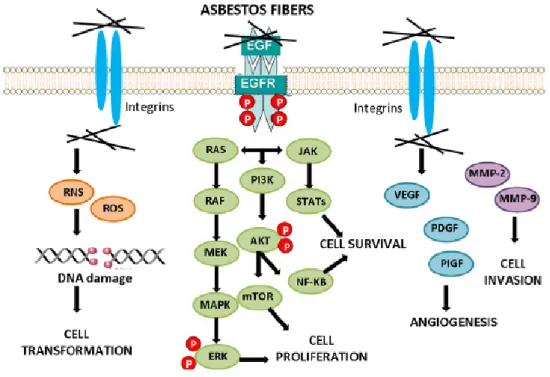
Moreover, in mesothelial cells the internalization of asbestos fibers via integrins or other receptors causes the activation of downstream signaling cascades conducing to cell transformation, cancer cell survival, proliferation, angiogenesis and invasion (Benedetti et al., 2015) (Figure 4). Finally, asbestos exposure induces in macrophages and mesothelial cells the
19
production of reactive oxygen species (ROS) and reactive nitrogen species (RNS), which are responsible of DNA damage, DNA mutations and genome instability (Liu et al., 2000).
Figure 4: Cell signaling activation by asbestos
1.1.3 Therapeutic approaches to MPM
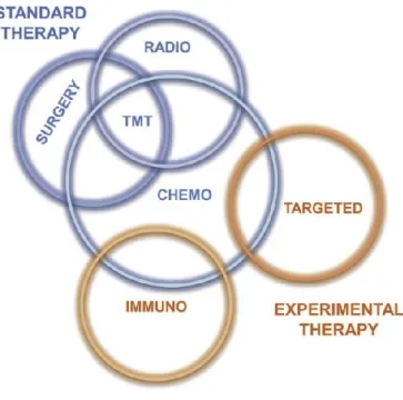
Conventional therapeutic approaches to MPM are surgical treatment, radiotherapy and chemotherapy (Figure 5). Several factors influence the choice of the best therapeutic strategy including tumour stage, histological differentiation and the patient’s status.
• Surgical approach: two surgical procedures are used in MPM treatment. Pleurectomy/decortication (P/D) that involves the radical removal of all visible disease of the pleura, both the inner and outer lung lining; extra-pleural pneumonectomy (EPP), a type of more radical surgical option which aims to eradicate all macroscopic tumours via the
20
removal of the areas surrounding it (Butchart et al., 1976). However, several studies demonstrate that surgery can bring benefits only in the early stages of the disease (Cao et al., 2014; Wald and Sugarbaker, 2016). In fact, only in stage I and II the tumour can be completely removed ensuring a favorable prognosis and a high survival rate (Ismail-Khan et al., 2006). On the contrary, surgery alone has proved ineffective in the treatment of advanced stage malignant mesothelioma because it does not improve survival rates; in fact, surgical procedures provide in these patients only a temporary relief reducing pain and controlling pleural effusions (Bibby et al., 2016).
• Radiotherapy: this approach alone, offer only symptom palliation because it does not affect tumour progression (Ung et al., 2006).
• Chemotherapy: in the past, this approach has been the only therapeutic approach used for the treatment of MPM in advanced states (Nowak, 2012). Chemotherapy alone is recommended for patients who are not operable (stage IV) and/or show sarcomatoid histology (Mott, 2012). All chemotherapeutic agents have been tested both in vitro than in vivo against MPM (Janne, 2003; Tomek et al., 2004). The most commonly used drugs for MPM are cisplatin, pemetrexed, methotrexate, gemcitabine, carboplatin, mitomycin, doxorubicin, epirubicin, vincristine, vinblastine cyclophosphamide, nintedanib, ifosfamide, ranpirnase and vinorelbine (Ismail-Khan et al., 2006; Sobhani et al., 2017). These drugs can be used alone or combined with each other increasing antitumor effect. For example, Vogelzang and colleagues demonstrated that pemetrexed/cisplatin combination is more effective in MPM than cisplatin alone (Vogelzang et al., 2003); in fact, in this phase III trial pemetrexed in combination with cisplatin compared to cisplatin monotherapy increases median survival from 9 to 12 months in advanced stage MPM patients (Vogelzang et al., 2003). Other chemotherapy combinations have been tested to treat MPM; however, the
21
combination between a platin compound and a folate antagonist represents the standard approach for advanced MPM in the last 15 years (Bonelli et al., 2017). In conclusion, chemotherapy may alleviate symptoms, improve quality of life and prolong survival in advanced stage MPM patients but is no curative (Bibby et al., 2016; Rossini et al., 2018). • Multimodality therapy (TMT): represents a more effective strategy and consists in
combining two or more different methods of treatment, such as surgery, radiation therapy, and chemotherapy (Baas et al., 2015; Rossini et al., 2018). For example, a recent study demonstrates that a trimodal approach, such as EPP with chemotherapy and radiotherapy, increase median survival to 18-24 months (Kishimoto et al., 2016). However, this approach can be applied only in patients with an operable tumour and a good performance status.
Figure 5: Therapeutic strategies to malignant mesothelioma
Conventional treatments for mesothelioma are ineffective and patient prognosis remains very poor. The failure of standard therapies pushed researchers to development alternative therapies which can be used alone or in combination with conventional treatments (Figure 5):
22
• Immunotherapy: it is a new therapeutic approach, based on the administration of drugs and therapeutic agents able to stimulate the immune system to target cancer cells promoting an antitumour immune effect (Farkona et al., 2016). This strategy can be used to treat many cancer types including MPM (Bograd et al., 2011; Voena and Chiarle, 2016). Immunotherapy represents a promising strategy for MPM treatment because it has been shown that lymphocyte infiltration to the tumour correlated with better prognosis in MPM patients (Leigh and Webster, 1982; Grégoire, 2010). Antitumour immunotherapy can be performed in two ways: passive immunotherapy and active immunotherapy. Active immunotherapy consists in administering one or several antigens through vaccination able to activate the immune system against cancer cells; passive immunotherapy relies on effectors isolated and activated in vitro and then re-injected (Grégoire, 2010). This last approach is used when the immune system is compromised. Numerous clinical trials of immunotherapy against MPM have been performed. For example, cytokines, monoclonal antibodies, and activated T lymphocytes have been used to stimulate the immune system against cancer cells (Astoul et al., 1998; Grégoire, 2010; Wong et al., 2014). Moreover, in other clinical trials have been used cell vaccines contain dendritic cells loaded with tumour-associated antigens to stimulate immune system against MPM cells (Hegmans et al., 2010). However, though immunotherapy gives excellent results in treating numerous cancer types, most clinical trials reporting negative results for MPM.
• Target therapy: consists in the targeting of pathways already known to be dysregulated in MPM, such as epidermal growth factor receptor (EGFR) pathway, vascular endothelial growth factor receptor (VEGFR) pathway, phosphatidylinositol-3 kinase (PI3K)/protein kinase B (AKT)/mammalian target of rapamycin (mTOR) pathway and Notch pathway, using small molecules with inhibitory activity (Sobhani et al., 2017). For
23
example, promising results have been obtained targeting Notch pathway (Rossini et al., 2018). In other clinical trials VEGFR or EGFR inhibitors have been tested alone or in combination with chemotherapy in MPM patients, but with poor results (Guazzelli et al., 2017). In a phase II trial a specific antibody anti-mesothelin (MORab-009) has been used in combination with pemetrexed and cisplatin in patients with advanced MPM; preliminary results shown a tumour regression in patients after treatment (Hassan et al., 2014). Target therapy represents a promising strategy in MPM therapy, but further studies are required.
1.1.4 Metabolic reprogramming and stromal functions
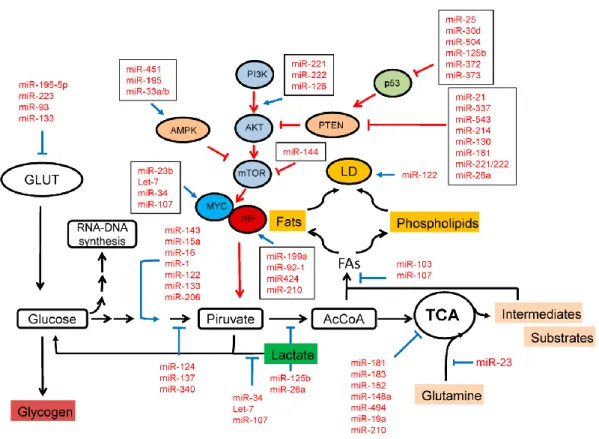
During malignant transformation cells modify their metabolism to support enhanced cell growth and proliferation (cell plasticity); the metabolic reprogramming (metabolic switch) sustains high energetic and metabolic demands and confers these cells a selective growth advantage and/or resistance to apoptosis (Pavlova and Thompson, 2016; Gentric et al., 2017). Many metabolic changes occur in cancer cells including increase of glycolysis and glutaminolytic flux, upregulation of amino acid and lipid metabolism, enhancement of mitochondrial biogenesis, induction of pentose phosphate pathway and macromolecule biosynthesis (Hanahan and Weinberg, 2011; Ward and Thompson, 2012) (Figure 6). During metabolic switch the most change is the increased of glycolysis, the main source of ATP for cancer cells (Gentric et al., 2017); in fact, cancer cells use glycolysis over mitochondrial oxidative phosphorylation (OXPHOS), to produce most of their ATP even under aerobic conditions (Warburg effect) (Warburg, 1956; Gogvadze et al., 2010; Xu et al., 2015). Cancer cells are characterized by an increased glucose uptake, a high glycolytic flux and an impaired mitochondrial respiration (Gogvadze et al., 2010; Gentric et al., 2017). Aerobic glycolysis helps cancer cells to sustain high energetic demand producing ATP in greater
24
quantities and at a faster rate compared with OXPHOS (Guppy et al., 1993). In cancer cells most of pyruvate (about 90%) is converted in lactate by lactate dehydrogenase (LDH). It has been shown that lactate production contributes to tumour formation and progression; for example, pyruvate conversion in lactate helps to regenerate NADH accelerating glycolysis (DeBerardinis et al., 2008); moreover, secreted lactate may act as fuel to other cancer cells (Semenza, 2008). Finally, lactate lowers the pH of extracellular microenvironment promoting cell invasion and metastasis (Bonuccelli et al., 2010; Martinez-Outschoorn et al., 2011). The high rate of glycolysis and suppressed gluconeogenesis help to maintain a low level of intracellular glucose, creating a gradient favouring the flux of glucose into the cell and create microenvironmental acidosis, compelling the evolution of phenotypes with stress resistance and metastatic capacity (Tomasetti et al., 2014). However, cancer cells exert the ability to switch from oxidative phosphorylation to aerobic glycolysis and vice versa (Tomasetti et al., 2014; Gentric et al., 2017). In fact, cancer cells exert increased metabolic plasticity that allows them to continuously adapt to changes in the tumour environment. Moreover, many non-glucose nutrients, such as amino acids, lactate, acetate, and macromolecules, can be used as alternative fuels for cancer cells confirming metabolic heterogeneity within tumour (Keenan et al., 2015; Gentric et al., 2017). For example, mitochondrial fatty acid oxidation (FAO) provides an alternative pathway to support cancer cell survival in glucose-limiting conditions by producing acetyl-CoA (Tomasetti et al., 2014). Moreover glycolysis, in addition to producing ATP, provides cancer cells necessary precursors for biosynthesis: for example, glucose-6-phosphate is often consumed by pentose phosphate pathway to synthesize nucleotides and NADPH (Shaw, 2006); similarly, cancer cells utilize 3-fosfoglicerate (3-PG) for de novo synthesis of amino acids and fatty acids (Jones and Thompson, 2009). Several factors promote glycolytic switch including oxidative stress, OXPHOS reduction, altered miRNA expression profile, tumour
25
suppressor downregulation (such as p53) and oncogenes overexpression (such as c-Myc) (Gogvadze et al., 2010; Nilsson et al., 2012; Bensinger and Christofk, 2012). Glutamine plays a key role in cancer cells supporting energetic demands (Griffiths and Keast, 1990; Cluntun et al., 2017). It has been shown that the rapid conversion of glutamine to lactate (glutaminolysis) produce sufficient NADPH to support fatty acid synthesis (DeBerardinis et al., 2007). Moreover, in cancer cells, glutamine assures the maintenance of the tricarboxylic acid (TCA) cycle function restoring oxaloacetate (anaplerosis) (DeBerardinis et al., 2007). In fact, glutamate derived from glutamine can be converted to α-ketoglutarate (α-KG) in mitochondria; then, α-KG enters in TCA cycle to restore oxaloacetate (OAA) (Dang, 2010). Finally, glutamine serves as an important source of reduced nitrogen for biosynthetic reactions, as a source of carbon to produce glutathione and as a precursor to nucleotides and lipid synthesis via reductive carboxylation (Cluntun et al., 2017). In cancer cells, TCA is primarily involved in the production of precursors for macromolecule biosynthesis (Jones and Thompson, 2009); for, example citrate can be exported to the cytosol to be converted into acetyl-CoA and OAA by the ATP citrate lyase (ACL) enzyme (DeBerardinis et al., 2008). Then, acetyl-CoA may be used for de novo synthesis of lipids (fatty acids, cholesterol, sphingomyelin, isoprenoids) and for protein acetylation (Jones and Thompson, 2009).
26
Figure 6: Metabolic reprogramming in cancer cells
Several oncogenes such as Myc, Ras, PI3K/AKT and hypoxia-inducible factor 1 (HIF-1) promote cancer reprogramming upregulating enzymes of glycolysis, pentose phosphate pathway and synthesis of lipids, proteins and nucleic acids (DeBerardinis et al., 2008; Phan et al., 2014). Moreover, oncogenes increase glucose and glutamine uptake promoting synthesis and translocation of their respective transporters (Phan et al., 2014).
27
Metabolic changes have been observed also in MPM during neoplastic transformation. Some studies show that the activation of PI3K/AKT pathway by insulin receptor substrate 1 (IRS1) protein in MPM cells plays a key role in neoplastic transformation promoting metabolic reprogramming, cell proliferation and angiogenesis (Zhou et al., 2014); moreover, MPM cells are characterized by high levels of enzymes lactate dehydrogenase (LDH) and ACL, a reduced gluconeogenesis, a high glucose uptake and an increased RC (Mullen et al., 2011; Tomasetti et al., 2016).
According to these notions, metabolic reprogramming plays a key role in tumour formation and progression; targeting cancer cell metabolism might be an effective strategy for cancer therapy.
Moreover, cancer cells during malignant transformation recruit from the local host stroma, several normal cell types, including vascular endothelial cells, pericytes, adipocytes, fibroblasts, and bone-marrow mesenchymal stromal cells, to become part of the tumour microenvironment (Werb and Lu, 2015). Tumour-associated stromal cells (TASCs) play a key role in tumour development and progression supporting tumour growth, angiogenesis, invasion, metastasis and therapy resistance (Bussard et al., 2016). In fact, these stromal cells surround the tumour to form a distinct microenvironment acting as a barrier which protect and sustain tumour. For example, cancer-associated fibroblasts (CAFs) maintain an optimal microenvironment for cancer cell survival and proliferation (Cirri et al., 2012; Marsh et al., 2013); activated endothelial cells govern tumour neoangiogenesis and promote cancer inflammation and metastasis (Franses et al., 2013); activated immune cells inhibit antitumor immune responses, induce chronic inflammation in the tumour microenvironment, promote angiogenesis, and cancer cell survival and proliferation (Zamarron and Chen, 2011). Moreover, during stromal cell activation, cancer cells induce a metabolic reprogramming in stromal components to sustain tumour energetic and metabolic demands (Cirri and Chiarugi, 2011;
28
Martinez-Outschoorn et al., 2014). For instance, it has been shown that cancer cells increase aerobic glycolysis (Warburg effect) in cancer-associated fibroblasts, resulting in the stromal production of energy-rich metabolites (such as L-lactate, pyruvate, ketone bodies) and precursors for biosynthesis of macromolecules (such as amino acids, nucleotides and fatty acids) (Balaraman, 2017). Moreover, TASCs collectively adapt, in a dynamic manner, their metabolism to cover the needs of cancer cells (Lopes-Coelho et al., 2018).
These evidences suggest that targeting stromal components represent a promising strategy against cancer. All these metabolic pathways are regulated by small non-coding microRNAs (miRNAs). It has been demonstrated that miRNAs play a key role in malignant transformation sustain metabolic reprogramming, proliferation, invasion, angiogenesis and communication among stromal components (Tomasetti et al., 2016; Rutnam and Yang, 2012; Gu et al., 2017) (Figure 7).
29
1.2 MiRNAs
MiRNAs are short single-strand non-coding RNAs of approximately 20-24 nucleotides,
processed from precursors with a characteristic secondary structure, that regulated gene
expression at transcriptional or post-transcriptional level (Ambros, 2004). MiRNAs regulate
the expression of large numbers of genes involved in crucial biological processes, including
development, cell proliferation, differentiation, apoptosis and maintenance of cellular
homeostasis (Alvarez-Garcia and Miska, 2005; Kloosterman et al., 2006). However, most miRNAs act at the post-transcriptional level by blocking the translation of target mRNAs
(Ambros, 2004).
1.2.1 MiRNAs and cancer: role in tumour formation and progression
Alterations in miRNA regulation lead to the development of genetic, metabolic and degenerative diseases, including cancer. Several studies demonstrate that miRNAs are involved in metabolic reprogramming of the transformed cells (Ward and Thompson, 2012) and in the regulation of the tumour-stroma cross talk (Wang et al., 2017; Rupaimoole et al, 2016). MiRNAs can function as tumour promoters or tumour suppressors by targeting cancer-related genes involved in important processes of cancer progression, including inflammatory response, angiogenesis, cell survival, proliferation, migration and invasion (Calin and Croce, 2006; Bhatti et al., 2009; Liu et al., 2018).
The “oncomirs” promote tumour development by negatively inhibiting tumour suppressor genes and/or genes that control cell differentiation or apoptosis. Oncomirs are significantly overexpressed in various tumours because of gene amplification, epigenetic mechanisms or transcriptional dysregulation (Zhang et al., 2007). For example, miR-17-92 cluster is overexpressed in many neoplastic diseases including breast, colon, lung, pancreas, stomach and
30
prostate cancer (Matsubara et al., 2007; Sylvestre et al., 2007). In humans, this cluster encodes six miRNAs (miR-17, miR 18a, miR-19a, miR-20a, miR-19b-1 and miR-92-1), is located at chromosome 13q31 and promotes angiogenesis, tumour growth and progression (Ota et al., 2004). Other miRNAs frequently overexpressed in cancer are miR-155, miR-21, miR-221, miR-222, miR-93 and many others (Kim et al., 2013; Baumhoer et al., 2012, Lu et al., 2012; Ganci et al., 2016). For example, increased expression of miR-103, miR-21 and miR-107 has been found in primary pancreatic tumours (Roldo et al., 2006; Asangani et al., 2008). Also, miR-155 acts as an oncogene through its action on tumour protein 53-induced nuclear protein 1 (TP53INP1) (Gironella et al., 2007). Furthermore, miR-141 and miR-200c, members of the miRNA-200 family, act as oncogenes by targeting and downregulating zinc-finger E-box binding homeobox 1 (ZEB1), a crucial inducer of epithelial-mesenchymal transition in various human tumours. High expression levels of let-7g, miR-181b, and miR-200c have been found in colon cancer (Nakajima et al., 2006).
Currently, many miRNAs act as oncosuppressors. Tumour suppressor miRNAs are miRNAs significantly downregulated in cancer diseases. Tumour suppressor miRNAs inhibit tumour development and progression by positively regulating tumour suppressor genes and inhibiting genes that promote proliferation, cell survival, migration, invasion and angiogenesis (Reddy et al., 2015; Lynam-Lennon et al., 2009). For example, the ectopic restoration of miR-340 inhibits migration, invasion, and metastasis of breast cancer cells by targeting Wnt pathway (Mohammadi-Yeganeh et al., 2016). Other known oncosuppressor miRNAs are let-7 and miR-34. Both miRNAs are frequently lost in cancer and negatively regulate multiple cell cycle-related oncogenes, such as Ras and Myc (Johnson et al., 2005; Sampson et al., 2007). Increasing evidences show that other miRNAs frequently downregulated in cancer are 145 and miR-126. It was reported that miR-145 acts as tumour suppressor in numerous human cancers,
31
including oral squamous cell carcinoma (OSCC) (Shao et al., 2013), nasopharyngeal carcinoma (Wu et al., 2015), non-small cell lung cancer (Ye et al., 2015), bladder cancer (Kou et al., 2014) and MPM (Cioice et al., 2014; De Santi et al., 2017). MiR-145 loss was reported to have pro-tumourigenic effects in MPM cells modulating clonogenicity, cell migration and resistance to drugs (Cioice et al., 2014). MiR-126 restoration produces a loss of malignancy in many cancer types including MPM, targeting vascular endothelial growth factor A (VEGF-A) (Chen et al., 2015), ADAM metallopeptidase domain 9 (ADAM9) (Wang et al., 2016), sex determining region Y-box 2 (Sox2) (Zhao et al., 2015), CRK, IRS1 (Tomasetti et al., 2014; Tomasetti et al., 2016) and phosphatidylinositol 3-kinase regulatory subunit beta (PI3KR2) (Du et al., 2014; Liu et al., 2014).
All stages of tumour progression, from neoplastic transformation to tumour growth and metastasis formation, are due to the concomitant downregulation of the oncosuppressor miRNAs and overexpression of the oncomirs (Calin and Croce, 2006; Xing et al., 2015). Evidences show that altered miRNA expression profile promotes metabolic reprogramming, including OXPHOS inhibition, lipid droplet (LD) accumulation, glycolytic shift and altered mitochondrial metabolism, during neoplastic transformation (Ward and Thompson, 2012; Tomasetti et al., 2016).
The tumour microenvironment (TME) or stroma is the cellular environment in which the tumour exists, and it is composed of blood vessels, extracellular matrix (ECM) and diverse types of non-malignant cells, including cancer-derived fibroblasts (CAFs) and immune cells (such as lymphocytes, macrophages, dendritic cells and mast cells) (Whiteside, 2008; Catalano et al., 2013). TME is essential for tumour growth and progression. It has been found that TME promotes cell proliferation, migration and invasion, supports extracellular remodelling and contributes to treatment resistance though production of various growth factors, chemokines
32
and cytokines (Bussard et al., 2016; Xiong et al., 2015). Recent studies have shown that miRNAs act as mediators between cancer cells and the TME (Kohlhapp et al., 2015; Chou et al., 2013; Wang et al., 2017). In fact, in tumour, neoplastic cells communicate with each other and with the other components of the stroma through the release of extracellular vesicles (such as exosomes) containing also miRNAs (Hu et al., 2015; Paggetti et al., 2015). Moreover, miRNAs in cancer associated-extracellular vesicles support metastasis by inducing pre-metastatic niche formation (Psaila et al., 2009). Cancer cells promote stroma formation by recruitment and reprogramming of somatic cells (such as normal fibroblasts and endothelial cells). Previous studies have shown that cancer cells modulate stromal cells by changing their miRNA expression profile. For example, Mitra and colleagues have found that ovarian cancer cells contribute to normal fibroblast-CAF transition by altering miRNA profile (Mitra et al., 2012). Moreover, through manipulation of the miRNA expression of endothelial cells, cancer cells can reprogram these cells to enhance their angiogenic potential (Png et al., 2012).
We can conclude that miRNA dysfunction contributes to tumour progression and development. An altered miRNA expression profile has been observed also in mesothelioma cells (Santarelli et al., 2011; De Santi et al., 2017). This alteration confirms the involvement of miRNAs also in MPM carcinogenesis. Among miRNAs which are upregulated in MPM cells, there are the miR-17-92 cluster, miR-625-3p and miR-34b/c; while the most important miRNAs which act as oncosuppressors are miR-31, miR-221, miR-222, miR-126, miR-1 and miR-15 and miR-145 (Xu et al., 2013; Bonci et al., 2008; Muraoka et al., 2013; Tomasetti et al., 2012; De Santi et al., 2017).
33
1.2.2 MiRNAs in diagnosis and therapy
The miRNA profiles are surprisingly informative and can be used in the diagnosis of cancer. Many studies show that miRNA expression profile can accurately differentiate tumour from benign tissue (Lu et al., 2005; von Brandenstein et al, 2012), distinguish cancer histological types (Calura et al, 2013) and diagnose the tissue source of metastatic cancer (Rosenfeld et al., 2008; Gilad et al., 2008); moreover, miRNA profile by reflecting cancer stages, can be used to diagnose cancer at the early stages (Pal et al., 2015). MiRNAs have also been reported as prognostic markers for a multitude of neoplastic diseases including ovarian (Eitan et al., 2009), pancreatic (Roldo et al., 2006), lung (Yu et al., 2008) and breast cancer (Lowery et al., 2009), and as predictive biomarkers to evaluate the efficacy of treatments (Dreussi et al, 2016).
Major factor contributing to the poor prognosis is that MPM is detected at advanced stage. Research is focused in finding miRNAs that can distinguish MPM at early stages (Santarelli et al., 2011; Micolucci et al., 2016). It was reported that miRNA profiles can distinguish MPM from non-neoplastic diseases, such as reactive mesothelial proliferation (RMP) (Andersen et al., 2012), and from other lung neoplastic diseases such as lung cancer adenocarcinoma (Andersen et al., 2014). Furthermore, Busacca et al., discovered that the expression of miR-17-5p, miR-21, miR-29a, miR-30c, miR-30e-5p, miR-106a, and miR-143 was significantly associated with the histopathological subtypes of MPM; the authors shown that it is possible to establish exactly the subtype of MPM by measuring the levels of these miRNAs in the biopsy samples (Busacca et al., 2010). Indeed, miRNA profiles can also be used as prognostic and predictive biomarkers in MPM. For example, patients that overexpressed hsa-mir-29c and miR-31 show a favourable prognosis after surgical resection (Pass et al., 2010).
34
1.2.3 MiRNAs in liquid biopsy
MiRNAs can be detected in biological fluids, primarily blood (plasma or serum), but also in saliva, cerebrospinal fluid, urine, and milk (Weber et al, 2010; Mitchell et al, 2008). Although the current gold standard of cancer diagnosis is represented by tissue biopsy, it is spreading the possibility to use the “liquid biopsy” (Larrea et al, 2016). Circulating miRNAs can be easily detected and can be used for cancer diagnosis at the early stages and to valuate efficacy of cancer therapies (Levy et al., 2016; Jung and Kirchner, 2018).
1.2.4 MiRNA-based cancer therapy
By playing a central role in cancer initiation, progression and metastasis, miRNAs might be potential therapeutic targets. Therefore, miRNA-based therapy is one of the challenges in the future (Shah and Calin, 2014). The therapy consists in the restoration of normal levels of miRNAs involved in carcinogenesis; for this purpose, there are two strategies of therapy: tumour suppressor miRNAs are replaced using synthetic miRNA-like molecules (mimics); oncogenic miRNAs, frequently overexpressed in cancers, can be silenced to restore the normal expression of their target tumour suppressor genes (Bhardwaj et al., 2010; Gambari et al., 2016) (Figure 11). Oncogenic miRNAs can be inhibited by using antisense oligonucleotides, antagomirs, sponges or locked nucleic acid (LNA) constructs (Garzon et al., 2009; Ma et al., 2010). Oncogenic miRNA inhibition can occur through two ways: antagomirs, antisense oligonucleotides or LNA constructs, complementary to the target miRNAs and able to silence its activity (Krutzfeldt et al., 2005). Instead, “sponge” constructs are sequences expressed from strong promoters, containing multiple binding sites that compete with the endogenous miRNA targets for miRNA binding (Ebert et al., 2007). The inhibition of oncogenic miRNAs seems to be a promising strategy to cancer treatment (Song and Rossi, 2013; Garzon et al., 2009). For
35
example, in mice antagomir-miR-10b, reducing miR-10b levels in cancer cells, inhibits metastasis to lungs (Melo and Kalluri, 2012). “Mimics” are synthetic miRNA-like molecules, which can replace downregulated miRNAs. MiRNA mimetics are small, chemically modified single-stranded RNA molecules designed to mimic endogenous mature miRNAs (Chorn et al., 2012). Numerous studies have demonstrated the efficacy of miRNA replacement therapy. For example, miR-16 restoration affects proliferation and increases drug sensitivity in MPM cells (Reid et al., 2013). Furthermore, Let-7 restoration inhibits the growth of lung, liver and pancreatic cancer cells (Takamizawa et al., 2004; Johnson et al., 2005).
Figure 11: MiRNA-based therapeutic strategies against cancer
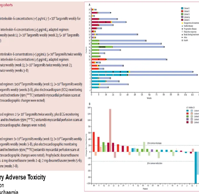
van Zandwijk and colleagues at the Asbestos Diseases Research Institute (ADRI), Australia (van Zandwijk et al., 2017), first performed a phase I trial aimed to evaluate safety and biological activity of miR-16 in patients with recurrent MPM. Patients (26) were given TargomiRs via 20 min intravenous infusion either once or twice a week in a traditional 3+3 dose-escalation design in five dose cohorts. The dose-escalation steps planned were 5×10⁹,
36
ischaemia in two patients. A patient showed anaphylaxis and cardiomyopathy who received 5×10⁹ TargomiRs once weekly, which was stablished as the maximum tolerated dose. In
addition, TargomiR infusions were accompanied by transient lymphopenia and temporal hypophosphataemia. In spite of the low toxicity, the proportion of patients who achieved an objective response was only 5%, and the duration of the objective response in that patient was 32 weeks. Median overall survival was 200 days (95% CI 94-358). During the trial, 21 deaths occurred, of which 20 were related to tumour progression and one was due to bowel perforation (Figure 12). A major hurdle in interpreting the data of the phase I study is related to the rapid disappearance of TargomiRs from the circulation after infusion. Moreover, immune reactions may occur shortly after the infusion of TargomiRs, and provide an explanation for the antitumour activity observed. The authors concluded that the unmet need of mesothelioma patients is very high. On the basis of these preclinical data combination therapy seems a logical next step in TargomiR development. Carefully planned trials with a smart/clean design and sufficient attention for future (predictive) biomarkers are obviously the way forward. As indicated earlier, optimal attention for the pharmacology of TargomiRs with pre-and post-treatment biopsies is needed to maximally explain the mechanism of action of this novel treatment approach, for mesothelioma as well as for other oncology indications.
37
38
1.2.5 MiR-126 as potential TargomiR
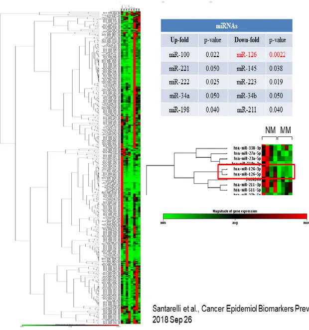
Recently, by screening MPM tissues and their non-malignant adjacent counterparts for 384 miRNAs a miRNAs panel was identified able to detect the asbestos-related malignancies including MPM and non-small cells lung cancer (NSCLC) from asbestos exposure (Santarelli et al., 2018). Among deregulated miRNAs in MPM, miR-126 was found significantly downexpressed in malignant tissues respect to non-malignant counterparts (Figure 13).
39
Low levels of miR-126 were found also in many other neoplastic diseases, including gastric cancer (Feng et al., 2010), breast cancer (Zhu et al., 2011), renal cell carcinoma cancer (Khella et al., 2015), pancreatic cancer (Jun et al., 2012), non-small cell lung carcinoma (NSCLC) (Yanaihara et al., 2006), colon cancer (Gou et al., 2008), squamous cell oral carcinoma (OSCC) (Sasahira et al., 2012), esophageal squamous cell carcinoma (Li et al., 2014), glioma (Luan et al., 2015), melanoma (Felli et al., 2013), osteosarcoma (Jiang et al., 2014) and bladder cancer (Jia et al., 2014). Given that miR-126 was downregulated in many malignancies highlights its role as tumour suppressor. In cancer cells downregulation of miR-126 occurs via several mechanisms. In many types of cancer, including MPM, downregulation of miR-126 was due to promoter hypermethylation of its host gene epidermal growth factor-like protein 7 (Egfl7) by DNA methyltransferase 1 (DNMT1) (Andersen et al. 2015; Saito et al., 2009; Zhang et al., 2013), which was aberrantly upregulated in these cells. Intriguingly, miR-126 restoration suppressed DNMT1, indicating the existence of a regulatory feedback circuit (Andersen et al. 2015). Furthermore, even the production of mitochondrial ROS in MPM cells would appear to be responsible of miR-126 downregulation (Tomasetti et al., 2014).
Some studies have shown that miR-126 inhibits tumour progression by negatively controlling cellular processes and signaling pathways. MiR-126 targets including proteins involved in cell cycle control (as PIKER2), in angiogenesis (as VEGF-A), in cell migration (as MERTK), in proliferation and development; furthermore, miR-126 also regulates the expression of some oncosuppressor genes such as p53 and nasopharyngeal carcinoma-associated gene 6 (NGX6). The transcription factor p53 is the master tumour suppressor; it binds to more than 4,000 sites in the genome and regulates the expression of more than 500 genes (Simabuco et al., 2018). This transcription factor regulates genes involved in almost all hallmarks of cancer. It
40
has been demonstrated that ectopic miR-126 increased p53 protein expression (Ebrahimi et al., 2015).
MiR-126 inhibits cancer progression via negative control tumour cell proliferation, migration, invasion and survival both in vitro than in vivo (Zhou et al., 2013; Yuan et al., 2016). Overexpression of miR-126 inhibits proliferation in many cancer types including colon cancer (Zhou et al., 2013; Yuan et al., 2016), osteosarcoma (Jiang et al., 2014), small cell lung cancer (Miko et al., 2011; Jia et al., 2018), breast cancer (Zhang et al., 2013), pancreatic cancer (Hamada et al., 2012), chronic myeloid leukaemia (Taverna et al., 2014), prostate cancer (Hua et al., 2018) and malignant mesothelioma (Tomasetti et al., 2014) via different mechanisms. For example, in lung cancer and MPM cells, miR-126 affects proliferation by targeting the PI3K/AKT/Snail signaling pathway (Jia et al., 2018; Tomasetti et al., 2014). Elevated miR-126 levels also impair cellular migration and invasion in these previous studies. Furthermore, in vivo experiments, ectopic miR-126 significantly reduces the volume of tumour, micro-vessel density and metastasis (Hua et al., 2018; Hu et al., 2016). Png and colleagues, discovered that miR-126 suppresses breast cancer metastasis to lung and bone. The authors found that this miRNA inhibits metastatic endothelial recruitment, metastatic angiogenesis and metastatic colonization targeting insulin-like growth factor binding protein 2 (IGFBP2), phosphatidylinositol transfer protein cytoplasmic 1 (PITPNC1) and tyrosine-protein kinase mer (MERTK), both in vivo and in vitro. IGFBP2 secreted by metastatic cells recruits endothelial cells (ECs) by modulating IGF1-mediated activation of the IGF type-I receptor on ECs; whereas MERTK receptor cleaved from metastatic cells promotes endothelial recruitment by competitively antagonizing the binding of its ligand growth arrest-specific protein 6 (GAS6) to endothelial MERTK receptors (Png et al., 2011). Moreover, miR-126 inhibits breast cancer metastasis to lung by suppressing the recruitment of mesenchymal stem cells and inflammatory
41
monocytes into the tumour stroma, both in vitro and in vivo (Zhang et al., 2013). Tomasetti and colleagues demonstrated that ectopic miR-126 inhibits tumorigenic properties of MPM cells reducing cell proliferation and the ability of the cells to form colonies in vitro (Tomasetti et al., 2014). Moreover, ectopic miR-126 inhibits tumour formation and progression also in vivo; the tumour formed by injection of malignant mesothelioma cells (H28) in mice was completely inhibited by miR-126 (H28-miR126) through a mechanism that involves angiogenesis (Figure
14).
Figure 14: Ectopic miR-126 affects tumour progression and angiogenesis
MiR-126 acts as a tumour suppressor by targeting VEGF, Sox2, IRS1 and ADM9 in several cancer types (Liu et al., 2009; Ebrahimi et al., 2014; Tomasetti et al., 2014; Ryu et al., 2011; Zhu et al., 2011). It was reported that VEGF-A, but not VEGF-B/C, is a target of miR-126 and
42
downregulation of miR-126 induces angiogenesis and lymphangiogenesis by activation of VEGF-A (Sasahira et al., 2012).
MiR-126 has been proposed to modulate the PI3K/AKT/mTOR signaling pathway targeting p85b of PI3K (Guo et al., 2008) and negatively regulating the IRS1 (Zhang et al., 2008) Activation of the PI3K/AKT/mTOR pathway is one of the most common events in cancer (Chen et al., 2014) and it has been shown to promotes angiogenesis (Zhu et al., 2011), proliferation (Adlung et al., 2017), migration (Xu et al., 2018), invasion (Xu et al., 2018) and metabolic re-programming (Elstrom et al., 2004; Ward et al., 2012). It has been found that miR-126 targeting IRS1 inhibits AKT pathway activation and produces a loss of malignancy in MPM cells (Tomasetti et al., 2014). Moreover, in lung cancer miR-126 targeting TGF-β1a inhibits PI3K/AKT/Snail signaling and blocks epithelial-to-mesenchymal transition (EMT) a crucial event in cancer development and progression (Jia et al., 2018).
ADAM9 was identified as a key target of miR-126 (Felli et al., 2013; Jiang et al., 2014). In esophageal squamous cell carcinoma and in osteosarcoma, it has been demonstrated that miR-126 restoration reduced cell proliferation, migration and invasion by inhibiting EGFR–AKT signaling pathway (Jiang et al., 2014; Liu et al., 2015); miR-126 targeting ADAM9 3’ UTR inhibits EGFR phosphorylation and activation: ADAM9 cleaves the pro-heparin-binding EGF-like growth factor (pro-HB-EGF) and releases soluble HB-EGF that binds to EGFR. In this way, the downstream EGFR signaling cascade is activated, such as the AKT pathway. Blockage of ADAM9 by miR-126 inhibits EGFR–AKT activation (an important pathway for cell growth and migration) (Liu et al., 2015).
Metabolic reprogramming of cancer cells is essential for their adaptation to tumour microenvironment and for maintenance of tumour growth (Cairns et al., 2011). MiR-126 has
43
been reported to suppress progression in several cancer types including MPM by affecting cellular metabolism (Tomasetti et al., 2014; Tomasetti et al., 2016) (Figure 15). MiR-126, targeting IRS1, inhibits insulin-signaling pathway and suppresses glucose uptake. Energy depletion promotes AMPK-dependent phosphorylation of ULK1 and the subsequent lysosomal autophagy (Tomasetti et al., 2016). Moreover, miR-126 targeting IRS1 induces nuclear translocation of (forkhead box O1) FoxO1 in MPM cells via the inhibition of IRS1/AKT pathway. In these cells, FoxO1 represses cell cycle and promotes the transcription of genes involved in glucose metabolism, in mitochondrial function and in gluconeogenesis, such as glucose-6-phosphatase (G6PC) and phosphoenolpyruvate carboxykinase 1 (PCK1) (Zhang et al., 2011; Dong et al., 2008). Furthermore, FoxO1 induces the expression of CAT and MnSOD genes with a reduction of mitochondrial ROS (Kops et al., 2002). Enhanced ROS production in cancer drives the onset of aerobic glycolysis, with lactate and ketone production promoting mitochondrial biogenesis and anabolic growth of tumour cells. Alleviation of mitochondrial oxidative stress via enhanced expression of antioxidant enzymes targeted to mitochondria was found to be sufficient to lower tumour severity (Sotgia et al., 2011). In addition, miR-126 reduces the ACL activity with accumulation of citrate, which becomes part of the TCA cycle as pyruvate. Citrate restoration blocks de novo synthesis of lipids and the reductive carboxylation (RC) of glutamine (Tomasetti et al., 2014); the RC of glutamine is activated by low citrate level in MPM cells and represents the most carbon source. Furthermore, miR-126 stimulates lipid droplets accumulation in a HIF1α-dependent manner (Tomasetti et al., 2016). In these cells the storage of fatty acids (FAs) in LDs and energy depletion increases autophagy flux that prevents cell proliferation and tumour growth both in vitro that in vivo (Tomasetti et al., 2014; Tomasetti et al., 2016). Moreover, it has been demonstrated that miR-126 affects mitochondrial metabolism inhibiting OXPHOS in MPM cells (Tomasetti et al., 2014).
44
Figure 15: MiR-126 affects cellular metabolism in MPM
In MPM cells, miR-126 alters cellular metabolism by regulating the expression of different target genes (Chakraborty et al., 2016). Main events induced by miR-126 are: repression of OXPHOS, block of lipid de novo synthesis, glycolytic shift, overexpression of CAT and MnSOD genes, and induction of gluconeogenesis and promotion of TCA (Tomasetti et al., 2014; Tomasetti et al., 2016). All these events have been associated with the reduction of malignancy in MPM cells (Tomasetti et al., 2014; Tomasetti et al., 2016).
45
1.2.6 MiR-126 and angiogenesis
MiR-126 gene is located within intron 7 of the Egfl-7 gene on chromosome 9 locus 34.3q (Wang et al., 2008) (Figure 16).
Figure 16: Localization of miR-126 gene within chromosome 9
MiR-126 is a miRNA known to be specifically expressed in endothelial cell lineage, hematopoietic progenitor cells and endothelial cell lines, such as HCAEC (Human Coronary Artery Endothelial cells) and HUVEC (Human Umbilical Vein Endothelial Cells) (Poliseno et al., 2006; McCall et al., 2011). In addition, this miRNA is highly expressed in highly vascularized tissues, such as heart, liver and lung. Hence, miR-126 is classified as “angio-miR” for its involvement in regulating angiogenesis and vascular integrity through the expression in endothelial cells (Wang et al., 2008; Fish et al., 2008). MiR-126 regulates many aspects of endothelial cell biology, including cell migration and proliferation, organization of the cytoskeleton, and capillary network stability (Wu et al., 2009). In vivo, the knockdown of miR-126 in zebrafish (Fish and Srivastava, 2009) and mice (Kuhnert et al., 2008) results in vascular leakage, haemorrhaging, and embryonic lethality. Moreover, it also has been shown that miR-126 downregulation impairs endothelial cell migration during vessel formation and compromises endothelial tube organization (Nikolic et al., 2010). MiR-126 promotes angiogenesis positively regulating the response of endothelial cells to VEGF. In endothelial cells, miR-126 represses negative regulators of the VEGF pathway, including sprouty-related EVH1 domain-containing protein 1 (SPRED1) and PI3KR2 (Sasahira et al., 2012; Zhu et al., 2011); miR-126 targeting SPRED1 and PIK3R2 promotes the activation of PI3K/AKT and
46
RAF1/extracellular signal-regulated kinase (ERK) pathways which positively regulate genes involved in endothelial cell proliferation and migration, and vessel formation (Sasahira et al., 2012; Zhu et al., 2011) (Figure 17).
Figure 17: MiR-126 promotes angiogenesis in endothelial cells
MiR-126 is considered a master regulator of physiological angiogenesis regulating embryonic angiogenesis, adult vascular homeostasis and vascular repair (Chistiakov et al., 2016). During embryonic development miR-126 promotes angiogenesis: EGFL7 and miR-126 are co-expressed from the common EGFL7 promoter and both support differentiation of embryonic stem cells to endothelial progenitor cells (EPCs) and endothelial cells (ECs), ECs maturation and neovessel formation (Le Bras et al., 2010; Nikolic et al., 2010). However, in mature vessels, this miRNA plays an opposite role; in fact, in mature ECs and EPCs miR-126 inhibits angiogenesis targeting EGFL7 (Sun et al., 2010) and VEGF-A (Ge et al., 2015) (Figure 18).
47
Figure 18: MiR-126, key regulator of angiogenesis
Furthermore, miR-126 maintains the quiescent endothelial phenotype associated by inhibiting proliferation and motility. Instead in mature vessels miR-126 exhibits vasculoprotective and atheroprotective properties and seems to be involved in the response of the cardiovascular system to injury and stress. In fact, in a case of vessel injury and/or hypoxia, miR-126 activates EPCs and ECs and contributes to vascular healing and neovessel formation. Several studies support the potential therapeutic use of miR-126 to treat vascular diseases and to improve neovascularization after ischaemia (Van Solingen et al., 2009).
Huang and colleagues have been shown that the cross talk between tumour cells and CAFs in the cancer stroma induces miR-126 downregulation in endothelial cells causing an increase of tube formation. They found that adrenomedullin (ADM) is a pro-angiogenic target of
miR-48
126 and that the concomitance presence of ADM and VEGF-A induces angiogenesis (Huang and Chu, 2014; De Giorgio et al., 2014;) (Figure 19).
Figure 19: Cancer cell–CAF cross talk induces downregulation of miR-126 in ECs
In conclusion, miR-126 acts as pro- or anti-angiogenic factor and its role in angiogenesis depends on cell types, developmental stage, microenvironment and external stimuli (Zhou et al., 2016; Chistiakov et al., 2016).
49
1.3 Exosomes
MiRNAs are involved in cross talk between the stromal populations. Mature miRNAs are released from most cells, often within extracellular vesicles (EVs) and disseminate through the extracellular fluid to reach remote target cells, including tumor cells.
Cell releases in the surrounding environment membrane-enclosed EVs with different intracellular origins, compositions, sizes, structures, functions and modes of formation (Raposo and Stoorvogel, 2013; Colombo et al.,2014). EVs consists of a lipid bilayer membrane that encase a small organelle-free cytosol and complex biological molecules, such as lipids, proteins and nucleic acids, derived from the cell of origin. EVs mediate cell-to-cell communication and thus, once released, modulate the response of their recipient cells in physiological and pathological conditions (Samuelson and Vidal-Puig, 2018; Gradilla et al., 2018). Exosomes are one sub-type of secreted vesicles. Three main types of EVs have been described based on their biogenesis pathways, size, cargo and function: exosomes, microvesicles and apoptotic bodies (Borrelli et al., 2018; Crescitelli et al., 2013) (Figure 20).