18
F-FDG PET/CT for Early Prediction of Response to
Neoadjuvant Lapatinib, Trastuzumab, and Their Combination
in HER2-Positive Breast Cancer: Results from Neo-ALTTO
Geraldine Gebhart1, Cristina Gámez2, Eileen Holmes3, Javier Robles2, Camilo Garcia1, Montserrat Cortés2,Evandro de Azambuja1,4, Karine Fauria5, Veerle Van Dooren4, Gursel Aktan6, Maria Antonia Coccia-Portugal7,
Sung-Bae Kim8, Peter Vuylsteke9, Hervé Cure10, Holger Eidtmann11, José Baselga5,12, Martine Piccart13, Patrick Flamen1*, and Serena Di Cosimo5,14*
1Nuclear Medicine Department, Institut Jules Bordet, Université Libre de Bruxelles, Brussels, Belgium;2PET Unit, IDI, Hospital Universitari de Bellvitge-IDIBELL, L’Hospitalet de Llobregat, Barcelona, Spain;3Frontier Science Scotland (FSS) Ltd., Kincraig, United Kingdom;4Breast European Adjuvant Study Team, Brussels, Belgium;5SOLTI Breast Cancer Research Group, Barcelona, Spain;6GlaxoSmithKline, Philadelphia, Pennsylvania;7Eastleigh Breast Care Centre, Pretoria, South Africa;8Asan Medical Center, University of Ulsan College of Medicine, Seoul, Korea;9Medical Oncology Department, Clinique Sainte Elisabeth, Namur, Belgium; 10Institut Jean Godinot, Reims, France;11University Hospital Kiel, Kiel, Germany;12Massachusetts General Hospital Cancer Center, Harvard Medical School, Boston, Massachusetts;13Department of Medicine, Institut Jules Bordet, Université Libre de Bruxelles, Brussels, Belgium; and14Department of Oncology, Istituto Nazionale dei Tumori, Milan, Italy
Molecular imaging receives increased attention for selecting patients who will benefit from targeted anticancer therapies. Neo-ALTTO (Neoadjuvant Lapatinib and/or Trastuzumab Treatment Optimisation) enrolled 455 women with invasive human epidermal growth factor receptor 2 (HER2)–positive breast cancer and com-pared rates of pathologic complete response (pCR) to neoadjuvant lapatinib, trastuzumab, and their combination. Each anti-HER2 ther-apy was given alone for 6 wk, followed by 12 wk of the same therapy plus weekly paclitaxel. The early metabolic effects of the anti-HER2 therapies on the primary tumors and their predictive val-ues for pCR were assessed in a subset of patients. Methods: Eighty-six patients underwent 18F-FDG PET/CT at baseline and
weeks 2 and 6 of anti-HER2 treatment. An imaging core laboratory provided central validation, and 2 independent reviewers, masked to assigned treatment arm and clinical outcomes, performed con-sensus18F-FDG PET/CT readings. Maximum standardized uptake
value (SUVmax) reductions from baseline were used to measure metabolic response. Results: Seventy-seven of the 86 enrolled patients presented an evaluable baseline 18F-FDG PET/CT scan;
of these, 68 and 66 were evaluable at weeks 2 and 6, respectively. Metabolic responses in the primary tumors were evident after 2 wk of targeted therapy and correlated highly with metabolic responses at week 6 (R25 0.81). pCRs were associated with greater SUVmax
reductions at both time points. Mean SUVmax reductions for pCR and non-pCR, respectively, were 54.3% versus 32.8% at week 2 (P 5 0.02) and 61.5% versus 34.1% at week 6 (P 5 0.02).18F-FDG
PET/CT metabolic response rates at weeks 2 and 6 were 71.6% and 60%, respectively using European Organization for Research and Treatment of Cancer criteria; pCR rates were twice as high for
18F-FDG PET/CT responders than nonresponders (week 2: 42%
vs. 21%,P 5 0.12; week 6: 44% vs. 19%, P 5 0.05). Conclusion:
Early metabolic assessment using 18F-FDG PET/CT can identify
patients with an increased likelihood of pCR after neoadjuvant trastuzumab, lapatinib, or their combination when given with chemotherapy.
Key Words: 18F-FDG-PET/CT; early response assessment;
anti-HER2 drugs
J Nucl Med 2013; 54:1862–1868 DOI: 10.2967/jnumed.112.119271
A
pproximately 20% of all breast cancers (BCs) overexpress or amplify the human epidermal growth factor receptor 2 (HER2), a characteristic associated with poor prognosis, increased prolif-eration, angiogenesis, and resistance to apoptosis (1–3).In combination with chemotherapy, the monoclonal antibody trastuzumab (Herceptin; Genentech), which specifically targets the HER2 extracellular domain, has been shown to improve survival in both the adjuvant and the metastatic settings (4–8). In addition, lapatinib (Tykerb; GlaxoSmithKline), a small-molecule inhibitor that targets the intracellular domain of HER2, has established its benefit in treating HER2-positive metastatic BC when combined with capecitabine or letrozole (9,10).
Preclinical and clinical studies showing that lapatinib resensi-tizes tumors that have progressed on trastuzumab (11–13) provided the first indication that combining the agents to create a dual anti-HER2 blockade may improve the current standard of care even further. Lapatinib and trastuzumab do have partly nonoverlapping mechanisms of action: trastuzumab inhibits ligand-independent HER2 and HER3 signaling (14) and triggers antibody-dependent cellular cytotoxicity (15). By contrast, lapatinib blocks ligand-induced heterodimer signaling and prevents signaling via a fre-quently expressed truncated version of HER2 that could render cells resistant to trastuzumab. Additionally, lapatinib leads to an accumulation of HER2 at the cell surface, enhancing trastuzumab-dependent antibody-trastuzumab-dependent cellular cytotoxicity (12).
Received Jan. 10, 2013; revision accepted May 14, 2013.
For correspondence contact: Geraldine Gebhart, Nuclear Medicine Depart-ment, Institut Jules Bordet, 121 Blvd. de Waterloo, 1000 Brussels, Belgium.
E-mail: [email protected] *Contributed equally to this work. Published online Oct. 3, 2013.
COPYRIGHTª 2013 by the Society of Nuclear Medicine and Molecular Imaging, Inc.
In the neoadjuvant setting, trastuzumab combined with chemo-therapy has been shown to significantly increase pathologic complete response (pCR) (16). According to recent data, higher pCR rates are in turn associated with improved outcomes (17).
We previously reported that treatment with the lapatinib and trastuzumab combination in Neoadjuvant Lapatinib and/or Tras-tuzumab Treatment Optimisation (Neo-ALTTO) resulted in a sig-nificantly higher pCR rate (51.3%) than with either trastuzumab (29.5%) or lapatinib (24.7%) alone. Neo-ALTTO randomized 455 patients from 86 sites in 23 countries to receive either lapatinib alone (n5 154), trastuzumab alone (n 5 149), or both drugs (n 5 152) for 6 wk, followed by the same assigned targeted therapy combined with weekly paclitaxel for 12 wk (18).
One of the trial’s key features was the initial 6-wk biologic win-dow, during which patients received only anti-HER2 drugs. This protocol enabled us to investigate the effect of anti-HER2 therapy alone and search for potential molecular and imaging biomarkers of response. Central to this were 3 serial PET/CT scans with18F-FDG performed in a subset of patients at baseline, week 2, and week 6.
A preplanned secondary objective of Neo-ALTTO was to assess both the early metabolic effects of anti-HER2 blockade on the primary tumors and their predictive value for pCR at the time of surgery. There was also interest in exploring whether 18F-FDG PET imaging would capture a higher rate of metabolic response with the dual HER2 blockade as opposed to single blockade. Neo-ALTTO thus represents the first investigation, to our knowledge, of18F-FDG PET/CT in patients treated with anti-HER2 therapies in the neoadjuvant setting.
MATERIALS AND METHODS Study Design
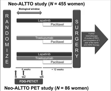
Patients were randomized to 1 of 3 treatment arms: oral lapatinib, intravenous trastuzumab, or their combination administered for 6 wk, followed by 12 wk of the assigned anti-HER2 therapy plus weekly paclitaxel (Fig. 1). Within 4 wk of the last administration of paclitaxel, patients un-derwent definitive breast surgery and were evaluated for pCR, defined as the absence of invasive cancer in the breast (19). The ethics committee and relevant health authorities at each participating institution approved the
study protocol (clinical trial registration number NCT00553358). All women gave written informed consent before study entry.
Evaluation of HER2/Hormone Receptor (HR) Disease
HER2/HR disease is described in the original manuscript of the Neo-ALTTO (18): in brief, HER2 and HR status were determined according to local pathology department criteria. HER2 status was assessed locally at participating institutions that had been accredited by the certified laboratory, Vall d’Hebron Institute of Oncology, Bar-celona.
Selection of18F-FDG PET/CT Study Sites
A feasibility questionnaire was sent to all sites before patient enroll-ment to assess their18F-FDG PET/CT capabilities. Thirty centers in 14
countries were ultimately selected. In these centers, the imaging substudy was proposed to all consecutive patients included in the Neo-ALTTO trial.
18F-FDG PET/CT Procedures
Three18F-FDG PET/CT scans were obtained for each patient: 1
before initiation of neoadjuvant treatment (baseline) and 2 more dur-ing treatment (weeks 2 and 6). All patients were studied usdur-ing a ded-icated18F-FDG PET/CT camera of the brand locally available (GE
Healthcare, Philips, or Siemens). All18F-FDG PET/CT images had to
be taken using the same scanner and identical acquisition parameters. Patients were required to have fasted for at least 6 h before the scan and to have blood glucose levels less than 150 mg/dL before18F-FDG
injection. The injected activity was 3.7–7.4 MBq/kg, with the differ-ence of net injected activity between the baseline and the 2 subsequent scans not to exceed 20%. For all scans, the delay between18F-FDG
injection and start of image acquisition had to be at least 50 min. For scans at weeks 2 and 6, the time interval between tracer injection and start of the PET/CT acquisition needed to fall within 20 min of the interval recorded for the baseline 18F-FDG PET/CT scan. Any18
F-FDG PET/CT scan showing a significant paravenous tracer injection defined as more than 10% of the injected dose was excluded from the analysis. In all 18F-FDG PET/CT centers, the same iterative
recon-struction method was used for the 3 time points, such as ordered-subset expectation maximization or row-action maximum likelihood algorithm, with and without CT-based attenuation correction.
Two nuclear medicine reviewers (in Barcelona and Brussels) retro-spectively assessed the quality of the18F-FDG PET/CT scans and
im-aging analysis. They verified and cross-checked the dedicated PET/CT imaging case report form and the DICOM (Digital Imaging and Com-munications in Medicine) headers. Face-to-face meetings were held by the 2 PET/CT reviewers to reach a consensus on assessments.
Selection of Target Tumor Lesions
Lesions were divided according to their location: breast, nodal (axillary, subclavicular, and internal mammary), and distant.
At baseline,18F-FDG PET/CT target lesions were defined as those
with a maximum standardized uptake value (SUVmax) $ 2.5 and a CT based maximal transaxial diameter$ 1 cm. Lesions not fulfilling these 2 criteria were classified as nontarget. Only target lesions were included in the statistical analysis.
Assessment of Metabolic Response
SUVmax was obtained by drawing a volume of interest in the lesions using the commercial PET VCAR software on an Advantage Workstation (GE Healthcare), which allows a practical approach to comparative analysis (20).
Lesion-Based Metabolic Response Assessment. SUV is a widely used18F-FDG PET/CT quantifier, calculated as the ratio of the
con-centration of the measured tissue radioactivity and the injected radio-activity normalized for patient body weight. SUVmax represents the highest pixel SUV within a tumor and is a continuous variable. For
FIGURE 1. Neo-ALTTO study design. B5 baseline; FEC 5 fluoroura-cil, epirubicin, cyclophosphamide; w25 week 2; w6 5 week 6.
each target lesion, metabolic response was calculated as the percent-age decrease of the baseline value according to the following formula: (SUV baseline – SUV response)/SUV baseline. PET responses were classified according to criteria of the European Organization for Re-search and Treatment of Cancer (EORTC) (21).
For the18F-FDG PET/CT scan performed at week 2, the selected
threshold was 15%, meaning that target lesions showing at least a 15% reduction of the SUVmax were considered responding. For the18
F-FDG PET/CT for week 6, the selected threshold was 25%, meaning that target lesions showing at least a 25% reduction of the SUVmax were considered responding. For both time points, lesions showing an increased activity of 25% were classified as progressive lesions. Fi-nally, also for both time points, if a lesion’s metabolic activity became visually identical to the local background activity, the response was classified as a complete metabolic response.
For this analysis, patients whose lesions showed either partial or com-plete metabolic responses were classified as responders, whereas met-abolically stable or progressive cases were considered nonresponders.
Region- and Patient-Based Metabolic Response Assessment. The least responding target lesion was used to categorize region- and patient-based response. Responses were assessed separately for the primary breast tumor, lymph nodes, and distant lesions. We also distinguished primary tumor response from overall response, the latter including the primary tumor and the nodes. Of note, the few distant lesions identified were considered as nodal lesions in the analysis.
Statistical Analysis
We hypothesized that with roughly 30 patients per arm undergoing an early18F-FDG PET/CT evaluation and by correlating early
meta-bolic changes with pCR at surgery, we would be able to differentiate patients with HER2-addicted tumors from those with tumors less de-pendent on HER2 signaling. Because the optimal timing of18F-FDG
PET/CT in this context was unknown, we decided to perform18F-FDG
PET/CT imaging at weeks 2 and 6. No formal statistical hypothesis was planned for this first exploratory analysis.
Metabolic response was first described as the change in SUVmax from baseline to weeks 2 and 6. The correlation between these changes was assessed, and the changes in SUVmax were then summarized by means and box plots.
Although most of the analyses of metabolic response based on EORTC criteria were descriptive, logistic regression models were fitted to assess the relationship between metabolic response, pCR, and stratification factors.
RESULTS
Evaluable Population
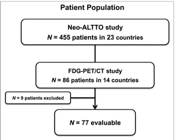
According to the predefined eligibility criteria, 77 of the 86 patients enrolled in Neo-ALTTO presented an evaluable baseline 18F-FDG PET/CT scan and were included; 68 and 66 of those patients had an evaluable18F-FDG PET/CT scan at week 2 and week 6, respectively. Ultimately, 62 patients were fully evaluable for all 3 time points (Fig. 2).
The main reason for exclusion was failure to comply with tim-ing for one or more of the 3 assessments (n5 12). Other reasons included 18F-FDG PET/CT not performed or lost (n5 9), insuf-ficient image quality (n 5 2), and scans obtained with different 18F-FDG PET/CT cameras (n5 1).
Among the 77 evaluable patients, 52 (67.5%) presented metabolic lymph node involvement, and 9 (11.7%) presented both metabolic lymph node involvement and clinically unsuspected distant lesions. Table 1 shows that the18F-FDG PET/CT cohort (77 patients) is re-presentative of the overall Neo-ALTTO population (455 patients), ex-cept for tumor size, which was found to be slightly larger in the imaging subgroup (54.5%) than in the general study population (39.8%).
Biologic Window
Correlation Between Week-2 and Week-6 18F-FDG PET/CT Results. A significant correlation (R25 0.81) was found between the changes of primary tumor SUVmax at weeks 2 and 6 (Fig. 3). Metabolic Response Rates at Weeks 2 and 6 According to EORTC Criteria. 18F-FDG PET/CT metabolic response rates in the primary tumor were 71.6% at week 2 and 60% at week 6. Similar results were found for overall response (primary tumor and nodes), with rates of 69% and 56%, respectively.
Absence of response in the primary tumor at week 2 was pre-dictive of nonresponse at week 6, with a negative prepre-dictive value of 90% (18/20 patients). The presence of response at week 2 was predictive of response at week 6, with a positive predictive value of 78.5% (33/42 patients). Figure 4 illustrates 2 examples.
Analysis of metabolic responses in the primary tumor according to treatment arm revealed the following: at week 2, responses were 66.7%, 56.5%, and 95% for the lapatinib, trastuzumab, and com-bination arms, respectively (P 5 0.016); at week 6, responses were 60.9%, 43.5%, and 78.9%, respectively (P5 0.065). Com-parison of the metabolic responses for single- versus dual-treatment arms showed 61.7% and 95% at week 2 (P5 0.013) and 52.2% and 78.9% at week 6 (P5 0.08), respectively.
Metabolic response rates were higher in patients with HR-negative tumors than in patients with HR-positive tumors: 82.1% versus 64.1% at week 2 (P5 0.18) and 78.5% versus 45.9% at week 6 (P5 0.016), respectively.
Of note, 5 (7%) patients (all with HR-negative tumors) showed complete metabolic response in their primary tumors at week 2, and 14 (21%) patients did so (10 with HR-negative tumors) at week 6.
Correlation Between Metabolic Response and pCR
Of the 77 patients included in this analysis, 27 (35.1%) achieved pCR at surgery.
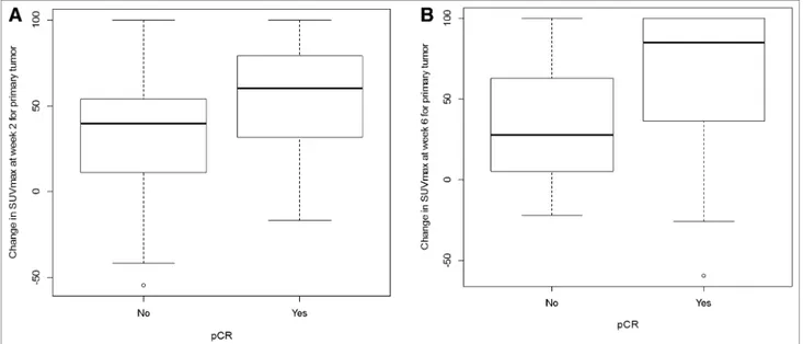
Relative Changes in SUVmax in Primary Tumor at Weeks 2 and 6 According to pCR. Patients with pCR at surgery had significantly greater reductions in SUVmax at weeks 2 and 6 (P5 0.02 for both time points) than those without a pCR. Mean SUVmax reductions were 54.3% versus 32.8% at week 2 and 61.5% versus 34.1% at week 6 for pCR and non-pCR, respectively. Box plots of the changes in SUVmax are shown in Figure 5.
pCR by Metabolic Response in Primary Tumor and Overall Response Using EORTC Criteria. Higher rates of pCR were observed in responders than nonresponders, independently of the selection
of the target lesion: primary tumor (week 2: 41.6% vs. 21.1%, P5 0.12; week 6: 43.6% vs. 19.2%, P5 0.05, respectively) with or without node involvement (Table 2). However, when adjustments were made for stratification factors and treatment arm in multi-variate logistic regression models, the odds ratio for metabolic re-sponse was reduced and no longer statistically significant (P 5 0.19 and 0.24 at weeks 2 and 6, respectively). This appeared pre-dominantly to be due to the adjustment for HR status, which was the strongest predictor of pCR.
pCR by Metabolic Response in Primary Tumor According to HR Status Using EORTC Criteria. HR-positive tumors had a lower rate of pCR than HR-negative ones: 8 of 43 (18.6%) versus 19 of 34 (55.9%). In addition, HR-positive tumors with metabolic responses classified as nonresponders had much lower pCR rates than those classified as responders (1/20 [5%] vs. 4/17 [23.5%] at week 6). This was not the case for HR-negative tumors: the pCR rates were comparable for the responders and the nonresponders, both for the primary tumor and for overall (13/22 [59.1%] vs. 4/6 [66.7%] at week 6) (Table 3).
Complete Metabolic Responses and pCR. Of the patients with HER2-positive/HR-negative BC, 10 showed a complete metabolic response at week 6; of these, 9 presented a pCR at the time of surgery. Of the patients with HER2-positive/HR-positive BC, only 4 showed complete metabolic response, of which 1 presented a pCR at surgery.
DISCUSSION
Although trastuzumab’s use both in advanced and in early disease is associated with improved clinical outcomes, so far not a single
FIGURE 3. Correlation between relative changes in SUVmax at week 2 and week 6.
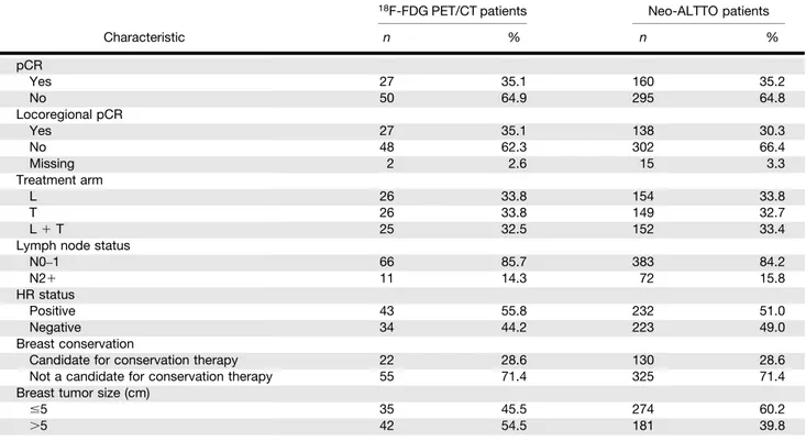
TABLE 1
Patient Characteristics According to Stratification Factors in18F-FDG PET/CT Population and in General Neo-ALTTO Population
18F-FDG PET/CT patients Neo-ALTTO patients
Characteristic n % n % pCR Yes 27 35.1 160 35.2 No 50 64.9 295 64.8 Locoregional pCR Yes 27 35.1 138 30.3 No 48 62.3 302 66.4 Missing 2 2.6 15 3.3 Treatment arm L 26 33.8 154 33.8 T 26 33.8 149 32.7 L1 T 25 32.5 152 33.4
Lymph node status
N0–1 66 85.7 383 84.2 N21 11 14.3 72 15.8 HR status Positive 43 55.8 232 51.0 Negative 34 44.2 223 49.0 Breast conservation
Candidate for conservation therapy 22 28.6 130 28.6 Not a candidate for conservation therapy 55 71.4 325 71.4 Breast tumor size (cm)
#5 35 45.5 274 60.2
.5 42 54.5 181 39.8
pCR5 pathologic complete response (breast); locoregional pCR 5 pathologic complete response (breast 1 lymph nodes); N 5 lymph node status; L5 lapatinib arm; T 5 trastuzumab arm; L 1 T 5 lapatinib 1 trastuzumab arm.
biomarker beyond HER2 has been validated to help us identify the women most likely to benefit from it. More recent research into anti-HER2 therapies indicates that using 2 anti-anti-HER2 agents to create a dual blockade offers further significant clinical advantages (22,23), but here also biomarker research has been unsuccessful (24).
Although PET/CT imaging has been established as a useful staging and prognostic instrument, the interest in functional imaging as a tool to predict pCR to neoadjuvant therapy emerged only a few years ago (25–29), with promising results shown particularly in tri-ple-negative BC (30). Neo-ALTTO is the first trial to test whether early18F-FDG PET/CT–based metabolic response assessment could be a clinically useful pharmacodynamic biomarker to select patients with a high versus low probability of response to a HER2-based
therapy combined with chemotherapy. Be-cause of Neo-ALTTO’s 6-wk biologic win-dow of anti-HER2 treatment alone, we could use18F-FDG PET/CT to assess the effect of anti-HER2 treatments without the interfer-ence of chemotherapy.
Our primary observation was the rapidity with which metabolic changes occurred in the tumors after exposure to the anti-HER2 blockade. Metabolic responses were seen at 2 wk in more than two thirds (71.6%) of the patients, including 5 with a complete meta-bolic shut-down. Metameta-bolic responses (21) were more frequent in the dual HER2 block-ade arm (95% at week 2) than in the single-agent arms (66.7% for lapatinib, 56.5% for trastuzumab at week 2), which is consistent with Neo-ALTTO’s overall results for 455 patients: twice the pCR rate was observed with trastuzumab plus lapatinib than with either agent used alone (18). Lapatinib and trastuzumab do have different mechanisms of action, possibly explaining the synergy of metabolic response seen in the combina-tion arm.
Moreover, the correlation between SUVmax changes at 2 and 6 wk is encouraging. These data clearly show that most of the decreased18F-FDG uptake is seen early during treatment. This is vital both to individualize treatment and to optimize future drug development.
Equally encouraging is the correlation between early metabolic response and the likelihood of achieving pCR, the main objective of the18F-FDG PET/CT analysis (19). The pCR rate was twice as high in patients showing an18F-FDG PET/CT response than it was in nonresponders. However,18F-FDG PET/CT nonresponders still showed a 20% pCR rate, which is clinically meaningful and may reflect the salvage activity of paclitaxel in some tumors that are not HER2-addicted. Interestingly, 20% is in line with the pCR rate
FIGURE 4. Representative examples of metabolic responder and nonresponder included in our study. (A) Metabolic responder achieving pCR: baseline axial fused PET/CT slice showing right breast tumor (left), week 2 PET/CT metabolic complete response (middle), and week 6 PET/CT metabolic complete response (right). (B) Metabolic nonresponder not achieving pCR: baseline axial fused PET/PT slice showing left breast tumor (left), week 2 PET/CT metabolic stable disease (mid-dle), and week 6 PET/CT metabolic stable disease (right).
of 22% observed in patients with HER2-positive tumors receiving only chemotherapy in the neoadjuvant setting (31).
Excellent concordance was also found between imaging results of the primary tumor alone and those of the primary tumor plus its adjacent lymph nodes, suggesting that further imaging studies of early response to preoperative anti-HER2 therapies may be based solely on primary tumor evaluation.
An unexpected observation was the marked heterogeneity of the 18F-FDG PET/CT response according to HR status. This un-planned analysis was conducted because of the growing evidence that positive/HR-positive disease is different from HER2-positive/HR-negative disease, with the former showing less dra-matic responses to chemotherapy plus anti-HER2 therapy.
In our study,18F-FDG PET/CT responses to HER2-therapy oc-curred less frequently in HR-positive tumors. This pattern also extended to metabolic complete responses, which were observed in only 4 HR-positive tumors but in 10 HR-negative tumors, and correlated less well to the achievement of pCR (1/4 pCRs for HR-positive tumors contrasting with 9/10 pCRs for HR-negative ones). Surprisingly,18F-FDG PET/CT response turned out to be a weaker predictor of pCR than HR status, with HR-negative tumors being more likely than HR-positive ones to reach a pCR. Also notable is the apparent greater salvage effect of chemotherapy in patients with HR-negative BC classified as nonresponders than in nonresponders with HR-positive BC. This may explain the much better negative predictive value of the early18F-FDG PET/CT in the latter group.
The potential weakness of the preoperative portion of Neo-ALTTO is that it lacked a blockade of the estrogen receptor pathway, which is a well-described escape mechanism in HER2-positive/HR-positive tumors (32,33). Our study corroborates these laboratory findings and suggests that HER2 blockade in the ab-sence of endocrine therapy has limited efficacy.
In future trials, we recommend the systematic stratification or even separation of the 2 distinct biologic subsets, HER2-positive/ HR-negative and HER2-positive/HR-positive. For the latter pa-tient group, a HER2 blockade combined with endocrine therapy should be explored. This has already been done by Chang et al., who reported a pCR of 21% when using trastuzumab plus lapatinib in combination with letrozole (34).
Our study was not powered to investigate the predictive value of the early metabolic complete response that occurred in 21% (14/ 66) of the patients at week 6, mostly patients with HR-negative tumors. However, 10 of 14 of these early complete18F-FDG PET/ CT responders showed pCR at surgery. Because pCR is a surrogate of long-term outcome in this patient subset, independent valida-tion of the apparently high correlavalida-tion between an early metabolic complete response in HER2-positive/HR-negative disease and pCR is required. Early metabolic complete response could become an in-novative way to separate patients well-served by existing anti-HER2 therapies from those who need additional or alternative ones.
Further work is required, however, and it includes determining a better 18F-FDG PET/CT response threshold to distinguish re-sponding from nonrere-sponding patients. In our study, we opted to use the EORTC threshold (15% at 2 wk), which is relatively low. However, threshold choice should depend on a study’s particular objective, with a high threshold most suited to identify the best responders and a low threshold to identify nonresponders.
The strength of this imaging trial is the multicentric effort. However, because of a lack of prior PET harmonization program, absolute baseline values of SUVmax were not comparable among cameras. Therefore, SUVs could not be pooled to study their predictive value but only their relative changes.
CONCLUSION
Our study is the first, to our knowledge, to demonstrate that early metabolic assessment using 18F-FDG PET/CT in patients with HER2-positive BC is informative and can identify patients with an increased likelihood of achieving pCR after anti-HER2 therapy combined with chemotherapy in the neoadjuvant setting.
DISCLOSURE
The costs of publication of this article were defrayed in part by the payment of page charges. Therefore, and solely to indicate this fact, TABLE 2
pCR and Metabolic Response
Metabolic response pCR Non-pCR pCR rate Overall week 2 Responder 19 28 40.4% Nonresponder 5 16 23.8% Overall week 6 Responder 16 21 43.2% Nonresponder 6 23 20.7% Primary tumor week 2
Responder 20 28 41.7% Nonresponder 4 15 21.1% Primary tumor week 6
Responder 17 22 43.6% Nonresponder 5 21 19.2% Overall metabolic response 5 breast 1 lymph nodes; re-sponder5 complete metabolic response 1 partial metabolic re-sponse according to EORTC criteria; nonresponder5 metabolic stable response1 metabolic progression.
TABLE 3
pCR Rate by Primary Tumor Metabolic Response and HR Status
HR-negative HR-positive
Metabolic response pCR Non-pCR pCR rate pCR Non-pCR pCR rate Primary tumor week 2
Responder 15 8 65.2% 5 20 20.0%
Nonresponder 2 3 40.0% 2 12 14.3%
Primary tumor week 6
Responder 13 9 59.1% 4 13 23.5%
this article is hereby marked “advertisement” in accordance with 18 USC section 1734. This trial was funded by GlaxoSmithKline. No other potential conflict of interest relevant to this article was reported.
ACKNOWLEDGMENTS
We thank Lorena de la Pen˜a (SOLTI Breast Cancer Research group) for her assistance in reviewing this manuscript and Carolyn Straehle for her editorial assistance. The Breast International Group, the Breast European Adjuvant Study Team, and SOLTI Breast Cancer Study Group were responsible for the design and coordination of the study, collection and management of data, medical review, analysis of the data, and reporting of the results. The members of the trial steering committee reviewed the report and were responsible for the decision to submit it for publication. The report was prepared by the members of the writing committee, who had unrestricted access to study data and made the final deci-sions about content. All authors and members of the Executive Committee (including a minority representation from Glaxo-SmithKline) reviewed the article and suggested changes. The data were analyzed by statisticians at Frontier Science Scotland.
REFERENCES
1. Slamon DJ, Clark GM, Wong SG, Levin WJ, Ullrich A, McGuire WL. Human breast cancer: correlation of relapse and survival with amplification of the HER-2/neu oncogene. Science. 1987;235:177–182.
2. Slamon DJ, Godolphin W, Jone LA, et al. Studies of the HER-2/neu proto-oncogene in human breast and ovarian cancer. Science. 1989;244:707–712. 3. Pegram M, Slamon D. Biological rationale for HER2/neu (c-erbB2) as a target
for monoclonal antibody therapy. Semin Oncol. 2000; 27(5, suppl 9):13–19. 4. Romond EH, Perez EA, Bryant J, et al. Trastuzumab plus adjuvant chemotherapy
for operable HER2-positive breast cancer. N Engl J Med. 2005;353:1673–1684. 5. Slamon DJ, Leyland-Jones B, Shak S, et al. Use of chemotherapy plus a mono-clonal antibody against HER2 for metastatic breast cancer that overexpresses HER2. N Engl J Med. 2001;344:783–792.
6. Piccart-Gebhart MJ, Procter M, Leyland-Jones B, et al. Trastuzumab after adju-vant chemotherapy in HER2-positive breast cancer. N Engl J Med. 2005;353: 1659–1672.
7. Cobleigh MA, Vogel CL, Tripathy D, et al. Multinational study of the efficacy and safety of humanized anti-HER2 monoclonal antibody in women who have HER2-overexpressing metastatic breast cancer that has progressed after chemo-therapy for metastatic disease. J Clin Oncol. 1999;17:2639–2648.
8. Smith I, Procter M, Gelber RD, et al. 2-year follow-up of trastuzumab after adjuvant chemotherapy in HER2-positive breast cancer: a randomised controlled trial. Lancet. 2007;369:29–36.
9. Geyer CE, Forster J, Lindquist D, et al. Lapatinib plus capecitabine for HER2-positive advanced breast cancer. N Engl J Med. 2006;355:2733–2743. 10. Johnston S, Pippen J Jr., Pivot X, et al. Lapatinib combined with letrozole versus
letrozole and placebo as first-line therapy for postmenopausal hormone receptor-positive metastatic breast cancer. J Clin Oncol. 2009;27:5538–5546. 11. Xia W, Gerard CM, Liu L, Baudson NM, Ory TL, Spector NL. Combining
lapatinib (GW572016), a small molecule inhibitor of ErbB1 and ErbB2 tyrosine kinases, with therapeutic anti-ErbB2 antibodies enhances apoptosis of ErbB2-overexpressing breast cancer cells. Oncogene. 2005;24:6213–6221.
12. Scaltriti M, Verma C, Guzman M, et al. Lapatinib, a HER2 tyrosine kinase inhibitor, induces stabilization and accumulation of HER2 and potentiates tras-tuzumab-dependent cell cytotoxicity. Oncogene. 2009;28:803–814.
13. Blackwell KL, Burstein HJ, Storniolo AM, et al. Randomized study of Lapatinib alone or in combination with trastuzumab in women with ErbB2-positive, tras-tuzumab-refractory metastatic breast cancer. J Clin Oncol. 2010;28:1124–1130. 14. Junttila TT, Akita RW, Parsons K, et al. Ligand-independent HER2/HER3/PI3K complex is disrupted by trastuzumab and is effectively inhibited by the PI3K inhibitor GDC-0941. Cancer Cell. 2009;15:429–440.
15. Hudis CA. Trastuzumab–mechanism of action and use in clinical practice. N Engl J Med. 2007;357:39–51.
16. Buzdar AU, Ibrahim NK, Francis D, et al. Significantly higher pathologic com-plete remission rate after neoadjuvant therapy with trastuzumab, paclitaxel, and epirubicin chemotherapy: results of a randomized trial in human epidermal growth factor receptor 2-positive operable breast cancer. J Clin Oncol. 2005; 23:3676–3685.
17. Untch M, Fasching PA, Konecny GE, et al. Pathologic complete response after neoadjuvant chemotherapy plus trastuzumab predicts favorable survival in hu-man epidermal growth factor receptor 2-overexpressing breast cancer: results from the TECHNO trial of the AGO and GBG study groups. J Clin Oncol. 2011;29:3351–3357.
18. Baselga J, Bradbury I, Eidtmann H, et al. Lapatinib with trastuzumab for HER2-positive early breast cancer (NeoALTTO): a randomised, open-label, multi-centre, phase 3 trial. Lancet. 2012;379:633–640.
19. Fisher ER, Wang J, Bryant J, Fisher B, Mamounas E, Wolmark N. Pathobiology of preoperative chemotherapy: findings from the National Surgical Adjuvant Breast and Bowel (NSABP) protocol B-18. Cancer. 2002;95:681–695. 20. Fox JJ, Autran-Blanc E, Morris MJ, et al. Practical approach for comparative
analysis of multilesion molecular imaging using a semiautomated program for PET/CT. J Nucl Med. 2011;52:1727–1732.
21. Young H, Baum R, Cremerius U, et al. Measurement of clinical and subclinical tumour response using [18F]-fluorodeoxyglucose and positron emission
tomog-raphy: review and 1999 EORTC recommendations. European Organization for Research and Treatment of Cancer (EORTC) PET Study Group. Eur J Cancer. 1999;35:1773–1782.
22. Guarneri V, Frassoldati A, Bottini A, et al. Preoperative chemotherapy plus trastuzumab, lapatinib, or both in human epidermal growth factor receptor 2-positive operable breast cancer: results of the randomized phase II CHER-LOB study. J Clin Oncol. 2012;30:1989–1995.
23. Gianni L, Pienkowski T, Im YH, et al. Efficacy and safety of neoadjuvant pertuzumab and trastuzumab in women with locally advanced, inflammatory, or early HER2-positive breast cancer (NeoSphere): a randomised multicentre, open-label, phase 2 trial. Lancet Oncol. 2012;13:25–32.
24. Gianni LBG, Kiermaier A, Bianchi G, et al. Neoadjuvant pertuzumab and trastuzumab: biomarker analyses of a 4-arm randomized phase II study (Neosphere) in patients with HER2-positive breast cancer. Cancer Res. 2011;71(24 suppl):S5–1.
25. Keam B, Im SA, Koh Y, et al. Early metabolic response using FDG PET/CT and molecular phenotypes of breast cancer treated with neoadjuvant chemotherapy. BMC Cancer. 2011;11:452.
26. Berriolo-Riedinger A, Touzery C, Riedinger JM, et al. [18F]FDG-PET predicts
complete pathological response of breast cancer to neoadjuvant chemotherapy. Eur J Nucl Med Mol Imaging. 2007;34:1915–1924.
27. Schelling M, Avril N, Nährig J, et al. Positron emission tomography using [18F]
fluorodeoxyglucose for monitoring primary chemotherapy in breast cancer. J Clin Oncol. 2000;18:1689–1695.
28. Rousseau C, Devillers A, Sagan C, et al. Monitoring of early response to neo-adjuvant chemotherapy in stage II and III breast cancer by [18
F]fluorodeoxyglu-cose positron emission tomography. J Clin Oncol. 2006;24:5366–5372. 29. Schwarz-Dose J, Untch M, Tiling R, et al. Monitoring primary systemic therapy of
large and locally advanced breast cancer by using sequential positron emission tomography imaging with [18F]fluorodeoxyglucose. J Clin Oncol. 2009;27:535–541.
30. Groheux D, Hindié E, Giacchetti S, et al. Triple-negative breast cancer: early assessment with18F-FDG PET/CT during neoadjuvant chemotherapy identifies
patients who are unlikely to achieve a pathologic complete response and are at a high risk of early relapse. J Nucl Med. 2012;53:249–254.
31. Gianni L, Eiermann W, Semiglazov V, et al. Neoadjuvant chemotherapy with trastuzumab followed by adjuvant trastuzumab versus neoadjuvant chemother-apy alone, in patients with HER2-positive locally advanced breast cancer (the NOAH trial): a randomised controlled superiority trial with a parallel HER2-negative cohort. Lancet. 2010;375:377–384.
32. Xia W, Bacus S, Hegde P, et al. A model of acquired autoresistance to a potent ErbB2 tyrosine kinase inhibitor and a therapeutic strategy to prevent its onset in breast cancer. Proc Natl Acad Sci USA. 2006;103:7795–7800.
33. Arpino G, Wiechmann L, Osborne CK, Schiff R. Crosstalk between the estrogen receptor and the HER tyrosine kinase receptor family: molecular mechanism and clinical implications for endocrine therapy resistance. Endocr Rev. 2008;29:217–233. 34. Rimawi MF, Mayer IA, Forero A, et al. Multicenter phase II study of neoadju-vant lapatinib and trastuzumab with hormonal therapy and without chemother-apy in patients with human epidermal growth factor receptor 2-overexpressing breast cancer: TBCRC 006. J Clin Oncol. 2013;31:1726–1731.