UNIVERSITA’ DEGLI STUDI DI ROMA “TOR VERGATA”
Facoltà di Medicina
Dipartimento di Medicina Sperimentale e Scienze Biochimiche
Cattedra di Virologia
Tesi di Dottorato in
Microbiologia Medica e Immunologia
(XIX ciclo)
New classes of anti-HIV-1 compounds active at
different stages of infection
Candidata Relatore
Dott.ssa Michela Pollicita Prof. Carlo-Federico Perno
ANNO ACCADEMICO 2006-2007
Abstract
It is widely recognized that monocytes/macrophages (M/M) represent a crucial target of HIV-1 in the body and play a pivotal role in the pathogenic progression of the HIV-1 infection. This strongly supports the clinical relevance of therapeutic strategies able to interfere with HIV-1 replication in M/M. The important role of M/M in HIV-1 transmission, dissemination of infectious virus throughout the body, and in virus persistence, even in patients treated successfully with HAART therapy, suggests the necessity to identify new treatments against HIV-1 replication active at different stages of virus infection. HIV-1 cellular entry inhibitors are a promising class of potential anti-HIV-1/AIDS drugs. By interacting with the viral envelope glycoproteins (gp120 or gp41), and/or with CD4 or the coreceptors, these inhibitors block different steps in the complex sequence of events leading to virus–cell fusion, counteracting in this way the HIV-1 infection of the target cells. We focused on two CCR5 inhibitors, DAPTA and TAK-779, both able to inhibit the R5 HIV-1 replication in M/M. Our results indicate that DAPTA and TAK-779 are potent anti-HIV-HIV-1 compounds able to block the virus entry of R5 HIV-1 strains in M/M, suppressing viral replication in the cells.In particular, DAPTA proved to be able to inhibit the virus replication at extremely low drug concentrations. The use of coreceptor inhibitors, such as DAPTA and TAK-779, could be important to contribute to a possible synergism with other antiretroviral treatments.Another class of compounds able to act before HIV-1 entry, is represented by carbohydrate-binding agents (CBAs). These agents are recently proposed as innovative anti-HIV compounds selectively targeting the glycans of the HIV-1 envelope glycoprotein gp120 and preventing DC-SIGN-directed HIV capture by dendritic cells (DC) and subsequent transmission of the virus to CD4+ T-lymphocytes. We now found that CBAs also efficiently prevent R5 HIV-1 infection of human primary M/M that do not measurably express DC-SIGN but markedly express the macrophage mannose-binding receptor (MMR).We observed also that pre-exposure of X4 HIV-1 to CBAs is able to prevent efficient virus capture by M/M and subsequent syncytia formation in co-cultures of uninfected CD4+ T-lymphocyte C8166 cells and CBA-X4 HIV-1 exposed M/M.The potential of CBAs to impair M/M in their capacity of hosting virus replication and chronic production of new virus particles, but also preventing M/M to efficiently capture and transmit HIV to T-lymphocytes might be an important property to be taken into consideration in the eventual choice to select microbicide candidate drugs for clinical investigation. Since M/M represent chronically infected cells, it will be also interesting to study new drugs acting at a post-integration stage in the replication cycle of HIV-1. A unique class of drugs that may contribute to the control of the latent HIV-1 reservoir includes the quinolone derivatives, first reported as an important class of broad-spectrum antibacterials. Two novel 6-desfluoroquinolone derivatives (6-DFQs), HM-12 and HM-13, were evaluated for their anti-HIV activity in acutely, chronically and latently HIV-1-infected cell cultures (including M/M) and found to behave as potent HIV-1 transcription inhibitors.Interestingly, in a murine in vivo model in which mice are inoculated with latently HIV-1-infected human cells, 6-DFQs were shown to efficiently prevent virus activation upon TNFα triggering.Thus, these compounds are able to slow down virus replication, and should be interesting candidate drugs to be combined with entry, integrase or reverse transcriptase inhibitors that acts prior to the proviral integration in the treatment of HIV-1 infections. Because it is known that HIV-1 infection induces a significant perturbation of the oxidative status of M/M, it can be interesting also to study new drugs able to counteract the cell damage correlated with this oxidative condition. In particular, we studied MnTBAP (Mn(III)tetrakis(4-benzoic acid)porphrin chloride), a synthetic peroxynitrite decomposition catalyst, able to reduce oxidative stress subsequent to peroxynitrite generation in HIV-1-infected M/M and found the compound efficient in inhibiting HIV-1 replication in M/M. In summary, the inherent properties of HIV-1 infection of M/M should be taken into account to design therapeutic strategies aimed at achieving an optimal therapeutic effect in all tissue compartments where the virus hides and replicates. We have investigated four possible new drug classes of compounds that represent interesting candidate drug leads for further (pre)clinical studies.
Table of contents
1. Introduction...5
2. Entry inhibitors ...8
2.1. CCR5 Inhibitors ...8
2.1.1. D-Ala-Peptide T-amide (DAPTA)...9
2.1.1.2. DAPTA inhibits R5 HIV-1 replication in macrophages...10
2.1.1.3. DAPTA reduces levels of HIV-1 DNA in macrophages ...11
2.1.1.4. DAPTA inhibits CCR5 binding in macrophages...13
2.1.2. TAK-779 ...14
2.1.2.1. TAK-779 inhibits intracellular calcium signalling ...14
2.1.2.2. TAK-779 potently inhibits HIV-1 replication in macrophages ...16
2.1.2.3. TAK-779 prevents syncytium formation in HIV-1 infected macrophages...17
2.1.2.4. HIV-1-infected macrophages induce severe CD4+ T cell depletion in hu-PBL-SCID mice...18
2.1.2.5. Effects of DAPTA and TAK-779 on CCR5 binding and gp120-induced apoptosis in neuronal cell lines ...19
2.1.3. Discussion ...21
2.1.4. Materials and Methods of the chapter ...24
2.1.4.1. Cells ...24
2.1.4.2. Virus...25
2.1.4.3. Compounds...26
2.1.4.4. Assessment of antiviral activity of DAPTA in acutely infected macrophages ...26
2.1.4.5. HIV-1 DNA analysis in presence of DAPTA...27
2.1.4.6. Interaction of DAPTA with CCR5...28
2.1.4.7. Measurement of intracellular calcium concentrations ...28
2.1.4.8. hu-PBL-SCID mouse model...29
2.1.4.9. Flow cytometric analysis of neuronal apoptosis ...29
2.2. Carbohydrate-Binding Agents (CBAs)...31
2.2.1. Capture of various HIV-1 strains by B-Lymphocyte Raji/DC-SIGN cells and macrophages...33
2.2.2 Inhibitory effect of CBAs on the ability of macrophages to capture HIV-1 particles ...34
2.2.3. Cocultivation of T Lymphocyte C8166 Cells and HIV exposed macrophages...35
2.2.4. Antiviral activity of CBAs in HIV-1 BaL infected macrophages...37
2.2.5. DC-SIGN and MMR expression in macrophages...38
2.2.6. Role of Mannose Receptor, DC-SIGN and CD4 on HIV-1 capture and transmission in macrophages...39
2.2.7. Inhibitory effect of mannan on the ability of macrophages to capture HIV-1 and transmit to T cells...41
2.2.8. Discussion ...42
2.2.9. Materials and Methods of the chapter ...44
2.2.9.1. Compounds...44
2.2.9.2. Cells ...44
2.2.9.3. Viruses...45
2.2.9.4. HIV-1-capture by Raji/DC-SIGN cells and macrophages...45
2.2.9.5. Effect of short exposure of HIV-1 to test compounds on HIV-1-capture by macrophages ... 45
2.2.9.6. Effect of short exposure of HIV-1 to test compounds on HIV-1-transmission from macrophages to T-cells ... 46
2.2.9.7. Antiviral effect of CBAs on HIV-1 BaL production ...46
2.2.9.9. Effects of CD4, DC-SIGN, MMR antibodies and mannan on HIV-1 capture and
transmission to T-lymphocytes... 47
3. HIV-1 transcription inhibitors...48
3.1. New 6-desfluoroquinolone derivatives (6-DFQs)...48
3.1.1. The 6-DFQ derivatives HM-12 and HM-13 inhibit HIV-1 replication in acutely and chronically HIV-1-infected macrophages...49
3.1.2. The 6-DFQ derivatives HM-12 and HM-13 inhibit HIV-1 transcription in latently infected macrophages...52
3.1.3. Effects of the 6-DFQ derivatives HM-12 and HM-13 on viral reactivation in an artificial in vivo model of HIV-1 latency... 54
3.2. Discussion ...56
3.3. Materials and Methods of the chapter...58
3.3.1. Compounds and plasmid constructs ...58
3.3.2. Cells and viruses ...58
3.3.3. Assessment of antiviral drug activity in acutely infected macrophages ...59
3.3.4. Assessment of antiviral drug activity in chronically infected macrophages...59
3.3.5. Assessment of antiviral drug activity in latently infected macrophages...60
3.3.6. Quantitative real-time PCR ...60
3.3.7. Animal experiments...61
4. Antioxidants compounds ...63
4.1. MnTBAP...63
4.1.2. Antiviral activity in acutely-infected macrophages ...64
4.1.3. Antiviral activity in chronically-infected macrophages...67
4.1.4. Acutely-infected PBL ...68
4.1.5. Drug toxicity ...68
4.1.6. HIV-1 p25 and p55 gag proteins analysis...68
4.1.7. Selective inactivation of peroxynitrite in HIV-1 infected macrophages...69
4.1.8. Effects of MnTBAP upon virus infectivity...70
4.1.9. Ultrastructural analysis of acutelly-infected macrophages treated with MnTBAP ...71
4.2. Discussion ...72
4.3. Materials and Methods of the chapter...74
4.3.1. Compounds ...74
4.3.2. Cell cultures ...75
4.3.3. HIV-1 isolates ...75
4.3.4. Drug toxicity ...75
4.3.5. Assessment of drug activity in acutely infected macrophages ...75
4.3.6. Assessment of antiviral drug activity in chronically infected macrophages...76
4.3.7. Assessment of drug activity in acutely-infected PBL ...76
4.3.8. Virus infectivity ...77
4.3.9. Western blot analysis ...77
4.3.10. Immunocytochemical Staining ...77
4.3.11. Ultrastructural studies ...78
5. Conclusions...78
1. Introduction
Although the introduction of highly active antiretroviral therapy (HAART) has led to a dramatic decrease of both the morbidity and the mortality of patients with Human Immunodeficiency Virus type 1 (HIV-1) infection (Murphy et al., 2001; Palella et al., 1998), the eradication of HIV-1 infection is not yet achievable and the main reason is the presence of virus reservoirs in infected patients. Monocytes/ macrophages (M/M) are a strategic reservoir of HIV-1 during the whole course of the infection, even in patients receiving HAART (Sonza et al., 2001). M/M play an important role in all phases of human immunodeficiency virus type 1 (HIV-1) infection. Once infected by HIV-1, M/M survive and produce large amounts of infectious viral particles (an average of ~ 400 virions released daily by each infected cell) and this for a long time period (Aquaro et al., 2002). They are widely recognized as the second cellular target of HIV-1, and represent a vehicle for virus dissemination in the body and the major reservoir for long term persistence of HIV-1 during HAART (Aquaro et al., 2002; Perelson et al., 1997; Sharkey et al., 2000). HIV-1 infected M/M are widely distributed in all tissues and organs (Koenig et al., 1986; McElrath et al., 1989; Tschachler et al., 1987), including the central nervous system (CNS) where they represent the majority of cells infected by HIV-1 (Gabuzda et al., 1986; Tyor et al., 1993). It was in fact demonstrated that M/M are highly efficient to enter in CNS by blood brain barrier (BBB) (they are 20 folds more efficient than non infected) and at this level they can free virions in outside environment, acting as a Troy horse (Kim et al., 2003; Verani et al., 2005). Microglia, local differentiated M/M, are the main source of virus in the brain, whose pathogenic secretory products cause neuro-AIDS (Guan et al., 2002; Kazmierski et al., 2003). HIV-1 enters into the target cells after binding of the viral envelope glycoprotein gp120 to the CD4, as principal receptor, and CCR5 (R5) (whose ligands are RANTES, MIP-1α, MIP-1β) and CXCR4 (R4) (whose ligand is SDF-1), as coreceptors (called R5-using and X4-using tropic strains, respectively) (Cohen, 1996, Deng et al., 1996, Dragic et al., 1996, Schols, 2004). HIV-1 can penetrate in the CNS rapidly (hours or days
genotypically and phenotypically: in addiction, they are in general macrophage tropic (M-tropic) (Gorry et al., 2001). However, quite recently some authors have described in CNS presence of X4- or dual-tropic strains which seem to be able to have access granted and replicate efficiently in M/M of HIV-related damage and that are thought to have a role in pathogenesis (Spudich et al., 2005, Yi et al., 1999, 2003, 2004).
HIV-1 replication in M/M is a crucial pathogenic event during the progression of HIV-1 infection. In fact, productively-infected M/M can fuse with CD4+lymphocytes and transfer the virus to these cells in the context of the antigen presentation and the immune response (Crowe et al., 1990); in addition infected M/M release cytotoxic factors that can mediate the activation of programmed cell death on bystander cells such as CD4+ and CD8+lymphocytes (Badley et al., 1997; Herbein et al., 1998; Garaci et al., 2003), neurons and astrocytes even without a direct infection of these cells (Aquaro et al., 2000; Mollace et al., 2002; Shi et al., 1996). In agreement with this result, as few as 500 HIV-1 infected M/M have been demonstrated to be able to completely deplete millions of autologous CD4+lymphocytes in a SCID mouse model (Garaci et al., 2003). HIV-1 infection in M/M is characterized by viral dynamics substantially different from that of CD4+lymphocytes. In fact, activated CD4+lymphocytes can sustain a rapid and exponential viral production followed by massive cell death (Bagnarelli et al., 1996). In contrast, M/M are resistant to the cytopathic effect of HIV-1(Gendelman et al., 1988; Orestein et al., 1988) and produce virus over a prolonged period, with dynamics that increases linearly during the first 1-2 weeks of infection, followed by a plateau of high level of replication (>108 copies of unspliced/spliced RNA produced) lasting at least up to 60 days after infection (Aquaro et al., 2002). M/M can survive HIV-1 infection for long periods of time. This is mainly related to autocrine secretion of the nerve growth factor (NGF) associated with enhanced expression of the high-affinity NGF receptor p140 trkA on their surface. This complex interaction enhances the ability of M/M to cope with HIV infection, thus transforming them in a powerful, long-term infected, viral reservoir (Garaci et al., 1999). This supports the role of M/M as an important source of HIV-1 and as a real cellular reservoir able to challenge the attempts to
eradicate the virus from patients (Orenstein et al., 1997; Perelson et al., 1997; Schrager and D’Souza, 1998). The dynamics of virus replication, quite different in M/M and CD4+lymphocytes, may suggest that anti-1 drugs act differently in these cells. The important role of M/M in HIV-1 transmission, dissemination of infectious virus throughout the body, and in virus persistence, even in patients treated successfully with HAART therapy, suggests the necessity to identify new treatments against HIV-1 replication and related cellular damage caused by these cells.
At present, treatment of HIV-infected individuals is based on combination therapy with HIV-1 reverse transcriptase (RT) and/or protease and/or gp41 inhibitors. Despite the notable success of HAART in reducing plasma viral loads to undetectable levels during HIV infection and slowing down clinical progression to AIDS, HAART fails to completely eradicate the virus in HIV-infected individuals. Additionally, emergence of multidrug-resistant viruses have increasingly been reported in patients receiving HAART, urging the need for new anti-HIV treatment strategies. On this basis, it is interesting study new and innovative anti-HIV-1 compounds working by mechanisms different from those of the existing HAART drugs and able to prevent or counteract the M/M infection and consequently the related cellular damages or to counteract the HIV infection in chronically infected M/M.
2. Entry inhibitors
2.1. CCR5 Inhibitors
HIV-1 enters into M/M after binding of the viral envelope glycoprotein gp120 to specific chemokine/HIV-1 coreceptors in conjunction with the CD4 receptor (Kaul et al., 2001; Mack et al., 1998). For this reason HIV-1 entry inhibitor are a promising new class of potential anti-HIV-1 drugs able to block different steps in the complex sequence of events leading to virus-cell fusion (De Clercq, 2002; LaBranche et al., 2001; Michael and Moore, 1999; Moore and Stevenson, 2000). In particular, M/M and microglia are infected primarily by HIV-1 strains that use the β-chemokine receptor CCR5 (R5 strains) (Albright et al., 1999; Choe et al., 1996; Ghorpade et al., He et al., 1997; 1998; Rana et al., 1997), and which predominant during the asymptomatic stages of HIV-1 infection (Baba et al., 1999; Connor et al., 1997). The clinical relevance of the CCR5 by HIV-1 is demonstrated by the impact of a naturally occurring CCR5 mutation, CCR5-Δ32, that generates a non-functional coreceptor (Berger et al., 1999; Liu et al., 1996; O’Brien and Moore, 2000; Seibert et al., 2006). Individuals who are homozygous for this mutation are significantly protected against HIV-1 infection and transmission, while infected, heterozygous individuals progress less rapidly to disease and death (Dragic et al., 2000; Seibert et al., 2006). Blocking CCR5 on the M/M surface with natural ligands for the CC-chemokine receptor (such as LD78β, RANTES or MIP-1 β), prevents HIV-1 infection of these cells (a crucial event during initial spread and early phases of infection) and consequently limits the spread of infectious viral particles during the whole course of the disease (Aquaro et al., 2002; Martin-Garcia et al., 2002; Ruff et al., 2001, 2003; Trkola et al., 2001). CCR5 is also expressed on neurons and astrocytes in the brain and, although neuronal cells are usually not productively infected by HIV-1, in vitro studies have shown that natural ligands of CCR5 protect neurons from gp120-mediated apoptosis (Brenneman et al., 1999; ; Cartier et al., 2003; Cocchi et al., 1995; Dragic et al., 2000; Kaul et al., 1999, 2001; Trkola et al., 2001). So, CCR5 is an attractive target both for inhibition of CCR5 mediated HIV entry in M/M (and consequently for virus transmission of infected particles in the body), and for prevention of
gp120-induced apoptosis in neuronal cell lines. For this reason, the identification of new CCR5-targeting antibodies, chemokines, chemokine analogs, small molecules and peptides, is an important step in the development of new antiviral drugs acting with different mechanism(s) of action, to synergistically control HIV-1 replication and damage directly or indirectly induced by the virus. Several different types of inhibitors for CCR5-mediated HIV-1 entry have now been identified and are in pre-clinical or clinical development as drug candidates (De Clercq 2002; Horuk 2003; Kaul et al., 2001; LaBranche et al., 2001; Michael and Moore 1999; Moore and Stevenson 2000; O'Hara and Olson 2002; Schwarz and Wells, 2002).
2.1.1. D-Ala-Peptide T-amide (DAPTA)
D-Ala-Peptide T-amide (DAPTA), or Peptide T, named for its high threonine content (ASTTTNYT), is a synthetic non-toxic peptide comprised of eight amino acids (185-192) of the gp120 V2 region, that functions as a viral entry inhibitor by targeting selectively CCR5 (Polianova et al., 2005 ; Ruff et al., 2001, 2003). Recently, in a small clinical trial, DAPTA has shown promising antiviral and immune benefits, and caused improvements in cognition in humans with HIV-1 infection, suggesting also its penetration into CNS (Heseltine et al., 1998; Polianova et al., 2005). A blind analysis of frozen stored plasma samples conducted by the NIMH in the early-1990’s from the randomized double-blind placebo-controlled trial of DAPTA for HIV-associated cognitive impairment (Heseltine et al., 1998) found a significant reduction (0.54 log 10, p= 0.037) in viral load between baseline and month 6 (Goodkin et al., 2006). In order to better define potency and potential mechanisms of action of DAPTA, we studied its effect on the inhibition of binding CCR5–gp120 in M/M, and HIV-1 induced apoptosis in neuronal cell lines.
The results indicate that DAPTA efficiently binds CCR5 and is able to inhibit HIV-1 entry, thus preventing HIV-infection of M/M, and in addition blocks HIV-1-M/M CCR5-mediated apoptosis in a neuronal cell line.
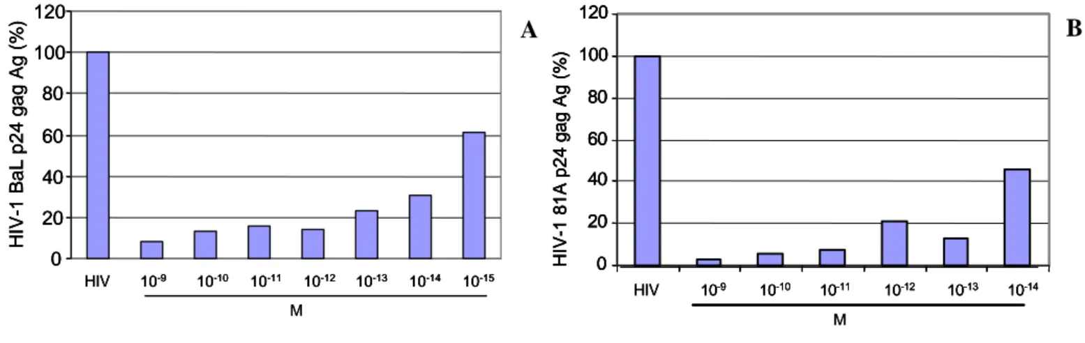
2.1.1.2. DAPTA inhibits R5 HIV-1 replication in macrophages
Viral replication and production in HIV-1 infected M/M treated with DAPTA was assessed 14 and 21 days after infection for p24 antigen production. A representative experiment is shown in the figure 1.At the day 14 after HIV-1 infection, the p24 gag antigen production in the supernatants of HIV-1 BaL infected M/M was drastically dose-dependently reduced in the presence of DAPTA, ranging from 10-9 to 10-15 M. In the figure 1A we can see the results, expressed as a percentage compared to positive control, in which M/M were HIV-1 infected without DAPTA treatment (100%). The maximal viral inhibition observed was around 90% with 10-9 M DAPTA concentration. With another CCR5-using HIV-1 strain, 81A, we obtained comparable results. In 81A HIV-1-infected M/M, DAPTA 10-9 M reaches viral inhibition around 97% at day 14 after the infection (Figure 1B). Comparable results were confirmed at day 21 after infection (data not shown).
Figure 1: Antiviral activity of DAPTA in acutely R5 HIV-1 BaL (Panel A) and 81A (Panel B) infected M/M
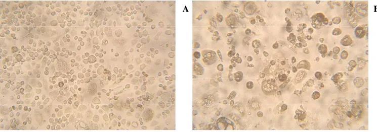
The protective effect of DAPTA was confirmed by figure 2. As we can see, the presence of DAPTA (Panel A) is able to reduce the cytopathic effect, with syncytia formation and aggregation of cells, induced by R5 HIV-1 in M/M after 14 days of infection (Panel B).
B A 0 20 40 60 80 100 120 HIV-1 Ba L p24 g ag Ag (% ) HIV 10-9 10-10 10-11 10-12 10-13 10-14 10-15 M 0 20 40 60 80 100 120 HIV-1 Ba L p24 g ag Ag (% ) HIV 10-9 10-10 10-11 10-12 10-13 10-14 10-15 0 20 40 60 80 100 120 HIV-1 Ba L p24 g ag Ag (% ) HIV 10-9 10-10 10-11 10-12 10-13 10-14 10-15 M 0 20 40 60 80 100 120 HIV -1 8 1 A p24 ga g Ag (%) HIV 10-9 10-10 10-11 10-12 10-13 10-14 M 0 20 40 60 80 100 120 HIV -1 8 1 A p24 ga g Ag (%) HIV 10-9 10-10 10-11 10-12 10-13 10-14 0 20 40 60 80 100 120 HIV -1 8 1 A p24 ga g Ag (%) 0 20 40 60 80 100 120 HIV -1 8 1 A p24 ga g Ag (%) HIV 10-9 10-10 10-11 10-12 10-13 10-14 M
Figure 2: Light microscopic pictures of M/M after 14 days of R5 HIV-1 infection in presence (Panel A) or in absence of DAPTA (Panel B).
Moreover, we tested the HIV-1 p24 antigen production in cells infected by CXCR4-using (X4) strains such as HIV-1 NL4.3. In particular we used C8166 infected with 1000 pg/ml of HIV-1 NL4.3 and treated, where requested, with several doses of DAPTA (ranging from 10-19 to 10-15 M), and analyzed the cytophatic effect starting by 3 days after the infection. We observed that DAPTA was not able to prevent the syncytia formation, due to the X4 HIV-1 infection of T cells. So, we can conclude, that DAPTA showed a potent antiviral activity against R5 strains but not against X4 strains of HIV-1.
2.1.1.3. DAPTA reduces levels of HIV-1 DNA in macrophages
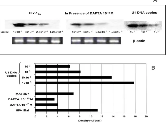
To further demonstrate that DAPTA blocks virus infection, M/M were analyzed for HIV-1 DNA formation. Eighteen hours post-infection, genomic DNA was extracted and two-fold dilution of cell equivalents (range 1 x 10⁶-1.25 x 10⁵) were amplified in an inverse/nested PCR specific for a conserved gag region of the viral genome. Semi-quantitative analyses of HIV-1 DNA in M/M were performed by comparison of DNA amplification products from infected cells, standardized by PCR for β-actin, to standards of amplified U1 DNA copies and cell numbers. The UN-SCAN IT-gel software (Silk Scientific Inc.) was used to determine band densities (Figure 3A). We observed that HIV-1 DNA per 2.5 x 10⁵ cells declined with 64% in the presence of peptide DAPTA (10 -7 M) and with 70% in the presence
of 10-10 M peptide DAPTA, compared with not-treated cells. Control infected cultures (HIV-1BaL), in the absence of peptide DAPTA or 2D7 mAb, had approximately 1 x 10⁴ HIV-1 copies per 105 M/M (i.e. 0.1 copy x M/M). The inhibition of HIV-1 DNA formation detected in M/M in the presence of mAb 2D7 at the maximum amount of 3 ug/ml was approximately 39% (Figure 3B). These data indicate that peptide DAPTA inhibits productive infection in M/M by blocking specifically the CCR5 dependent entry with a potency greater than that of the specific anti-CCR5 antibody 2D7.
β-actin
Cells: 1x10 6 5x10 5 2.5x10 5 1.25x10 5 1x10 6 5x10 5 2.5x10 5 1.25x10 5 10 5 10 3 10 2
HIV-1Bal In Presence of DAPTA 10-10 M U1 DNA copies
A B ? 0 2 4 6 8 10 12 14 16 18 20 HIV-1Bal DAPTA 10 -7M DAPTA 10 -10M MAb 2D7 U1 DNA copies 1x10 6 5x10 5 10 3 10 2 Density (%Total )
Figure 3: Reduction of 1 DNA formation in M/M in presence of DAPTA. A, M/M were infected with HIV-1 BaL in presence or in absence of DAPTA at HIV-10-7 M–10-9 M doses and mAb 2D7 (3000 pg/ul). HIV-1 DNA was extracted from M/M 18 h after infection and 1 x 10⁶-1.25 x 10⁵ cell equivalents were detected by Southern hybridization. HIV-1 DNA extracted from U1 cells, containing two integrated HIV copy, was used as positive standard. B, Band density was measured by UN-SCAN-IT (Silk Scientific Inc).
2.1.1.4. DAPTA inhibits CCR5 binding in macrophages
To confirm that DAPTA binding is specific for CCR5, a competition experiment between CCR5-FITC antibody 2D7 and DAPTA in M/M was done. 2D7 antibody recognizes a conformation-dependent epitope in the second extracellular loop of CCR5, and is a potent inhibitor of R5 virus cell entry. Flow cytometric analysis showed that 35% of mock-treated M/M are CCR5+ positive. DAPTA treatment cells showed reduced binding of the 2D7 mAb to CCR5 in a dose dependent manner, with maximal reduction of CCR5 expression (9%) occurring at 10-12 M. (Figure 4) (p≤0.001). Overall, the inhibition of CCR5-binding by several DAPTA doses is about 43% and reaches a maximum of 73% with 10-12 M. These results suggest that DAPTA reduced the CCR5 antibody binding to the receptor in M/M by masking the binding-site.
DAPTA 10-9M DAPTA 10-10M DAPTA 10-12M DAPTA 10-11M DAPTA 10-14M DAPTA 10-13M
Negative control Mock-treated
Figure 4
Figure 4: Binding of DAPTA to CCR5 in M/M. M/M were incubated with DAPTA for 30 min at 4°C. Surface CCR5 was detected with CCR5-FITC mAb (2D7) and the cells were analyzed with a FACScan flow cytometer. M1 represents the % of CCR5 positive cells.
2.1.2. TAK-779
One of the first CCR5 inhibitors described is TAK-779, a non-peptidic compound with small molecular weight (Mr 531.13), known to interact mainly with CCR5 (Baba et al., 1999; Dragic et al., 2000; Shiraishi et al., 2000; Takashima et al., 2001). TAK-779 acts as antagonist and binds predominantly within a cavity formed between the transmembrane helices 1, 2, 3 and 7 (Dragic et al., 2000). Binding interactions of TAK-779 to the CCR5 receptor probably induce conformational changes of the second extracellular loop and further obstruct the interaction between gp120 and the coreceptor (Baba et al., 1999; Dragic et al., 2000). The anti-HIV-1 activity in PBMCs and CCR5-transfected cell lines are described now in several papers, but no data are available yet in M/M. Here, we wanted to study the anti-HIV-1 activity of TAK-779 in M/M and, in order to apply these concepts to an in vivo model, we used severe combined immunodeficient (SCID) mice, engrafted with human peripheral blood lymphocytes (hu-PBL-SCID mice) in which immunological and viral parameters can be easily monitored. These mice represented a reliable system to study the pathogenesis of HIV-1 infection that may shed light on events not yet highlighted in primate models and in humans (Garaci et al., 2003). Our results indicate that TAK-779 efficiently binds to CCR5, preventing in this way, the R5 HIV-1 infection in M/M and, importantly, the treatment of M/M with TAK-779 is able to prevent the CD4+T-lymphocytes depletion.
2.1.2.1. TAK-779 inhibits intracellular calcium signalling
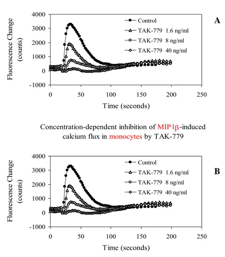
To address the receptor specificity of TAK-779, we evaluated its ability to inhibit chemokine-induced intracellular calcium signalling mediated through CCR5. We performed calcium flux experiments with receptor-specific chemokine ligands and cells expressing the corresponding chemokine receptor, CCR5-transfected U87.CD4 cells and primary monocytes, respectively. After preincubation with or without TAK-779 at dose-dependent concentrations, the cells were stimulated with a CCR5-binding chemokine and the changes in intracellular calcium concentration were recorded by the use of the FLIPR system.
We observed that TAK-779 dose-dependently inhibited the calcium signalling induced by the CC-chemokine MIP-1α and RANTES, in monocytes and in CCR5-transfected U87.CD4 cells, respectively (EC50: 4-40 ng/ml) (Figure 5). These results show that TAK-779 potently blocked the CCR5-mediated Ca2+-signaling, not only in CCR5-transfected cells (Figure 5; Baba et al., 1999), but also in M/M.
Figure 5: Dose-dependent inhibition of RANTES-induced calcium flux in U87.CD4.CCR5 cells by TAK-779 (Panel A) and dose-dependent inhibition of MIP 1β-induced calcium flux in monocytes by TAK-TAK-779 (Panel B).
Concentration-dependent inhibition of MIP1β-induced calcium flux in monocytes by TAK-779
-1000 0 1000 2000 3000 4000 0 50 100 150 200 250 Time (seconds) F luorescence C hange (c ount s) Control TAK-779 1.6 ng/ml TAK-779 8 ng/ml TAK-779 40 ng/ml
Concentration-dependent inhibition of MIP1β-induced calcium flux in monocytes by TAK-779
-1000 0 1000 2000 3000 4000 0 50 100 150 200 250 Time (seconds) F luorescence C hange (c ount s) Control TAK-779 1.6 ng/ml TAK-779 8 ng/ml TAK-779 40 ng/ml A B
2.1.2.2. TAK-779 potently inhibits HIV-1 replication in macrophages
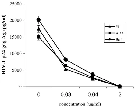
Anti-HIV-1 activity of TAK-779 was evaluated in M/M obtained from the blood of several healthy donors and infected in vitro by different R5 viruses (HIV-1 BaL and ADA) 14 days after infection for p24 antigen production. TAK-779 showed a potent and dose-dependent antiviral activity against HIV-1 BaL and ADA, and also against an R5 clinical isolate (#5), with an average EC50 of 50 ng/ml. The results indicate that TAK-779 suppresses the viral replication in a dose dependent fashion, revealed by the profound inhibition of HIV-1 p24 gag Ag production. This inhibition reached the value of about 99% at 2 ug/ml concentration of TAK-779 (Figure 6). Comparable anti-viral potency was still achieved even after 21 days after the start of the experiment.
Figure 6: Potent antiviral activity of TAK-779 against two R5 strains of HIV-1 (BaL and ADA) and clinical isolate #5. Supernatants were collected at day 14 and tested for p24 antigen production.
0 5000 10000 15000 20000 25000 0 0.08 0.04 2 concentration (ug/ml) H IV -1 p24 gag A g ( p g/ m l) #5 ADA Ba-L
2.1.2.3. TAK-779 prevents syncytia formation in HIV-1 infected macrophages
We also evaluated the effect of TAK-779 on syncytia formation in HIV-1-infected M/M. In our experiments we observed that the infection by R5 viruses (300 TCID50/ml BaL and ADA) induced the formation of syncytia in M/M at day 14, with aggregation of cells, often with circular arrangements of the corresponding nuclei and an evident cytopathic effect compared to uninfected M/M. M/M infected with the same amount of BaL and ADA, but treated with TAK-779 (1 ug/ml), clearly showed a reduced cytopathic effect and much less syncytia (Figure 7). These results also show that TAK-779 profoundly inhibited the syncytia formation and cytopathic effects in R5 HIV-1-infected M/M.
Figure 7: Light microscopic pictures of M/M after 14 days of R5 HIV-1 (BaL and ADA) infection in presence or in absence of TAK-779 (1 ug/ml).
Mock-infected
ADA + TAK-779 ADA
BaL + TAK-779 BaL
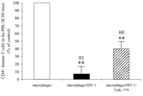
2.1.2.4. HIV-1-infected macrophages induce severe CD4+ T cell depletion in hu-PBL-SCID mice
To apply these concepts to an in vivo model, we used SCID mice, engrafted with hu-PBL-SCID mice. These mice represented a reliable system to study the pathogenesis of HIV-1 infection that may shed light on events not yet highlighted in primate models and in humans. In this model the hu-PBL-SCID mice can be successfully infected with HIV-1 and the immunological and viral parameters of which can be monitored. We known that mice, reconstituted with PBMC from healthy donors and inoculated with infected M/M showed a dramatic depletion of CD4+ T-lymphocytes, with the presence of HIV-DNA in spleen and lymph nodes, and both an overwhelming infection and a sustained plasma viremia (Garaci et al., 2003). The ability of M/M to communicate with and transfer HIV-1 to, CD4+ lymphocytes is based on their anatomical location in lymph nodes and tissues and on their physiological interaction with CD4+ lymphocytes in the context of antigen presentation and immune response (Garaci et al., 2003). In this model, we wanted to evaluate the effect of TAK-779 on HIV-1 infection of M/M, injected i.p. in hu-PBL-SCID mice. We observed that inoculation in hu-PBL-SCID mice of 5000 untreated R5 HIV-1 infected M/M, after one week of infection, resulted in a dramatic depletion of 93% of the CD4+ human T-lymphocytes. In sharp contrast, the CD4+ T-lymphocytes were only partially depleted when the mice received 5000 R5 HIV-1-infected M/M treated with TAK-779 (2 ug/ml), reaching a percentage of depletion of 60% compared to the mice inoculated with uninfected M/M (Figure 8).
Figure 8: TAK-779 inhibits CD4+ T- lymphocytes depletion induced by HIV-1 infected M/M in hu-PBL-SCID mice. Percentages of CD4+T-lymphocytes depletion was measured in hu-PBL-SCID mice at day 14 after challenge with 5000 HIV-infected M/M, 5000 TAK-779-treated HIV-1 infected M/M, and 5000 mock-infected M/M; (** p < 0.001; * p=0.001) when compared with mice challenged with uninfected M/M.
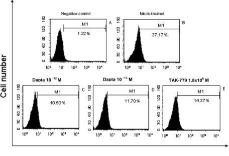
2.1.2.5. Effects of DAPTA and TAK-779 on CCR5 binding and gp120-induced apoptosis in neuronal cell lines
To assess CCR5 expression on the surface of a neuronal cell line, SK-N-SH cells were stained with 2D7 mAb in presence or in absence of DAPTA (at different doses) and TAK-779. The SK-N-SH line has the potential of differentiating to neural cells in the presence of retinoic acid, and it has been used as a model of primary neurons (Speth C et al., 2000; Trillo-Pazos et al., 2000; Yeung et al., 1998). The results indicate that CCR5 expression in these differentiated cells is limited and further reduced in the presence of DAPTA (Figure 9); indeed an inhibition of CCR5 expression of 68.5% and 72% in presence of 10-13 M and 10-12 M DAPTA concentration respectively was observed in comparison with unexposed SK-N-SH (p<0.001). In the presence of TAK-779 (1.8 x 10⁻⁶ M) the inhibition is about 61%.
0 10 20 30 40 50 60 70 80 90 100
macrophages macrophages/HIV-1 macrophages/HIV-1/
TAK-779 CD4+ human T cells in hu-PBL-SCID mice (% of control)
**
**
93 60Figure 9: Binding of DAPTA to SK-N-SH cells. Differentiated SK-N-SH cells were incubated with DAPTA for 30 min at 4°C. Surface CCR5 was detected with CCR5-FITC mAb (2D7) and the cells were analyzed with a FACScan. Negative control of SK-N-SH not stained with CCR5-FITC antibody (A). 2D7 stained untreated cells (B). SK-N-SH treated with 10-12 M (C) or with DAPTA 10-13 M (D) or with TAK-779 1.8 x 10⁻⁶ M (E). The percentage of CCR5 positive cells are indicated in each histogram.
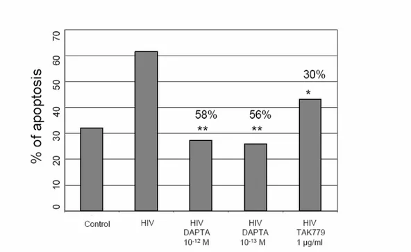
Finally, we exposed differentiated SK-N-SH cells to the R5 HIV-1 BaL, in the presence or absence of DAPTA and TAK-779, and assessed neuronal apoptosis. Time-course studies revealed that cell apoptosis in this cellular line occurred between 5 and 6 days after addition of the virus. These results shown at day 5 (Figure 10). In particular, when SK-N-SH were incubated with HIV-1 BaL, a dramatic reduction of cell viability was seen by flow cytometric analysis. The cytopathic effect, observed in SK-N-SH exposed to R5 HIV-1 released from infected M/M, was mainly related to apoptosis. Indeed, flow cytometric analysis showed apoptosis in 60% of SK-N-SH cells exposed to HIV-1 BaL compared to 28% and 26% observed in DAPTA 10-13 M and 10-12 M treated cells, respectively. Moreover, SK-N-SH cells treated with 1.8 x 10⁻⁶ M TAK-779 (a concentration able to strongly inhibit virus replication in M/M) resulted in a 30% inhibition of apoptosis compared to the cells not treated with TAK-779 (Figure 10). These data show that DAPTA potently blocks R5 gp120-mediated neuronal apoptosis, and DAPTA is even more potent in preventing the neuronal apoptosis compared to TAK-779 and provide a rationale
Dapta 10 -13M
for DAPTA to be evaluated as a potential therapeutic agent for the neuropsychiatric and neurological sequelae of AIDS.
Figure 10: Levels of apoptosis in R5 HIV-1 BaL exposed and DAPTA/TAK-779 treated SK-N-SH cells. The apoptotic cells were stained with PI and analyzed with a FACScan flow cytometer. Statistical analysis was by χ2 Test (** p < 0.001;* p=0.001), for DAPTA or TAK-779 vs. control of infected but not drug-treated cells. Numbers over bars represent the % of apoptosis inhibition.
2.1.3. Discussion
The important role of M/M in HIV-1 transmission, dissemination of infectious virus throughout the body, and in virus persistence, even in patients treated successfully with HAART therapy, suggests the necessity to identify new treatments that can act working by mechanism different than those of existing HAART drugs against HIV-1 replication and related cellular damage caused by these cells.Since HIV-1 needs coreceptors after CD4 binding to entry in M/M, it is clear that these receptors became obvious targets for antiviral drug development. CCR5 and CXCR4 are the principal 1 coreceptors for HIV-1 entry in target cells (Berger et al., HIV-1999; Dragic et al., HIV-1996; Feng et al., HIV-1996).M/M, in whose HIV-1 infection is persistent and productive for long periods of time, are infected primarily by HIV-1 strains
that use CCR5 as coreceptor (Kaul et al., 2001). Actually, several types of CCR5 inhibitors have been identified and are in pre-clinical or clinical development as drug candidates (De Clercq, 2002; Horuk, 2003; Kazmierski et al., 2003; LaBranche et al., 2001; Michael and Moore, 1999; Moore and Stevenson, 2000; O’Hara and Olson, 2002). The present study aimed to determine the effects of DAPTA, a synthetic peptide comprised of eight amino acids of the gp120 V2 region (Polianova et al., 2005; Ruff et al., 2001, 2003), and of TAK-779, a non peptidic and antagonistic compound of CCR5 (Baba et al., 1999; Dragic et al., 2000), proposed to function as a viral entry inhibitor selectively targeting the chemokine receptor CCR5.
Both with DAPTA as with TAK-779 we observed an inhibition of R5 HIV-1 replication in M/M. This is probably correlated with a reduction of viral entry in the cells, due to a binding and obstruction of CCR5 by the coreceptor inhibitors.
DAPTA showed a potent antiviral activity against HIV-1 R5 strains (a dose response anti-HIV-1 activity was observed in a low dose range both with BaL as with 81A, Figure 1) but not X4 strains. This effect is here shown to be mediated in part by a down-expression of CCR5-coreceptor by DAPTA, in line with previous studies demonstrating that binding of HIV-1 coreceptors by their natural ligands contribute to the inhibition of viral replication (Amara et al., 1997; Mack et al., 1998). Inhibition of viral replication may be ascribed to high affinity binding of DAPTA to CCR5 and subsequent impediment of gp120 binding to this receptor and thus inhibit the entry of HIV-1 in host cells.
TAK-779 also interacted with a dose-dependent antiviral activity against R5 HIV-1 strains, both against R5 isolates (BaL and ADA) as against clinical R5 isolates (#5) in M/M. Binding interactions of TAK-779 to the CCR5 receptor probably induce conformational changes of the second extracellular loop and further obstruct the interaction between gp120 and the coreceptor (Baba et al., 1999; Dragic et al., 2000) and consequently the viral entry in the cells, inhibiting, in this way, R5 HIV-1 replication in M/M. The effect of HIV-1 entry inhibition is also seen in the remarkable decrease of HIV-1 DNA detected in DAPTA-treated M/M. This is particularly relevant to the long-term persistent virus replication typical of infected M/M, which are able to sustain production of virus particles for weeks or months after virus
integration (Aquaro et al., 2002). Under these circumstances of chronic viral production, the activity of reverse transcriptase inhibitors is absent in persistently-infected M/M, and that of protease inhibitors is limited (Aquaro et al., 1997, 1998). The inhibition of entry of HIV-1 in M/M is then particularly relevant, and may represent a better way to control the progression of the disease.
It is known that M/M, not only sustain a long-term persistent virus replication, but also play a role in the profound and selective death of CD4+ T lymphocytes. M/M, in fact, transfer virus particles and produce factors that can mediate, directly or indirectly, the activation of programmed cell death on bystander cells both in vitro and in vivo (Aquaro et al., 2000; Badley et al., 1997; Garaci et al., 2003). In our experiments we observed that the pre-treatment with TAK-779 (2 ug/ml) of R5 HIV-1 infected M/M dramatically reduces the depletion of CD4+ T-lymphocytes in hu-PBL-SCID mice. So, the impediment of gp120 binding to CCR5 can prevent the entry of HIV-1 in M/M, the viral reservoir in which HIV-1 replicate and survive for long time, and thus to control the progression of the disease, particularly if used at early stages of the disease, when virus spreading is still limited. Moreover, because M/M can recruit lymphocytes and trigger their cell death through the release of virus proteins, chemokines and other factors (Garaci et al., 2003; Kaul et al., 2001) the use of entry inhibitors, as TAK-779, can prevent the bystander phenomenon that leads to the death of the majority of lymphocytes.
Moreover, the HIV-1 entry mediated by M/M in the brain, where these cells are key elements in the pathogenesis of HIV-encephalitis, causes several pathological abnormalities that can have a clinical appearance in AIDS Dementia Complex (ADC), also because in this anatomical privileged site HAART drugs are not able to enter the brain. The availability of binding inhibitors is therefore even more important, since it can prevent the bystander phenomenon that leads to death of the majority of lymphocytes and neurons even if those cells are not directly infected.
Indeed, CCR5 expression in neurons is responsible to apoptosis induced by gp120-CCR5 binding (Cartier et al., 2003; Cocchi et al., 1995; Dragic et al., 2000; Kaul et al., 2001; Trkola et al., 2001). CCR5-binding by inhibitors, is able to significantly prevent apoptosis in neuronal cell lines exposed to HIV-1 R5 strains. DAPTA is a more potent inhibitor than TAK-779, of neuronal cell lines apoptosis
induced by HIV-1 R5 strains. DAPTA, likely binds CCR5 at a site different than TAK-779, whose direct antiviral effect is clearly more pronounced than that induced by DAPTA. Due to the putative differences in CCR5-binding, it is conceivable that the phenomenon of apoptosis induction and that of virus entry, are mediated by different parts of HIV-gp120, and thus differently contribute to the pathogenesis of HIV-1 infection. Additionally, signalling effects through CCR5 may differentially affect entry compared to apoptosis of chemokine receptor targeted drugs. Recent tests in an animal model revealed that DAPTA, currently in phase II trials for HIV-1 disease, is able to block the release of the pro-inflammatory cytokines TNF-α and interleukin-1, counteracting in this way, the inflammatory state in the brain associated with HIV-1 infection in ADC (Tuttle et al., 2001). This may open the way to antiviral approaches that combine various inhibitors of HIV-1 entry, acting with different mechanism(s) of action, to synergistically control HIV-1 replication and damage directly or indirectly induced by the virus. Targeting such approaches to M/M, which are pivotal cells in the progression of HIV-related damage, may provide a major antiviral effect, particularly if used at early stages of the disease, when virus spreading is still limited.
In conclusion, the usage of coreceptor inhibitors, as DAPTA or TAK-779, may synergistically contribute to the control of HIV-1 replication and to the damage directly or indirectly induced by this virus.
2.1.4. Materials and Methods of the chapter
2.1.4.1. Cells
M/M were prepared and purified as described in published procedures (Aquaro and Perno, 2005). Briefly, peripheral blood mononuclear cells (PBMCs) were obtained from the blood of healthy HIV-seronegative donors. PBMCs were separated by Ficoll-Hypaque gradient centrifugation and seeded in plastic 48-well plates (Costar, Cambridge, Mass.) at a density of 1.8 × 10⁶ cells/ml in RPMI 1640
L-glutamine, and 20% heat-inactivated, mycoplasma- and endotoxin-free fetal calf serum (HyClone, Logan, Utah) (complete medium). Cell cultures were incubated in a humidified atmosphere with 5% CO2 at 37°C. Non adherent cells were removed, 6 days after seeding by repeated gentle washing with warmed RPMI 1640, leaving a monolayer of adherent cells, which are incubated in complete medium as previously described. Adherent cells obtained with this technique consisted of >95% differentiated M/M.
Peripheral blood lymphocytes (PBL) were purified from PBMC by repeated adherences to remove monocytes, and then cultured with the same medium as M/M, supplemented with 2 ug/ml phytohemagglutinin (PHA). Stimulation was carried out for 72 hours; afterward, the medium was discarded, cells were washed three times with RPMI 1640 and the concentration was adjusted to 5 x 10⁵ cells per ml of medium supplemented with 50 U/ml recombinant interleukin-2 (IL-2).
The neuroblastoma cell line SK-N-SH (Koenig et al., Science 1986) was obtained from American Type Culture Collection (ATCC HTB-11) and maintained in RPMI 1640 supplemented with 10% fetal calf serum, 50 U of penicillin/ml, 50 ug of streptomycin/ml, 2 mM L-glutamine. To differentiate these cells to a neural cell phenotype they were exposed to 1 M retinoic acid (Sigma, Chemical Co., St. Louis, MO) for 4 days.
CCR5-transfected human astroglioma U87.CD4 cells were kindly provided by Dr. Dan R. Littman (Skirball Institute of Biomolecular Medicine, New York, NY, USA) and were cultured in Dulbecco’s modified Eagle’s medium (Life Technologies, Paisley, UK) containing 10% heat-inactivated fetal bovine serum (FBS) (BioWhittaker Europe, Verviers, Belgium), 0.01 M HEPES buffer (Life Technologies), 0.2 mg/ml geneticin (G-418 sulfate) (Life Technologies) and 1 ug/ml puromycin (Sigma-Aldrich, St. Louis, MO, USA).
2.1.4.2. Virus
Different R5 viral strains, BaL, 81A and ADA whose characteristics and genomic sequence have been previously described, were used (Cenci et al., 1997; Gartner et al., 1986; Popovic et al., 1984). The
clinical R5 HIV-1 # 5 isolate was isolated from the plasma of an HIV-1-infected patient and passaged only once in PBMC. The viruses were expanded in M/M, supernatants were collected, ultracentrifuged for two hours at 22 000 rcf at 4°C, and stored at -80°C before use (Perno et al., 1993). Characteristics of viral stocks used for this study were 2.1x10⁸ HIV-RNA genomes/ml (corresponding to 35 ng of p24 antigen) and 5 x 10³ tissue culture infectious doses 50% per ml (TCID50/ml) as assessed by virus
titration in other primary M/M cultures.
2.1.4.3. Compounds
D-Ala-Peptide-T-Amide (DAPTA) was synthesized under GMP conditions and obtained from Bachem (Torrence, CA). A stock solution, diluted in sterile water was made fresh for each experiment.
The CCR5 antagonist TAK-779 (N,N-dimethyl-N-(4-[[[2-(4-methylphenyl)-6,7-dihydro-5Hbenzocycloheptenyl]carbonyl]amino]benzyl)-tetrahydro-2H-pyran-4-amonium chloride; Mr=531.13) (Baba M et al., PNAS 1999) was obtained from Takeda Chemical Industries (Osaka , Japan).
The anti-CCR5 monoclonal antibody (mAb) (clone 2D7) was purchased from BD Pharmingen (San Diego, CA).
2.1.4.4. Assessment of antiviral activity of DAPTA in acutely infected macrophages
Two days after isolation (i.e. 7 days after plating) M/M were exposed to various concentrations of DAPTA (10-15, 10-14, 10-13, 10-12, 10-11, 10-10, 10-9 M) for 20 min, and then, without washing out the drug, challenged with the R5 HIV-1 strains BaL or 81A (3000 pg/ml of p24 gag Ag). For the TAK-779 experiments M/M were treated with the compound (0.08, 0.4 and 2 ug/ml of TAK-779) for 20 min, and then they were challenged with 300 TCID50/ml, a virus dose affording a maximal virus production from M/M, HIV-1 BaL , HIV-1 ADA and HIV-1 R5 clinical isolate # 5. Two hours after virus challenge at 37°C in a humidified atmosphere supplemented with 5% CO2, M/M were washed extensively with warm RPMI 1640 to remove the excess virus and complete medium containing the appropriate drugs (DAPTA or TAK-779) was replaced. Fresh complete medium and drugs were added at 7 day. Supernatants were collected at different time points and at day 14 assessed for virus production by
analysis of HIV-1 p24 gag Ag production with a commercially ELISA kit (Biorad, France). Moreover, we analyzed by light microscopy the cytopathic effect in M/M at day 14 after infection. The experiment was run in triplicate, with six positive controls for each experiment. The geometric mean of p24 gag Ag production of replicates in each experiment was used to determine the effective drug concentration where 50% and 90% of viral replication is inhibited (EC50 and EC90, respectively). Statistical analysis was performed by χ2 Test (** p < 0.001; * p=0.001), for drug treated vs. control of infected but not drug-treated M/M.
2.1.4.5. HIV-1 DNA analysis in presence of DAPTA
PCR analysis of integrated HIV-1 proviral DNA was performed on differentiated M/M. M/M were obtained from peripheral blood by adherence, cultured for 5 days and infected with HIV-1 BaL (30 pg/ml) in presence or in absence of DAPTA (10-9 M and 10-7 M doses) and anti-CCR5 mAb 2D7 (3000 pg/ul). For HIV-1 proviral integration analysis genomic (total) DNA was isolated in from M/M after 18 h of HIV-1 infection (Qiagen DNA isolation and purification kit) and 1 x 10⁶-1.25 x 10⁵ cell equivalents were amplified in an inverse/nested PCR specific for a conserved region within gag gene (primers pair SK39/SK38; Gene Bank accession numbers A24318/A26625, synthesized by Gibco BRL/Invitrogen Life Technologies Carlsbad, CA). The 115-bp PCR products were detected by oligomer hybridization using 3’-fluorescein labelled probes SK19 (Gene Bank accession number A24328). The probe was labelled with Gene Images 3’-oligolabelling Module (Amersham, Piscataway, NJ) according manufacturer’s procedure and specific target sequences immobilized on the Hybon-N nylon membrane (Amersham, Piscataway, NJ) were detected by Gene Images ECL Detection Kit (Amersham, Piscataway, NJ) by exposing to blue-light sensitive X-ray film (Pegasus Scientific Inc., Burtonsville, MD). Amplification of β-actin housekeeping gene was utilised to evaluate the efficiency of the extraction procedure and to estimate the concentration of isolated DNA. DNA isolated from U1-cells in which two HIV-1 proviral copies are integrated in each cell genome was used as a positive control and
semi-quantitative analysis. Band density was measured by software program UN-SCAN-IT-gel software (Silk Scientific Inc., Orem, Utah, USA).
2.1.4.6. Interaction of DAPTA with CCR5
To verify the specificity of DAPTA-CCR5 binding we assessed the percentage of CCR5 expression in presence of DAPTA in M/M and in the differentiated neuronal cell line SK-N-SH. M/M were detached gently from the plates with trypsin/ EDTA (0.02%), centrifuged at 1600 rpm for 10 min, counted and resuspended at a density of 2 x 10⁵ cells /ml in complete medium. The M/M were then incubated with DAPTA, at several doses, for 20 min at 4°C or at 37°C and then stained with FITC-labelled anti-CCR5 mAb (2D7, BD Pharmingen) for 30 min at 4°C in the dark. After incubation, stained cells were washed with PBS and analyzed with a FACScan flow cytometer (Becton Dickinson). Ten thousand events were collected for each sample. The data were acquired and analyzed by the Lysis II program (Becton Dickinson). The same staining procedure was repeated for the neuronal cell line SK-N-SH.
2.1.4.7. Measurement of intracellular calcium concentrations
Adherent CCR5-transfected U87.CD4 cells and freshly isolated primary monocytes were seeded in 0.1% gelatin-coated 96-well black-wall microplates (Costar, Cambridge, MA, USA) at 2×10⁴ cells per well for U87.CD4 cells and at 2×10⁵ cells per well for primary monocytes on the day prior to the experiment. On the day of the experiment, the plated monolayers were loaded with the fluorescent calcium indicator Fluo-3 acetoxymethyl (Molecular Probes, Leiden, The Netherlands) at 4 uM for 45 min at 37°C. After thorough washing with calcium flux assay buffer (Hanks’ balanced salt solution with 20 mM HEPES buffer and 0.2% bovine serum albumin (BSA), pH 7.4), the cells were pre-incubated for 15 min at 37°C with TAK-779 at the indicated concentrations in the same buffer. Then, the intracellular calcium mobilization in response to the appropriate chemokine (RANTES or MIP-1α) was measured at 37°C by monitoring the fluorescence as a function of time simultaneously in all the wells using a Fluorometric Imaging Plate Reader (FLIPR) (Molecular Devices, Sunnyvale, CA, USA) (Princen et al., 2003).
2.1.4.8. hu-PBL-SCID mouse model
M/M, infected with R5 HIV-1 in the presence or in the absence of TAK-779, were gently scraped from plastic plates, suspended in PBS, and washed twice. After counting, M/M were resuspended in RPMI medium 1640 and injected i.p. in SCID mice, engrafted with human peripheral blood lymphocytes (hu-PBL-SCID mice) at different concentrations. For the experiment were used CB17 scid/scid female mice at 4 weeks of age and were kept under specific pathogen-free conditions. These SCID mice were housed in microisolator cages; all food, water, and bedding were autoclaved before use.
The hu-PBL-SCID mice were killed 14 days after the injection with the M/M, and cells were collected from the peritoneal cavity. At each time, a two-step peritoneal lavage was done. The first washing was performed with 1 ml of cold RPMI medium 1640. The recovered volume was centrifuged, and the supernatant was stored at -20°C while the cells were pooled with those obtained from a second 4-ml washing as described (Rizza et al., 1996). Cells recovered from the peritoneum of the hu-PBL-SCID mice were resuspended in PBS and incubated with the appropriate fluorochrome-conjugated mAbs for 30 min. The cells were then washed with a mixture of PBS, 2% FCS, and 0.1% sodium azide and fixed with 2.5% paraformaldehyde. Three-color flow cytometry was performed with a FACScan fluorescence-activated cell-sorter cytometer (Becton Dickinson) and cells were analyzed with CellQuest (Becton Dickinson) software. A total of 20000 events per sample were collected. Cells were analyzed according to forward and side scatter properties to gate the live cell populations. The mAbs used were directly-labeled anti-human CD45-PerCp, CD4-FITC and CD8-PE (all obtained from Becton Dickinson).
2.1.4.9. Flow cytometric analysis of neuronal apoptosis
We assessed neuronal apoptosis in the neuroblastoma cell line, SK-N-SH, after retinoic acid-induced differentiation. To avoid overgrowth, SK-N-SH were seeded in Petri plates at a density of 60 000 cells/ wells in RPMI 10% medium and exposed, at day 1 after seeding, to 1 M retinoic acid, for 4 days. After differentiation, culture medium containing retinoic acid was completely removed from the Petri plates, and replaced with fresh RPMI 10% medium containing 8000 pg/ml of p24-gag of HIV-1 BaL grown in
M/M cultures, and DAPTA (at 10-13 and 10-12 M). As control, we used the CCR5 antagonist TAK-779 (1.8 x 10⁻⁶ M). The cells were then incubated at 37°C in humidified air containing 5% CO2 for 5 days. On the day of analysis, the cells were gently detached with trypsin-EDTA (0.02%) and centrifuged at 1600 rpm for 10 min. Pellets were washed with phosphate buffered saline (PBS), placed in ice, and permeated with ice-cold 70% ethanol for 30 min. The aliquots were centrifuged at 1500 rpm for 10 min, the pellets were washed with PBS, incubated with Propidium Iodide (PI; 100 ug/ml) and RNase (250 ug/ml Qiagen) at 4°C for 2 hours in the dark. Then the samples were washed twice with PBS and PI-stained cells were analyzed by monitoring the incorporation of PI intracellular with a FACScan flow cytometer. Ten thousand events were collected for each sample. Data were acquired and analyzed by the Lysis II program (Becton Dickinson). Statistical analysis was performed by χ2 Test (** p < 0.001; * p=0.001), for DAPTA vs. control of not drug-treated neuronal cell lines.
2.2. Carbohydrate-Binding Agents (CBAs)
HIV-1 is an enveloped virus whose surface glycoprotein gp120 binds CD4 and a co-receptor on the target cell membrane to initiate infection. Many of the gp120 glycosylation sites are terminally mannosylated, a pattern common to many pathogens (Nguyen and Hildreth, 2003). Carbohydrate-binding agents (CBA) have been recently proposed as innovative anti-HIV compounds selectively targeting the glycans of the HIV-1 envelope glycoprotein gp120. Short pre-exposure of HIV-1 to CBAs prevents the DC to efficiently bind HIV-1 and no syncytia formation occurs upon subsequent co-cultivation with T-lymphocytes. Thus, the mannose-specific CBAs (i.e. the plant lectins HHA, GNA, NPA and CA; the procaryotic cyanovirin-N (CV-N)) and the GlcNAc-specific (i.e. the plant lectin UDA) but not other entry inhibitors, or polyanionic compounds, efficiently abrogate the DC-SIGN-directed HIV-1 capture and subsequent transmission to T-lymphocytes. Such compounds should have the potential to impair the ability of DC to capture HIV and to transmit HIV to T lymphocytes (Balzarini et al., 2007). The aim of our study is to demonstrate the ability of CBAs to inhibit HIV-1 capture also in M/M, one of the major cellular targets for HIV-1 infection and virus reservoir, and subsequent virus transmission to CD4+ T-lymphocytes. M/M contribute to the transmission and the pathogenesis of HIV-1 infection throughout the progression of HIV-HIV-1 infection especially at late stages when CD4+ T lymphocytes have been extensively depleted (Williams et al., 2002; Tomkowicz et al., 2006; Herbein et al., 2002). In fact, productively-infected M/M can fuse with uninfected CD4+ T lymphocytes and transfer the virus to these cells, thus further contributing to depletion of CD4+ T lymphocytes (Crowe S et al., 1990); in addition, HIV-1 infected M/M may induce the apoptosis on bystander uninfected cells, such as CD4+ and CD8+ T lymphocytes, neurons and astrocytes by releasing cytotoxic factors (Aquaro et al., 2000; Badley et al., 1997; Herbein et al., 1998; Mollace et al., 2002; Shi et al., 1996). Recently, it has been demonstrated that as few as 500 HIV-exposed M/M cause complete depletion of several millions of autologous CD4+ T-lymphocytes, sustained HIV-viremia and spreading of HIV-1-DNA in mouse lymphoid organs (Garaci et al., 2003). Therefore, M/M sustain persistent and continuously productive HIV infection (Li et al., 1999). We have looked at the ability of Macrophage Mannose
Receptor (MMR) on M/M, that lack expression of DC-SIGN, a a dendritic cell-specific ICAM-3 grabbing receptor/HIV-1-binding protein responsible of the adhesion between DC cells and resting T-cells (Geijtenbeek et al., 2000), to bind to HIV and to enable the transmission to T-T-cells in co-culture. The MMR is a 175-kDA transmembrane glycoprotein containing three types of characterizing domains, two of which have distinct carbohydrate recognizing properties. The amino-terminal cystein-rich domain plays a critical role in binding sulphated glycoproteins. The C-type lectin domains facilitate carbohydrate-dependent uptake of mannosylated protein antigens on micro-organisms including bacteria, yeast, enveloped viruses and protozoans (Reading et al., 2000). The MMR shows high affinity for mannose and fucose, intermediate affinity for N-acetylglucosamide and glucose and low affinity for galactose (Stahl et al., 1998). Because these terminal sugars are rarely found on mammalian cell-surface, the MMR could be responsible for recognition of self and nonself antigens (Engering et al., 1997). It also plays a key role in pathogen-related acquired host defence by mediating antigen internalization and delivery to MHC class II compartments for antigen presentation (Engering et al., 1997; Tan et al., 1997). Our data show that the HIV-1-association with M/M, that lack expression of DC-SIGN, is MMR mediated, as evidenced by inhibition with soluble mannose-binding lectin and with MMR antibody. The outcome of this type of studies would be very helpful to guide the choice of potential candidate microbicide drugs (Balzarini et al., 2004; Balzarini and Van Damme, 2007).
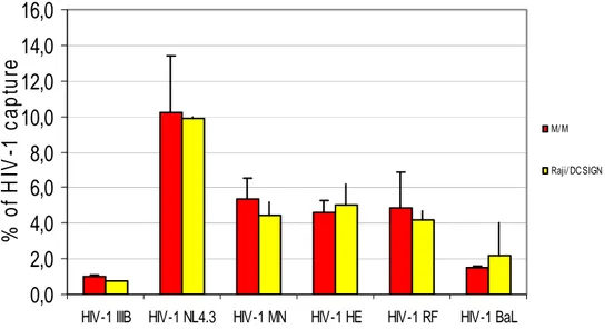
2.2.1. Capture of various HIV-1 strains by B-Lymphocyte Raji/DC-SIGN cells and macrophages
Exponentially growing DC-SIGN-expressing B-lymphoblast Raji cells were suspended in cell culture medium at 6 x 10⁶ cells/400 ul. M/M were obtained by Ficoll-Hypaque. Raji-DC-SIGN and M/M were exposed to HIV-1 for 1 or 2 hours, respectively. Then, the unabsorbed virus was carefully removed by several washing steps. It was calculated that after the serial washing steps, we removed the HIV-1 p24 remained in the supernatant.
Then we analyzed the HIV-1 capture by Raji cells and M/M (previously detached from the plates) by a HIV-1 p24 ELISA.
The results represented in the figure represent the percentage of captured HIV-1 p24 calculated compared with the input of HIV-1 24. HIV-1 p24 associated with Raji/DC-SIGN cells and M/M could be reliably measured. HIV-1 NL4.3 was most efficiently captured (10% of input virus), whereas HIV-1 IIIB was least efficiently captured (1% of input virus). For each individual strain, similar capture efficiency was observed for DC-SIGN-expressing cells and for M/M.
Figure 11: Capture HIV-1 p24 by M/M and Raji/DC SIGN. Raji/DC-SIGN and M/M were exposed to several strains of HIV-1. After the time of infection the cells were washed to be sure to remove the unbound virus. The Raji/DC-SIGN cell cultures and M/M were then analysed for p24 content by a p24ELISA.
0,0
2,0
4,0
6,0
8,0
10,0
12,0
14,0
16,0
HIV-1 IIIB HIV-1 NL4.3 HIV-1 MN HIV-1 HE HIV-1 RF HIV-1 BaL
%
of
H
IV
-1 c
a
p
tu
re
M/ M Raji/ DC SIGN2.2.2 Inhibitory effect of CBAs on the ability of macrophages to capture HIV-1 particles
High amounts of HIV-1 particles (100 ul; 67000 pg/ml) were exposed to serial dilutions of the test compounds for 30 min. Then, the drug-exposed virus suspensions were added to M/M (100 ul/ well) for 2 hours at 37°C after which the cells were thoroughly washed four times with 1 ml RPMI 20% as described above. M/M were then detached by ELISA analysis for p24 content (Panel A). The analysis of the HIV-1 p24 Ag from the last wash was negative and this revealed the there were not virus particle in the medium and that the HV-1 p24 Ag revealed was only for the virus captured from M/M. The percentage of captured p24 Ag HIV-1 (pg/ml) calculated compared with beginning p24 Ag HIV-1 (pg/ml) .
The results show that the CBAs dose-dependently inhibited capture of HIV-1 by the M/M
Figure 12: Inhibitory effect of CBAs on the ability of to capture HIV-1 particles. HIV-1 (NL4.3) particles were exposed to various dilutions of the test compounds (30 min) prior to administration to M/M for 2 hours. After removal of unbound virus by several washing steps, cell-associated virus was qualified by p24 ELISA.
0,0 20,0 40,0 60,0 80,0 100,0 120,0 NL4.3 UDA 500 ug/ml UDA 100 ug/ml
UDA 20 ug/ml NPA 100
ug/ml
NPA 20 ug/ml PRMA 500 ug/ ml PRMA 100 ug/ml % of H IV-1 c a pt ur e
2.2.3. Cocultivation of T Lymphocyte C8166 Cells and HIV exposed macrophages
Because M/M can capture HIV-1 particles and transmit HIV-1 to T-cells we wanted to see if CBAs are able to prevent the transmission of the virus particles to C8166 T lymphocytes (cocultivation) by HIV-1 NL4.3-exposed M/M (200000/ well; 1 ml). As described for the capture, 67000 pg/100 ul of HIV-1 were exposed to the serial dilutions of the test compounds for 30 min. CBA-exposed virus suspensions were added to M/M (100 ul/ well) for 2 hours at 37°C after which the cells were thoroughly washed four times with RPMI 20% to be sure to remove unabsorbed virus particles. Then C8166 (200.000/ well; 1 ml) were added to M/M and cocultivated for 3-4 days. The supernatant analysis of the HIV-1 p24 from the last wash was negative and this revealed the absence of virus particles in the medium able to infect the T cells. The coculture of the CBA/ virus-exposed M/M and C8166 did not result in increased p24 amounts in the cell cultures after 3 and 4 days. The percentage of HIV-1 p24 (pg/ml) transmission was expressed as a percent of the value of the positive control (100%), in which M/M were incubated with HIV-1 NL4.3 without the CBAs. As we can see in the figure 13 the CBAs, both UDA (Panel A) as NPA (Panel B), are able to prevent the transmission of HIV-1 from M/M to T cells at 3 and 4 days.
Figure 13: Inhibitory Effect of CBAs UDA (Panel A) and NPA (Panel B) on the Ability of M/M to Transmit captured HIV-1 Particles. HIV-1 (NL4.3) particles were exposed to various dilutions of the test compounds (30 min) prior to administration to M/M for 2 hours. After removal of unbound virus by several washing steps M/M were cocultivated with C8166 cells. The virus production was qualified by p24 ELISA after 3 and 4 days of coculture
0,0 0,2 0,4 0,6 0,8 1,0 1,2 1,4 1,6 NPA 100 ug/ml NPA 20 ug/ml NPA 100 ug/ml NPA 20 ug/ml % of H IV -1 t rans mi tt ed Day 3 Day 4 1.6 1.4 1.2 1.0 0.8 0.6 0.4 0.2 0.0 % of HIV-1 tra n s m itte d 0,0 0,2 0,4 0,6 0,8 1,0 1,2 1,4 1,6 NPA 100 ug/ml NPA 20 ug/ml NPA 100 ug/ml NPA 20 ug/ml % of H IV -1 t rans mi tt ed Day 3 Day 4 1.6 1.4 1.2 1.0 0.8 0.6 0.4 0.2 0.0 % of HIV-1 tra n s m itte d Day 3 Day 4 0,0 0,5 1,0 1,5 2,0 2,5 3,0 3,5 UDA 500 ug/ml UDA 100 ug/ml UDA 20 ug/ml UDA 500 ug/ml UDA 100 ug/ml UDA 20 ug/ml % of H IV-1 tr a n s m itted 3.5 3.0 2.5 2.0 1.5 1.0 0.5 0.0 % of HIV-1 tra n s m itte d A Day 3 Day 4 0,0 0,5 1,0 1,5 2,0 2,5 3,0 3,5 UDA 500 ug/ml UDA 100 ug/ml UDA 20 ug/ml UDA 500 ug/ml UDA 100 ug/ml UDA 20 ug/ml % of H IV-1 tr a n s m itted 3.5 3.0 2.5 2.0 1.5 1.0 0.5 0.0 % of HIV-1 tra n s m itte d A B
Indeed, we observed in figure 14 that in cocultures of uninfected C8166 cells and HIV-1–exposed M/M giant cells appear at days 3 and 4 after the cocultivation (Panel A). On the contrary, no syncytia were microscopically observed to appear in the C8166 coculture with uninfected M/M (Panel B). Interestingly, uninfected C8166 cells were predominantly clustered at M/M locations.
A
B
Figure 14: cytophatic effect induced by HIV-1 NL4.3 exposed M/M cocultured with T cells.
Panel A: 3 days cocultures of uninfected C8166 cells + HIV-1-exposed M/M; Panel B:
2.2.4. Antiviral activity of CBAs in HIV-1 BaL infected macrophages
M/M preincubated with several doses of CBAs (Panel A: HHA, Panel B: NPA, Panel C: UDA, Panel D: PRM-A) were infected with HIV-1 BaL (3000 pg/ml) and supernatants were collected at day 7 and 14. The HIV-1 p24 analysis by ELISA at day 14 revealed that CBAs are able to prevent the HIV-1 BaL replication in M/M, perhaps blocking the virus glycosylation sites responsible of the binding and, consequently, the virus entry and infection of M/M. Results are expressed as a percent of the value of the positive control, in which M/M were incubated with HIV-1 BaL without the CBAs. The values of EC50 (ug/ml) and EC90 (ug/ml) were shown in the table. The geometric mean of p24 gag production was used to determine the effective drug concentration where 50% and 90% of viral replication is inhibited (EC50 and EC90, respectively), by linear regression of the log of the percent HIV-1-p24 production (compared to untreated controls) versus the log of the drug concentration.
Figure 15: Antiviral activity of CBAs in HIV-1/ BaL infected M/M. The virus production was qualified by p24 ELISA at day 14 after the infection.
CBA EC50a (uM) EC90b (uM) HHA 0.040 0.132 GNA 0.038 0.666 NPA 0.026 0.174 CA 0.01 0.10 UDA 0.207 0.415 CN-V 0.0005 0.002 PRMA 3.8 9.5
2.2.5. DC-SIGN and MMR expression in macrophages
We examined the expression of DC-SIGN and MMR receptors in M/M by flow cytometry. We observed that M/M don’t express DC-SIGN (0.8% positive), indicating that this protein doesn’t contribute to HIV-M/M binding, while M/M express MMR (11.5% positive).
Table 1: Inhibitory activity of CBAs on the HIV-1 BaL infection of M/M
a50% Effective concentration. b90% Effective concentration.