FACOLTÀ DI SCIENZE M.F.N.
DIPARTIMENTO DI SCIENZE E INNOVAZIONE
TECNOLOGICA
CORSO
DIDOTTORATO
DIRICERCA
IN:SCIENZE AMBIENTALI (Acque interne e agroecosistemi)
CICLO XXVI
ISOLATION, STRUCTURAL AND BIOCHEMICAL
CHARACTERIZATION OF PLANT PSII-LHCII
SUPERCOMPLEXES
Candidato : Simone BARERA
Responsabile scientifico: Prof. Roberto BARBATO
Responsabile scientifico esterno: Dott.ssa Cristina PAGLIANO Coordinatore del corso: Prof. Giorgio MALACARNE
2
LIST OF ABBREVIATIONS………..5
ABSTRACT………..6
LIST OF THE ORIGINAL PUBLICATIONS………..7
1. GENERAL INTRODUCTION………...8
1.1. Photosynthesis (Light reactions)………9
1.1.1. Thylakoids organization………...9
1.1.2. Linear electron transfer………..12
1.1.3. Cyclic electron transfer………12
1.2. PSII structure and functions………14
1.2.1. General overview………14
1.2.2. PSII polypeptide composition and arrangement………16
1.2.3. Light Harvesting Complex and PSII Ultrastructure (PSII-LHCII
supercomplex)……….32
1.3. Detergents and thylakoids solubilization………35
2. G
ENERAL
M
ATERIALS AND
M
ETHODS………37
2.1. Plants growth……….38
2.2. Isolation of thylakoid membranes………38
2.3. Thylakoids solubilization with
-DM and
-DM………...38
3
2.6. Biochemical characterization of solubilized membranes and of isolated
supercomplexes……….………...39
2.7. Mass spectrometry………...40
2.8. Oxygen evolution measurements………42
2.9. Transmission electron microscopy 2D and 3D single particle image
Analysis………42
3. R
ESULTS………..44
3.1. Comparison of the
and
isomeric forms of the detergent n dodecyl-D-
maltoside for solubilizing photosynthetic complexes from pea thylakoid..45
membranes………45
3.1.1. Aim of the work………45
3.1.2. Overview of the results………..45
3.2. Characterization of PSII-LHCII supercomplexes isolated from pea thylakoid
membranes by one-step treatment with α- and β-dodecyl-D-maltoside……48
3.2.1. Aim of the work………..………..48
3.2.2. Overview of the results………48
3.3. Proteomic characterization and three dimensional electron microscopy study
of PSII-LHCII supercomplexes from higher plants………52
3.3.1. Aim of the work………52
3.3.2. Overview of the results………..52
4. G
ENERAL
D
ISCUSSION………...58
4
thylakoid membranes
………
59
4.2. Characterization of PSII-LHCII supercomplexes isolated from pea thylakoid
membranes
by
one-step
treatment
with
α-
and
β-dodecyl-D
maltoside………60
4.3.
Proteomic characterization and three dimensional electron microscopy study
of PSII-LHCII supercomplexes from higher plants………61
REFERENCES………63
PUBLICATIONS IN ATTACHMENTS………..81
5
APS Ammonium persulphate
ATP Adenosine triphosphate
BCIP 5-bromo-4- chloro-indolyl phosphate
Chl Chlorophyll Cyt Cytocrome DCBQ 2,6-dichloro benzoquinone DCMU 3-(3,4-dichlorophenyl)-1,1-dimethylurea DM dodecyl-D-maltoside DTT Dithiothreitol EM Electron microscope ESI Electron Spray Ionization EDTA Ethylenediaminetetraacid
FeCN Ferrycianide (K3Fe(CN)6)
FNR Ferredoxin-NADP+-Reductase
GTP Guanosine triphosphate
HEPES N-2-hydrossiethilpiperazine N‟-2-etansulphonic
LHCI-II Light Harvesting Complex I-II LMM Low Molecular Masses
MALDI-TOF Matrix-assisted laser desorption ionization/time of flight
MES 2-[N-morpholino]ethanesulfonic
NADP Nicotinamide adenine dinucleotide
NBT Nitro blue tetrazolium
OEC Oxygen-evolving complex
P680 Primary electron donor in PS II
PC Plastocyanine
PQ Plastoquinone
Pheo Pheophytin, localized in D1 protein
PSI Photosystem I
PSII Photosystem II
QA The first quinone electron acceptor in PS
QB The second quinine electron acceptor in
SDS-PAGE Sodium dodecyl sulfate polyacrylamide
Temed N,N,N- tetramethylethylendiamine
TMBZ Tetramethyl benzidine
Tricine N-[2-hydroxy-1,1- bis(hydroxymethyl)
6
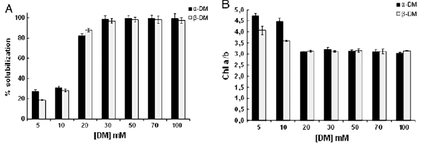
In the first part of this thesis work, they have been investigated the solubilizing properties of -DM and
-DM on the isolation of photosynthetic complexes from pea thylakoids membranes maintaining their native architecture of stacked grana and stroma lamellae. Exposure of these stacked thylakoids to a single step treatment with increasing concentrations (5–100 mM) of -DM or -DM resulted in a quick partial or complete solubilization of the membranes. Regardless of the isomeric form used: 1) at the lowest DM concentrations only a partial solubilization of thylakoids was achieved, giving rise to the release of mainly small protein complexes mixed with membrane fragments enriched in PSI from stroma lamellae; 2) at concentrations above 30 mM a complete solubilization occurred with the further release of high molecular weight protein complexes identified as dimeric PSII, PSI-LHCI and PSII– LHCII supercomplexes. It has been identified in 50 mM of both detrgents the minimal concentration that fully solubilized PSII-LHCII supercomplexes. In the second part of this work they have been characterized, in terms of polypeptide composition, purity and functionality by using biochemical techniques, two forms of PSII-LHCII supercomplexes isolated via sucrose gradient centrifugation from solubilized Pisum sativum thylakoid with 50 mM- and -n-dodecil maltoside. In according to the previous results, to isolate the supercomplexes they have been solubilized directly thylakoids instead of PSII membranes (BBYs), in order to minimize the possibility of detergent-induced artefacts and they have been used stacked membranes in order to avoid destabilization of the interaction of LHCII with the PSII core. The isolated supercomplexes haven‟t shown any ATP-ase and PSI contaminations. Both supercomplexes have shown an identical set of LMM subunits. The supercomplex isolated with -DM was larger and more intact, in terms of OEC and Lhcb subunits, than the supercomplex isolated with -DM. The supercomplex isolated with -DM has shown an higher activity than of those isolated with -DM. In the last part of the work they have been structurally characterized both isolated supercomplexes by using EM single particle analysis. The EM data have shown that the supercomplexes isolated with
-DM was a C2S2M2 while those isolated with -DM was a C2S2 supercomplex. They have been
obtained the 2D projection maps and the 3D reconstructions maps and angular reconstitution at 30 Å (for the C2S2M2) and at 28 Å (for the C2S2).
7
This thesis is based on the following publications (in attachment):
Pagliano, C., Barera, S., Chimirri, F., Saracco, G., Barber, J. (2012). Comparison of the α and β
isomeric forms of the detergent n-dodecyl-D-maltoside for solubilizing photosynthetic complexes from pea thylakoid membranes. Biochim. Biophys. Acta 1817: 1506-1515.
Barera, S., Pagliano, C., Pape, T., Saracco, G., Barber, J. (2012). Characterization of PSII-LHCII
supercomplexes isolated from pea thylakoid membrane by one-step treatment with - and -dodecyl-D-maltoside. Phil. Trans. R. Soc. B 367:3389-3399.
Pagliano, C., Nield, J., Marsano, F., Pape, T., Barera, S., Saracco G.,Barber J. (2013).
Proteomic characterization and three-dimensional electron microscopy study of PSII-LHCII supercomplexes from higher plants. Biochim. Biophys. Acta Nov 16. doi:pii: S0005-2728(13)00189-8. (in press).
8
9
1. GENERAL INTRODUCTION:
1.1 Photosynthesis (Light reactions):
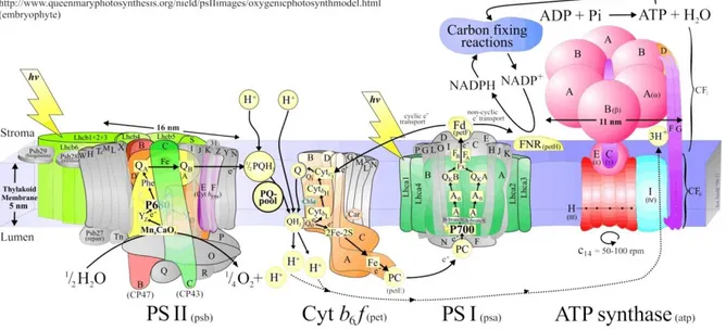
Photosynthesis is the process by which light energy is converted into chemical energy. The overall chemical reaction of oxygenic photosynthesis is the sunlight-driven, chlorophyll-sensitised transformation of water and atmospheric carbon dioxide to make energy-storing carbohydrates, and oxygen as a by-product. Photosynthetical reactions take place inside the thylakoid membranes and they are catalized by four multi-subunit membrane protein complexes (PSII, cytochrome b6f, PSI and
ATP-ase).
Figure 1.1 Cartoon model of the functional organization of photosynthetic thylakoid membranes in oxygenic organisms ( Copyright © Jon Nield, Queen Mary, University of London, UK).
The most important reaction of photosynthesis is the water oxidation carried out by Oxygen Evolving Complex (OEC), the oxidizing side of PSII.
1.1.1 Thylakoids organization:
Thylakoid membranes are organized in two main regions: staked region (grana) and unstacked region (stroma lamellae). In higher plants grana are a well organized piles of stacked thylakoid membranes in wich are located PSII complexes (dimer and supercomplexes). Grana stacks are linked by stroma lamellae in wich are located mainly PSI and ATP-ase. The 3D architecture of the granum–stroma
10
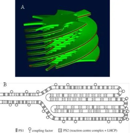
assembly and the entire thylakoid membrane network is complicate and interesting, especially because of the ability of the membranes to undergo reversible changes in folding and organization under varying environmental conditions. Recently many efforts are aimed at determining of the 3D organization of the thylakoid membranes, in particular the capacity to form grana stacks. One of the main consequences of stacking is the physical separation of PSI and PSII. The chloroplast ultrastructure changes from shade to sun plants, in fact as an adaptation to low light conditions, some plants have evolved extensive granal stacking in order to maximize the thylakoidal area involved in capturing solar energy (Anderson et al. 1975). The sun plants instead, living under excess light, have less appressed thylakoid membranes (Boardman, 1977), which could be an adaptation to maximize the space for the enzymes that are required for carbon fixation reactions. The organization of the thylakoid membranes rapidly and reversibly responds to changes in light intensity and quality not only in palnts growth in different light conditions, but the changes are visible in the alternation from day to night. The dynamics in the lateral distribution of the photosynthetic protein complexes in the thylakoid membrane most probably have a vital role in regulation of the functions, maintenance, and efficiency of the photosynthetic apparatus. Recent studies (Danielsson et al, 2006) have demonstrated that PSII distribution is well defined and it is correlated to the chloroplast ultrastructure. In the grana core are located mainly PSII-LCHII supercomplexes, while from these region towards the lamellae region are located PSII dimer and PSII monomer. The regulation of the chloroplast ultrastructure and the disposition of the water-splitting cofactors of the PSII complex inside the thylakoid membranes are mechanisms finely regulated and are essential to limitate photodamage. When the excess of light damage PSII, the damaged protein migrates from grana to stroma membranes, where the non-functional complexes are disassembled, damaged subunits are replaced with newly synthesized ones, and after reassembly the PSII complex finally migrates back to grana membranes. (Mulo et al. 2008). They have been proposed two models of the chloroplast ustrastructure the elical model and the fork model rapresented in figure 1.2 A and B.
The helical model was derived from EM studies of thin sections and serial sections of chemically fixed thylakoid membranes. As it is visible in figure 1.2, the stroma thylakoids are enfolded around the granal stacks in the form of multiple right-handed helices. The helical arrangement of the stroma thylakoids around grana was further supported by electron microscopy techniques including scanning electron microscopy and freeze-fracture EM (Staehelin et al. 1996). More recently tomography data revealed that there are fewer connections between one particular granum thylakoid and its surrounding
11
stroma thylakoids. Also the number of connected grana thylakoids in a stack by one spiral stromal thylakoid seems to be lower than previously thought.
Figure 1.2 A. Two current models for the 3D organization of thylakoid membranes in plant chloroplasts. (A) The helical model (Mustàrday et al. 2003) (B) The fork model (Andersson and Anderson, 1980).
Electron tomography studies have demonstrated that the dimensions of the connections between the stroma and grana thylakoids are more variable in size (Daum et al. 2010,Austin and Staehelin, 2011). The fork or folded membrane model was proposed at the beginning of 80‟s to illustrate the lateral heterogeneity in the distribution of PSII and PSI in the thylakoid membrane (Andersson and Anderson, 1980). In this model, represented in figure 1.2 B, the stroma membranes bifurcate and make a fork, which connects two adjacent grana thylakoids within a stack. The fork model was proposed not based on direct structural data but the fork motif was observed in many micrographs, including those of thin serial sections (Mustàrdy et al. 2008). Strong evidences for the existence of the fork at the granum– stroma assembly were provided by cryo tomography on cryo immobilized and freeze-substituted sections of intact leaves (Shimoni et al. 2005).
12
1.1.2 Linear electron transfer
Each of the four complex embedded in the thylakoidal membranes plays a distinct role in the electron transfer chain (ETC). PSII cointains a special pair chl called P680 that generates strong oxidant able to extract electrons from water and split it in O2 and protons (H+) using Calcium-Manganese cluster
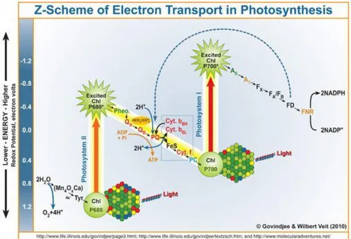
(donor side). The electrons come from water are collected by plastoquinone pool. The plastoquinoles are then reoxidized by the Cyt b6f complex and the electrons are collected by plastocyanin. During electron transport, Cyt b6f releases H+ in the lumen generating a H+ electrochemical gradient across the thylakoidal membrane. This ΔH+ proton motive force (pmf) is dissipated by the ATP-ase to form ATP. The plastocyanin, reduced by Cyt b6f, donates the electrons to the photo-oxidized P700 of the PSI which, on photon absorption, reduces ferredoxin. In conclusion ferredoxin is oxidized by FNR to obtain NADPH. Linear electron transfer is described by the Z scheme, in wich it is possible, observing the redox potential, the relationships between the involved electron acceptors and donors (figure 1.3).
Figure 1.3 Representation of the Z-scheme (Copyright © Govindje & Wilbert Veit 2010).
1.1.3 Cyclic electron transfer
During the light phase of the photosynthetical process not all the harvested light is adsorbed, a fraction is dissipated as fluorescence, the process by which chlorophylls release, with a longer wavelength, the energy derived from light absorption. Another fraction is dissipated as heat. The remaining portion is used for photochemical reactions. The percentage of not adsorbed light changes as a consequence of
13
light intensity and environmental conditions such as temperature, salt stress or drought. The excess of light absorption during the photosynthesis light reaction could lead to damage the photosynthetic apparatus via ROS generation, accordingly, it must be tuned and balanced. There are three mechanisms in the oxygenic photosynthetic organisms by which the excess of light is dissipated an the synthesis of ATP and NADPH is regulated: cyclic electon transfer (CET), state transition and water-water cycle. Cyclic electron transport have been discovered fifty years ago, until now they have been characterized two redundant routes for electrons cycling around PSI: the first one involves an NAD(P)H dehydrogenase (NDH) encoded by chloroplast and the other one is dependent on the PGR proteins (DalCorso et al. 2008). The electrons derived from the PSI reducing side are transferred back to the plastoquinones pool in both cases. In this way the proton gradient across the thylakoid membrane is enhanced generating more ATP.
With the first route mediating by NDH, called Chlororespiration, plants controls ATP/NADPH ratio in order to low the stromal over-reduction due to excessive light absorption with respect to the need of reducing equivalents (Shikanai, 2007).
The second one (PGR mediated route), instead, is less characterized, but seems to be connected to the thermal dissipation mechanisms of excessive absorbed light called nonphotochemical quenching (NPQ). A major component of NPQ, measured as a quenching of fluorescence emission, is energy-dependent thermal dissipation quencing (qE), which is related to the xanthophyll cycle. Thermal dissipation mechanisms involves, during the xanthophyll cycle, Violaxanthin deepoxidase (npq1) and protonation of PsbS, a subunit of PSII which is also known as npq4, and, as a consequence, this latter reaction is pH dependent (Li et al. 2000). In mediated PGR pathway PGR5, a membrane protein, is involved in the transfer of electrons from ferredoxin to plastoquinone. This alternative electron transfer pathway, whose molecular identity has long been unclear, is known to function in vivo in cyclic electron flow around photosystem I. It has been proposed (Shikanai, 2007) that the PGR5 pathway contributes to the generation of a ΔpH that induces thermal dissipation when Calvin cycle activity is reduced. Under these conditions, the PGR5 pathway also functions to limit the overreduction of the acceptor side of PSI, thus preventing photosystem I photoinhibition. This pathway is Antimicyn A-sensitive, indicating a direct connection with the Q cycle of Cyt b6f in higher plants (Takahashi, 2009).
The pgr5 mutant of Arabidopsis thaliana has been described as being deficient in cyclic electron flow around PSI, however, the precise role of the PGR5 protein remains unknown. Recently it has been examined PGR pathway by measurements of the kinetics of P700 oxidation in far red light and re-reduction following oxidation in the presence of DCMU in intact leaves of pgr5 and wild type A.
14
thaliana. The measurements has demonstrated that the mutant is able to perform cyclic electron flow at a rate similar to the wild type. The PGR5 protein is therefore not essential for cyclic flow. However, cyclic flow is affected by the pgr5 mutation under conditions where this process is normally enhanced in wild type leaves, high light or low CO2 concentrations resulted in enhancement of cyclic electron
flow. This suggests a different capacity to regulate cyclic flow in response to environmental stimulation in the mutant. The pgr5 mutant is inefficient in maintaining the right plastidial redox potential, in fact, the electron transport chain is reduced in most cases. In addition to the membrane protein PGR5, there is another transmembrane protein called PGRL1 present in thylakoids of Arabidopsis thaliana. It has been demonstrated that plants lacking PGRL1 shown a similar beahvior to PGR5-deficient plants. Recent studies confirm that PGRL1 and PGR5 interact physically and associate with PSI.
1.2 PSII structure and functions:
1.2.1 General overview:
PSII is a membrane protein complex embedded in the thylakoid membrane, it is a water-plastoquinone oxido-reductase. The reaction catalized by this enzyme is the one of most important for the life on our planet. The release of dioxygen, during light phase of photosynthesis, created an oxygenic atmosphere allowing a widely diversified life, based on mitochondrial-oxidative phosphorylation. The great importance of PSII for the life on earth justifies the considerable efforts that have been made to obtain further details, using biochemical, biophysical and genetic approaches, about its structure and the oxygene evolution reaction.
Recently, the attention of many researchers has been driven towards the possibility of mimic the water splitting activity. In order to reach this goal it is necessary a deep knowledge of the structure of the PSII catalytic site. In the past the first direct structural information about PSII was derived from electron microscopy (EM) via freeze fracture studies on thylakoid membranes (Staehelin et al. 2003), followed by the imaging isolated PSII particles (Hankamer et al. 1997). PSII dimeric structure, and its water-splitting activity, are conserved among all the photosynthetic oxygenic organisms. Thus the sequences of the intrinsic PSII subunits are highly conserved in all oxygen evolving autotrophs, are encoded by plastidial genes in green algae and higher plants, whereas the extrinsic proteins are nuclear-encoded and show high variability among oxygenic photosynthetic organisms (Ishihara et al. 2007). The little variability between plant PSII and cyanobacteria PSII has allowed to complement the electron
15
crystallographic studies of the higher plant PSII core dimer with X-ray crystallographic data obtained from cyanobacterium PSII crystals.
Several X-ray crystallographic structures are available for cyanobacterial PSII (Zouni et al. 2001; Jordan et al. 2001; Ferreira et al. 2004; Kamiya and Shen, 2003; Guskov et al. 2009;) and also the detailed structure of LHCII has been resolved (Liu et al. 2004; Standfuss et al. 2005; Yan et al. 2007). Actually, the crystal structure of PSII isolated from T. vulcanus has been refined to 1.9 Å giving more information about the positioning of the water molecules and giv further detail about the OEC (Umena et al. 2011).
Figure 1.4. Crystal structure of PSII isolated from T. vulcanus at 1.9 Å (Umena et al. 2011).
Unfortunaltely the complete structure of higher plant PSII hasn‟t been resolved. Therefore the combination of structural information about the cyanobacterial PSII core and higher plant LHCII with experimental biochemical data has made it possible to model the structure of the PSII-LHCII complex of higher plants (Nield and Barber, 2006). In general, solving of the structure of PSII at the highest resolution possible has become one of the most important goals of modern biology. Knowing the exact structure of the water-splitting apparatus of PSII would pave the way to development of photolytic bio-mimicking devices to produce hydrogen or electricity from water and sunlight.
Even if the high resolution 3D structure of cyanobacteria PSII dimer revealed all the positions of the atoms inside the oxygen evolving complex and the water molecules, the water splitting reaction carried out by manganese cluster, is still an unclear process and it needs further studies.
16
1.2.2 PSII polypeptide composition and arrangement
It has been widely believed that the PSII complex normally functions as a dimer and the monomeric complex may be an intermediate form in the normal assembly pathway or in the damage-repair cycle (Barbato et al. 1992; Hankamer et al. 1997).
PSII complexis composed by more than 20 subunits and many cofactors. PSII subunits can be divided in 3 groups: core complex proteins, low molecular mass subuinits, oxygen evolving extrinsic subunits.
Core complex proteins:
PSII reaction center (RC) is composed by two homologues five- helix trans membrane proteins with molecular mass around 39 kDa D1 and D2 (encoded from psbA and psbD genes rispectively), it contains all the cofactors involved in the electron transfer chain, from water to plastoquinones. Proteins D1 and D2 are higly conserved in fact they are remarkably similar to the L and M subunits of the purple bacteria Rhodopseudomonas sphaeroides reaction center (Barber et al. 1987). They contain a pheophytin molecule, a carotene molecule (Loll et al. 2005), three chlorophyll a molecules. Inside the loops between the IV and V helices on the stromal side of these proteins there are binding sites for a plastoquinone molecule. Protein D1 binds the majority of the cofactors involved in PSII electron transport chain: Y Z is Tyr161, P680 is Chl a molecule ligated by His198 (called PD1), Pheo D1 is molecule of pheophytin bounded by Tyr126 and Glu130 and a Q B site where plastoquinone molecules are bounded. D1 contains an other Chl molecule called Chl D1 and bind the manganese cluster in the C-terminal region. All of residues implicated in this bind, located close to the surface of the CD lumenal -helix of the D1 subunit, are nearly absolutely conserved with very little variation observed in dinoflagellates (Takishita and Uchida, 1999). Moreover D1 protein binds two chloride atoms near to the manganese cluster (Murray et al. 2008; Umena et al. 2011) and a non-haem Fe, normally redox inactive, which is positioned equal distance between Q A and Q B.
Another important property of D1 is the rapidly assembly-disassembly in the thylakoid membrane. This is due to the fact that PSII is susceptible to photoinibition and to protect entire system, D1 is degradated in order to regulate the electron transport chain. The degradation, synthesis and reinsertion of this protein into the PSII in plants is finely regulated. During the turnover Met1 is cleaved and its N-terminal Thr can be acetylated and reversibly phosphorylated. (Michel et al. 1988; Pursiheimo et al. 1998; Vener et al. 2001; Turkina et al. 2006).
17
The D2 proteins conatins two Chl a molecules, P D2 and Chl D2 , which together with P D1 and Chl D1 form the cluster constituting P680 and the QA site. The D2 protein also contributes ligands for the non-haem Fe, and is involved in binding chloride through side chain.
Similarly to D1, in higher plants and green algae (but not in cyanobacteria) the D2 undergoes reversible acetylation and phosphorylation of its N-terminal Thr residue (Michel et al. 1988; Pursiheimo et al. 1998; Vener et al. 2001; Turkina et al. 2006).
CP47 and CP43, products of the psbB and psbC genes, are the core complex antenna proteins, they transfer excitons derived by light absorption to P680 at two opposite sides of the RC. They are structurally homologous, having a molecular weight of ~ 56 and 50 kDa respectively, six helices each of them and sixteen and fourteen chlorophyll a molecules respectively.
CP47 protein has 16 Chl a molecules and 5 -carotenes. It has a very large lumenal loop joining transmembrane between helices V and VI, consisting of 200 amino acids arranged in two long and four short helices as well three -sheets. The large loop indirectly takes part in the water oxidation reaction by interacting with the D2 protein, which is adjacent to it.
CP43 (PsbC), after post-translational processing, contains 470 amino acids it is homologous to CP47 in that it has six transmembrane a-helices arranged in the same way but located on the D1-side of the RC. It also contains 13 Chl a molecules and binds 5 b-carotene molecules. It has a large lumenal loop like CP47 but slightly smaller. CP43 protein contains two long and three short helices one of which contains the highly conserved motif which provides a ligand to the manganese cluster. Altough there are some differencese with CP47, the main function remains the same: CP43 acts as a light-harvesting system for PSII and its presence is also necessary for water-splitting activity.
Low molecular mass subuinits:
Inside the PSII core complex, apart from D1, D2, CP43 and CP47 there are many subunits called Low Molecular Mass (LMM) because of their molecular weight its from about 3 kDa to 12 kDa. Up to now the knowledge about their function is very low; it seems, however, that most of these LMM polypeptides are not involved in the electron transfer but, instead, are structural components.
The first evidence for low molecular mass subunits associated with the PSII complex was presented by Ljungberg and his co-workers who isolated a hydrophilic 5-kDa protein (corresponding to PsbTn) from PSII and shown that the PSII complex contained several other small, unidentified subunits. This small polypeptides were named, according to their apparent molecular masses, the 10, 8, 7, 6.5, 5.5-, 5 and
4-18
kDa proteins. Recently they have been added 6.8, 3.7 and the 3.2 kDa proteins to the list of small subunits in PSII.
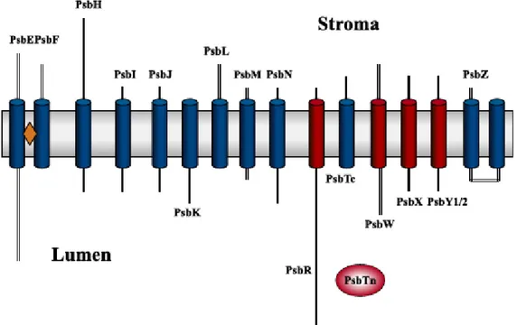
Several of the low molecular mass subunits of PSII were sequenced and the first corresponding gene products, PsbK and PsbI were identified. The protein nomenclature was correlated to the gene names to avoid the confusing names related to the apparent molecular masses of the proteins. Dramatic improvements and developments have been made in protein detection and identification techniques during the last decade. Highly sensitive fluorescence techniques combined with the latest improvements in ion mass spectrometric techniques, which allow the detection and identification of some femtomoles of proteins (roughly corresponding to 0.1 ng), now complement the traditional Coomassie brilliant blue staining. These new techniques have confirmed that PSII contains a large set of low molecular mass subunits. Molecular biological methods have also yielded new insights into this group of PSII subunits. The LMM proteins identified are encoded by the psbE, psbF, psbH, psbR, psbI, psbJ, psbK, psbL, psbM, psbN, psbT, psbX, psbW and psbZ genes.
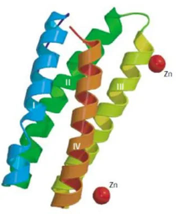
Figure 1.5 Cartoon representation of the transmembrane elices of the LMM proteins (Shi and Schroeder, 2004).
19
PsbE and PsbF
The genes psbE and psbF encode for and subuinits of Cytochrome b 559 (Cyt b 559) with molecular masses of 9 and 4 kDa, respectively and each of these polypeptides has one transmembrane helix. The Cyt b 559 is closely associated with PSII in all oxygenic photosynthetic organisms. The
and subunits of the Cyt b 559 are components of the minimal PSII reaction center complex that is still capable of primary charge separation (Nanba et al. 1987). Cyt b 559 is a b-type cytochrome, but it has a unique feature with respect to its midpoint reduction potential (Em). High-, low- and intermediate-potential forms of Cyt b 559 have been observed in intact chloroplasts, PSII membranes and isolated D1/D2/ Cyt b 559 complexes. The function of Cyt b 559 has been unknow for a long time, but structural details suggest that Cyt b 559 could participate in a secondary electron transfer pathway within PSII (Heber et al. 1979). These observations and the results of other studies on Cyt b 559 mutants indicate that Cyt b 559 is involved in protecting PSII from photoinhibition (Poulson et al. 1995). In addition, evidence from site-directed mutagenesis in Synechocystis 6803 suggests that Cyt b 559 is required for the assembly of functional PSII complexes. (Morais et al. 1998).
PsbH
The PsbH protein was identified in higher plant chloroplasts and initially called 10- or 9-kDa phosphoprotein. It is encoded by the plastome in algae and higher plants and it is present also in cyanobacteria. The protein contains 63-90 amino acids, depending on the species, with molecular masses between 7.0 and 9.9 kDa. PsbH is an intrinsic membrane protein with a single transmembrane helix and its N-terminal region has been suggested to be exposed to the stromal side of the thylakoid membrane. In higher plants, the protein is located close to Cyt b 559 and PsbX (Buchel et al. 2001), its phosphorylation sites of PsbH are located at the N-terminus and this reversible phosphorylation is light-dependent and redox-controlled (Michel et al. 1987). Recent studies on PSII complexes isolated from spinach suggest that PsbH, together with other PSII phosphoproteins, may be required for D1 protein turnover by regulating dimeric and monomeric PSII transition through their phosphorylation and dephosphorylation. This has been further substantiated by the recent demonstration that PsbH is required for the rapid degradation of the photodamaged D1 protein and insertion of newly synthesized D1 into the thylakoid membrane. In summary, the PsbH protein is essential for PSII activity in eukaryotes. It may play a role in regulating PSII assembly/stability and may also be involved in repair of photodamaged PSII under high light.( Kruse et al. 1997, Bergantino et al. 2003).
20
PsbI
The PsbI protein, called also 4.8-kDa protein, is encoded by the plastome. PsbI is highly conserved in higher plants and cyanobacteria. The protein contains at least 38 amino acids with molecular masses ranging between 4.1 and 4.5 kDa. Sequence analysis indicates that it has a single transmembrane span. The N-terminus of this protein is formyl-Met and is located on the stromal side of the thylakoid membrane and analysis of its cross-linked proteins indicates that the nearest neighbors of PsbI are D2 and the a subunit of Cyt b 559 (Tomo et al. 1993). The function of PsbI has been investigated in Cyanobacteria. Inactivation of the psbI gene in Synechocystis 6803 and T. elongatus strain BP-1 caused similar reductions in PSII activity. In the case of T. elongatus DpsbI mutant, no dimeric PSII could be isolated. It was suggested that while PsbI is not essential for PSII photochemistry in cyanobacteria, it may be involved in processes that help to optimize PSII function, such as the dimerization of PSII or stabilization of PSII dimers (Ikeuchi et al. 1995).
PsbJ
PsbJ is one of the most hydrophobic proteins in the thylakoid membrane, containing 39-42 amino acid residues, with a molecular mass between 4.0 and 4.4 kDa. Sequence analysis suggests that the protein has a single transmembrane helix with its N-terminus extending to the stromal side. The PsbJ function is controversial even if there are many evidence that the inactivation of the psbJ gene reveal phenotype with low PSII oxygen evolution caused by the reduced amount of functional PSII complexes. (Lind et al. 1993, Regel et al. 2001).
PsbK
The chloroplast PsbK protein has been found in PSII core complexes from the thermophilic cyanobacteria and higher plants. The PsbK precursor contains a hydrophobic presequence ranging from 5 to 24 amino acids long, and some of the longer presequences have a putative transmembrane span that may play a role in the insertion of the protein in the thylakoid membrane. In all species examined so far mature PsbK protein contains 37 amino acids and has molecular masses between 4.1 and 4.3 kDa. Accordingly, it is the most conserved small protein in PSII. The Arabidopsis PsbK shares 76% identity with the Synechocystis 6803 homolog. PsbK has been recently found to be tightly associated with CP43 (Sugimoto and Takahashi, 2003). Deletion of the psbK gene in Chlamydomonas destabilized the PSII complex, and the amount of PSII in the resulting mutants was less than 10% of
21
wild-type levels.The transformate bacteria were unable to grow photoautotrophically and no PSII activity was detected, suggesting that this protein is indispensable for PSII function in Chlamydomonas (Takahashi et al. 1994). Biochemical analysis of subcore complexes of PSII from spinach has shown that dimeric CP47-reaction center complexes contain the small proteins PsbK and PsbL, together with 1-2 molecules of plastoquinone.The monomeric complexes instead do not contain PsbK.According to these results, it has been suggested that the PsbK protein is involved in binding plastoquinone and in maintaining the dimeric organization of PSII (Zheleva et al. 1998).
PsbL
PsbL protein is encoded by the plastome, it contains 37-39 amino acid residues with molecular masses between 4.3 and 4.5 kDa depending on the species. Primary sequence analysis suggests that PsbL has one transmembrane helix and it has also been suggested that the N-terminus of the protein is located at the stromal side of the thylakoid membrane. PsbL is present in both PSII core complexes of higher plants and cyanobacteria only in the dimeric form, indicating that the PsbL protein may have a role in maintaining PSII dimeric organization (Zheleva et al. 1998). The Synechocystis 6803 PsbL mutant shown the total loss of PSII-mediated oxygen evolution, and it appear to be unable to grow photoautotrophically. Interestingly, no binding of herbicides that normally bind D1/D2 proteins was detected in these mutants, despite immunological detection of D1 and D2 protein. This was suggested to be due to changes on the acceptor side of PSII (Anbudurai and Pakrasi 1993). Further studies of PsbL mutants from tobacco has showned that this plants are unable to grow photoautotrophically, and if sucrose was added they only grew slowly with pale green leaves: no photosynthetic activity was detected in them. According to these studies we can conclude that PsbL is essential for the normal function of PSII. Its deletion causes fatal damage to PSII in both cyanobacteria and tobacco plants, especially for donor side electron transfer, PSII core assembly and maintenance of the dimeric form of PSII.
PsbM
The PsbM (chloroplast encoded) contains 31-38 amino acid residues, with calculated molecular masses between 3.5 and 4.2 kDa. Sequence analysis suggests that the PsbM protein is one of the most hydrophobic proteins in the thylakoid membrane. The PsbM protein from Arabidopsis shares 54% identity to that from Synechocystis 6803. The PsbM protein has been detected in PSII complexes isolated from Cyanobacteria and higher plants in particular from pea using proteomic techniques. It is
22
located with PsbL and PsbTc at the monomer–monomer interface of the dimeric core complex and it contribute to the stabilization of PSII dimers (Ferreira et al. 2004). PsbM is not the only protein responsible for dimer stabilization in fact knock-out of psbM in tobacco plants revealed that PsbM is not required for biogenesis of higher order PSII complexes in plants, but it alters the properties of the Q B site and the electron flow within PSII (Umate et al. 2007).
PsbN
The PsbN protein contains 43 amino acids and has a molecular mass of 4.5-5.0 kDa in most species. The effects of deleting both PsbN and PsbH on PSII activity in Synechocystis 6803 were the same as those of solely deleting the PsbH protein, indicating that PsbN is not essential for photoautotrophic growth and normal PSII function. PsbN protein has not been detected directly in plant PSII, and it is not present in the crystal structures of PSII isolated from cyanobacteria. The only evidence of its presence in a PSII preparation from the cyanobacterium Synechococcus vulcanus (Ikeuchi et al. 1989a) was reexamined by Kashino and colleagues (Kashino et al. 2002), who revealed that the protein originally regarded as PsbN in that preparation was actually PsbTc. These results strongly suggest that PsbN is not a PSII protein.
PsbR
The PsbR protein also named the 10-kDa polypeptide (with a molecular mass of 10.3 kDa) contains 99 amino acids. Studies have indicated that PsbR could not be removed from the membrane by treatment with high salt concentrations. Ou Infact predictions indicate that it contains a transmembrane span close to its C-terminus and during purifications PsbR presents hydrophobic property (Ljungberg et al. 1986; Lautner et al. 1988). Recent studies have shown that PsbR has a charged domain. These particular features suggest that PsbR is a docking protein, in particular it has been proposed that PsbR has a binding site for the extrinsic PsbP protein. Nevertheless, it has been also found that when PsbS and PsbR proteins were removed from PSII, the extrinsic PsbO, PsbP and PsbQ proteins were retained suggesting that not only PsbR is involved in the docking of the PsbP protein. Further studies on the function of PsbR have demonstrade that the PsbR mRNA is accumulate upon light exposure and the PsbR protein was undetectable in etiolated spinach, in contrast to the expression patterns of the PsbO, PsbP and PsbQ extrinsic proteins (Lautner et al. 1988).
23
PsbS
The PsbS protein is not part of LMM subunits in fact it has 205 amino acids and a molecular mass of 22 kDa. This subunit is nuclear encoded and it has been predicted to have four transmembrane helices. PsbS protein has a high sequence homology with the light-harvesting proteins of the Lhc family (Funk et al. 1995), buti t has a transmembrane helix more and for its still controversial ability in the native form to bind Chl and carotenoid molecules (Funk et al. 1995; Dominici et al. 2002). Recent studies have shown that PsbS is able to bind exogenous zeaxanthin (Aspinall-O‟Dea et al. 2002). This binding may be associated with the formation of nonphotochemical quenching centres of Chl fluorescence (qE) functioning to dissipate excess light energy absorbed by PSII lightharvesting system, a phenomenon in which PsbS is known to play a crucial role (Li et al. 2000). Recently, an explanation for this role has been provided showing that PsbS controls the macro-organization of the grana membranes in higher plant chloroplasts. The regulation of PsbS interaction between the antennae and the PSII core in the thylakoid membranes of higher plants seems to be via pH induced conformational changes (Kiss et al. 2008). As it was difficult to locate PsbS to any particular place in membrane fractionation studies, it was suggested that it is mobile in the thylakoid membrane (Teardo et al. 2007) and that its location can be determined by a reversible dimerization (Bergantino et al. 2003).
PsbTc and PsbTn
The psbTc gene is highly conserved among cyanobacteria, algae and higher plants. The lower case c in the name of this protein indicates that in higher plants and algae the protein is chloroplast-encoded. The product of this gene contains 31-38 amino acids with molecular masses ranging from 3.5 to 4.4 kDa. PsbTc is an intrinsic protein with a single transmembrane helix close to the N-terminus. PsbTc has been detected in C. reinhardtii and spinach.It has been proposed that the C-terminal tail of the protein is exposed to the stromal side of the thylakoid membrane. The predicted orientation of PsbTc, as well as PsbK, is similar to that of nuclear-encoded PsbW and PsbX, but opposite to that of other small, chloroplastencoded PSII proteins. PsbTc has been proposed to have an intimate association with the D1 and D2 proteins (Zheleva et al. 1998). The function of this subunit has been studied in a deletion mutant from C. reinhardtii where the growth and PSII function were impaired under high light conditions. Therefore, it has been suggested that the protein is required for maintaining optimal PSII activity under adverse conditions, such as high light stress. Further studies revealed that this chloroplast-encoded protein undergoes degradation upon high light, but does not play a role in photoprotection. Instead, it is required for efficient recovery of photodamaged PSII. It has been
24
proposed that PsbTc in C. reinhardtii is involved in posttranslational events during photoinhibition. (Monod et al. 1994, Ohnishi and Takahashi 2001).
PsbTn (nuclear encoded protein) has been purified and partially sequenced from PSII core complexes of spinach and wheat, it is a hydrophilic protein with an apparent molecular mass of 5.0 kDa according to polyacrylamide gel analyses, and was previously named the 5.0-kDa. After the maturation process , the protein has only 28 amino acids and a molecular mass of 3.2 kDa in Arabidopsis, making it the smallest known polypeptide in the PSII complex to date. The function of PsbTn protein is unknown but there is a bipartite presequence in its amino acid sequence characteristic of the lumenal proteins, so it has been suggested that the protein could be an extrinsic protein.
PsbW
The nuclear psbW gene is present in green algae and in higher plants. This gene encodes a protein (PsbW) with a long transit peptide (59-83) amino acid residues in length depending on species) .The mature PsbW protein, previously known as the 6.1-kDa protein, is composed of 54 amino acid residues in spinach and Arabidopsis. The PsbW protein has one transmembrane span and the N-terminus of the protein was demonstrated to be located on the lumenal side and the C-terminus on the stromal side of the thylakoid membrane. The PsbW protein is tightly associated with the PSII reaction very close to the large subunit of Cyt b 559. Expression of the PsbW gene is light-regulated at both mRNA and protein levels in Arabidopsis, in fact study on photoinhibition demonstrated that the repair of photoinhibitory damaged PSII involves the rapid turnover of the PsbW protein. Recent studies have showned that in knock-out Arabidopsis mutants the supramolecular organization of PSII–LHCII supercomplexes is widely compromise, suggesting a more peripheral positioning close to the minor antenna complexes in PSII (García-Cerdán et al. 2011).
PsbX
The PsbX protein is nuclear-encoded in higher plants and in some algae where it is located in the chloroplast or cyanelle. PsbX precursor from Arabidopsis has a bipartite presequence of 74 amino acid residues, which is absent in its plastid-encoded counterpart in algae. This evidence could suggests that the gene has been transferred from the plastid to the nucleus and acquired the transit peptide (Kim et al. 1996). The mature PsbX protein contains 38-42 amino acid residues with a calculated molecular mass of 4.0-4.2 kDa, depending on the organism. Predictions according to its primary sequence indicate that PsbX has a single transmembrane span and that the N-terminus is located at the lumenal side of the
25
membrane. The higher plant PsbX is located within the PSII core, but it was undetectable in either PSIIRC or LHCII preparations. Recent studies has been swooned that the expression of the psbX gene is light-regulated at both the mRNA and protein levels in Arabidopsis. The regulation of psbX mRNA in Synechocystis 6803 instead, under normal light conditions has been found to be independent of light, although it is expressed strongly during high light treatment. Inactivation of the psbX gene in Synechocystis 6803 caused a reduction in the amount of PSII and a slight uncoupling of the antenna. In the psbX deletion mutant the donor and acceptor sides of PSII functioned properly under normal and stress conditions (such as high light, and salt stress) but the decrease of the PSII amount might suggest that the PsbX protein are directly or indirectly involved in regulation of the amount of PSII (Shi et al. 1999, Funk 2000).
PsbY
The psbY gene in higher plant encodes a much larger polypeptide precursor (19.5 kDa in Arabidopsis and 20.7 kDa in spinach). The precursor is synthesized in the cytosol, imported into the chloroplast, then subjected to several steps of processing and maturation to generate two thylakoid membrane proteins, PsbY-1 and PsbY-2 with molecular masses of 4.7 and 4.9 kDa respectively. In Arabidopsis, both PsbY proteins have been predicted to have one transmembrane span with the N-terminus at the lumenal side of the thylakoid membrane. The twin PsbY proteins have been isolated from spinach PSII membrane fractions (BBY particles). Further studies have reported that the PsbY protein have catalase-like manganese-dependent activity and an L-arginine metabolizing activity that converts L-arginine into ornithine and urea (Gau et al. 1998).
PsbY mRNA was found in leaves, but not roots, stems or etiolated materials. The amount of mRNA increased during the first couple of hours of illumination, and then decreased.
Deletion mutagenesis in Synechocystis 6803 has showned that the psbY mutant cells have normal photosynthetic activity and that PsbY protein does not play an essential role in the photosynthetic water oxidation process, in accordance with its localization in the periphery of PSII, far from the OEC (Meetam et al. 1999). A discrete function for this subunit is still unknown, and a higher plant psbY knockout mutant is needed.
PsbZ
The PsbZ protein is encoded by a chloroplast gene, psbZ, highly conserved among organisms, contains 62 amino acid residues and its theoretical molecular mass, in Arabidopsis is 6.6 kDa. PsbZ has been
co-26
purified with LHCII complexes from tobacco plants. Thus, it has been suggested that the location of this protein seems to be at the interface of PSII and LHCII complexes. Recent studies have pointed out in evidence that psbZ knockout tobacco plants developed pale leaves under standard heterotrophic growth conditions due to a reduction in chlorophyll content; the plants also have shown a dwarf phenotype in low light conditions. Moreover the level of the antenna proteins CP26 and CP29 were found to be markedly reduced in psbZ knockout plants. The lack of the PsbZ protein and the consequently decrease of the amounts of CP26 and CP29 caused structural changes in PSII that prevented the isolation or detection of PSII–LHCII supercomplexes in electron microscopy studies. (Swiatek et al. 2001).
However, as the loss of PsbZ leads to reduced amounts of CP26, these effects may be secondary. To summarize, PsbZ is likely to be a structural factor that stabilizes PSII–LHCII supercomplexes. It may also be involved in photoprotective processes under suboptimal growth conditions. In cyanobacteria, where the light-harvesting system is completely different from higher plants, a role for PsbZ in the regulation of electron transfer activity through the two photosystems with implication in photoprotection has been suggested (Bishop et al. 2007).
Oxygen evolving extrinsic subunits:
The catalythic site of higher plants PSII, which is also known as OEC, is composed by a Ca-Mn cluster and three extrinsic subunits namely PsbO, PsbP and PsbQ.
The OEC proteins are nuclear-encoded but PsbP and PsbQ are present only in higher plants and in green algae. In cyanobacteria and in red algae they are substituted by PsbU and PsbV although there are some differences in the function and binding properties. Concidering recently study, wich have been found in the cyanobacterial PsbQ and PsbP homologues, it has been proposed that the ancestor of the oxygenic photosynthetic organism had all the extrinsic proteins (Roose et al. 2007; Enami et al. 2005; Ishihara et al. 2007). At the present state, after several years of evolution, Enami and their coworkers, individuate three evolutionary branch: and green algae and higher plants, which have PsbO, PsbP and PsbQ; Red algae and Diatoms, which have PsbO, PsbQ homologous (in stoichiometric amount) PsbU and PsbV and Cyanobacteria, which have PsbO, PsbU and PsbV.
This phylogenetic division reflect their adaptation to different ecological niches, in fact type and Chl content changes through these three groups. Another evidence of the evolution of PsbP from the ancestor to primitive green algae and higher plants it has been found in two PsbP-like proteins (PPL1 and PPL2) in the lumen of Arabidopsis (Ishihara et al. 2007). As it has been said before, all these
27
proteins in eukaryotes have their coding region in the nuclear genome, Ishihara and their co-workers claim that probably the gene transfer from the chloroplast genome to the nuclear in order to allow a greater diversification of the gene product structures and functions.
The expression of the OEC genes is finely regulated and in fact, it has been found that, in the extreme halophyte Salicornia veneta, the psbQ and psbP genes are strongly underexpressed (Pagliano et al. 2009). Thus, some PSII subunits, like OEC proteins, could be involved in an evolutional adaptation to an extreme ecological niche.
PsbO
PsbO protein also called 33 kDa Manganese Stabilizing Protein is a component of the PSII OEC in every type of oxygenic photosynthetic organism. Biochemical and genetic studies have shown that the PsbO protein play a crucial structural and functional role in many different types of OECs. The most important functions carried out by PsbO are: stabilize the manganese cluster and modulate the Ca2+ and Cl- requirements for oxygen evolution (Bricker, 1992).
This extrinsic subunit is conserved from cyanobacteria to higher plants, where it is nuclear-encoded. PsbO protein is often referred to as the 33 kDa protein, but after processing it has a calculated molecular mass of 26.5 kDa and its sequences is composed by 240-257 residues, depending on species. The crystal structure of this protein shows an elongated shape with two main domains: a -barrel structure composed of eight antiparallel -strands and an other domain, formed by a loop joining -strands 5 and 6. The -barrel plays an important role in stabilizing the manganese cluster and in maintaining an optimal environment for water oxidation (De Las Rivas and Barber, 2004). Other studies have been shown that PsbO mantain the optimal Ca2+ and Cl- levels at the catalytic site, but it does not bind Mn directly.(Briker et al. 2008). The loop binds to the lumenal surfaces of the D1, D2, CP43 and CP47 proteins. The deletion of the psbO gene in cyanobacteria did not prevent water oxidation, although increased susceptibility to photoinhibition was observed (Philbrick et al. 1991; Mayes et al. 1991). In Arabidopsis there are two PsbO proteins called PsbO1 and PsbO2. They differ only by 10 amino acids in their mature form and also for their expression, in fact PsbO1 isoform is four- to fivefold more abundant than PsbO2 (Murakami et al. 2002). An Arabidopsis mutant with an impaired psbO1 gene shown retarded growth, although the level of the PsbO2 protein was significantly increased (Murakami et al. 2002, 2005). Two T-DNA insertion mutant lines of Arabidopsis deficient in either the PsbO1 or the PsbO2 protein has helped to understand the different functions of the two
28
isoforms: PsbO1 seems to mainly support PSII activity, whereas the interaction of PsbO2 with PSII regulates the turnover of the D1 protein, increasing its accessibility to the phosphatases and proteases involved in its degradation, a mechanism possibly mediated by its GTP hydrolytic activity (Lundin et al. 2007, 2008).
The absence of the PsbO protein has varying effects on PSII depending on the organism. For example, plants, not in alla cases, and the green alga Chlamydomonas reinhardtii are not capable of photoautotrophic growth without PsbO (Mayfield et al. 1987; Yi et al. 2005). Cyanobacterial PsbO mutants can grow photoautotrophically, although at lower levels relative to wild type (Philbrick et al. 1991). One of the major controversy associated with PsbO, involves its stoichiometry within the PSII complex. Structural analyses of cyanobacterial PSII indicate one PsbO per PSII monomer (Kamiya and Shen 2003; Ferreira et al. 2004; Loll et al. 2005), but reconstitution studies in plants indicate the molar ratio of PsbO in PSII is two (Bricker, 1992; Popelkova et al. 2002b). This data can be reconciled by the recent observation that plant PsbO contains two sequence motifs at its N-terminus which determine binding stoichiometry, while cyanobacterial PsbO has only one such sequence (Popelkova et al. 2002a). However, structural studies of plant PSII show there is not sufficient electron density to accommodate two copies of the PsbO protein (Nield et al. 2002; Nield and Barber, 2006).
PsbP
The PsbP protein (also called 23 or 24 kDa protein) was first determined to be a component of PSII in plants, but psbP gene can even be found in the primitive cyanobacterium Gloeobacter violaceus which lacks thylakoids, suggesting an ancient role for this protein in PSII. Arabidopsis has 10 copies of the psbP gene in its genome and eight of these were found to be expressed proteins in the thylakoid lumen (Schubert et al. 2002). PsbP was first identified during release–reconstitution experiments in higher plants. The PsbP and PsbQ proteins are removed by 1 M NaCl treatment with a concomitant decrease in oxygen evolution. Addition of Ca2+ and Cl– to the assay medium restores PSII activity without the addition of these proteins (Ghanotakis et al. 1984; Miyao and Murata, 1985). Specifically, PsbP was found to modulate the Ca2+ requirement for PSII activity and has been hypothesized to act as a Ca2+ concentrator and prevent release of Ca2+ during turnover of PSII (Miyao and Murata, 1984). Multiple studies have shown that PsbP has implicated, with PsbQ, also in the regulation of the Cl– requirement. Clearly the PsbP protein functions to modulate the ionic requirements for oxygen evolution, but it also plays a structural role to protect the manganese cluster from attack by exogenous reductants (Ghanotakis et al. 1984). Altought PsbP and PsbQ proteins are extremely important in the ionic
29
recruitment, recent studies have shown that in the extreme halophyte Salicornia veneta the psbQ and psbP genes are strongly underexpressed but the oxygen evolving activity is not compromised (Pagliano et al. 2009).
In mutagenesis studies, plants lacks the PsbP protein, accumulates PSII centers, high concentrations of Cl– were necessary to promote oxygen evolution and there were significant defects in the light-driven assembly of the manganese cluster . This inefficient photoactivation process and decreased Cl– affinity resulted in a substantial amount of competing donor side damage. These data suggest a specific role for PsbP in the functional assembly of the manganese cluster and stability of PSII.
The PsbP knock-down plants exhibited a reduced variable fluorescence yield, lower oxygen evolution and decreased amounts of PsbQ, which has been shown to require PsbP for binding to PSII in plants. RNAi studies shown that all of the isoforms are required for optimal activity, but generally PSII activity was correlated with the total amount of PsbP protein. Therefore the PsbP protein is essential for normal in vivo PSII activity in plants because it is critical for OEC stabilization. (Ishihara et al. 2005). The essential role of PsbP for normal thylakoid architecture was further emphasized in Arabidopsis mutants, where dramatic alterations in thylakoid structure were found in plants expressing low and intermediate amounts of the PsbP protein under normal growth light conditions (Yi et al. 2009). Similar results were obtained in tobacco mutants, where the knockdown of PsbP impaired also the accumulation of PSII supercomplexes (Ido et al. 2009).
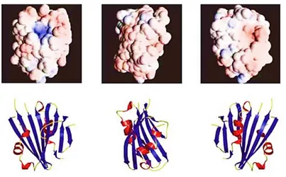
Thornton and his co-workers have determined amount of PsbP by using quantitative immunoblot comparisons of the proteins relative to antigen standards and it seems to be approximately 3% of that of CP47 in the thylakoid membranes. In plants, binding of the PsbP protein requires PsbO and is hypothesized to be a largely electrostatic interaction (reviewed in Seidler, 1996). In contrast, PsbP from green algae has been shown to bind independently of the other extrinsic proteins (Suzuki et al. 2003). These variations in the binding properties of PsbP highlight structural adaptations for associating with distinct combinations of proteins in the OECs of different organisms. Refer to the sections for the organismspecific OECs for further discussion. Currently, a high resolution (1.6 Å) crystal structure of PsbP from Nicotiana is available (Ifuku et al. 2004). The crystal structure of PsbP has revealed that the core is an anti-parallel -sheet with -helices on either side (Figure 1.5).The electron densities of the N-terminal 15 residues, and two loop regions were not resolved.
30
Figure 1.5. PsbP crystal structure at 1.6 Å (Ifuku et. al 2004).
This result may indicate possible stabilizing conformational changes in PsbP upon binding. Further studies on PsbP N-terminus indicate that this portion of the protein is critical for ion retention in PSII (Ifuku and Sato 2001), but it hasn‟t been reported any mechanism for its function. PsbP structure suggest a non-conventional role of the protein in plant PSII; in fact PsbP is very similar to that of Mog1p, a regulatory protein for a Ran GTPase suggesting it may be a possible GTP/GDPsensitive regulator (Ifuku et al. 2004; De Las Rivas and Roman, 2005).
PsbQ
The psbQ genes have been identified in a number of different photosynthetic organisms. Arabidopsis contains multiple copies of psbQ genes, four of which have been identified as expressed proteins in the thylakoid lumen (Peltier et al. 2002; Schubert et al. 2002). Arabidopsis contains two expressed PsbQ proteins (Peltier et al. 2000; Schubert et al. 2002), encoded by two genes, psbQ-1 and psbQ-2.
The protein PsbQ (known also as 16-18 kDa protein), in its mature form, contains 149 amino acids and has a molecular mass of 16.5 kDa. Like PsbP, it is located close to PsbO and the Mn4CaO5 cluster of
the OEC in higher plants and green algae, where it is nuclearencoded. Its gene has been found in a number of different photosynthetic organisms including cyanobacteria, where this subunit (known as PsbQ-like protein) can be stoichiometric with PSII (Thornton et al. 2004) and isolated as such (Roose et al. 2007b).
High-resolution crystal structures of isolated spinach PsbQ protein (Calderone et al. 2003; Balsera et al. 2005) have revealed that the PsbQ protein can be divided into two structural domains: a C-terminal four-helix bundle with an asymmetric charge distribution and a flexible terminus. The PsbQ
N-31
terminal region include two short b-strands surrounding a large flexible loop region (residues 14-33) and a polyproline type II motif (Balsera et al. 2005), which may obtain a more rigid conformation upon binding PSII.
Figure 1.6. PsbQ crystal structure at 1.95 Å (Calderone et al. 2003).
The crystal structures of PsbQ show two bound Zn2+ atoms, where the coordinating residues of one atom are entirely conserved in the plant PsbQ proteins (Calderone et al. 2003; Balsera et al. 2005). While Zn2+ was specifically required for crystal growth, the biological significance of this result has yet to be determined.
Figure 1.7. Zn2+ binding domain of the PsbQ crystal structure (Balsera et al. 2005).
The cumulative data on PsbQ indicate it is a key structural component of OECs from a number of different organisms, but many questions remain regarding its function and its mode of association with PSII. High-resolution structures of PSII complexes containing PsbQ are necessary to provide insights
32
into its role in the ionic requirement for oxygen evolution and its location relative to the other protein components of the OEC.
In higher plants, the binding of PsbQ to PSII requires the presence of PsbO and PsbP (reviewed in Enami et al. 2008). Biochemical in vitro studies have shown that PsbQ, like PsbO and PsbP, is involved in modulating the Ca2+ and Cl- level for optimal oxygen evolution activity (reviewed in Seidler 1996; Roose et al. 2007a; Bricker et al. 2012). In particular, PsbQ, in concert with PsbP, functions to lower the Cl- requirement for optimal activity. (Ghanotakis et al. 1984; Miyao and Murata, 1985). This finding fits well with the evidence that in some halophytic plants, growing in extremely salty environments, the presence of PsbQ is dispensable (Pagliano et al. 2009; Trotta et al. 2012).
1.2.3 Light Harvesting Complex and PSII Ultrastructure (PSII-LHCII supercomplex)
Light harvesting complex II
In photosynthetic organisms PSII core dimer is serviced by a number of extra chlorophyll binding proteins which transfer the excitation energy to CP43 and CP47. The number and type of this supplementary antennae complexes vary among the different types of oxygenic photosynthetic organisms. To efficiently harvest the available spectrum of light energy that drives photosynthesis, different photosynthetic organisms use diverse proteins, each one binding specific pigment molecules. In cyanobacteria and eukaryotic red algae, light harvesting for PSII is carried out primarily by a group of pigmented proteins, called phycobiliproteins, encoded by apc and cpc genes that become constituents of the extrinsic macromolecular complex called phycobilisome (PBS).
In higher plants the polypeptides which bind the pigments are called Chl a/b binding proteins (also known as Cab or Lhcb proteins). They are part of the Lhc gene super family which consists of the lhcb1, lhcb2, lhcb3, lhcb4 (CP29), lhcb5 (CP26) and lhcb6 (CP24) genes products are collectively called LHCII (Jansson, 1999; Dekker and Boekema, 2005). Lhcb1, Lhcb2, and Lhcb3 are associated with various combinations in order to form trimeric structures, while CP29, CP26 and CP24 are monomers which are closely associated with PSII. The number of associated Lhcb trimers differs as a consequence of the light growing conditions and among the different species (Jackowski et al. 2003). They have been resolved the crystal structure of the LHCII trimer at 2.7 Å (Liu et al. 2004) and the monomeric Lhcb protein CP29 at 2.8 Å (Pan et al. 2011). These crystal structures have revealed that all the Lhcb polypeptides span the membrane by means of three highly conserved helices. Furthermore, they bind various chlorophyll b molecules, besides chlorophyll a molecules and xanthophylls and
33
carotene. It has been calculated that 200-300 pigment molecules are associated with LHCII along with CP43 and CP47 per PSII monomer (Barber, 2006). The LHCII proteins are important not only for the light harvesting activity, but also for the protective role under high light intensity too. The core complex has to be protected from excess light. This is mainly achieved by the harmless dissipation of the excitation energy into heat by a process that is known as high- energy quenching (qE), a component of the NPQ. The process is triggered by acidification of the thylakoid lumen, which activates violaxanthin de-epoxidase (VDE), which, in turn, converts violaxanthin into zeaxanthin; PsbS is also involved in this process. It is worth noting that the Lhcb proteins bind 78% of the total violoxanthin associated with the PSII antenna system. The trimeric LHCII complexes are present also in the green alga C. reinhardtii, where the polypeptide components are encoded by 9 lhcbm genes (Lhcbm1-6, Lhcbm8, Lhcbm9 and Lhcbm11). In addition, green algae contain also the monomeric polypeptides Lhcb4 and Lhcb5, but not Lhcb6.
PSII-LHCII supercomplexes
In higher plants, in stacked grana region, PSII is organized in PSII-LHCII Supercomplex which are composed by a dimeric core complex of PSII (C2), which is associated with two copies of each minor
light-harvesting protein CP26 and CP29, strongly bound two LHCII trimers called trimer S. (Boekema et al. 2000; Yakushevska et al. 2001; Dekker and Boekema, 2005). This PSII-LHCI supercomplex called C2S2 contains almost all PSII core proteins but the peripheral antenna the Lhcb3 and CP24 proteins appear to be absent. A threedimensional structure of this supercomplex from spinach was constructed using a low-resolution 3D electron density map obtained by single particle cryo electron microscopy (Nield and Barber et al. 2006). They have been found larger PSII-LHCII supercomplexes, containing two extra copies of the monomeric Lhcb6 with two additional LHCII trimers (M-trimer) moderately bound to the C2 via Lhcb4 and Lhcb6, are known as C2S2M2. Occasionally, they have been
also observed in isolated spinach thylakoids fragments, supercomplexes with one or two additional LHCII trimers (L-trimer) loosely bound to the dimeric PSII core dimer. These supercomplexes have been called C2S2M2L1-2 (Boekema et al. 1999). PSII-LHCII supercomplexes have been isolated with
-DM and -DM mild treatment, from thylakoids or PSII membranes (BBY). They have been isolateddirectly from thylakoids, a variety of different-sized PSII-LHCII structures, including the C2S2M2 and C2S2 supercomplexes. (Bumba et al. 2004 and Eshaghi et al. 1999). Recently, by using