FACULTY OF MEDICINE AND SURGERY
PhD in
“Experimental and Regenerative Medicine”
XXIX cycle
“Dysregulation of EGFR pathway in EphA2 cell
subpopulation significantly associates with poor prognosis
in colorectal cancer”
Tutor PhD Student
Prof. Vito Michele Fazio Luisa Loiacono
________________________________________________________________ Final Dissertation Academic Year 2015-2016
INDEX
SUMMARY ... 1 1.1 Colorectal Cancer ... 2 1.1.1 Epidemiology ... 2 1.1.2 Molecular etiology ... 2 1.1.3 Models of carcinogenesis ... 31.1.4 Colon Cancer Stem Cells... 7
1.1.5 Molecular pathways of Colorectal Cancer ... 11
1.1.6 MiRNAs in colorectal cancer ... 15
1.2 EPHA2 and EPHB2 ... 19
1.2.1 Structure and signaling of EPHA2 and EPHB2 ... 19
1.2.2 EphA2 and EphB2 in the intestinal epithelium ... 22
1.2.3 EphA2 and EphB2 in Colorectal Cancer... 24
1.3 Colorectal Cancer therapies ... 32
1.3.1 Chemotherapeutic agents ... 33
1.3.2 Angiogenesis inhibitors ... 34
1.3.3 EGFR inhibitors ... 35
1.3.4 EphA2 and EphB2 based targeted therapy ... 38
1.4 Experimental models of colorectal cancer: the AOM/DSS mouse model ... 46
2. AIM OF THE PROJECT ... 49
3. MATERIALS AND METHODS ... 50
3.1 Achievement and characterization of the AOM/DSS murine model ... 50
3.2 Isolation of EphA2 and EphB2 cell populations in AOM/DSS murine model ... 52
3.3 Total RNA extraction and molecular analysis in murine sorted cells ... 54
3.4 Immunohistochemistry of murine tissue samples ... 57
3.5 Selection of CRC patient cohorts and genomic data from TCGA and GEO datasets60 3.6 Bioinformatic and statistical analysis ... 61
ii
4.1 Histopathological analysis of the AOM/DSS model ... 62
4.2 Molecular characterization of murine CRC EphA2 and EphB2 cell subpopulations 63 4.3 EGFR/EphA2 related genes and miRNAs expression analysis of murine CRC EphA2high cell populations. ... 67
4.4 Prognostic significance of EphA2 and EphA2/EGFR downstream genes in CRC patients ... 68
4.5 Association between EphA2/Efna1/EGFR gene expression status and poor response to cetuximab treatment in CRC patients ... 81
4.6 Correlation between EphA2/Efna1/EGFR gene expression level and KRAS genetic status ... 83
5. DISCUSSION ... 89
6. CONCLUSIONS AND PERSPECTIVES ... 94
iii TABLES’ ABBREVIATIONS
CI Confidence interval CR Complete remission CSS Cancer specific survival HR Hazard ratio
N Number NA Not available OS Overall survival PD Progressive disease PFS Progression free survival PR Partial remission
SD Stable disease WT Wild type
1 SUMMARY
The general experimental project object of the research activity of my PhD course was directed to investigate the complex hierarchical scenario of the altered pathways in colorectal carcinogenesis. In this perspective, the pathway activated by EphA2 and EphB2 have fundamental but opposite roles. Ephs (Ephrin receptors) are the largest group of Receptor Tyrosine Kinases (RTK) and detailed biochemical studies have revealed them as very attractive drug targets and diagnostic biomarkers. EphA2high
cells in normal mucosa are positioned at the crypt top, where differentiated cells lie, while EphB2high cells are restricted to the crypt base and behave as intestinal stem
cells. In colorectal cancer (CRC) progression, EphA2 expression is significantly increased exerting a crucial role in migration and invasion. On the contrary, EphB2 expression is significantly reduced in the tumor bulk. Nevertheless, as already demonstrated, EphB2high cancer cells do persist and retain stem-like signature, in vitro
organoid formation ability and in-vivo high tumorigenic activity in orthotopic xenograft. This issue constitutes the “Eph paradox” that we tried to unveil and study. Our hypothesis was that gene expression signatures of EphB2, EphA2 and other tumor cell subpopulations might help characterize their functional roles in the contest of the progressive hierarchical organization of the tumor, throughout the different phases of CRCarcinogenesis. Our experimental strategy predicted that moving from animal models to clinical specimens might help assess whether and to what extent EphA2high and EphB2high cells contribute to CRC progression.
With this aim we first developed and characterized the murine AOM/DSS model, a platform that reliably reproduces the causal progression of every single phase of CRCarcinogenesis, to study the pathways involved in the initiation and evolution of the malignancy (Oncotarget 2015: Novel insights into Notum and glypicans regulation in colorectal cancer).
In this landscape, my thesis work focused on the analysis of the genetic and epigenetic features of the EphA2high cell population in the context of colorectal cancer (CRC). Particularly, we investigated a possible correlation between EphA2 and EGFR (Epidermal Growth Factor Receptor) pathways in tumor development, finding an association to mechanisms of resistance to therapy in colorectal cancer patients (Clinical Cancer Reaserch 2017: Dysregulation of EGFR pathway in EphA2 cell subpopulation significantly associates with poor prognosis in colorectal cancer).
2 1. INTRODUCTION
1.1 Colorectal Cancer
1.1.1 Epidemiology
Colorectal cancer (CRC) is the third most common cancer worldwide, with between 1,4 million new cases being diagnosed every year and 700,000 deaths per year. CRC is the second most common cancer in women (9.2%) and the third in men (10%)1. Its incidence has risen by more than 200,000 new cases per year from 1990 to 2012 and predictions for 2016 are not encouraging, with 134,490 new cases and 49,190 death related to this cancer expected.
Worldwide, the probability of suffering from CRC is about 4%-5%, but this percentage can be raised by a number of risk factors. Between the most common non-modifiable risk factors of colorectal cancer we can enumerate age2, a familiar or a personal history of colorectal cancer3 and a personal history of inflammatory bowel disease (IBD) like ulcerative colitis or Crohn’s disease4. Lifestyle-related risk factors include inactivity, obesity, smoking and alcohol consumption5-7.
1.1.2 Molecular etiology
The molecular etiology of colorectal cancer can be found in point mutations involving oncogenes, tumor suppressors or DNA repair mechanisms. The nature of these mutations has determined the classification of CRCs in sporadic, hereditary and familial. 70% of colon cancers are sporadic, generated by point mutations that casually occur during life and in most cases follow a specific succession that leads to a specific morphologic sequence, evolving from adenoma to carcinoma state. Typically, the first mutation occurs in a tumor suppressor gene, Adenomatous Polyposis Coli (APC), and causes the formation of non-malignant adenomas, or polyps. 15% of these lesions undergoes mutations at the level of KRAS, TP53 and DCC, and in ten years is expected to evolve to carcinoma state8. Only the 5% of colorectal carcinomas is caused by inherited mutations and is classified in polyposis and non-polyposis forms. The most common non-polyposis form is the Familial
3
Adenomatous Polyposis (FAP), characterized by the presence of numerous potentially malignant polyps in the colon9. The non-polyposis form, or Hereditary Non-Polyposis Colorectal Cancer (HNPCC), is caused by mutations in the DNA repair mechanism’s genes (MSH2, MLH1, MLH6, PMS1, PMS2), mainly related to the Lync Syndrome10. The familial class of colorectal cancers, finally, includes all the inherited variants that cannot be assigned to any of the inherited cancer categories11.
Genomic instability underlies all the variants of colorectal cancer and includes pathogenic mechanisms like chromosomal instability (CIN), microsatellite instability (MSI) and CpG island methylator phenotype. The most common (80%-85%) instability pathway is CIN12 characterized by aneuploidy and loss of heterozygosity caused by alterations in chromosome segregation, telomere dysfunction and DNA damage response. This aberrant phenotype affects critical genes involved in the physiological function of the cell including APC, KRAS, PI3K and TP53, leading to tumor proliferation, invasion and metastasis13.
Loss of DNA repair mechanisms, caused by spontaneous events or germinal mutations, is at the basis of MSI pathway and underlies a hypermutable phenotype affecting non-coding regions and codifying microsatellites. Generally MSI tumors have a better prognosis than sporadic tumors10. CIMP tumors are characterized by epigenetic instability: CpG island hypermethylation of oncogene promoters leads to genetic silencing and loss of protein expression. Genetic and epigenetic alterations are not mutually exclusive and together contribute to the development of colorectal cancer 14.
1.1.3 Models of carcinogenesis
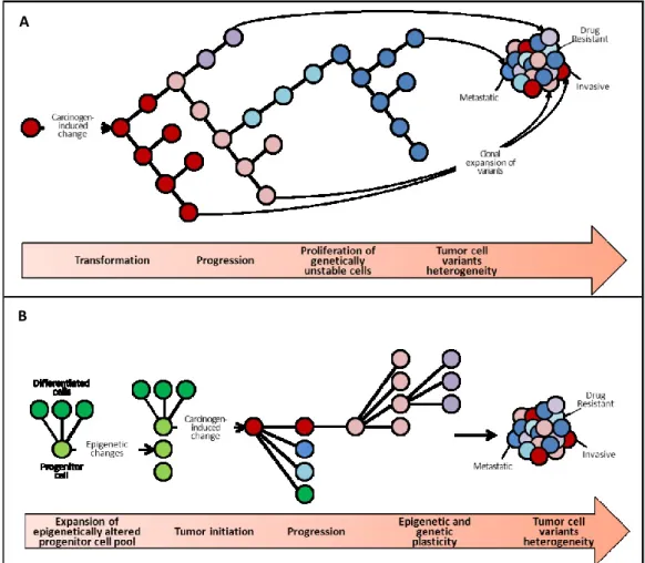
CRC evolves from adenoma to dysplastic adenoma and adenocarcinoma through distinct phases of genetic and morphologic alterations. Taking as paradigmatic model the colorectal carcinogenesis Fearon and Vogelstein introduced in 1988 a multiphasic-multigenic clonal model15 (Fig.1 A). According to them, cancer arises from a single cell and develops following progressive genetic mutations. Each mutation provides a selective advantage to the cell that proliferates and gives rise to a monoclonal population of cancer cells. The typical features of cancer biology, including invasion, metastasis and
4
pharmacoresistance, may be attributed to each of these mutations, whose accumulation, more than the frequency, has an essential importance in carcinogenesis process. Fearon and Vogelstein identified three phases in the carcinogenetic process: initiation, promotion and progression8. In the elaboration of this model, epigenetic alterations are only considered as an alternative to the “classical” genetic mutations that involve two classes of genes with different roles in tumor: oncogenes, that promote an autonomous cellular growth independently from external mytogenic stimuli, and oncosuppressor genes that, on the contrary, block cellular proliferation. Oncogenes and tumor-suppressor genes are named “gatekeepers” as they regulate the entrance of the cell in the oncogenic process. Among the oncogenes, which act in a dominant pattern and are switched on by point mutations, translocation, fusions and amplifications, we can enumerate transcription factors (MYC), chromatin modifiers (EZH2, Enhancer of Zeste Homologue 2), growth factors (TGFα, Tissue Growth Factorα) and their receptors (EGFR Epidermal Growth Factor Receptor), signal transductors (RAS, Rat Sarcoma) and apoptosis mediators (BCL-2, B-Cell Lymphoma 2).
Tumor-suppressor genes are inactivated through point mutations, deletions and translocations, as they negatively influence cellular growth regulating cell cycle (RB, Retino Blastoma gene), inhibiting cell duplication in presence of a genetic alteration (p53), or blocking cell migration and invasion (CDH1, Cadherin 1).
Caretakers genes are involved in DNA repair and genome stabilization and include: mismatch repair genes (MSH2, MutS homolog; MLH1, MutL-homolog), nucleotide excision repair genes (XP, Xeroderma Pigmentosum), recombination repair genes (ATM, Ataxia Telangiectasia Mutated)16.
This model has been very useful for the comprehension of the different mechanisms involved in carcinogenesis, stating that the genetic alterations necessarily arise in the first phases of the disease. Thanks to this theory the most important therapeutic agents in anticancer therapy are now available acting versus the so-called gatekeeper genes to block tumor growth, as for example the monoclonal antibodies Bevacizumab®, against VEGF, Cetuximab® and Panitumumab® against EGFR, largely used in colorectal cancer therapy.
However, the model presents some important limitations: the absence of a mutation that is necessary and sufficient to trigger specific stages of tumor
5
progression; the impossibility to explain the extended latency period only with the succession of multiple mutations; the lack of a correlation between genes and environment.
Following a deeper comprehension of the epigenetic mechanisms involved in tumor progression, such as genome-wide demethylation (Vogelstein 1983), hypoacetylation of histonic proteins and gene-specific hypomethylation, in 2006 this globally accepted model was surpassed by Feinberg’s epigenetic progenitor model7 (Fig.1 B). Observing that stem cells are at the origin of cancer and that the principal difference with the somatic cell resides in the epigenetic status, Feinberg speculated that early epigenetic alterations of stem
A
B
Figure 1: Models of carcinogenesis: (A) the clonal genetic model of
6
cells could be the basis of cancer pathogenesis. Its model comprises three steps: for first, the stem cells of a given tissue undergo an epigenetic alteration mediated by tumor progression genes (TPGs) deregulation, triggered by an environmental damage or particular events involving the stem cell itself or the stromal compartment. TPGs are usually involved in stemness regulation: IGF2 (Insulin-like Growth Factor 2), when hit by loss of imprinting, promotes the expansion of the progenitor cells’ compartment; APOBEC (Apolipoprotein B mRNA-editing Enzyme, Catalytic polypeptide) could be the responsible of the genome-wide demethylation of tumors; the transcription factors OCT4 (Octamer-binding Transcription factor 4), FOXD3 (Forkhead box D3) and Nanog normally maintain the balance between self-renewal and differentiation in the stem cell compartment; EZH2 influences chromatin structure. The perturbed equilibrium between non differentiated progenitors and differentiated cells causes the advent of a polyclonal precursors able to give rise to neoplasia. Tumor initiation is the second step of Feinberg’s model and is triggered by a monoclonal mutation of gatekeeper genes in the context of the epigenetically deregulated progenitors. This mutation is cancer type-specific, and in colorectal cancer involves APC (Adenomatous Polyposis Coli) or β-catenin. Finally, the third step consists in genetic and epigenetic instability that sustains cancer evolution and explains tumor heterogeneity.
This model has important implications in the study and treatment of cancer. The presence of epigenetically deregulated progenitors implies that the early phases of carcinogenesis take place when a preneoplastic lesion is still not identifiable. Moreover the reversibility of epigenetic mutations makes them an interesting therapeutic target, as demonstrated by FDA approved drugs with demethylating activity like Azacitidine (Vidaza; Celgene, Summit, NJ, USA) and Decitabine (Dacogen; SuperGen, Dublin CA, USA), and the histone deacetylase inhibitors like Vorinostat and Romidepsin. So it could be possible to identify and treat tumors in the very early steps of their development, leading to disease remission.
Under this theory, the typical features of advanced tumors like invasion, metastasis and pharmacoresistance are not determined by the progressive mutations occurring during carcinogenesis, but are inherent in the epigenetically perturbed progenitors at the origin of cancer.
7
Moreover according to Feinberg cancer heterogeneity is attributable to the presence of tumor progenitor cells epigenetically modified at gatekeepers’ level, which are phenotypically different from the tumor bulk and more similar to the early progenitors.
In this model is finally elucidated the role of the environment in the decade-lasting process of determining epigenetic alterations, that explains the insurgence of cancer mostly in adult age.
1.1.4 Colon Cancer Stem Cells
The failure of therapies directed against the proliferating fraction of cancer cells and the physiological loss of mutated cells during tissue renewing questioned the clonal model of carcinogenesis. More likely, cancer derives from less represented cells with a long half-life and the ability to undergo self-renewal, clonal expansion and accumulating mutation, namely the stem cells (SCs). When a tumorigenic mutation alters SCs’ self-renewal program, they transform in Cancer Stem Cells (CSCs) to trigger and sustain cancer development. Tumor assumes the features of a neo-organ, sustained by a small fraction (<1%) of stem cells and mostly constituted by a bulk of cell populations at different levels of differentiation18.
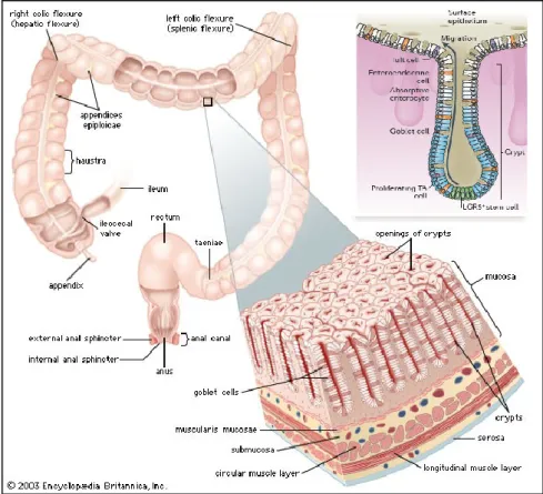
In physiological conditions, the colonic tissue consists of four distinct layers: mucosal, submucosal, muscular and serous. The mucosal epithelial layer faces the lumen and is made of a single sheet of columnar epithelial cells that form digitiform invaginations supported by the lamina propria to build the functional unit of the intestine, named Lieberkühn crypt. Intestinal stem cells (ISCs) are restricted to the crypt basis and give rise, through asymmetric cell division, to the transient amplifying cells (TA), characterized by a high proliferation rate but a reduced half-life. TA cells migrate to the top of the crypt, proliferate and differentiate in one of the three epithelial cells types that populate the intestinal wall: colonocytes, muciparous cells and enteroendocrine cells. Intestinal stem cells are sustained by a stem cell niche that in colon is composed by myofibroblasts at the crypt basis (Fig.2).
8
The main properties of stem cells are: differentiation, or the ability to give rise to an heterogeneous population of mature cells with short half-life that progressively specialize following a hierarchical process; self-renewal, or the capability to give rise to new stem cells with identical proliferative potential; homeostatic control, or the ability to balance the differentiation and self-renewal processes indulging environmental stimuli or tissue damages19. Stem cells perform symmetrical and asymmetrical cell divisions to maintain the exact number of stem cells in a population, generating respectively two identical stem cells or one stem cell and one more differentiated cell20.
To identify these cells in the heterogeneous context of the colonic tissue, a number of markers have been proposed and validated in the last years.
The first putative marker of ISCs is Msh-1 (Musashi-1), a protein that controls at the post-transcriptional level the genes involved in maintaining the
Figure 2: the structural organization of colon. Schematic representation
of colon histology and crypt organization. Edited from Encyclopedia Britannica, 2008.
9
undifferentiated status of the stem cells21. Nishimura and colleagues localized Msh-1+ cells at the crypt bottom, exactly where ISCs lie22.
Lgr5 (Leucine-rich repeat containing G protein-coupled Receptor 5), is a target of the fundamental Wnt intestinal pathway and has been proposed as marker of the colon stem cells. Lgr5 is a transmembrane G-coupled protein whose expression is limited to proliferating intestinal cells at the bottom of the crypt. Lgr5+ cells are able to give rise to all the colonic epithelial cell lineages23. EphB2, another Wnt target, has been recently proposed as a stem cell marker. Its activity is essential for cell positioning during intestine development and high levels of EphB2 expression correlates with a stem-like phenotype in normal colon, as will be discussed later24.
Also the integrin subunit β1 (CD29) is a candidate surface marker for the proliferative zone of the human colon crypt, that overexpresses this protein respect to the other colonic cells25.
Cancer Stem Cells are defined as cells able to self-renew and maintain the ability to give rise, through asymmetric cell division, to tumorigenic and non-tumorigenic cancer cell offsprings. The complex tumor cellular system includes cell subpopulations with distinct tumorigenic ability: the high percentage of cells that form the tumor bulk, unable to initiate cancer, and the rare tumor initiating cells (TICs) that, when implanted in a xenograft, are able to generate a tumor that histologically and phenotypically resembles the original one.
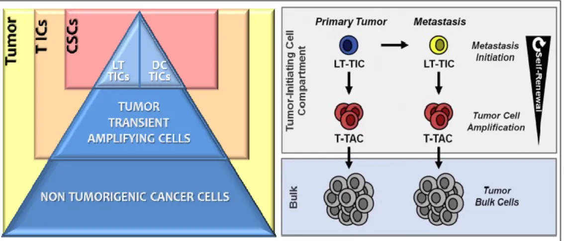
Dieter and colleagues demonstrated in an animal xenograft model the presence of distinct stem cell subpopulations in CRC26: colorectal cancer biopsies-derived cells cultured in suspensions formed tumor spheres that, when xenografted, were able to generate the original tumor. These spheres consisted of a conspicuous number of cells unable to proliferate, that corresponded to the tumor bulk; a smaller cell populations with an intermediate proliferative potential (tumor transient amplifying cells, T-TACs) with a predominant role in tumor formation; a minimal cell population with high proliferative potential (long term tumor initiating cells, LT-TICs) capable of self-renewing and metastasis. Serial xenografts gave rise to a third stem population, named delayed contributing tumor initiating cells (DC-TICs), with late recruitment in the carcinogenesis process. Cancer stem cells are therefore included in the two cell subpopulations of LT-TICs and DC-TICs able to
self-10
renew, differentiate through asymmetric cell division and give rise to all the different cancer cell lines (Fig. 3).
Among the different markers currently in use for colorectal CSCs, the transmembrane glycoprotein CD133 is one of the first proposed. Its role is still not totally clarified, but it is likely involved in asymmetrical cell division and self-renewal. Serial xenografts in immunodeficient mice demonstrated a marked increase of the tumorigenic potential in the small CD133+ cellular fraction (2.5%) respect to the non-dissociated tumor. However, a number of studies questioned CD133’s specificity, showing an unexpected expression of the protein in intestinal cells distributed all along the crypt axis and a metastatic potential also in CD133- cell population27.
The transmembrane glycoprotein CD44, restricted to the basolateral membrane of the colonocytes at the bottom of the crypt, is involved in cell survival, growth, differentiation and migration. CD44 is widely used as CSCs biomarker in a number of solid tumors, including CRC: CD44+ colon cancer cells are highly tumorigenic, even more if also CD133+, conversely CD44- colon cancer cells are unable to form tumors in immunodeficient mice28.
CD166 is a mesenchymal stem cell marker with a role in cell-cell contact formation and has been related to negative prognosis in CRC. Cells that are positive for CD44 and CD166 show elevated tumorigenity in immunodeficient mice compared to CD44-CD166+, CD44+CD166- or CD44-CD166-29.
Figure 3: Hierarchy of colon cancer stem cells. Edited from Zeuner A and De Maria R
11
Three molecules involved in the Wnt pathway are now in use as marker of colon CSCs.
Lgr5 has been found on the surface of colon cancer stem cells and is considered as a CRC-SC marker. Spheroid cultures derived from primary tumors were enriched for Lgr5 expression and Lgr5+ cells form CRC cell lines displayed colony forming, tumorigenic, and therapy resistance abilities30.
Ascl2, homologous to the Drosophila Achaete-scute complex gene, is a transcription factor expressed in a Wnt-dependent and highly restricted fashion in intestinal stem cells. Ascl2 acts as a master regulator of crypt stemness by interpreting Wnt levels and specifying stem cells. When overexpressed, it induces stem cell genes and crypt neogenesis in vivo31.
EphB2 has been firstly proposed as an intestinal stem cell marker by Battle’s group as they demonstrated that EphB2+ colon cancer cells not only display a gene expression profiles that overlaps with the one of the intestinal stem cells, but also show organoid formation ability in vitro and high tumorigenic activity in vivo25.
Finally also aldehyde dehydrogenase 1 (ALDH1) is in use as CSCs marker. It is a detoxifying enzyme that oxidizes intracellular aldehydes and identifies cells resistant to alkylating agents, which are protected from oxidative stress32.
1.1.5 Molecular pathways of Colorectal Cancer
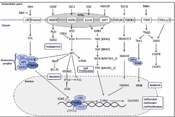
Genomic alterations that underlie colorectal cancer progression affect the main pathways involved in cell proliferation, migration and survival (Fig.4).
The Wnt signaling exerts a major role in developmental processes, influencing cell proliferation, differentiation and polarity. Under basal conditions, the cytosolic protein β-catenin (CTNNB1) binds to a destruction complex formed by APC, Glycogen Synthase Kinase3β (GSK3β), axin, casein kinase 1 (CK1) and is consequently phosphorylated, ubiquitinated and destroyed in the proteasome. Following Wnt binding to the Lipoprotein Receptor-related Protein (LRP) and Frizzled, the cytosolic Disheveled (DSL) protein is activated and can consequently inhibit β-catenin phosphorylation and degradation. CTNNB1 accumulates in the cytosol and translocates in the nucleus, where it activates the transcription of target genes involved in processes of tissue development and homeostasis33. Excessive binding of Wnt
12
ligands with Fz receptors and their coreceptors or malfunction of the destruction complex causes aberrant accumulation of free β-catenin in the cytoplasm and translocation in the nucleus, where it targets oncogenes related to invasive growth like c-MYC, CD44 and uPAR. The most common alterations of this pathway are inactivating mutations of APC and activating mutations of CTNNB1, which confer a selective advantage to transformed cells34. Recently a renewed interest has raised about the involvement of the Wnt inhibitor Notum and its related molecules glypicans in the modulation of Wnt signaling. Our group demonstrated for the first time Notum over-expression in early and late lesions of the AOM/DSS murine model of sporadic CRC and in human colorectal adenocarcinomas. Notum expression levels were correlated to β-catenin abnormal distribution, indicating that Notum expression is associated with canonical Wnt signal modulation in CRC pathogenesis. Moreover, Glypican-1 and Glypican-3 dysregulation were related to Notum and β-catenin alterations35.
Figure 4: Main Pathways affected in Colorectal Cancer. Palma S From Molecular Biology to
Clinical Trials:Toward Personalized Colorectal Cancer Therapy Clinical Colorectal Cancer, Vol.
13
The p53 protein, encoded by the tumor suppressor gene TP53, leads a downstream pathway that plays a crucial role in regulating cell cycle and apoptosis. DNA damage and oncogenic stress activate p53 signaling to either induce cell cycle arrest through p21, facilitate DNA damage reparation, or promote apoptosis through PUMA, Bax, Bak and Bcl-2, among others36. Under physiological conditions, p53 signaling pathway inhibits tumor formation through modulating DNA reparation, cell cycle and apoptosis37. Accumulating evidence has indicated that p53 signaling is frequently dysregulated in CRC progression and the aberrant signaling is associated with poor prognosis. Indeed inactivating mutations in TP53 gene or conformational alterations in p53 protein cause the loss of the tumor suppressive activity, promoting tumorigenesis and progression38.
COX, also named prostaglandin hyperoxide synthase, is the key enzyme of the pathway that regulates the metabolism of eicosanoids: it catalyzes the conversion of arachidonic acid to PGH2 which finally converts in PGs and thromboxane A2. The COX-1 isoform of the enzyme is expressed in a numbers of cells and tissues in their physiological activity; whereas the COX-2 isoform is inducible by cytokines, growth factors ant tumor promoters. COX‐2 signaling regulates angiogenesis, apoptosis and invasion: increased COX-2 expression is related to advanced stages and reduced survival rate in CRC according to clinical retrospective trails. COX-2 is also an independent prognostic CRC metastasis39.
TGF-β/Smad signaling pathway is triggered by two serine–threonine kinase receptors, TGF-βR I and TGF-βR II and has been found to be implicated in CRC carcinogenesis, acting as a tumor suppressor in the early stage, and as a metastasis promoter in the later stage40. The activated receptors promote the phosphorylation of the Smad2/3 dimers, that dissociate from the receptors and together with Smad4 form Smad2/3/4 complex. The complex translocates into the nucleus to modulate the transcription of multiple target genes, leading to cell growth inhibition. TGFBR2 mutation and loss of SMAD2 and SMAD4 are frequent aberration in colorectal cancer to remove the antitumoral effect of TGF-β signaling41.
Tyrosine Kinases Receptors (RTKs) are cell membrane proteins with intrinsic enzyme activity. Physiologically, they regulate a wide variety of
14
cellular processes including cell proliferation, apoptosis and migration. An enhanced activity of RTKs has been linked to development and progression of various types of cancer.
Between the more than 20 different RTK families, the most described include hepatocyte growth factor receptor (HGFR, or MET), ErbB receptors, insulin receptor, insulin-like growth factor receptors (IGF-R), platelet-derived growth factor receptors (PDGFR), fibroblast growth factor receptors (FGFR), vascular endothelial growth factor receptor (VEGFR) and Eph-receptors.
Insulin Growth Factor-1 Receptor (IGF1-R) has been found to be overexpressed in CRC: receptor activation by ligands such IGF2 leads to activation of PI3K-AKT pathway with increased cell growth and proliferation42.
Vascular Endothelial Growth Factor (VEGF) is the main promoter of tumor neo-angiogenesis, a crucial mechanism in cancer development to sustain the rapid and uncontrolled growth of cancer cells. Five VEGF family members have been identified in mammals, including VEGFA, VEGFB, VEGFC, VEGFD (FIGF) and placenta growth factor (PIGF or PGF). VEGF receptor tyrosine kinases include three high-affinity receptors named VEGFR1 (FLT1), VEGFR2 (FLK1/KDR) and VEGFR3 (FLT4), and two coreceptors, neuropilin 1, NP1 (NRP1) and 2 NP2 (NRP2). The binding of VEGF ligands to the different VEGFRs activates distinct downstream signaling pathways, including MAPK and PI3K-AKT, that regulate different cellular functions from proliferation to cytoskeletal reorganization and migration, all contributing to the angiogenetic process. VEGF upregulation has been associated with CRC progression and survival43.
Activation of EGFR and ErbB2 are early events during colon carcinogenesis. EGFR belongs to the ErbB family of related cell membrane receptors whose members include HER1 (ErbB1), HER2/neu (ErbB2), HER3 (ErbB3) and HER4 (ErbB4). EGFR is also known as HER1. A multiplicity of ligands binds these receptors, including EGF, TGF, amphiregulin, epiregulin, betacellulin, heparin-binding EGF and epigen. The receptor-ligand bound, following the recruitment of the PTPN12-regulated adaptor protein SHC, activates a complex multilayered network generated by receptor cross-talk and lateral signaling that converge on the classical MAPK and PI3K routes of signal transduction, which trigger transcription factors like ATF2 to express genes that maintain cell division, proliferation, differentiation and migration44.
15
Although their diverse functions, all the RTKs share common signaling cascades triggered by adaptor proteins such SHC, that are often deregulated in the malignant progression.
The PI3K-AKT-mTOR cascade is one of the most studied in tumor biology. Following by the activation by RTKs, AKT is phosphorylated and activated by PI3K, PDK and MTORC2. Direct consequences of AKT activations are: inhibition of the pro-apoptotic activity of BCL2, degradation of p53 by MDM2, activation of mTOR, that lead to increased cell growth, survival and proliferation. Activating mutations of PI3KA, inactivation of the suppressor PTEN and overexpression of AKT are commonly found in CRC45,46.
MAPK cascade is another crucial way of the complex RTKs network. It starts with RAS activation by SOS, complexed with a docking protein to the activated tyrosine kinase receptor, that displaces guanosine diphosphate (GDP) molecules from RAS and thus allowing guanosine triphosphate (GTP) molecules to bind and activate it. Active GTP-RAS recruits and removes the constitutive inhibition from the RAF proteins, which are then capable of binding and activating the KSR1 enzyme. KSR1 enzyme phosphorylates and activates MEK which in turn phosphorylates and activates ERK that enters the cell nucleus to activate a range of transcription factors, with the consequent expression of genes involved in cell proliferation. Hyperactivation of this signaling pathway is one of the most common aberrations in colorectal cancer47.
1.1.6 MiRNAs in colorectal cancer
MicroRNAs (miRNAs) are non-coding RNAs which regulate the gene expression at a post-translational level. Their biogenesis begins in the nucleus, with the enzymatic activity of RNA polymerase II, which transcribes genes located in intragenic regions, and RNA polymerase III, which transcribes the rare miRNA genes surrounded by repetitive DNA sequences. The transcription product is named pri-miRNA (primary-miRNA), a molecule of hundreds of nucleotides which includes a 5’ cap and a polyadenylated tail. Pri-miRNAs are still processed in the nucleus by the RNase DROSHA complexed with its cofactor DGCR8 to produce a hairpin molecule of 70 nucleotides termed pre-miRNA (precursor-pre-miRNA). The product is exported in the cytoplasm to be
16
processed by the endonuclease Dicer in a double stranded RNA of 18-25 nucleotides, which includes a leading strand (miR) and a passenger strand (miR*). One of these strands is destroyed by argonaute proteins (AGO) while the other is incorporated in the RNA-induced silencing complex (RISC) to exert its silencing activity48.
Target identification is based on complementary base-pairing between the miRNA and a region usually located in the 3’UTR of the mRNA, named “seed sequence” and is followed by target degradation, in case of perfect complementarity, or steric translational repression, in case of imperfect complementarity. MiRNAs’ pathway of expression is strictly ruled: the huge power of this class of molecules resides in the fact that a single miRNA is able to inhibit a number of targets, exerting its function on a wide range of physiological and pathological processes.
MiRNAs that play a key role in cancer, targeting genes involved in development, apoptosis, differentiation and cell proliferations, are named oncomirs. These molecules are usually located in genomic regions subject to deletion, duplications or mutations and are often deregulated during carcinogenesis, influencing hundreds of genes and pathways. They can act as tumor promoter, if their target is a tumor suppressor gene, or as tumor suppressors, if their target is an oncogene49.
Particularly, in colorectal cancer miRNAs control the main pathway involved in CRC development and progression (Fig.5).
WNT signals are crucial for the regulation of the stem cell activity at the base of intestinal crypt and for epithelial cells renewal. Incongruous activation of this pathway leads to development of gastrointestinal polyps and adenocarcinoma, as described above. MiR-135a/b targets the tumor suppressor APC, enhancing WNT pathway activation and consequent premalignant colorectal adenoma development50. MYC, a downstream transcript of this pathway, exerts its oncogenic function also through the upregulation of the expression of the cluster miR17-92 (17, 18a, miR-19a, miR-20a, miR-19b-1, miR-92-1), whose most important target is E2F1, a cell cycle transcription factor involved in a pro-apoptotic pathway51. Mir-26b inhibits LEF1, a member of the transcription complex activated by CTNNB1, a key player of WNT52. TCF/LEF complex is repressed also by members of mir-34 family, whose transcription is activated by TP53, linking these two oncogenic
17
signaling53. CTNNB1 function is also perturbed by mir-143 and -145, which target CTNNG1, acting on CTNNB1 translocation54, and are usually downregulated in CRC.
MiRNAs are also involved in the complex network of p53. This protein is a tumor suppressor that responds to diverse stress signals by directing specific cellular responses including senescence, cell cycle arrest, apoptosis, invasion and metastasis. Its fundamental role in tumor suppression has been extensively reviewed and comprises also CRC55. TP53 is able to regulate directly miRNA transcription: p53 binding site has been identified in the promoters of let-7i, 20a, 21, 25, 34a/b/c, 145, miR-181b, miR-183, miR-195, miR-215, miR-45156. Particularly, mir-34 is a well-known p53 inducible miRNA57 with a role in the inhibition of genes involved in cell cycle regulation, cell proliferation, apoptosis and DNA repair, like CDK4/6 (Cyclin-dependent kinase 4/6), Cyclin E2, E2F5, BIRC3 (Baculoviral IAP Repeat-Containing 3) e Bcl-2. A positive feedback loop is generated when mir-34 silences the TP53 inhibitor SIRT1 (Silent mating Type Information Regulation 2 Figure 5: miRNA and CRC pathways. An overview of WNT, EGFR, TP53 and TGFb signaling
pathways in CRC and the regulation of their key molecules by miRNAs. Edited from Mohammadi A The role of microRNAs in colorectal cancer Biomed & Pharmacoth 84 (2016) 705–713
18
homolog 1). Also miR-192, 194, 195, whose expression is regulated by p53, activate a positive feedback loop with a complex network of miRNAs that induce p53 accumulation58.
EGFR signaling pathway is involved in the most important mechanisms altered in the process of carcinogenesis, including cell proliferation, survival and migration. The most common alterations perturbing this pathway are EGFR mutations and KRAS mutations, described in 30-60% of CRCs59. Mir-143 targets KRAS and is often downregulated in CRC60. Also the let-7 family of miRNA targets KRAS, which in turn is able to regulate the expression of this cluster, removing its inhibiting activity and promoting carcinogenesis61.
The MAPK cascade is also affected by epigenetic silencing by miR-26b, which targets and inhibits the transcription factor ATF62: its downregulation is very common in CRC tumor development.
Also PI3K/AKT signaling is targeted by epigenetic silencing by the activity of miR-520a and -525a, which inhibits PI3KA subunit. However a point mutation the 3’UTR of PI3KA gene escapes miRNA silencing and promotes the oncogenic activity of this signaling63. Moreover, the expression of mir-126 and mir-30a that respectively target PIK3R2 and PI3KCD is usually reduced in CRC64,65; while mir-19- miR-21, miR-32 and miR-92-1-5p activates PI3K/AKT signaling targeting the negative regulator PTEN66-68.
A number of miRNAs, including miR-17-5p, miR-20a, miR-21, miR-23b, miR-106a and miR-301a, have been reported to target the transcript of TGF-β, a growth factor that in colon cells controls a wide spectrum of cellular functions including proliferation, differentiation, apoptosis and migration, with a role in CRC carcinogenesis suppression. In particular miR-21 is activated by WNT pathway and is involved in stemness regulation69. Another connection with WNT signaling involves miR-17-92 cluster that is modulated by MYC and inhibits SMAD and TGF-β70. MiR-106a/363 and miR-106b/25 clusters are involved in the inhibition of TGFBR2 and SMAD2/SMAD471. Also miR-130a, miR-301a, and miR-454 target SMAD4 and are commonly upregulated in CRC72. Mir-25 on the contrary is able to inhibit SMAD7, a well-known negative regulator of TGF-β signaling, and its expression is preferentially decreased in colon carcinogenesis73.
19
MicroRNAs are also involved in metastasis, targeting the principal genes involved in the process. ZEB1, an EMT (Epithelial to Mesenchymal Transition) inducer, downregulates miR-200 miRNA family members (miR-200a, miR-200b, miR-200c, miR-141, miR-429), known MET (Mesenchymal to Epithelial Transition) promoters that, in turn, target TGF-β2 and ZEB1, triggering a feed-forward loop74. The oncogene MET (Mesenchymal-Epithelial Transition factor) induces tumor growth, angiogenesis and metastasis and is negatively regulated by miR-133b and miR-175. Finally, COX-2, which promotes the apoptosis, angiogenesis and tumor invasion, is target of miR-101, a microRNA commonly downmodulated in CRC76.
1.2 EPHA2 and EPHB2
Studies on Eph receptors and their ephrin ligands have significantly improved in the last years. This rapid development is not only because the Ephs are the largest group of Receptor Tyrosine Kinases (RTK), but also because detailed biochemical studies have revealed them as very attractive drug targets and diagnostic biomarkers. EphA2 and EphB2, in particular, seem to play a key role in CRC initiation and progression and are currently under observation for their therapeutic value.
1.2.1 Structure and signaling of EPHA2 and EPHB2
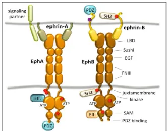
The Eph (erythropoietin-producing human hepatocellular receptors) superfamily is the largest group among tyrosine kinase receptor families. Eph family comprises 16 receptors classified in two subclasses, EphA or EphB, depending on their sequence homology and their binding affinity for their ephrin ligands77. Although Eph receptors preferentially bind ligand of the same class, cross-binding has been shown for EphA4, which can also bind to ephrin-B ligands77, and EphB2 which can bind to ephrinA578. All Eph receptors contain an extracellular region, with a conserved N-terminal globular ligand-binding domain (LBD), a cysteine-rich domain which comprises a Sushi and an epidermal growth factor (EGF)-like domain and two fibronectin type-III repeats (FN1 and FN2). The intracellular region contains a juxtamembrane region (JM), a tyrosine kinase domain, a sterile alpha motif (SAM) domain,
20
and a (PDZ) domain-binding motif79-81. The ectodomain and the intracellular domain are linked by a transmembrane helix (TM) (Fig.6).
Ephrins (Eph receptor interacting proteins) are also divided into EphrinA and EphrinB subclasses79,80. EphrinA proteins (A1-A6) are anchored to the plasma membrane via a glycosylphosphatidylinositol (GPI) linkage while ephrinB members (B1-B3) are transmembrane proteins containing a cytoplasmic domain with several conserved Tyrosine residues and a terminal PDZ-binding motif (Fig. 6).
Eph–ephrin binding occurs on the surface of the same cell (in cis) or at the site of contact of two opposing cells (in trans) and results in bidirectional signaling into both the receptor cell (‘‘forward signaling’’) and the ligand cell (‘‘reverse signaling’’)82,83. The trans bound requires cell-cell direct contact and it results in cell repulsion or adhesion, depending on a complex interaction of factors. Signal transduction by the Eph family is a multistep process leading to the assembly of higher-order signaling clusters in the interacting cells79.
The first step in the formation of Eph-ephrin cluster is the binding 1:1 between Eph receptor and an ephrin ligand on opposing cell surface that leads to the formation of an ephrin-Eph dimer that aggregates successively in a heterotetramer. Eph and ephrin complexes aggregate into larger clusters through the recruitment of ephrin-bound Eph receptors and additional Eph receptor in an ephrin-independent lateral mode. Only the association of
Figure 6: Schematic representation of Eph/ephrin structure.
Edited from Barquilla A Eph receptors and ephrins: therapeutic
21
signaling/adaptor proteins elicit Eph receptor signaling, the strength of which correlates with the size and composition of the clusters84, and which partly explain the myriad of cellular response that are elicited by Eph activation.
Effects induced by Eph/ephrin binding involve their interaction with specific intracellular proteins, including phosphoinositide 3-kinase (PI3K), Src family kinases, Vav2, Vav3 and ephexin which coupling to Rho GTPases trigger cytoskeleton modulation85,86.
Forward signaling involves autophosphorylation of Eph receptor and successively activation of the tyrosine kinase intracellular domain through Src family kinase–mediated phosphorylation87. Phosphorylation of downstream substrates is mediated by adaptors like proteins containing Src-homoloy 2 (SH2) domains and then by a number of Rho guanidine nucleotide exchange factors (GEFs), e.g. Vav2, Tiam, Kalirin, and Intersectin88.
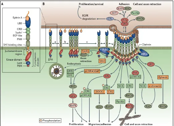
Eph signaling interplays with several molecules and signaling pathway, including members of Rho family of GTPases (RhoA, Cdc42, and Rac)88, focal adhesion kinase (FAK), the PI3 kinase pathway and Jak/Stat pathway89 (Fig. 7).
In particular, EphA2 is preferentially expressed on the membrane of epithelial cells, including small intestine, and colon where it regulates tissue development and maintains epithelial tissue homeostasis90,91.
Unlike other receptor tyrosine kinases, EphA2 receptor does not require ligand binding for some of its activities, and can directly activate GTPases of the Rho family through the GEF Ephexin92. EphA2 has diametrically opposite roles in regulating cell migration and invasion, depending on its ligand dependent or independent activity: if activation of EphA2 with its ligand ephrin-A1 inhibits chemotactic migration, EphA2 overexpression promotes migration in a ligand-independent manner. EphA2 ligand independent activity requires receptor phosphorylation on serine 897 by Akt. Ephrin-A1 stimulation of EphA2, on the contrary, inhibits Akt activation with a negative feedback mechanism and causes EphA2 dephosphorylation on the serine residue, as demonstrated by Miao and colleagues in human astrocytoma93.
Co-clustering with other RTKs and cross-class transphosphorylation of Eph receptors have also been reported, with the contribution of either receptor-type to the signaling outcome depending on relative abundance of the two receptors.
22
EphB2 receptor forward signaling is mediated by specific GEF, intersectin and kalirin which regulate the EphB2-mediated cytoskeleton reorganization, mesenchymal invasion and migration94,95.
The most important and best described EphB2 forward signaling influences the regulation of cell positioning and cell proliferation, activated by PI3K and Abl pathways, respectively96. In addition, EphB activation can also reduce cells adhesion through a negative modulation of the MAPK pathway via R-Ras97.
1.2.2 EphA2 and EphB2 in the intestinal epithelium
In the intestine, Eph/ephrin signaling regulates a number of biological processes including cell proliferation, differentiation, migration and tissue morphogenesis. Eph receptors play a key role in tissue organization, particularly in maintaining the appropriate structure and preventing cell intermingling.
Figure 7: Eph signaling. Boyd AW Therapeutic targeting of EPH receptors and their
23
EphA2 and its ligand ephrinA1 are mainly involved in maintenance of intestinal barrier and in colon epithelial homeostasis with functions in stress response. Eph/ephrin system co-works with junctional molecules to accomplish cell sorting processes and modulate epithelial integrity.
Several members of EphA family are largely expressed in intestinal epithelium. High expression of EphA1, EphA4 and Eph7 are found at the crypt basis, in cell with high proliferative activity; whilst EphA2, EphA5 and ephrinA1 are highly expressed in differentiated cells in the crypt top98.
EPHA2 and E-cadherin co-localize along the lateral membrane at site of cell-cell contact and ensure morphologic maintenance of epithelial cells. A reciprocal regulatory positive loop between EPH receptors and E-cadherin has been demonstrated. E-cadherin regulates phosphorylation and localization of EphA299 and stabilizes cell-cell contacts facilitating EphA2 association with its ligands. Moreover, ligand-mediated activation of EphA2 promotes E-cadherin–based cell-cell adhesion100 (Fig. 8).
As described above, in the intestine context, stem cells localize at the bottom of crypt, where they divide and give rise to progenitor cells, which continue to divide as they migrate up the crypt axis. As cells leave the crypt, they also abandon their cycling activity and start to differentiate. Wnt signaling is a pivotal mitogenic regulator for intestinal stem cells and it also transcriptionally regulates the expression of EphB receptors and negatively regulates expression of their ligands101,102. EphB receptors and their ephrin-B ligands are expressed in counter gradients along the crypt-villus axis in the intestine, where EphB2 and EphB3 are present at high levels in stem cells at the bottom of the crypt and ephrin-B1 and ephrin-B2 are predominantly expressed by differentiating cells in the upper portion of the crypt103. In differentiating cells low levels of EphB2 favor their migration up to the gradient of ephrin-B1 expressing cell, in contrast up regulation of ephrin-B1 drives cells down the EphB2 gradient. In this way Eph/ephrin system controls the correct positioning of cells in the intestine through a unidirectional flow mediated by repulsive mechanism101(Fig. 8).
EphB receptors (EphB2 and EphB3) regulate proliferation and migration in the intestinal stem cell niche by two independent EphB signaling103. Proliferation is mediated by tyrosine kinase dependent signaling, via Abl and
24
cyclin-D1, whereas cell positioning is mediated via PI3K in a kinase independent fashion96. Studies on null mice showed that Ephrin-B1 null mice displays distorted cell positioning104, whilst EphB2- and EphB3-null mice result in decreased stem/progenitor cell proliferation and in distorted migration101,103.
1.2.3 EphA2 and EphB2 in Colorectal Cancer
Eph receptors and their ligands have emerged as integral players in the pathogenesis of cancer: aberrant expression of Eph and ephrin genes have been identified in a wide range of human tumors such as neuroblastoma, carcinomas of the breast, lung, gastric, prostate, ovarian, melanoma and colon.
Ephs and ephrins were thought to play an oncogenic role in human cancer, as initially demonstrated by their first isolation from a hepatocellular carcinoma cell line, where they were found to be at least 10-fold overexpressed compared with non-malignant tissue. However, recent Figure 8: EphA2 and EphB2 in the intestinal crypt. Edited from Scoville DH Current View:
25
evidence demonstrates a tumor suppressive role for Ephs in some instances, suggesting that the role of Ephs and ephrins is far more complex than first assumed105.
As evidence, their downstream signaling pathways control processes such as cell growth, proliferation, organization of the cytoskeleton, cell-matrix and cell-cell attachment, the dysregulated function of which contributes to an invasive and metastatic tumor phenotype.
EphA2 in Colorectal Cancer
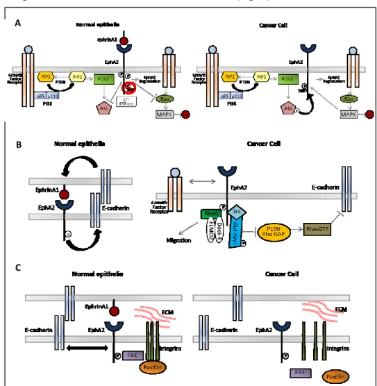
In particular, EphA2 overexpression has been documented at mRNA and protein level in a number of human malignancies such as lung, breast, liver, gastric, renal, prostate, ovary, esophagus, bladder, pancreas, cervical, melanoma, glioblastoma, SCCHN and colon106. Moreover, in CRC genetic ablation of EphA2 in ApcMin/+ mice has been found to result in significant reduction in number and size of intestinal tumors107. The pro-oncogenic role of EphA2 resides in its ligand independent activity: a number of studies have documented low levels of EphA2 phosphorylation in malignant cells compared to normal cells despite its overexpression108. A crucial initial step of colorectal carcinogenesis is reduction of E-cadherin expression and function, resulting in decreased cell-cell adhesion and destabilization of the epithelial architecture with loss of cell-cell attachment. As a consequence, the interaction between EphA2 and ephrinA1 on neighboring cells is inhibited, abolishing its tumor suppressing function mediated by Tyr phosphorylation, internalization and degradation of EphA2 receptor109. Moreover, the ephrinA1 inhibiting activity on AKT is removed and EphA2 ligand-independent effect is switched on by AKT-mediated phosphorylation on Serine897. This signaling promotes cancer cell migration, by means of the association between EphA2 and Focal Adhesion Kinase (FAK), a tyrosine kinase involved in EphA2/Integrins crosstalk: FAK phosphorylation results in active conformation of integrins and triggering of integrin-mediated adhesion, cell spreading and migration110. Unligated EphA2 is also able to destabilize adherent junctions via Rho-GTP activation: on one hand, it enhances the low molecular weight phosphotyrosine phosphatase (LMW-PTP) activity that in turns hinders p190 RhoGAP, a Rho-GTP inhibitor111,
26
on the other hand it interacts with Ephexin4, one of guanine nucleotide exchange factors for RhoG, and activates RhoG109(Fig. 9).
When ephrinA1-EphA2 bound is broken, the negative feedback loops existing between EphA2 and Ras/MAPK and PI3K/Akt pathways are removed, so EphA2 can exerts its pro-oncogenic activity also through direct crosstalk with EGFR network stimulating cancer cell proliferation and survival. In turn, EphA2 expression is upregulated in response to cell adhesion by EGFR, MEK and SRC family kinases112(Fig. 10).
Recently, EPHA2 receptor has been found to play an important role in many aspects of EMT, including induction of a mesenchymal-like phenotype,
Figure 9: EphA2 molecular pathways in normal and cancer cells. (A) AKT and MAPK; (B) GAP and cadherins; (C) integrins. Edited from Beauchamp A Ephs and Ephrins in
Cancer: Ephrin-A1 Signaling Semin Cell Dev Biol. 2012 23(1): 109–115.
A
C B
27
inhibition of epithelial characteristics and crosstalk with EMT-related signal transduction pathways such as the previously described E-cadherin, RAS/MAPK and Akt/mTOR networks.
The role of EphA2 in tumor metastasis has been widely investigated in a number of tumors including melanoma, ovarian, lung, renal, prostate179, but less is known about colon cancer. An immunohistochemical study of Saito et al. found a direct relation between EphA2/E-cadherin expression and colorectal cancer lymph node metastases131.
EphA2 is also involved in tumor cell-extrinsic, microenvironmental mechanisms of tumor progression: EphA2 and ephrin-A1 expression were correlated with MVD in human CRC samples, suggesting they might regulate neovascularization as well as tumorigenesis. These clinical observations are consistent with data derived from cell culture and animal studies113: EphA2-ephrinA1 system has been demonstrated playing a key role in tumor angiogenesis with a clearly distinct mechanism from which this system plays in affecting the behavior of tumor cells. Indeed EphA2 expressed on cancer cell is not the principal actor, but rather the EphA2 receptor localized on the endothelial cells that, stimulated by the tumor-derived ephrin-A1, is able to induce expression of VEGF and subsequently activate distant host endothelial cells, leading to angiogenesis and metastasis114. Moreover recent evidences found that EphA2 expressing cells participate to the process of vasculogenic Figure 10: EphA2 and EGFR crosstalk. Schematic representation of the crosstalk existing
between EphA2 and EGFR in (A) normal and (B) cancer cells. Edited from Larsen AB
Activation of the EGFR Gene Target EphA2 Inhibits Epidermal Growth Factor-Induced Cancer Cell Motility Mol Cancer Res 2007;5:283-293.
28
mimicry, where aggressive and dedifferentiated tumor cells form fluid-conducting channels not lined by endothelial cells115.
EphB2 in Colorectal Cancer
Deregulated mRNA and protein expression of EphB2 have also been reported in human colon cancer. Although increased EphB RTK expression was detected in the initial phases of CRC, subsequent expression analyses coupled with genetically engineered mouse models suggest tumor suppressive functions for EphB receptors: a number of studies report a direct correlation between loss of EphB2 with CRC progression116,117 and EphB2 overexpression with prolonged survival118.
So, if EphA2 has a clear pro-oncogenic function in colorectal carcinogenesis, the role of EphB2 seems to be dual, or biphasic. In the first phases of CRC progression, EphB2 expression is upregulated and the receptor acts as tumor-promoter. As the cancer evolves from adenoma to carcinoma, EphB2 expression is gradually lost, due to the prevalence of its onco-suppressor function. The EphB2 tumor onco-suppressor activity was demonstrated in a mouse model of adenomatous polyposis (APCMin/+) where an invasive adenocarcinoma developed when EphB2 signaling was inhibited116.
This duality resides in two distinct pathways in which EphB2 is involved: proliferation and positioning of intestinal stem cells. The principal regulator of proliferation in the intestine is Wnt, through the activation of c-Myc and inhibition of p21. Recently EphB2 has been identified as direct transcriptional target of TCF/β-catenin complex and a third mediator of Wnt proliferating effect. The Wnt pathway is over-activated in the 70% of CRCs, which show homozygous inactivation of Adenomatous Polyposis Coli (APC) tumor suppressor gene that inhibits catenin nuclear translocation. When β-catenin/Tcf complex migrates to the nucleus it switches on EphB2 transcription in the stem cells located at the base of the colonic crypt. EphB2 expression and kinase function activate a signaling cascade that involves Abl and CyclinD1, direct effector of cell cycle regulation. As the tumor progresses from adenoma to carcinoma, the proliferating kinase-dependent function of EphB2 becomes less relevant, to the advantage of the kinase-independent function of cell-positioning. At the same time, CyclinD1 expression becomes independent from EphB signaling, keeping on exerting its mitogenic activity.
29
The second activity of EphB2 receptor consists in the regulation of cell positioning along the crypt axis. Its kinase-independent signaling inhibits PI3K, suppressing migration and invasion of cancer cells. Moreover EphB-ephrinB1 bound regulates the formation of E-cadherin-based adhesions thanks to the interaction with the metalloproteinase ADAM1096.
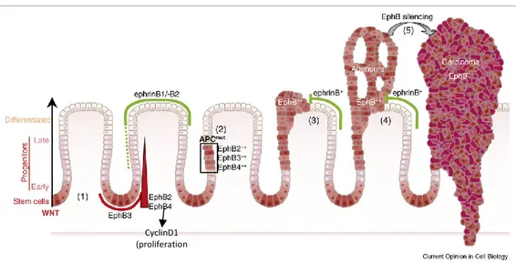
Battle and colleagues hypothesizes a model for EphB2 activity during colorectal cancer progression: in the first phases, APC-mutant cells populate the stem cell niche at the bottom of the crypt forming the so-called dysplastic niche. Cancer cells proliferate laterally, in strict contact with adjacent normal crypts, where the exposed ephrinB1 can bind the EphB2 overexpressed on tumor cells in response to constitutively activated Wnt signaling. This bound compartmentalizes EphB2+ cells expansion in the niche context and activates CyclinD1 pathway of proliferation, resulting in in situ adenoma growth. At the transition from adenoma to carcinoma EphB2 expression in lost, so the cancer cell is free to exit the crypt and invade the surrounding tissue. This model is coherent with a study of 2005 where reduction or loss of EphB2 and EphB4 expression correlated with the shift from adenoma to invasive carcinoma in a panel of 108 human CRC samples116. Moreover, a positive correlation between EphB2 expression and better overall and recurrence-free survival in human CRC patients has been identified in three independent studies111 and reduced EphB2 expression has been associated with metastasis both in CRC metastatic cell lines and in human colorectal cancer132,133 (Fig. 11).
EphB2high cell population at the bottom of normal colon crypt represents a
small percentage of the overall amount of intestinal cells, expressing the classical stemness markers like Ascl2 and Lgr5. In colorectal cancer this proportion is maintained, leading to ascribe a less important role to this population in cancer development. However a study of Suarez et al. demonstrated a high tumorigenic activity for a mouse colorectal cancer FACS sorted EphB2high cell population: these cells were able to form organoids when
cultured in vitro and to generate xenograft when injected into NOD/Scid mice in histological patterns that largely resembled the primary tumor, demonstrating to hold high tumor-initiating potential as well as long-term self-renewal and differentiation capacity120. Also Feng et al. validated the CSCs identity of human CRC cells overexpressing EphB2 by the mean of a sphere formation assay121.
30
So we could speculate that EphB2high cell population in CRC constitutes the
cancer stem cell niche that represents the reservoir of the tumor itself, fueling its expansion and invasion.
EphA2 and EphB2 role in metastasis
Circulating tumor cells (CTCs) are cancer-derived cells that are able to detach from the tumor, enter the circulation, reach and invade the target organ and form metastasis122. To gain these competences, CTCs undergo epithelial to mesenchymal transition, a reversible process in which cells decrease expression of epithelial markers and acquire mesenchymal features. Once completed the metastatic process, cells undergo mesenchymal to epithelial transition, resembling again primary tumor genetically and phenotypically. Circulating tumor cells have been found in the peripheral blood of patients with a wide range of solid tumors, including colorectal cancer, and their detection in liquid biopsy could represent a less invasive sampling for tumor diagnosis and follow up123.
The association of EphA2 with cancer, its exclusive overexpression on tumor cells and its involvement in EMT make it a potential surface marker to isolate CTCs. In 2008 Scarberry and colleagues successfully used magnetic nanoparticle conjugated with the EphA2-specific peptide YSA to target and remove metastatic ovarian cancer cells from the fluid of the abdominal cavity
CyclinD1 (proliferation
)
Figure 11: EphB–ephrinB interactions during CRC progression. Edited from
Merlos-Suárez A Eph-ephrin signalling in adult tissues and cancer. Curr Opin Cell Biol. 2008 Apr;20(2):194-200.
31
or circulatory system124. EphA2 was also found on the surface of CTCs isolated from diverse stage III-IV tumor types including colorectal cancer123.
The role of EphB2 in CTCs is less investigated: only recently Hamilton et al. identified EphB2 expression on a CTC cell line expanded ex vivo from human small cell lung cancers125.
Exosomes are microvesicles containing an array of proteins, DNA, mRNAs and microRNAs that are normally released from many cell types in the microenvironment to influence target cells with their content. Cancer cells secrete higher concentration of exosomes in order to suppress the immune system, modulate the angiogenesis and condition the metastatic niche, to generate a pro-tumor environment for the adhesion and growth of distant tumors126. Researchers’ efforts are focused not only on the cargo of these microvesicles, but also on the recognition molecules they express to selectively target recipient cells.
A number of studies identified different types of Eph receptors on exosomes’ surface, inducing tumor promotion or suppression with different effects from the classical bidirectional signaling. Sun and colleagues127 recently recognized an essential bound between ephrinA2 expressed on osteoclast-derived exosomes’ surface and EphA2 expressed on osteoblast to permit osteoclasts/osteoblast crosstalk. Moreover, EphA2 seems to be crucial for sorting molecules in multivesicular bodies and so in exosomes128. Tauro et al.129revealed in colorectal cancer-derived exosomes the expression of EphA2 and ephrin-B1 and Gong130 found also EphB2 in vesicles extracted from HEK293 and HeLa cells overexpressing EphB2 and from U251 glioma cells and cultured primary cortical neurons expressing endogenous EphB2.
Both EphA2 and EphB2 seem to exert a key role in the metastatic process: on the one hand, EphA2 has been demonstrated to be involved in neoangiogenesis and epithelial to mesenchymal transition and its presence on CTCs’ and exosomes’ surface could be crucial to target the metastatic niche and establish distant tumors; on the other hand, EphB2 is a validated marker of intestinal stem cells and its stemness function is maintained during tumor progression, so its expression could probably characterize circulating tumor cells committed to form metastasis, even if not yet demonstrated as in exosomes. So, circulating tumor cell committed to metastasize could have an
32
intermediate EphA2/EphB2 phenotype, showing in part epithelial and in part mesenchymal features fine-tuned in the ongoing process of epithelial to mesenchymal transition.
1.3 Colorectal Cancer therapies
In CRC management tumor-related features, including number and localization of metastases, tumor progression, presence or absence of biochemical markers, and patient-related factors, like co-morbidity and prognosis, influence the choice of the first-line treatment. According to this, CRC patients have been classified in four distinct risk groups to match the best treatment strategy135. Group 0 includes patients with no metastatic disease or with resectable liver or lung metastases and lack of poor prognostic signs and is treated with surgical resection of the metastasis. Chemotherapy has not been found to provide a great advantage in the overall survival of this group. Group 1 includes patients initially treated with induction chemotherapy to reduce the number and size of the metastases and enable subsequent surgical resection. Recommended chemotherapy for these cases comprises combinations of cytotoxic agents with targeted therapy. Group 2 includes patients with disseminated unresectable disease. Treatment is palliative rather than curative and should induce metastatic regression in a short time to reduce the symptoms, aggressiveness and extension of the disease: the preferred option comprises a cytotoxic doublet in combination with a targeted agent (anti-VEGF or anti-EGFR strategies). In oligometastatic patients who respond to treatment, additional ablative methods may be considered to increase the progression-free interval. If ablative methods cannot be used, de-escalation or discontinuation of the initial combination should be studied as a maintenance treatment. Group 3 includes patients with unresectable disease. In this case the purpose of the treatment will be to prevent tumor progression and increase treatment-free life: the most commonly used strategies comprise a fluoropyrimidine as cytotoxic agent combined, or not, with a biological targeted agent (Fig. 12).
33 1.3.1 Chemotherapeutic agents
Fluoropyrimidines are anti-metabolite agents whose main mechanism of action consists in the inhibition of thymidylate synthase activity. 5-fluorouracil (5-FU), developed in 1957, was the first studied compound: it inhibits tumor cell division by blocking the conversion of deoxyuridine monophosphate (dUMP) to deoxythymidine monophosphate (thymidylate). 5-FU is commonly given either as a bolus injection with leucovorin (folinic acid) or a continuous infusion, but if 5-FU bolus treatment favors RNA damage, continuous treatment with 5-FU favors DNA damage136.
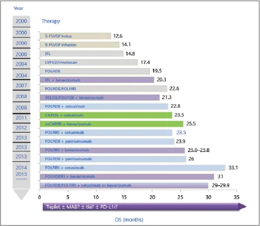
Figure 12: Improvement of OS in stage IV mCRC. Examples of phase II/III studies
between 2000 and today with improvement of OS in the therapy of mCRC * Phase II studies. Edited from Pohl M Therapeutic Strategies in Diseases of the Digestive
Tract – 2015 and Beyond Targeted Therapies in Colon Cancer Today and Tomorrow