UNIVERSITY OF FOGGIA
PhD RESEARCH IN
“H
EALTHF
OODI
NNOVATION ANDM
ANAGEMENT’”
(cycle XXX)
“Evaluation of the effects of alternative physical approach on
the metabolism and functional traits of useful
microorganisms"
Supervisor: Prof.ssa Maria Rosaria Corbo Co-supervisor: Prof. Antonio Bevilacqua
Prof.ssa Claudia Piccoli
PhD Student:
Dott.ssa An gela Racioppo
A c a d e m i c T r i e n n i u m
STATEMENT OF ORIGINALITY
This work contains no material which has been accepted for the award of any other degree or diploma in any university or other tertiary institution, and contains no material previously published or written by another person, except where due reference has been made in the text.
Angela Racioppo December 21, 2017
ABSTRACT
Scientific background: Probiotics in foods could lead to significant changes in food
flavor and rheology, due to their active metabolism. A possible way to overcome this
problem is the attenuation of probiotics through a physical or chemical method. Some authors studied homogenization as a way to attenuate/modulate the metabolism of starter cultures in dairy products (Lanciotti et al., 2004, 2006, 2007); in this project I have used a new emerging technology, the ultrasounds. (i) A screening of the ultrasound (US) (power/duration) on different probiotic microorganisms was perfomed, and were studied the probiotic and technological characteristics after US-exposure. (ii) The effects of US on the release of intracellular components, was investigate. (iii) The interaction of attenuated strains with gut microbiota was evaluated, using in vitro batch culture fermentation.
Open questions: (i) Few data are available on effects of US on probiotics and technological characteristics of probiotic strains; (ii) few data are available on the effect of attenuation with ultrasound on the sub-lethal injury; (iii) no data are available on the interaction of attenuated strains with gut microbiota.
Aims: (i) Choice of the best combination of ultrasound to avoid post-acidification without affecting the viability of the strains, and study of the probiotic and technological characteristics to evaluate if attenuation could change them; (ii) study the release of intracellular components (nucleic acids and proteins) after the application of ultrasound; and (iii) evaluation the effects of attenuated strains on gut microbiota.
Planning of the research: In the first part three different genera of probiotics were used:
Lactobacillus, Bifidobacterium, Propionibacterium. The strains were treated with
ultrasound and studied for technological and probiotic characteristics.
In the second part, the effects of US on the release of intracellular components, was investigated. The strains were studied after physical treatment to assess the release of intra-cellular constituents (nucleic acids, proteins) and injury of the membrane.
In the last part, the interaction of attenuated strains with gut microbiota, was studied. This study was carried out at University of Roehampton (UK).
Materials and Methods: (i) Technological traits: acidification in lab medium, growth at different temperatures, pHs and salt content; probiotic traits: antibiotic-resistance, survival at pH 2.5 and in the presence of 0.3% bile salt, hydrophobicity, and biofilm formation. (ii) Injury characterization was evaluated by leakage of UV-absorbing
substances. (iii) The interaction of attenuated strains with gut microbiota was evaluated,
using in vitro batch culture fermentation.
Results: (i) The best combinations to avoid post-acidification were the following: power, 60%; time, 6 min; pulse, 2 s for Lactobacillus and Bifidobacterium, 40%, 8 min P.
jensenii; 60%, 4 min P. freudenreichii subsp. freudenreichii. US did not affect viability
at 45 °C or at pH 9, but it determined a decrease of microbial growth to pH 4 (lactobacilli and bifidobacteria). However, the US did not affect the GI of propionibacteria. The effect of attenuation could be enhanced by the storage under refrigeration. US-treatment did not affect most of the technological traits, but generally caused an increase of susceptibility to some antibiotics.
Concerning probiotic traits, US caused an increase of hydrophobicity for L. reuteri and
P. freudenreichii spp. freudenreichii, after US-exposure. These results were confirmed
with adhesion to Caco-2 cells for L. reuteri. US-attenuated L. reuteri experienced a significant increase of hydrophobicity (from 3 to 25%) and a higher adhesion to Caco-2 cells. Moreover, US improved the stability of the biofilm over the time, and this result confirmed the data obtained with hydrophobicity. (ii) The release of nucleic acids and proteins was found, highlighting that cell membrane could be another target physical treatments. (iii) Concerning the effects of US on gut microbiota, the ultrasound didn’t affect the gut microbiota, but in some cases, it could have a positive effect.
Significance and Impact of PhD research: A main drawback of probiotics in foods can
relate to their active metabolism, some strains of lactic acid bacteria continue to produce lactic acid and cause post-acidification (the decrease of pH within the storage). Therefore, it is important to control their metabolism. A possible way to control the metabolism of probiotic in foods is the attenuation through physical or chemical methods. One of the emerging technologies is ultrasound (US). This approach was used to avoid post-acidification in a commercial rice drink (Bevilacqua et al., 2016). The present PhD thesis contributed to evaluate the effects of attenuation with ultrasound, on some technological and probiotic strains, testing three different genera of probiotic strains. Moreover, this
PhD project has investigate the changes that may affect probiotic strains after attenuation; the release of proteins, nucleic acids. Finally, the novelty of this PhD thesis was the study of the effects of attenuated strains on gut microbiota.
Future trends: A future perspective could be a focus on the use of US to improve or modulate the adhesion of probiotic strains, considering the increase of hydrophobicity
and the higher adhesion to Caco-2-cells. It is important to investigate the effects of other
attenuated strains on gut microbiota by modulating the variables of the treatment.
Key words: Hydrophobicity, acidification, growth, attenuation, gut-microbiota, proteins,
TABLE OF CONTENTS
Chapter 1. INTRODUCTION………... 1
1.1.Probiotics………...…….... 1
1.1.1. Definition of probiotics……….………...………... 1
1.1.2. The genus Lactobacillus ……….... 3
1.1.3. The genus Bifidobacterium………..……….. 4
1.1.4. The genus Propionibacterium……… 5
1.1.5. Probiotics: guidelines for use in foods and food supplements…………... 6
1.2. Probiotics in foods: attenuation and modulation of metabolic activity ……. 8
1.3. Attenuation by sonication……… 10
1.4. Other methods of attenuation………. 14
1.4.1. Attenuation by hydrostatic high pressure and high-pressure homogenization……… 14
1.4.2. Attenuation by microfluidization……… 15
1.4.3. Attenuation by heat treatment……… 15
1.4.4. Attenuation by freezing-thawing……… 16
1.4.5. Attenuation by spray and freeze drying………. 16
1.4.6. Lysozyme treatment……… 17
1.4.7. Solvents………... 17
1.4.8. Lactose-negative mutants as attenuated starters……… 18
Chapter 2. AIMS AND PLANNING OF RESEARCH………...…. 19
Chapter 3. MATERIALS AND METHODS………...…… 20
3.1. Reference Cultures………..…….…. 20
3.2. Optimal culture conditions………..….……. 20
3.3. US-treatment………...…………... 20
3.4. Effect of attenuation on same technological and functional properties …… 21
3.4.1. Acidification……… 21
3.4.2. Effect of NaCl, pH, temperature………. 21
3.4.3.Survival at pH 2.0, 2.5 and with 0.3% of bile salt added……… 22
3.4.5. Hydrophobicity……… 22
3.4.6. Biofilm formation………. 23
3.5. Adhesion to Caco-2 cell lines……… 23
3.6. US - treatments and sub-lethal injury detection……… 24
3.6.1. Targets of the experiments………... 24
3.6.2 Injury characterization……….. 24
3.7. Evaluation of the effects on gut microbiota……….. 25
3.7.1 Targets of the experiments……… 25
3.7.2 Batch culture fermentation……… 25
3.8. Statistical analysis………. 27
Chapter 4. RESULTS AND DISCUSSION……… 28
4.1. Attenuation: preliminary screening……….. 28
4.1.1.Acidification and viability ………... 28
4.2.Effects on some technological properties: growth profile………. 30
4.3. Effects on some probiotics properties………... 31
4.3.1. Resistance to low pH and bile salts………. 31
4.3.2. Susceptibility to antibiotics……….. 31
4.3.3. Hydrophobicity and adhesion assay……… 33
4.3.4. Biofilm formation………. 35
4.4. Injury characterization…….……..……… 36
4.5. The effects of US on gut microbiota………. 37
Chapter 5. CONCLUSIONS………. 39
5.1. Significance and impact of PhD research……….. 39
TABLES………...……… 41
FIGURES………. 49
REFERENCES……… 57
PUBLICATIONS ARISING FROM THIS PhD PROJECT
- Angela Racioppo, Maria Rosaria Corbo, Claudia Piccoli, Milena Sinigaglia,
Antonio Bevilacqua (2017). Ultrasound attenuation of lactobacilli and
bifidobacteria: effect on some technological and probiotic properties.
- 1 -
Chapter 1. INTRODUCTION
1.1.Probiotics
1.1.1. Definition of probiotics
The term probiotic was used for the first time at the beginning of the last century by the Nobel laureate Elie Metchnikoff who, following a number of observational studies, suggested that the longevity of the Caucasian shepherds could be due to the assumption of foods containing live lactic bacteria (Metchnikoff, 1907).
These observations gave rise to a long series of scientific and research studies aimed to study the composition of the human intestinal microbiota and the potential use of bacterial strains with probiotic action to stimulate and secure the natural well-being of our body.
The definition of Probiotics, as provided by current guidelines published by the Ministry of Health, is the one adopted in 2001 by the Expert Consultation FAO / WHO. It defines Probiotics as "live and vital microorganisms which, when administered in adequate amounts as part of a food or a supplement, gives benefit on the human health (FAO/WHO, 2001).
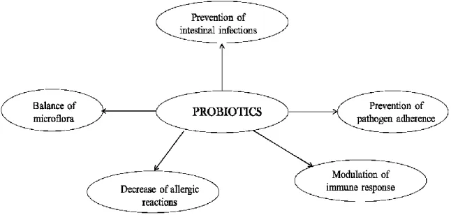
Several studies show that probiotic bacteria are able to positively influence the state of health, thanks to the numerous activities that they carry out: maintaining a balance in the intestinal microflora, the protection against intestinal pathogens and modulation immune response leading to an improvement in allergies food and autoimmune disorders (Fig. 1.1)
- 2 - Figure 1.1: Mechanisms of action of probiotics
To be a probiotic, a bacterial strain has to fulfill several criteria. It has to be healthy, to resist acid and bile, to adhere to intestinal epithelial cells, to be able to persist long enough in the digestive tract, to produce anti-microbials, to modulate immune responses and to resist technological processes.
A careful reading of the guidelines prepared by FAO/WHO (2001) is absolutely necessary for the selection of strains to be used in probiotic preparations; they require a the taxonomic identity, the specific phenotypic characteristics, security of use and their potential effectiveness.
The international scientific literature shows a growing number of microorganisms considered as probiotics. Notwithstanding, it is difficult to list thoroughly all the bacterial species in use considered as probiotics.
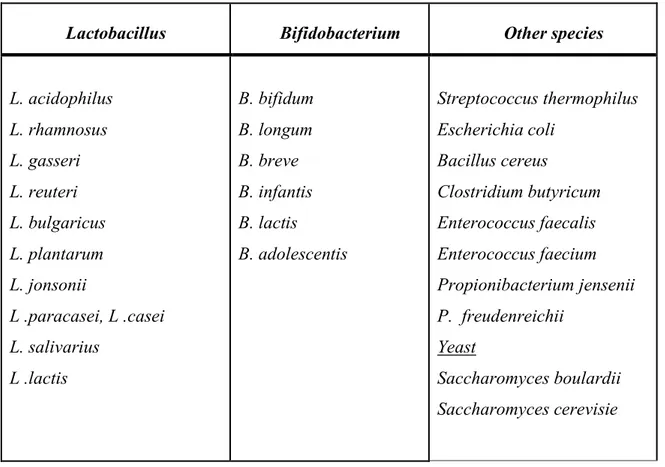
Table 1.1 shows groups of microorganisms used as probiotics. The widely used probiotic bacteria include strains from the genera Lactobacillus and Bifidobacterium. These are the most abundant genera in probiotic containing food products. Moreover, other species of microorganisms have been suggested for probiotic effects, such as Saccharomyces,
- 3 -
Table 1.1 List of the most frequently used probiotic microorganisms (Heyman,2002).
1.1.2. The genus Lactobacillus
The Members of Lactobacillus genus are heterogeneous, Gram-positive, non-spore-forming rods or coccobacilli, catalase-negative (Felis et al., 2007). The genus
Lactobacillus belongs to the phylum Firmicutes, class Bacilli, order Lactobacillales,
family Lactobacillaceae and its closest relatives, being grouped within the same family, are the genera Paralactobacillus and Pediococcus. This genus comprises close to 200 species with a G+C content usually below 50 mol% (Tannock, 2004). Lactobacilli are at the interface of aerobic and anaerobic life. Many lactobacilli retain the conditional capacity for respiration, but their ecology and physiology are mainly related to the fermentative conversion of sugars to organic acids, with lactic acid as the primary fermentation end product (Axelsson, 2003; Zheng et al., 2015). They are almost ubiquitous: they are found in environments where carbohydrates are available, such as food (dairy products, fermented meat, sourdoughs, vegetables, fruits, beverages). The human body hosts various Lactobacillus species in different anatomic regions (oral cavity, gut and female genital tract) entailing different interactions with the host (Munson
Lactobacillus Bifidobacterium Other species
L. acidophilus L. rhamnosus L. gasseri L. reuteri L. bulgaricus L. plantarum L. jonsonii L .paracasei, L .casei L. salivarius L .lactis B. bifidum B. longum B. breve B. infantis B. lactis B. adolescentis Streptococcus thermophilus Escherichia coli Bacillus cereus Clostridium butyricum Enterococcus faecalis Enterococcus faecium Propionibacterium jensenii P. freudenreichii Yeast Saccharomyces boulardii Saccharomyces cerevisie
- 4 -
et al., 2004; Rossi et al., 2016). The historical subdivisions of the genus Lactobacillus
based on the type of fermentation have been excellently reviewed by Pot et al. (1994), who have underlined how terms such as “homofermentative”, “heterofermentative”, “obligately homofermentative”, “facultatively heterofermentative” and “obligately heterofermentative” have been given different meanings by different authors and may be misleading. The accepted ‘modern’ definition is that given by Hammes et al. (1995):
- obligately homofermentative lactobacilli are able to ferment hexoses almost exclusively to lactic acid by the Embden–Meyerhof–Parnas (EMP) pathway while pentoses and gluconate are not fermented as they lack phosphoketolase; - facultatively heterofermentative lactobacilli degrade hexoses to lactic acid by
the EMP pathway and are also able to degrade pentoses and often gluconate as they possess both aldolase and phosphoketolase;
- obligately heterofermentative degrade hexoses by the phosphogluconate pathway producing lactate, ethanol or acetic acid and carbon dioxide; moreover, pentoses are fermented by this pathway.
Lactobacilli play a crucial role in maintaining the ecological equilibrium of these environments, through direct antimicrobial effects, enhancement of mucosal barrier integrity, and immune modulation (Patel et al., 2015). In addition, lactobacilli are important bacteria in food microbiology and human nutrition due to their contribution to fermented food production and their use as probiotics in food and pharmaceuticals (Bernardeau et al., 2006; Bourdichon et al., 2012).
1.1.3. The genus Bifidobacterium
Bifidobacteria are Gram-positive polymorphic branched rods that occur singly, in chains or clumps. They are non-spore-forming, non-motile, and non-filamentous. They are anaerobic and chemoorganotrophs, having a fermentative type of metabolism. They produce acid but not gas from a variety of carbohydrates. They are catalase negative, with some exceptions (B. indicum and B. asteroides when grown in presence of air). They were first included among the family Lactobacillaceae, but in 1924 the species L.
bifidum was reclassified into the new genus Bifidobacterium. The species of the genus Bifidobacterium form a coherent phylogenetic group and show over 93% similarity to the
- 5 -
phylum Actinobacteria, class Actinobacteria, subclass Actinobacteridae, order
Bifidobacteriales, family Bifidobacteriaceae.
Bifidobacteria have been isolated from a variety of ecological niches, such as sewage, fermented milk and anaerobic digestion facilities, yet are most frequently associated with the GIT of humans and animals (in general where the offspring of the bifidobacterial host is raised with parental care which may ensure direct transmission from mother to child/progeny) (Scardovi, 1974; Dong et al., 2000).
They are normal inhabitants of the gastrointestinal tract of humans, making up to 25% of the cultivable faecal bacteria in adults and 80% in infants (Picard et al., 2005). Strains most commonly found in human intestines and faeces are those belonging to the species
B. catenulatum, B. pseudocatenulatum, B. adolescentis, B. longum, B. breve, B. angulatum, B. bifidum and B. dentium, and the typical species isolated from functional
foods is B. animalis subsp. lactis (Masco et al., 2005). Often in commercial products are present species other than those typical of the human gut. An example is given by B.
lactis, a species widely used in commercial products thanks to its greater capacity for
survival and resistance to stress. They are commonly used as probiotics, with a long history of safe use in fermented dairy products. Their positive effects on human health include prevention of infection by pathogenic bacteria, immunostimulatory and anti-carcinogenic capabilities, protection against infectious diarrhoea, lowering of serum cholesterol and alleviation of lactose intolerance (Russell et al., 2011).
1.1.4. The genus Propionibacterium
Propionibacteria belong to the Actinobacteria class with a high G + C content (64-68%); they are mesophilic, Gram-positive, catalase positive, motile pleomorphic rods, non-spore-forming, and anaerobic to aerotolerant bacteria. Some cells may be elongated, bifid or arranged in “Chinese characters” (Zaratè, 2012). They grow at 15-40° C and at pH 5.1-8.5; the optimal temperature for growth is 30° C (Zaratè, 2012; Cousin et al., 2011). The current taxonomy describes 13 species that can be divided into two groups: “dairy or classical” and “cutaneous”; the classical propionibacteria (P. acidipropionici, P.
cyclohexanicum, P. freudenreichii, P. jensenii, P. microaerophilum, P. thoenii) are
generally isolated from milk and dairy environments, whereas the cutaneous propionibateria (P. acidifaciens, P. acnes, P. australiense, P. avidum, P. granulosum, P.
humerusii, P. propionicus) are from skin/intestine of human and animals.
- 6 -
and others), alcohols (glycerol, erythritol and others) and organic acids (lactic and gluconic acids) and produce propionic and acetic acids and carbon dioxide as final products. They are also able to produce a variety of beneficial compounds for human health, such as vitamin B12 and folic acid (Hugenholtz et al., 2002), and some strains have been proposed as probiotics, due to their ability to modulate intestinal microbiota through their bifidogenic effect and to anti-inflammatory effects and antimutagenic properties, among others (Cousin et al., 2011).
Due to their antimicrobial activity, propionibacteria are used to enhance the technological properties of various food products, e.g. they are used to prolong the shelf life of bread, cakes, cheeses, fruits, vegetables and tobacco, as they suppress the growth of moulds and spoilage microorganisms (Zaratè, 2012). Propionibacteria also stimulate the immune system and limit cancer progression although the mechanism involved is not defined. Cousin et al. (2011) reported that dairy propionibacteria are able to prevent infections and allergies, promote immune system maturation, and reduce the risk of cancer because they bind carcinogenic compounds (mycotoxins, plants lectins and heavy metals).
1.1.5. Probiotics: guidelines for use in foods and food supplements
In order to protect the health of consumers, foods containing probiotics are subject of attention, as confirmed by the elaboration of international guidelines aimed to safeguard the quality of the products. Ministerial guidelines (revision of 2013) define first the requirements required to use microorganisms as probiotics. They must safe, be alive and vital in such quantity as to allow multiplication and their activity in the intestine. The minimum quantity of microorganisms able to temporarily colonize the intestine is 109 cfu for strain and for day. The recommended daily dose of the product must therefore possess a charge in viable cells equal to 109cfu for at least one of the strains present in the product. The number of vital microorganisms in the product is, in fact, very important because beneath certain levels the beneficial effects cannot be achieved. For this reason, Italy has set a minimum concentration of 106 cfu, which must be maintained during shelf-life of the product.
To ensure the safety of the microorganism used, it is necessary a taxonomical identification of the strain. First of all, for safety and efficacy reasons it is important that each strain under study is clearly identified; in fact, different bacterial strains belonging to the same species may play different beneficial actions in the host. The species can be identified by determination and analysis of the DNA sequence coding for the 16S rRNA
- 7 -
or through the hybridization of nucleic acids, the strain can be characterized by PFGE (Pulse Field Gel Electrophoresis).
For the evaluation of the bacterial safety EFSA (European Food Safety Authority) has introduced the concept of QPS ("Qualified Presumption of Safety") it was developed a list of bacterial groups suitable for QPS assessment; for them it is not necessary safety assessment but only the determination of sensitivity to antibiotics (EFSA, 2013).
The phenomenon of bacterial resistance to antibiotics is often based on the presence in the bacterial cells of mobile genetic elements, such as plasmids and transposons, which can be transmitted from one organism to another promoting the horizontal spread of resistance. Particularly the intrinsic resistance to antibiotics is not a problem, the problem arises when the resistance determinants may be transferred to other bacterial strains, therefore, evaluation of antibiotic sensitivity is necessary to ensure the absence of acquired or potentially transmissible resistances.
The ability to survive to stressful gastrointestinal tract (GIT) conditions, (low pH and bile tolerance), are essential for probiotic strains to colonize the small intestine (Huang et al., 2004). The transit of probiotics present in food through the GIT takes variable times and is submitted to different stressful conditions. After mastication, the first barrier that bacteria must overcome is the low pH of the stomach with values ranging from 1 to 3. Into the duodenum the pH value rises to 6-6.5 but bile salts reach concentrations ranging from 1.5 to 2% during the first hour of the digestion (Noriega et al., 2004). Usually they do not colonize the intestinal mucosa for long periods of time, and are eliminated within few days after the subject stops ingesting them; however, a few subjects have been shown to be colonized for long periods by some strains (Marteau, 2001). For the screening of probiotics so it is important to simulate in vitro these GIT conditions.
- 8 -
1.2. Probiotics in foods: attenuation and modulation of metabolic activity
Consumption of probiotics through food products is the most viable approach at present. Most probiotic food products are categorized as functional foods, and represent a significant portion of this product category. Among functional foods, certain products have received great attention due to their importance as suitable vehicles for probiotic bacteria. Scientific evidence indicates that conditions such as reduction in lactose intolerance, prevention of colon cancer, inflammatory bowel disease, reduction in allergies, cholesterol and blood pressure have all been reported as benefit from the ingestion of specific probiotic strains (Patrick, 2012). Functional foods may provide benefits in health terms, but should not be seen as an alternative to a varied and balanced diet and a healthy lifestyle.
The supplementation of probiotics in foods requires special technologies, because of the active metabolism of probiotics, which could lead to a significant changes in the flavour and in the rheology of the products. Therefore, it is important to control the metabolism of probiotic and starter cultures in foods, without adversely affecting their viability and functional properties. A possible way to overcome this problem is the use of attenuated strains.
Attenuation can be defined as a technological method to enhance the total pool of intracellular enzyme released into the matrix, positively influencing flavour and quality of the final product. Principally, attenuated cultures are lactic acid bacteria (LAB) that do not have the ability to synthesize lactic acid during ripening (Klein et al., 1999).
The first preparation and use of attenuated starters as a cheese additive, was proposed by Petterson et al. (1975) to accelerate the ripening of Svecia, a Swedish semi-hard cheese, by thermal treatments (e.g., 69°C for 15 s). At this time, their main purpose was to accelerate proteolysis and shorten ripening time. A similar approach can be found in many other papers (Di Cagno et al., 2012; Tabanelli et al., 2013). Attenuation of the cells is also necessary to eliminate the overproduction of lactic acid during cheese making (Johnson et al., 1995).
Attenuated starters can be prepared through different treatments. Besides thermal treatments, Klein et al. (1999) report attenuating treatments such as, spray drying, freeze drying, fragilization using lysozyme or solvents, or the selection of lactose negative mutants treatment. A number of chemical and physical techniques intended for such use have been extensively reviewed (Klein et al., 1999; El-Soda et al., 2000; Geciova et al., 2002).
- 9 -
According to Yarlagadda et al. (2014) these techniques can be generally divided into two types of treatment:
Chemical treatments: such as the use of hexadecyltrimethylammonium bromide (CTAB), ethylenediaminetetraacetic acid (EDTA), isopropyl alcohol (IPA), sodium dodecyl sulphate (SDS) or n-butanol (Exterkate, 2006; Doolan et al., 2009); Physical treatments including heat or freeze shocking, and/or mechanical
treatments such as sonication (Exterkate, 2006), bead mill, high-pressure homogenization and microfluidization (Geciova et al., 2002).
Each method has its own advantages and disadvantages. Chemical treatments using chelating agents, such as EDTA, have a strain-specific effects affected by buffers and are more effective towards Gram-negative bacteria. SDS is used mainly for Gram-negative bacteria; however, it can also cause denaturation of proteins. The use of alkanols such as n-butanol increases the permeabilization process of lactoccocal cells; however the enzymes are sensitive to irreversible inactivation (Exterkate, 2006). The use of mechanical treatments like bead milling is more efficient in yeasts or moulds compared with bacteria but the effectiveness is dependent upon the size of beads. One of the emerging technologies, is the ultrasound. It is applied to impart positive effects in food processing such as improvement in mass transfer, food preservation, assistance of thermal treatments and manipulation of texture and food analysis (Knorr et al., 2011). Industrial use of attenuated strains is not widespread, due to some limits. In fact, the attenuation is often strain dependent, each methods shows benefits and limits, and the approximate costs for the use of attenuated strains are hard to ascertain.
- 10 - 1.3. Attenuation by sonication
Sonication is a laboratory-scale method that not only increases cell lysis, but also significantly increases the degradation of enzymes by heat denaturation (Geciova et al., 2002), as ultrasonic waves have the potential to exert a significant effect on microorganisms and living cells (Tabatabaie et al., 2010).
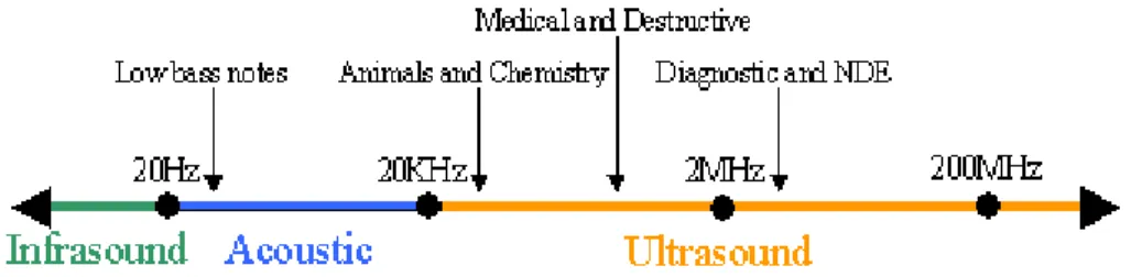
Ultrasound (US) is defined as pressure waves with a frequencies of 20 kHz or more (Fig. 1.2).
Generally, it uses frequencies from 20 kHz to 10 MHz. It can be divided into three frequency ranges:
1. Higher-power US at lower frequencies (16-100 kHz), called “Power Ultrasound”, used in food processing to inactivate microorganism and chemicals;
2. Low intensity US at high frequencies (from 100 kHz to 1 MHz) used in non-invasive imaging, sensing and analytical tools;
3. Diagnostic US (1-10 MHz), used for medical imaging.
It is important to highlight that the use of the this new technology in industrial processes has two main requirements:
- a liquid medium, even if the liquid element forms only 5% of the overall medium; - a source of high-energy vibrations (the ultrasound) (Patist et al., 2008).
Figure 1.2: Ultrasound frequencies
It is a destructive approach because it generates mechanical and shear forces, as well as localized high temperatures and pressures which lead to physical disruption and
- 11 -
promotion of chemical reactions, such as oxidation (Abela et al., 2014; McClements, 1995). High intensity US is used in food industry for many applications, such as emulsification, modification and control of crystallization processes, degassing of liquid foods, enzyme inactivation, enhanced drying and filtration, induction of oxidation reaction, and more recently for food preservation (Gao et al., 2014; Knorr et al., 2004; Zheng et al., 2006).
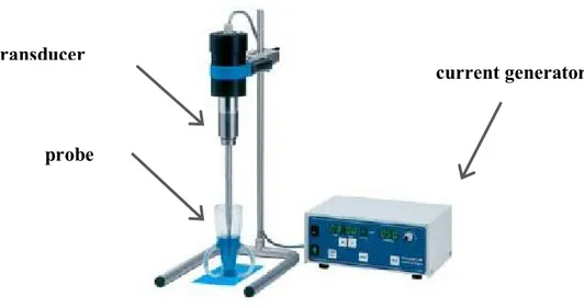
Figure. 1.3: Components of US production system.
Figure 1.3 shows the US production system; it consists of:
a current generator that supplies electricity at the desired frequency to the transducer;
a transducer or converter, which converts electrical energy into mechanical vibrations (pressure waves); they, in turn, are conveyed into a probe;
a probe that amplifies the vibration produced; this is the sonication site that can be continuous or discontinuous.
Nowadays ultrasound technology uses the electrostrictive transformer principle. This is based on the elastic deformation of ferroelectric materials within a high frequency electrical field, caused by the mutant attraction of the molecules polarised in the field. For the polarization of molecules a high-frequency alternating current will be transmitted
transducer
probe
- 12 -
via two electrodes to the ferroelectrical material; then, after the conversion into mechanical oscillation, the sound waves are transmitted to an amplifier, to the sound radiating sonotrode and finally to the treated medium (Knorr et al., 2004).
The principal effect of ultrasound on a fluid medium is to impose an acoustic pressure (Pa) in addition to the hydrostatic pressure acting on the fluid.
This acoustic pressure is a sinusoidal wave that depends on: time (t), frequency (ƒ) and the maximum pressure amplitude, Pa,max. (eq.1) (Di Benedetto et al., 2010).
Pa = Pa,maxsin (2πƒt) (eq. 1.)
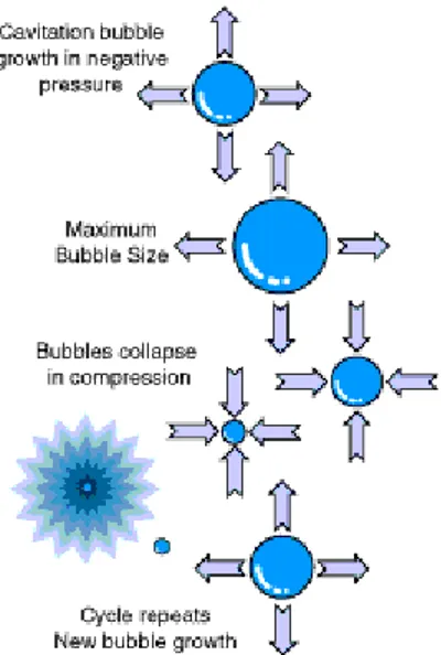
The maximum pressure amplitude of the wave is directly proportional to the power input of the transducer. In fact, at lower intensity the pressure wave induces motion and mixing within the fluid; this phenomenon is called acoustic streaming; while, at higher intensities the local pressure causes the formation of tiny bubbles. A further increase generates negative transient pressures within the fluid, enhancing bubble growth and producing new cavities by the tensioning effect on the fluid; then, during the compression cycle, the bubble shrinks and their contents are absorbed back into the liquid. This process of compression and rarefaction of the medium particles and the consequent collapse of the bubbles describes the well-known phenomenon of cavitation (Fig. 1.4). Pressure resulting from these implosions causes the main bactericidal effect of ultrasound.
- 13 -
The efficacy of ultrasonic treatment depends on the type of microorganism treated, in fact, for example, spores are relatively resistant to the effects of ultrasound. Moreover, positive cells have been found to be more resistant to ultrasound than Gram-negative cells and this may due to the structure of the cell wall.
Other factors that influence the antimicrobial effectiveness are the amplitude of the waves ultrasound, the time of exposure/contact, the volume of processed food, the composition product and the temperature of treatment.
Thus sonication, might be used as an alternative method to conventional thermal treatment for pasteurization and sterilization of foods products. On the other hand, various research have shown how sonication might affect physicochemical parameters of products. Generally, sonication may break down the cell wall and cause autolysis (Tabatabaie et al., 2010), so thus it increases the level of bioactive compounds.
A positive effect of sonication on beneficial bacteria was also reported by Suckova et al. (2014); they used this approach to increase the yield of vitamin B12 produced by
Propionibacterium shermanii. This effect was attributed to an increase in the
permeability of membrane with enhanced lactose metabolism. On the other hand, Nguyen et al. (2009) recovered a stimulating effect on bifidobacteria, probably associated with an acceleration of lactose hydrolysis and transgalactosylation in milk (Nguyen et al., 2012). This effect on sugar metabolism was also evidenced by a significant change in acid profile, with a decrease of acetic and propionic acids.
Furthermore, free radical formation is another proposed mode of action of ultrasound inactivation with bactericidal effects. Application of ultrasound to a liquid can lead to the formation of free radicals that provoked the breakages along the length of the DNA causing the formation of small DNA fragments (Betts et al., 2000).
Another approach, is the use of sonication to control the metabolism of probiotics and starter cultures in foods. Bevilacqua et al. (2016), studied the use of US-attenuated L.
plantarum, L. casei LC01 and B. animalis subsp. lactis Bb12 inoculated in a commercial
rice drink, to reduce acidification. Attenuation did not affect viability of probiotics or the sensory scores of the beverage. Moreover, some preliminary experiments performed by these authors showed that probiotic traits were not affected by attenuation.
- 14 - 1.4. Other methods of attenuation
1.4.1. Attenuation by hydrostatic high pressure and high-pressure homogenization
The High Pressure (HP) treatment is defined as non-thermal treatment that uses the pressure (300-700 MPa, in some cases up to 1000 MPa) as the main preservation method. Due to the fact that pressure increase is achieved through a fluid (for example water), this process has been also referred to as high hydrostatic pressure (HHP) as opposite to the High Pressure of Homogenization (HPH), where the increase of the pressure is obtained forcing the product through a small valve (homogenizing valve) (Bevilacqua et al., 2010). A homogenizer consists of a positive displacement pump and a homogenizing valve. In the homogenizing valve (often referred to as radial diffuser), the fluid is forced under pressure by pump, through a small orifice between the valve and the valve seat (Diels et al., 2006). This treatment was proposed for the non-thermal fluid food microbial decontamination (Bevilacqua et al., 2010; Lanciotti et al., 2007). Cavitation and viscous shear have been identified as the primary mechanisms of microbial cell disruption during HPH treatment (Kleinig et al., 1998; Middelberg, 1995). Some studies have suggested the use of this treatment to accelerate cheese ripening (O’ Reilly et al., 2000a-2003; Saldo et al., 2002), and to control spoilage organism in cheese (O’ Reilly et al., 2000b). This technology is also active on food constituents, especially proteins, leading to changes in their functional properties and activities (Kheadr et al., 2002; Vannini et al., 2004). Lanciotti et al. (2007) showed that HPH is able to modify, in relation to the strain and to the treatment applied, both the fermentation kinetics and the enzymatic activities of starter and no starter lactic acid bacteria (LAB) without detrimental effects on cell viability.
Regarding in vitro functional properties, Muramalla et al. (2011) studied the use of some low homogenization pressures (up to 13.8 MPa for 5 passes) to improve certain probiotic characteristics of yogurt bacteria and L. acidophilus LA-K. These authors demonstrated that treatments at 13.80 and 6.90 MPa, repeated for 5 times, improved acid tolerance and bile tolerance, respectively, of L. acidophilus LA-K but had no effect on protease activity and its growth, recommending this technique for improvement of certain probiotic characteristics.
Among the processes involving the pressure, the High Hydrostatic Pressure (HHP) is one of the most studied (Kelly et al., 2009; Senorans et al., 2003;Wan et al., 2005) and its
- 15 -
efficacy in inactivating different microbial species is well documented (Ananta et al., 2003; Desmond et al., 2001; Knorr et al., 2001). During HHP treatment, a sample receives instantaneous and uniform pressure. Furthermore, HHP can cause perturbation of the bacterial cell wall and membranes (Cheftel, 1995).
1.4.2.Attenuation by microfluidization
A microfluidizer (Microfluidica, Newton, MA, USA) operates on a principle different from that of the high-pressure valve homogenizer. A flow of cell suspension is impacted at high velocity with adjustable pressure against a stationary surface in an interaction chamber that disrupts cell integrity. The operation pressure is a function of flow rate and thus the rate of cell disruption increases with increasing pressure and the number of passes through the chamber. The reduction of enzyme activity by thermal degradation can be minimized by controlling the temperature within the chamber (Bevilacqua et al., 2017). This technique could be used to create specific populations of live, permeabilized
or lysed cells for use as adjuncts in cheese ripening (Yarlagadda et al., 2014).
1.4.3. Attenuation by heat treatment
The most studied form of starter attenuation is heat treatment. This method is based on the sub-lethal heat treatment of a cell suspension. A critical point for heat attenuation was to define the correct temperature/time combination, so that acidification was eliminated or delayed without excessively denaturing potential ripening enzymes (e.g. proteases, peptidases, esterases, or others). Petterson et al. (1975) recommend a temperature of 59°C for 15s for mesophilic starters (lactococci) and a temperature of 69°C for 15s for thermophilic (lactobacilli such as L. helveticus); thus, they found a delay in acid production between 5 and 10 hours.
According to Frey et al. (1986) attenuation by heat may occur at different levels, such as damage to the enzymes involved in the transport of the substrates through the cell wall and membrane, physical damage to the membrane, or the denaturation of β-galactosidase. The greatest contribution of heat-attenuated starter to cheese making was the hydrolysis of medium-sized peptides (Ardö et al., 1988; Vafopoulo et al., 1989). Peptides were shown to be intracellular in lactic acid bacteria (Kunji et al., 1996), therefore their presence in cheese would indicate that cell lysis occurred (Lortal et al., 1997).
- 16 -
1.4.4. Attenuation by freezing-thawing
Freezing cells in suboptimal conditions reduces the viability of lactic acid bacteria. Stressed cells do not contribute significantly to lactic acid production during cheese making, but they may retain protease and peptidase activity (El-Tanboly et al., 2010a). The physico-chemical-biochemical effects of freezing on cells are multiple and complex. Bacterial cells can freeze by intracellular ice formation or dehydration; freezing removes water from both the external and internal environments and increases the concentration of solutes both inside and outside the cells (Ray et al., 1973). Ice crystals can cause damage to the cell membrane. After freezing and thawing treatment, bacterial cells can lose many micro and macromolecular cellular components (Klein et al., 1999).
The first cheese study involving freeze-shocked cells, was performed by Bartels et al. (1987). This treatment involved the washing of a tenfold concentration of cells, freezing at -20°C and thawing at 40°C prior to the addition of milk. A similar study was conducted by El-Tanboly et al. (2010a) to determine the effects of L. acidophilus on the sensory attributes, ripening time and composition of Gouda cheese, as well as the survival of the best microorganism subjected to freeze shocking at -10/-20° C for 24/96 hours. They suggested that the combination of attenuated lactobacilli and rennet exerted a positive effect on maturation. Moreover, attenuated starters reduced bitterness due to their enhanced proteolysis (El-Tanboly et al., 2010b).
1.4.5.Attenuation by spray and freeze-drying
Johnson et al. (1995) compared the attenuation of L. helveticus CNRZ-32 by spray drying and freeze-drying to freezing. In this case, the attenuation was the result of both dehydration and heat treatment. The heat applied to the drying rate may be the cause of the delay in acid production in lyophilized cells. They noted that frozen starters occupy large storage volumes, and result in higher storage and shipping costs. Dried cultures, in contrast, offer an economical and practical alternative. In addition, freeze-dried adjuncts might have delayed acid production because cells are subjected to attenuation by dehydration effects and thermal effects when heat is applied to speed drying. Furthermore, the frozen and freeze-dried cells had the highest viability, compared to SDHT (spray-dried at a high outlet air temperature of 120°C) cells. In addition, in SHDT samples, aminopeptidase and β-galactosidase activities were almost completely deleted, lactic acid production was delayed by 5 hours and permeability was increased more than in the other samples. Instead, for frozen, freeze-dried and SDLT (spray-dried at a low
- 17 -
outlet air temperature of 82°C) cells, lactic acid production was not delayed and the rates of lactic acid production were similar, despite the drastic reduction in vitality. Finally, aminopeptidase and β-galactosidase activities of the SDLT cells were several time higher than those of the frozen and freeze-dried cells.
1.4.6.Lysozyme treatment
Attenuation using lysozyme consists of treating a cell suspension with this enzyme (Ristagno et al., 2012). Lysozyme is active throughout a wide pH range (4-10); however, high ionic strength (>0.2 M salt) was shown to have an inhibitory effect on its activity (Chung et al., 2000). The cell treated with lysozyme break down in the curd at the salting step and thus release their intracellular enzymes into the cheese matrix (Briggs, 2003). Lysozyme belongs to a class of enzymes that lyses the cell walls of Gram-positive bacteria, breaking the bond between N-acetylglucosamine and N-acetyluramic acid of peptidoglycan in the bacterial cell wall. Peptidoglycan hydrolysis, by exogenous lysozyme, causes cell lysis and death in a hypo-osmotic environment, but some types of exogenous lysozyme can also cause lysis of bacteria by stimulating autolysin activity on interaction with the cell surface (Nakimbugwe et al., 2006).
1.4.7.Solvents
A Chemical treatment with chelating agents, such as EDTA, has strain-specific effects that are influenced by buffers and are more most effective towards Gram-negative bacteria. SDS is used mainly for Gram-negative bacteria, and it can cause denaturation of
proteins (Geciova et al., 2002; Middelberg, 1995). The use of alkanols such as n-butanol
increases the permeabilization process of lactoccocal cells; however the enzymes are sensitive to irreversible inactivation
Exterkate (2006) found that the use of alkanols like n-butanol increased the permeabilization process of lactococcal cells.
Attenuation with various solvents showed changed the lipid structure of the cell membrane (Jain et al., 1978), in particular n-butanol, making the cell unable to produce lactic acid (Klein et al., 1999; Ristagno, 2012). Exterkate (1984) also reported increases in peptidase activity.
- 18 -
1.4.8. Lactose-negative mutants as attenuated starters
Klein et al. (1999) considered the use of lactose-negative mutant strains as attenuated starter. According to Briggs (2003) a lactose-negative mutant can be defined as an organism that is unable to ferment lactose; therefore, it is unable to produce lactic acid, but is still able to provide the necessary of enzymes that will slowly be released as the cell lyse, which will be available to enhance proteolysis, lipolysis, glycolysis, leading to a final product with overall reduced bitterness and a reduction of ripening time. However, these natural cultures, which lose their ability to convert lactose into lactic acid, are difficult to isolate and therefore, most of them are genetically engineered.
- 19 -
Chapter 2. AIMS AND PLANNING OF RESEARCH
Attenuation is a tool to control/modulate the acidification of active drinks due to the metabolism of probiotics throughout storage, without affecting their viability. Nowadays, various attenuation methods are known. An attenuation method is based on the use of ultrasound. Different authors have studied the use of ultrasound as attenuation methods. This approach has been used for starter cultures of dairy products and cereal beverages, avoiding post-acidification during storage at 4°C and maintaining a high vitality of microorganisms until consumption (Bevilacqua et al., 2016). However, the effects of this methodology on the functional and technological characteristics of the various microbial strains are not known.
The main goal of this PhD thesis was to study the metabolic activity and functional properties of probiotics from the genera Lactobacillus, Bifidobacterium and
Propionibacterium, after attenuation with physical treatments, such as ultrasound.
In particular, the first phase of my PhD thesis was focused on the evaluation of technological and probiotic traits. In this step three different genera of probiotics were used: Lactobacillus, Bifidobacterium, Propionibacterium. The strains were treated with ultrasound and studied for the acidification in lab medium, growth at different temperatures, pHs and salt content, antibiotic-resistance, survival at pH 2.5 and in the presence of 0.3% bile salt, hydrophobicity, and biofilm formation.
The aim of the second part of PhD thesis, was to investigate the effects of US on the release of intracellular components. The strains were studied after physical treatment to assess the release of intra-cellular constituents (nucleic acids, proteins) and injury of the membrane.
In the last part of this PhD thesis, the interaction of attenuated strains with gut microbiota was evaluated, using in vitro batch culture fermentation. This study was carried out at University of Roehampton (UK).
- 20 -
Chapter 3. MATERIALS AND METHODS
3.1. Reference Cultures
This study focused on 2 strains of Lactobacillus:
- L. plantarum L-12, isolated from a sourdough, and belonging to the Culture Collection of the Department of the Science of Agriculture, Food and Environment, University of Foggia (Corbo et al., 2014);
- L. reuteri DSM 20016, purchased from DSMZ (Deutsche Sammlung von Mikroorganismen und Zellkulturen GmbH, Germania);
on 2 species of Bifidobacterium:
- B. longum Bb-46 and B. infantis Bb-02, purchased from Chr. Hansen (Hørsholm, Denmark).
and on 2 species of Propionibacterium:
- P. freudenreichii subsp. freudenreichii DSM 20271;
- P. jensenii DSM 20535 (now Acidipropionibacterium jensenii; Scholz et al., 2016).
3.2. Optimal culture conditions
The strains of Lactobacillus and Propionibacterium were stored at -20°C in MRS broth (Oxoid, Milan, Italy) added with 33% of sterile glycerol (J.T. Baker, Milan, Italy). The working cultures were prepared by inoculation with 6-7 log cfu/ml in 5 ml of fresh MRS broth, and incubated at 37±1°C for 24 h.
Bifidobacterium were preserved at -20°C in MRS broth supplemented with 0.5% cysteine (cMRS) (Sigma-Aldrich, Milan, Italy) added with 33% of sterile glycerol (J.T. Baker, Milan, Italy). The working cultures were prepared by inoculation with 6-7 log cfu/ml in 5 ml of fresh cMRS broth, and incubated at 37±1°C for 24 h
3.3. US – treatment
The strains were treated with ultrasound (US) through a VC Vibra Cell Ultrasound equipment; model VC 130 (Sonics and Materials Inc., Newtown, CT, USA). The main variables of the treatment were the net power (40, 60 and 80%) and the duration of the treatment (2, 4, and 6 min); pulse was set to 2 s. Before each treatment, the ultrasonic
- 21 -
probe was washed with sterile distilled water; immediately after processing, the sample was cooled in ice. Technological and probiotic properties were assessed both on US-treated and unUS-treated bacteria. UnUS-treated bacteria were used as control.
3.4. Effect of attenuation on some technological and functional properties
3.4.1.Acidification
Aliquots of MRS broth (Oxoid, Milan, Italy) and MRS+cysteine (cMRS, Oxoid) (Bifidobacterium) were individually inoculated with each strain to 7 log cfu/ml and incubated at 4, 15, 25, 30 or 37°C; the pH of the medium was evaluated after 6, 24 and 48 h through a pH-meter Crison (Crison Instruments, Barcelona, Spain). Data from pH were modelled as pH decrease. The experiments were performed in three different batches.
3.4.2.Effect of NaCl, pH, temperature
Growth profile of strains was assessed as follows.
The strains were individually inoculated in MRS or cMRS broth, at level of 7 log cfu/ml. Microbial growth was evaluated after 24 and 48 h as absorbance at 600 nm using a spectrophotometer UV–Vis DU 640 Beckman (Fullerton, CA, USA).
The data were modelled as growth index, as reported by Bevilacqua et al. (2009): 100
Abss Absc GI
where:
Abss is the absorbance of US-treated microorganisms and Absc is the absorbance of the controls (untreated bacteria).
GI was analyzed following the approach proposed by Bevilacqua et al. (2009):
NaCl pH Temperature
Lactobacillus 7% 4-9 15-37-45°C
Bifidobacterium 7% 4-9 15-37-45°C
- 22 - - GI <25% complete inhibition;
- 25%<GI<75% partial inhibition; - GI>75% no inhibition.
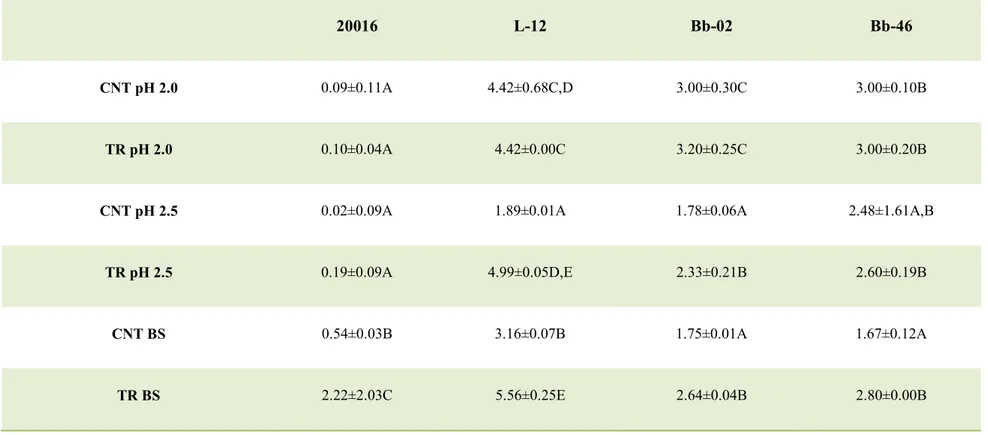
3.4.3.Survival at pH 2.0, 2.5 and with 0.3% of bile salt added
Acidified distilled water (pH 2.0 and 2.5) or distilled water supplemented with 0.3% bile salts (Oxoid) were individually inoculated with each strains at level of 7 log cfu/ml and stored at 37 °C. The viable count was determined after 3 h.
3.4.4. Antibiotic-resistance
Antibiotic-resistance was carried out through an E-test approach using the antibiotic strips produced by Liofilchem (Roseto degli Abruzzi, Italy). The following compounds were used: Ampicillin (0.016-256 µg/ml), Ciprofloxacin (0.002-32 µg/ml), Clarithromycin (0.016-256 µg/ml), Chloramphenicol (0.016-256 µg/ml), Erythromycin (0.016-256 µg/ml), Gentamicin (0.064-1024 µg/ml), Tetracycline (0.016-256 µg/ml), Trimethoprim (0.002-32 µg/ml), Vancomycin 0.016-256 µg/ml). The strains were plated onto the surface of MRS agar or cMRS agar; thereafter, the strip containing the antibiotic was placed onto the plates. The plates were incubated at 30°C or 37°C for 24 h. The tests were performed at least in duplicate.
3.4.5. Hydrophobicity
Bacterial cell surface hydrophobicity was assessed by measuring microbial adhesion to xylene using the method described by Bautista-Gallego et al. (2013). Briefly, US-treated and untreated bacteria were centrifuged (1500g for 10 min). The resulting pellet was washed twice in PBS (Phosphate Buffer saline, Sigma-Aldrich), re-suspended in 10 ml of 0.1 M KNO3 (C. Erba, Milan, Italy) and the absorbance at 600 nm was measured (A0). Three milliliter of xylene were then added to forma two-phase system. After a 10-min pre-incubation at room temperature, the two-phase system was mixed by vortexing for 20 s. Then, the samples were stored under static conditions at room temperature for 20 min, 1, 2 or 3 h. At each sampling point, the aqueous phase was carefully removed and the absorbance at 600 nm was assessed.
The percentage of the cell surface hydrophobicity (H%) was calculated using the following formula:
1
1 0 0% A1 A0
- 23 - where:
A1 and A0 are the absorbance at the time t and the initial value.
3.4.6. Biofilm formation
Glass slides (25.4 mm × 76.2 mm) were used as surfaces to get the biofilm attached. All slides were cleaned with acetone before soaking in 3.5% sodium hypochlorite (V/V) at 75˚C for 5 min. Then they were rinsed and transferred into 7.0 g/l phosphoric acid solution for 5 min. Slides were rinsed in distilled water, air dried and autoclaved at 121 ˚C for 15 min (Arizcun et al., 1998). This cleansing was required to remove fingerprints, oils, grease and other soils that may have been on glass.
The samples were prepared pouring 45 ml of MRS broth into sterile tubes and vertically dipping sterile slides in; the inoculum with 107 cfu/ml was performed in each of them and the samples were incubated at 37˚C, without agitation, for 7 days. The populations in planktonic and sessile state were periodically determined.
The populations in planktonic state were determined by a standard plate count procedure with MRS Agar, incubated at 37˚C for 48 h. Regarding the sessile state, the slides were aseptically removed from the medium and rinsed with sterile distilled water to remove the unattached cells. Then each slide was placed into a test-tube containing 20 ml of sterile saline solution (0.9% NaCl) and sonicated with a 20 Hz “Vibra Cell” Ultrasound equipment (Sonics and Materials Inc., Newtown, CT, USA) for 3 min in order to detach and collect the sessile cells. The obtained results were expressed as log cfu/cm2.
3.5. Adhesion to Caco-2 cell lines
The Caco-2 human colon adenocarcinoma cell line (kindly provided by Prof. M. Landriscina, University of Foggia) was grown in Dulbecco's Modified Eagle Medium (DMEM Glutamax, Gibco, Rockville, MD, USA) containing 4.5 g/l glucose, and supplemented with 25m MHEPES buffer, 10% (v/v) fetal bovine serum (Gibco) and antibiotics. For adhesion assay, the Caco-2 cells were seeded in 12-well standard tissue culture plates (culture area 4 cm2), the medium was changed every alternate day and the incubation was carried out at 37°C in 5% CO2 atmosphere until 7 days after reaching confluency. Afterwards, the cells were washed twice with 3 ml phosphate-buffered saline (PBS, pH 7.4) and an aliquot of 1.5 ml of DMEM lacking antibiotics and serum was added to each well and incubated at 37°C for 30 min. US attenuated and untreated bacteria were collected by centrifugation (4000g for 5 min at 4°C) immediately after
- 24 -
sonication, washed twice in sterile PBS (pH 7.4) and resuspended in DMEM without antibiotics and serum at 106 and 105 cfu/ml: a volume of 1 ml of each working suspension was added to different wells (total volume 2.5 ml) and incubated at 37°C for 3 h. After the incubation, the media were removed, the wells were washed twice with 3 ml PBS, cells from monolayers were detached by trypsinization (1 ml 0.25% trypsin-EDTA solution to each well-incubation at room temperature for 15 min). Cell suspension was then serially diluted with saline solution (0.9% NaCl) and plated on MRS agar. The plates were incubated for 48–72 h at 37°C and colonies were counted. Bacterial cells initially added to each well of 12-well plates were also counted taking off aliquots before and after incubation time
3.6. US - treatments and sub-lethal injury detection 3.6.1. Targets of the experiments
This phase focused on P. freudenreichii subsp. freudenreichii DSM 20271, P. jensenii DSM 20535.
3.6.2. Injury characterization
Sub-lethal injury was studied after the application of US-treatments:
- P. freudenreichii subsp. freudenreichii (power 60%, time 4 minutes, pulse 2
seconds);
- P. jensenii (power 40%, time 8 minutes, pulse 2 seconds)
To quantify the intracellular material released from the strains, untreated and treated samples (6-7 log cfu/ml) were centrifuged at 6.000 x g for 10 min. The UV absorbance of the supernatant was measured at 260 nm and at 280 nm with a spectrophotometer (spectrophotometer UV-VIS DU 640 Beckman (Fullerton, CA) (Virto et al., 2005). The data were modelled using the following formula:
((Absc-Abss)/Absc)*100 where:
Abss is the absorbance of US-treated microorganisms and Absc is the absorbance of the controls.
- 25 - 3.7. Evaluation of the effects on gut microbiota
3.7.1. Targets of the experiments
These experiments were done on treated and untreated L. reuteri (DSM 20016).
3.7.2. Batch culture fermentation
Collection and faecal sample preparation. Faecal samples were obtained from three
healthy human volunteers who were free of known metabolic and gastrointestinal diseases (e.g., diabetes, ulcerative colitis, Crohn’s disease, irritable bowel syndrome, peptic ulcers and cancer). All healthy faecal donors had the experimental procedure explained to them and were given the opportunity to ask questions. All faecal samples from healthy were collected on site, stored in an anaerobic cabinet (10% H2, 10% CO2 and 80% N2) and used within a maximum of 15 min after collection. The samples were diluted 1/10 wt/vol in anaerobic phosphate-buffered saline (PBS, 0.1 M phosphate buffer solution, pH 7.4) and homogenised (Stomacher 400, Seward, West Sussex, UK) for 2 min (460 paddle-beats/min). To maintain the anaerobic conditions, the PBS was maintained in anaerobic cabins until the time of use. The resulting faecal slurries from each individual were used to inoculate the batch-culture systems.
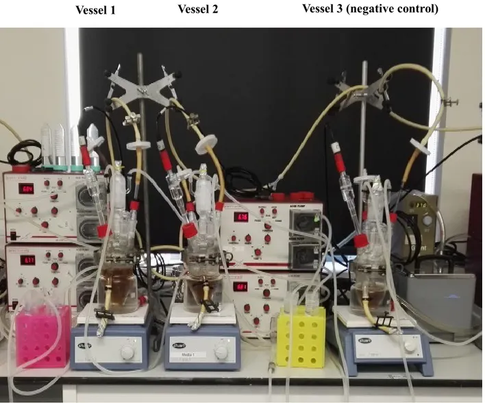
Batch culture fermentations. Previously sterilized batch culture fermentation vessels
(280 mL working volume) were filled with 45 mL of sterile complex colonic model growth medium. The composition of this medium included peptone water (5 g/L), yeast extract (4.5 g/L), starch (5 g/L), tryptone (5 g/L), NaCl (4.5 g/L), KCl (4.5 g/L), mucin (4 g/L), casein (3 g/L), pectin (2 g/L), xylan (2 g/L), arabinogalactan (2 g/L) and inulin (1 g/ L) (Tejero-Sarinena et al., 2012). All media and chemicals were purchased from Oxoid and Sigma. Then, the vessels were connected to a circulating water bath at 37°C and sparged with O2-free N2 gas overnight to attain anaerobic conditions. The pH was adjusted to between 6.7 and 6.9 using pH meter controllers with
NaOH or HCl (Electrolab260; Electrolab Ltd., Tewkesbury, UK), and 5 mL of faecal slurry was then inoculated in each vessel. In total, three vessels were prepared and supplemented with 1 mL of each strain including a negative control, i.e., a sample containing faecal slurry but without probiotic bacteria. The batch cultures were run for 24 h, and 5 mL of the samples were removed at 0, 2, 4, 6, 8 and 24 for analysis of bacterial populations by fluorescence in situ hybridisation (FISH).
- 26 -
Fig. 3.1: Batch cultures fermentation used at University of Roehampton, London (UK).
- 27 -
Enumeration of bacterial populations by fluorescence in situ hybridisation (FISH)
FISH was performed as described by Costabile et al. (2014). A 375 μL aliquot of the batch culture samples was fixed in three volumes of ice-cold 4% (w/v) paraformaldehyde for 4 h at 4°C, centrifuged at 13.000 g for 5 min and washed twice in 1 mL of sterile PBS. The cells were again pelleted by centrifugation and re-suspended in 150 μL of sterile PBS, to which 150 μL of ethanol was added. The samples were then mixed and stored at -20°C until used. All probes were synthesised by Sigma-Aldrich. The following bacterial groups were identified using synthetic oligonucleotide probes that target specific regions of the 16S ribosomal RNA molecule, labelled with the fluorescent dye Cy3:
- Clostridium hystolyticum clusters I/II (Chis150, TTATGCGGTATTAATCTYCCTTT)
(Franks et al., 1998);
- Lactobacillus/Enterococcus spp. (Lab158, GGTATTAGCAYCTGTTTCCA) (Harmsenet al., 1999);
- Clostridium clusters XIVa+b (Erec482, GCTTCTTAGT CARGTACCG) (Franks et al., 1998);
- Bacteroides/Prevotella group (Bac303, CCAATGTGGGGGACCTT) (Manz et al., 1996);
-Bifidobacterium spp. (Bif164, CATCCGGCATTACCACCC) (Langendijk et al., 1995); - Total bacterial with 4’,6-diamidino-2-phenylindole (DAPI).
3.8. Statistical analysis
The results were analyzed through t-student’s test (paired comparison) or one-way ANOVA using Tukey’s test as the post-hoc test (multiple comparison) (P<0.05). Statistic was performed through the software Statistica for Windows (Statsoft, Tulsa, and Okhla.).
- 28 -
Chapter 4. RESULTS AND DISCUSSION
4.1. Attenuation: preliminary screening
Attenuation should fulfill two basic requirements: avoid the acidification by probiotic without affecting their viability. At this scope candidate probiotics belonging to the genera Lactobacillus, Bifidobacterium, Propionibacterium were treated with ultrasound. Acidification and viability were preliminary tested; then, in order to understand whether attenuation could influence the characteristics of the strains, after the attenuation some selected technological and probiotics traits were evaluated.
4.1.1. Acidification and viability
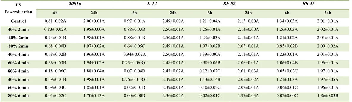
Table 1 (a, b) shows the acidification by lactobacilli, bifidobacteria and propionibacteria after sonication at various power levels/times. After 6 h, an increase of the power and/or US-duration decreased acidification; however, the complete attenuation was achieved only in some combinations:
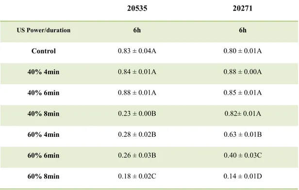
- 60%/6 min, 80%/4–6 min, for both Lactobacillus spp. and Bifidobacterium spp.; - 40% /4-8 min, for P. jensenii;
- 60%/4-8 min, for P. freudenreichii subsp. freudenreichii.
After 24 h acidification occurred in both the controls and US-treated samples, without significant differences (P > 0.05).
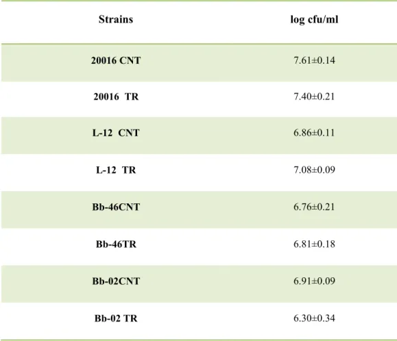
The second basic requirement was the viability; thus the combinations able to avoid acidification (40%/4 min; 40%/8 min; 60%/4 min; 60%/6 min; 60%/8 min; 80%/4 min; 80%/6 min;) were tested and the viable count was determined before and after the sonication.
For Lactobacillus spp. and Bifidobacterium spp., a power of 80% unfortunately caused a significant reduction of cell count (1–2 log cfu/ml) (P< 0.05) (data not shown), whereas the combination at 60% did not affect cell viability (Table 2a), thus it was chosen as the best combination to achieve attenuation for these genera.
- 29 -
Concerning propionibacteria, a power of 40% did not cause a reduction of cell count (Table 2b), but the attenuation was influenced by the duration of the treatment; in fact, an increase of the duration of the treatment reduced the acidification. Therefore, for P.
jensenii the treatment of 40%/8 min was chosen as the best combination, while for P. freudenreichii subsp. freudenreichii, was 60%/4 min.
The best combinations (60%/6min Lactobacillus and Bifidobacterium; 40%/8min P.
jensenii; 60%/4min P. freudenreichii subsp. freudenreichii) were used to test the effect
of attenuation on some technological and functional properties.
US-attenuation has been proposed by Bevilacqua et al. (2016) to avoid post-acidification by some probiotics in an organic rice drink; they reported the beneficial effect of this technique to control the phenomenon of post-acidification. In addition, they also performed some preliminary experiments to test the effect of US on the survival at pH 2.0/2.5 and on hydrophobicity, but the actual effect of US on some other probiotic properties or the influence on the overall “technological profile” has been never addressed. US can act on the cells by causing either an external cavitation or an internal cavitation, or both (Mawson et al., 2011). These effects are responsible of different kinds of injuries on the membranes along with the leakage of intracellular components (Wu et
al., 2015). These injuries could be lethal or not; it depends on the energy level, which is a
function of both the net power and the duration of the treatment (Jomdecha et al., 2010). Thus, high power levels caused an irreversible effect with a viability loss, whereas at 40%/60% the effect was lower and determined a delay of acidification. In addition, this effect could be transient for a probable repair of the damage (Hurst, 1984), as suggested by the extent of acidification of treated bacteria after 24 h of incubation at 37 °C.
To assure a prolonged effect of attenuation, the treated bacteria were stored at 4-15 °C. As expected, the acidification did not occur at 4 °C both for untreated and attenuated bacteria. On the other hand, some significant differences were found at 15 °C (Fig. 1).
After 2 days, the control-cultures decreased the pH of the medium by 0.47–1.42 (for B. longum and L. plantarum, respectively), whilst US-attenuated strains did not
experience a significant acidification. After 14 days, ΔpH was ca. 2 for untreated
L. plantarum and L. reuteri and 1.5–1.6 for bifidobacteria; acidification for treated
bacteria was always significantly lower. This effect could be due to the viable count of the target strains (ca. 8 log cfu/ml in the control samples and 7.0–7.3 log cfu/ml).