1
Scuola Normale Superiore
Ph.D. Thesis
In Biophysical Sciences
Structural and functional alterations of the kinase domain: impact
on membrane trafficking of receptor tyrosine kinases
Candidate
Rosy Amodeo
Advisor
Laura Marchetti
Supervisor
Stefano Luin
2020
2
“La saga del NGF prospettata con la dovuta umiltà come paradigmatica del decorso a tappe successive delle ricerche scientifiche
ha seguito un percorso tortuoso non programmato e imperfetto. Come tale avvalora il concetto che l’imperfezione e non la perfezione sono alla base dell’operato umano.”
3
Foreword
This thesis is the result of my research activity at the NEST Laboratory of Scuola Normale Superiore in Pisa. I started to work on TrkA receptor tyrosine kinase during my master thesis in 2014. I continued and extended my studies during my PhD in Biophysical Sciences at NEST since 2015. This research activity was performed within a joint PhD program sponsored by Scuola Normale Superiore and Istituto Italiano di Tecnologia.
4
C
ONTENTS
LIST OF ABBREVIATIONS 7
LIST OF PUBLICATIONS 10
INTRODUCTION 11
1.TRKA AS A PARADIGM OF HUMAN TYROSINE KINASE RECEPTORS 14
1.1 Tyrosine Kinase Receptors ... 14
1.1.1 Structural architecture of RTKs 15
1.1.2 The Tyrosine kinase domain 16
1.1.3 Mechanisms of activation and autoinhibition of RTKs 19
1.1.4 The family of pseudokinases 23
1.2 TrkA receptor and the Neurotrophin signaling network ... 24
1.2.1 TrkA structure and signaling 26
1.2.2 TrkA mechanism of activation 29
1.2.3 TrkA trafficking 30
1.2.4 TrkA ubiquitination 32
1.2.5 The involvement of TrkA receptor in HSAN IV disease 33
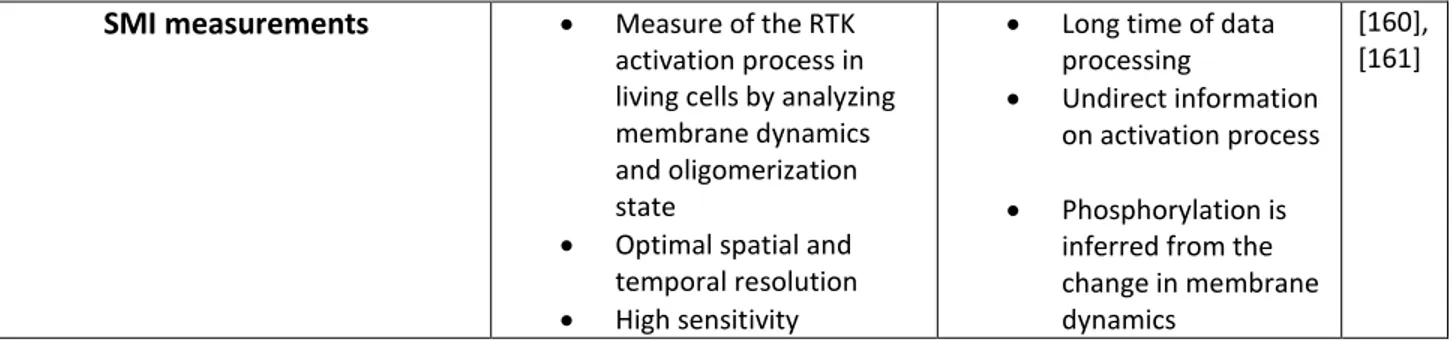
1.3 VEGFR2 Receptor structure and mechanism of activation ... 36 1.4 Experimental strategies to detect RTKs kinase activity: from biochemical to advanced imaging approaches ... 37
1.4.1 Single-molecule imaging and tracking of membrane receptors 40
2.
MOLECULAR INSIGHT ON THE ALTERED MEMBRANE TRAFFICKING OF TRKA KINASE DEAD MUTANTS48
5
2.2 valuation of membrane mobility and surface exposure of dead-kinase receptors ... 49
2.3 A structural rearrangement is responsible for TrkA-K547N membrane immobilization ... 54
2.4 TrkA-K547N membrane mobility depends on the integrity of cortical actin ... 60
2.5 Role of TrkA post-translational modifications on NGF-induced membrane-related dynamics and functions ... 62
2.6 Concluding remarks ... 66
3.
THE HSAN IV RELATED TRKA-R649W MUTANT: EVIDENCE FOR DELAYED DEGRADATION AND CYTOSOLIC ACCUMULATION OF AUTOPHAGOSOMAL VESICLES68
3.1 BACKGROUND AND RATIONALE OF THE WORK ... 683.2 Membrane and intracellular trafficking of TrkA-R643W in DRG neurons ... 69
3.3 Involvement of TrkA-R649W in the alteration of autophagic flux ... 73
3.4 Concluding remarks ... 75
4.
MATERIALS AND METHODS76
4.1 Constructs ... 764.2 Immortalized and primary cell cultures and transfection ... 77
4.3 Preparation of viral stocks for neurotrophin receptors ... 79
4.4 Transduction of immortalized and primary cells ... 79
4.5 Immunoblotting and Immunoprecipitation ... 80
4.6 TrkA detection by immunofluorescence ... 81
4.7 Drug treatments ... 82
4.8 FluoNGF binding assay ... 83
6
4.10 SFP synthase production and purification ... 84
4.11 Single molecule labeling and imaging of surface receptors ... 85
4.12 Single molecule internalization assay ... 86
4.13 Single step photobleaching assay ... 86
4.14 TIRF microscopy ... 87
4.15 SPT data analysis ... 88
4.16 Structural MD simulations ... 88
4.17 Statistical Analysis ... 89
APPENDIX AFLUOROLABELLING OF THE PPTASE-RELATED CHEMICAL TAGS: COMPARATIVE STUDY OF DIFFERENT MEMBRANE RECEPTORS AND DIFFERENT FLUOROPHORES IN THE LABELLING REACTIONS
91
A.1 Comparative Qdot- and fluorophore- labelling of different S6-tagged membrane receptors ... 93A.2 Comparative study of S6-TrkA labelling by different CoA-fluorophore substrates in the PPTase labelling reaction 96 APPENDIX B AN OPTIMIZED PROCEDURE TO SCALE UP SAMPLING IN SMI EXPERIMENTS
100
B.1 Schematic timeline for investigation of TrkA-wt and TrkA-K547N membrane dynamics in response to actin cytoskeleton alterations. ... 102B.2 Schematic timeline for investigation of p75NTR membrane dynamics in response to membrane cholesterol modulation. ... 103
7
L
IST OF ABBREVIATIONS
ACP: Acyl carrier protein A-loop, AL: Activation loop APE: Alanine-Proline-Glutammate AVs: Autophagosomal vesicles CD: Cytochalasin D
CIPA: Congenital Insensitivity to Pain and Anhidrosis CNS: Central Nervous System
CoA: Coenzyme A
CRD: cysteine rich domain
CSD: Cumulative Square Displacement C-tail: Carboxyl-tail
DAG: Diacylglycerol
DFG: Aspartate-Phenylalanine-Glycine DRG: Dorsal Root Ganglia
ECD: Extracellular domain EGF: Epidermal Growth factor
EGFR: Epidermal Growth factor receptor
EM-CCD: Electron-multiplying charge coupled device FGFR: Fibroblast growth factor receptor
FPs: Fluorescent proteins
FRAP: Fluorescence Recovery After Photobleaching FRET: Fluorescence Resonance Energy Transfer GAB-1: GRB2-associated binding protein 1
GBR2-SOS: Growth factor receptor-Bound protein-2- SOS G-loop: Glycine- rich loop
HRD: Histidine-Arginine-Aspartate
HSAN: Hereditary Sensory and Autonomic Neuropathy ICD: Intracellular domain
8 IGFR-1: Insulin-like Growth factor
INSR: Insulin receptor IP: Immunoprecipitation IP3: Inositol Triphosphate JK: Jasplakinolide
JM: Juxta-membrane domain KDR: Kinase Insert Domain receptor KID: Kinase insert domain
KIT: Tyrosine protein kinase KIT KM: Kinase mutant
LB: Latrunculin B
LRR: Leucine rich repetition
MAPK: Mitogen-activated protein kinase MDS: Molecular Dynamic Simulation MSD: Mean Square Displacement MSS: Moment Scaling Spectrum NGF: Nerve Growth factor
NTRK1: Neurotrophic Receptor Tyrosine Kinase 1 NTs: Neurotrophins
PAABD: Phospho-amino acid binding domain PI3K: Phosphoinoside 3-kinase
PIP2: Phosphatidylinositol 4,5-bisphosphate PKA: Protein kinase A
PLC-: Phospholipase C- PNS: Peripheral Nervous System
PPTase: Phosphopantetheinyl transferase PTB: Phosphotyrosine binding
PTMs: Post Translational Modifications RM: Recruitment mutant
RTKs: Receptor Tyrosine Kinases SH2: Src homology-2
SHC: SH2 containing protein
9 SPA: Scintillation Proximity Assay
SPT: Single Particle Tracking
TIRF: Total Internal Reflection Fluorescence TKD: Tyrosine Kinase Domain
TMD: Transmembrane domain
TNFRs: Tumor Necrosis Factor receptors TrkA: Tropomyosin related kinase A Ub: Ubiquitin
VAIK: Valine-Alanine-Isoleucine-Lysine
VEGFR2: Vascular Endothelial Growth factor receptor type 2 WB: Western blot
10
L
IST OF
P
UBLICATIONS
“Fluorolabelling of the PPTase-related chemical tags: comparative study of different membrane receptors and different fluorophores in the labelling reactions”. Amodeo R*, Convertino D, CalvelloM, Ceccarelli L, Bonsignore F, Ravelli C, Cattaneo A, Martini C, Luin S, Mitola S, Signore G, Marchetti L. Under revision on Frontiers Molecular Biosciences.
“Molecular insight on the altered membrane trafficking of tyrosine kinase dead receptors.” Amodeo R*, Nifosì
R, Giacomelli C, Ravelli C, La Rosa L, Callegari A, Trincavelli ML, Mitola S, Luin S, Marchetti L.Biochim Byophis Acta Mol Cell Res.2019. 1867(2):118614.
“Fast diffusing p75NTR monomers support apoptosis and growth cone collapse by neurotrophin ligands”.
Marchetti L*, Bonsignore F*, Gobbo F*, Amodeo R, Calvello M, Jacob A, Signore G, Schirripa Spagnolo C, Porciani D, Mainardi M, Beltram F, Luin S, Cattaneo A. Proc Natl Acad Sci U S A. 2019. 116(43):21563-21572.
“Site-Specific Direct Labeling of Neurotrophins and Their Receptors: From Biochemistry to Advanced
Imaging Applications.” Gobbo F*, Bonsignore F*, Amodeo R, Cattaneo A, Marchetti L. Methods in
Molecular Biology. 2018. 1727:295-314.
“Probing labelling-induced lysosome alterations in living cells by imaging-derived mean squared displacement analysis”. D’Urso W*, D’Autilia F, Amodeo R, Marchetti L, Cardarelli F. Biochemical and
Biophysical Research Communications (BBRC). 2018. 503(4):2704-2709.
11
I
NTRODUCTION
Human receptor tyrosine kinases (RTKs) are master regulators of the principal cell functions during embryonic development and adult homeostasis [1]. Their mechanism of action and structural organization are maintained between different species [2], highlighting the importance of a deeper understanding of the role of their catalytic core, the Tyrosine Kinase Domain (TKD). Mutations in the TKD are frequently related to the pathogenesis of cancer, type 2 diabetes, cardiovascular, neurodegenerative and developmental disorders [3]–[6]; for these reasons commonly employed drugs act through the modulation of RTKs activity and/or target specifically their ATP binding site [7], [2], [8]. However, little is known about the specific aberrant mechanisms brought by different mutations of RTKs in these diseases. In particular, while it is established that several hyper-activating oncogenic mutations cause disruption of auto-inhibitory interactions [9], [10] and alterations in membrane disposition, cytoskeletal organization and intracellular trafficking of RTKs leading to prolonged RTKs signaling [11], the role of inactivating mutations looks more heterogeneous. For example, an increased expression of Ryk, ROR and several ephrin (Eph) receptors was found associated with cell motility and metastasis in different cancers, despite the lack of kinase activity of these proteins [12], [13]; on the other hand, other inactivating mutations of RTKs lead to a loss-of-function responsible for neuronal disorders [14]. It appears clear that understanding the role that different mutations have in the onset of diseases represents a big turn on how to treat patients with these specific RTKs alterations [15].
My thesis is focused on the different impact that several mutations inside the TKD have on the structure, membrane distribution, subcellular trafficking and post-translational modifications (PTMs) of one member of the RTK family, the Tropomyosin-related receptor kinase A (TrkA), the high-affinity nerve growth factor receptor, essential in development and survival of selected neuron populations in the nervous system.
More in details, I found that the mutation of lysine 544 (numeration of the human sequence), crucial to allocate ATP and thus to kinase activity, is responsible for the altered membrane dynamics and distribution of TrkA
receptor in absence of Nerve Growth Factor (NGF) stimulation. Using a single particle tracking (SPT) approach I discovered that this mutation causes a restricted membrane mobility and an increase of cell surface pool, without affecting the membrane oligomerization state of the receptor. Molecular dynamics simulations (MSD) revealed that this mutation is predicted to drive a breakage of a salt bridge between Lys544 in the β3 sheet and the Glu560 in the αC helix of the N lobe, responsible for different arrangements of this helix with the C lobe; this conformational change probably leads to new interactions of the mutant with the cortical actin cytoskeleton, causing the increase of TrkA membrane pool and its slow dynamics. On the other side, other mutations for TrkA
12
catalytic, scaffolding and ubiquitination activities revealed no alterations in membrane dynamics in resting conditions; however, none of these mutants, despite being able to bind the ligand, immobilizes its membrane pool after NGF treatment, thus allowing us to conclude that not only phosphorylation, but also ubiquitination are fundamental for NGF-dependent membrane immobilization of TrkA at the plasma membrane.
In parallel, during my thesis I also found that the TrkA-R649W mutation (corresponding to the human R643W residue), positioned in the HRD (Histidine-Arginine-Aspartate) domain of the catalytic loop and responsible for the onset of Hereditary Sensory and Autonomic Neuropathy type IV (HSAN IV) disease, is characterized by an altered membrane distribution of receptors in both neuroblastoma and Dorsal Root Ganglia (DRG) neurons in an actin-dependent manner. Furthermore, despite its absence of kinase activity, TrkA-R649W is rapidly internalized in response to ligand stimulation and probably not recycled as its wt counterpart. Finally, the increased number of autophagosomal vescicles (AVs) found in neuroblastoma cells overexpressing the mutant, can candidate the alteration of the autophagic flux as a possible molecular effect characterizing the neuropathy.
My thesis is organized in four chapters plus two appendixes; in Chapter 1 I introduce the structural organization, mechanisms of action and auto-inhibition of RTKs and of a subclass of kinase receptors, able to signal also with an impaired kinase activity. The second part of the chapter is focused on the structure, dynamics, activation and PTMs of TrkA receptor. Finally, in the third part I describe biochemical and biophysical methods adopted to evaluate the RTKs activation, giving particular importance to the state of the art of advanced imaging techniques studies applied to RTKs.
Chapter 2 deals about the evaluation of membrane dynamics and PTMs in response to different mutations inside
the TKD of TrkA receptor. First, I described the membrane dynamics and exposition of a dead-kinase mutant, TrkA-K544N, analyzing possible causes for its changes in membrane dynamics also exploiting the results of computational analysis, using Molecular Dynamic Simulation (MDS). I then tested the interactions of cytoskeleton with TrkA-K544N, analyzing its changes in dynamics and membrane exposition in response to pharmacological treatments. Finally, I moved on with the evaluation of the dynamics, membrane exposition and PTMs of mutants for TrkA kinase, recruitment and ubiquitination activity.
In Chapter 3 I collect the characterization of the membrane distribution, intracellular trafficking, signaling and protein turn-over of the TrkA-R649W, a kinase inactive mutant found associated with HSAN IV congenital neuropathy. I evaluated changes in dynamics of TrkA-R649W in response to cytoskeleton alterations, such as the internalization of this receptor in response to NGF treatment. Finally, I also measured the number of AVs shown by cell overexpressing this mutant in resting condition, to evaluate possible involvement of the disease with dysfunction of autophagic flux.
13
In Chapter 4 I describe into details the methods and materials used for biochemical experiments, the methods adopted to perform MDS, the microscopy set-up used for our imaging experiments and the relative adopted data analysis.
Finally, in Appendix A I report a methodological work that allowed me to optimize the phosphopanteithenyl transferases labelling method used to fluorolabel molecules for single molecule imaging. In details, I compared the labelled cell fraction for two types of labelling strategies on TrkA and VEGFR2 receptors, belonging to the RTKs family and P75NTR, member of the Tumor Necrosis Factor receptor (TNFR). I also evaluated the
physico-chemical properties of different fluorophores used in this labelling reactions and their suitability for high-end microscopy techniques such as Total Internal Reflection Fluorescence (TIRF) microscopy, with particular respect to signal-to-background ratio and fluorophore aspecific adhesion to or internalization in cells.
Appendix B presents a methodological scheme of an experimental timeline that I used during the preparation of
samples to be imaged at the TIRF microscope during my thesis work. This allowed me to maximize the number of samples that can be collected in one-day-work, so as to ensure an optimal sampling and reproducibility in Single Molecule Imaging (SMI) experiments.
14
1. T
RK
A
AS A PARADIGM OF
H
UMAN TYROSINE KINASE
RECEPTORS
1.1
T
YROSINE
K
INASE
R
ECEPTORS
Human tyrosine kinase receptors constitute a large family of 58 single-pass transmembrane proteins, divided in 20 subfamilies, which through their intrinsic enzymatic activities are able to catalyze the phosphorylation of tyrosine residues of target proteins using ATP as a phosphate donor. RTKs are known to be fundamental components of cellular signal transduction, regulating the main cellular functions as survival, proliferation, differentiation, cell-cell communications and metabolism [1]. Moreover their structural organization, mechanism of action and interaction with specific intracellular effectors are highly conserved from Caenorhabtitis Elegans to humans [2], highlighting the importance of understanding the biological role of each conserved protein domain of the TKD [2]. It is now well known that RTKs transduce signals coming from the extracellular environment to the interior of the cell, starting kinase-dependent signaling cascades leading to different and specific cellular responses. However, the first fundamental contribution to the RTKs knowledge dates back to the 1950 when Rita Levi Montalcini and Stanley Cohen discovered the first soluble growth factor, called Nerve Growth Factor , for its ability to induce neurites outgrowth when administered to cells [16]. Only few years later Cohen discovered another growth factor, the Epidermal Growth Factor (EGF), but the mechanisms underlying the cellular outputs induced by these molecules remained unknown until 1978, when he demonstrated the existence of the first RTK named EGF receptor (EGFR). EGFR was discovered as a membrane protein that resulted to increase its phosphorylation status after the binding with EGF [17]. The current concept of RTKs occurred around the 80s with the discovery of kinases and phosphatases roles, which are enzymes able to reversibly phosphorylate and de-phosphorylate proteins, respectively, modifying their functions thereby regulating their activities [17], [18]. Just for the important role played by these enzymes in regulating different cellular responses, aberrations in the activation of RTKs leads to the onset of a wide range of diseases including cancer, type 2 diabetes, cardiovascular and neurodegenerative diseases [19]. About these, the most famous example is probably the EGFR, that was the first RTK discovered to be linked to human cancer [20], leading the way to the development of drugs with important roles in cancer therapy through the modulation of RTKs activity [2], [8].
15
1.1.1 Structural architecture of RTKs
The structural architecture of RTKs is maintained among the members of this family and reflects their functional organization composed by three main domains (Fig. 1.1):
i) the extracellular domain (ECD) is the portion exposed to the external side of the plasma membrane, responsible for ligand binding. The ECD represents the most variable portion among the RTKs, generally divided in different sub-domains like immunoglobulin (Ig-) like domains, cysteine-rich domains, fibronectin type III-domains or EGF-like domains [21].
ii) The transmembrane domain (TMD) is the single-pass -helix that connects the ECD with the intracellular domain of the receptor, contributing to the stability of dimers and sometimes playing also an active role in signaling [22].
iii) The intracellular domain (ICD) has a structural organization conserved among the RTKs, composed by the juxta-membrane region (JM), the catalytic TKD and the carboxy (C-) terminal. The TKD as well as the JM and C-term regions typically contain Tyr residues that are auto-phosphorylated upon ligand binding to the receptor [21].
In the absence of stimulation, the majority of RTKs exists as a single polypeptide chain organized in a monomeric form, and the ligand binding induces dimerization and receptor activation [23]. However Met, INSR-1 and insulin receptors are exceptions: Met receptor is a heterodimer composed by an extracellular short -chain connected by a disulfide bond to a membrane spanning -chain, while insulin receptor and IGFR-1 are expressed at plasma membrane already in form of disulfide linked-(2 heterodimers (Fig. 1.1) [1], [24]. Furthermore, there are
several studies reporting also that EGF molecules activates EGFR through binding to pre-existing oligomers, and also that Tie2 and Eph receptors require the formation of larger oligomers for their activation [25]–[28]. In any case, ligand binding is always necessary for RTKs activation.
16
Figure 1.1. Schematic representation of the 20 subfamilies of human tyrosine kinase receptors; different ECD (legend in box on the bottom
of the figure) are identified by structure determination or sequence analysis, ICD are shown as red rectangles. L: leucine, Ig: immunoglobulin, SAM: sterile alpha motif, PSI: plexin-semaphorin-integrin, WIF: Wnt inhibitory factor, FZ: Frizzled domain, LDLa: low-density lipoprotein receptor class A, YWTD: Tyr-Trp-Thr-Asp, SEMA: structural domain of semaphoring, Mam: mephrin/A5-protein/PTP mu. Taken from [2].
1.1.2 The Tyrosine kinase domain
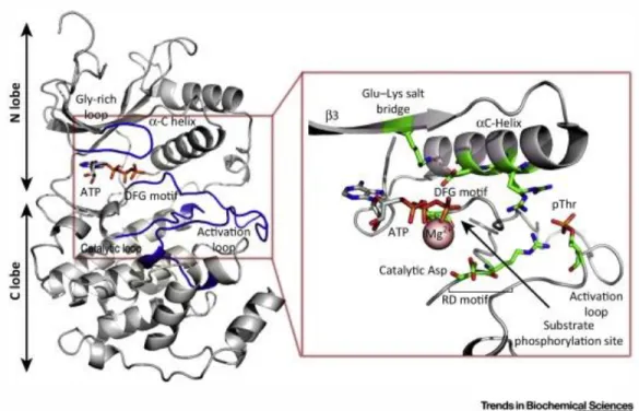
The TKD is an amino-acid domain approximately 300-500 aa long, responsible for the catalysis of the transfer of the -phosphate from ATP to the hydroxy groups of Tyr residues on the target protein. From a structural point of view, the TKD is organized in two lobes, the N-terminal small lobe (N-lobe) and the C-terminal large lobe (C-lobe), connected by the kinase insert domain (KID) characterized by a variable length, spanning from few to up to 100 amino acid (Fig. 1.2) [29]. The N-lobe, structured in antiparallel β-sheets and one single α-helix denoted as αC helix, has the important role of binding and stabilizing the ATP complexed with Mg2+ ions [30]. The C lobe
is mainly composed by -helices and loops [31], and enhances the chelation of Mg 2+ ions with ATP, binds the
intracellular protein to be phosphorylated and finally transfers a phosphate group from ATP to the Tyr residues [21]. Between the N-lobe and the C-lobe resides the substrate cleft, where ATP, divalent cations, and peptide
17
substrate are bound. More in details, the TKD is characterized by the following defined and conserved structural motifs (Fig. 1.2):
Figure 1.2. Representation of functional elements in the TKD of the active p38/ (PDB 3PY3). The active site, which is zoomed, is located
between the N-lobe and the C-lobe, with the ATP-binding domain positioned in the cleft between these two lobes. During the receptor activation, the phosphorylation of residues in the Activation loop (A-loop) promotes the interaction with an Arg located in the catalytic loop and with the N-terminal residues of the C helix, inducing the alignment of the DFG motif and Glu–Lys salt bridge. Image taken from [32].
i) αC-helix positioned in the N-lobe, containing a glutamate, whose residue is fundamental for ATP binding and catalytic activity, because it is involved, in the active configuration of protein kinases, in the formation of a conserved salt bridge with a residue of lysine (Lys) positioned in the 3 sheet [31]. This Lys is also involved in the
interaction with the α- and β-phosphates of the bound Mg-ATP, necessary for its positioning for the phosphotransfer. In inactive kinases, this specific C-helix can assume non-canonical conformations [33] and
mutations at the β3 Lys are typically related to ‘kinase dead’ variants of canonical kinases [34].
ii) Glycine-rich loop (G-loop or P-loop), containing the consensus sequence GxGxxG between strands β1 and β2 and associating closely with the phosphate groups of bound ATP through backbone interactions [34].
iii) Activation loop (A-loop, AL), a region with high flexibility characterized by different phosphorylation sites that lead to efficient receptor catalytic activity [35]. Its specific phosphorylation and orientation regulates the switch between inactive and active configuration of TKD and is responsible of substrate binding [30]. This motif is usually 20-30 amino acids long [36] and is limited by the DFG motif at the N-term and the APE (Ala-Pro-Glu) motif at the C-term.
18
iv) DFG motif present in the first part of the A-loop, and composed by aspartate-phenylanine-glycine triplet; it has an important role in the binding of the phosphates of ATP. The Asp residue critically coordinates divalent cations within the A-loop, and is one of the most important, conserved residues in TKD [34].
v) Catalytic loop, a conserved motif positioned between 6 and 7 sheets, containing the HRD domain formed by a sequence of histidine-arginine-aspartate residues; the aspartate residue therein (7 Asp) is thought to function as a catalytic base and/or to correctly orients the hydroxyl group of the Tyr to be phosphorylated [32], [34].
vi) APE motif is a conserved alanine-proline-glutammate motif involved in the anchoring of the A-loop with the kinase domain core and in substrate binding.
Generally, after receptor-activation-driven protein phosphorylation, the AL interacts with the positive residues of the C-helix and of the catalytic loop. This process induces conformational changes of the DFG motif and of the C-helix; in particular the rotation of the C-helix promotes the formation of the Glu-Lys salt bridge and the start of catalytic activity (Fig. 1.3) [32].
Previous studies made on the active and inactive IRK and Protein Kinase A (PKA) structures obtained from crystal X-ray studies revealed the existence of an open conformation referred to as the inactive receptor configuration, and a close one as the active counterpart (Fig. 1.3) [37] (Hubbard, 1997; Hubbard et al., 1994). It was reported that one of the mechanisms regulating the switch between active and inactive forms of RTKs depends on the specific orientation of the C helix towards the C-lobe and in particular the A-loop [36]. A specific auto-inhibitory conformation between the C helix and the A-loop ensures the receptor inactivation in absence of stimulation; on the contrary, ligand binding induces a conformational switch that allows the ATP allocation in its binding site and the beginning of the catalysis.
19
Figure 1.3. Upper panel: ribbon representation of the crystal structure of PKA and IRK. Here the functional structures and residues of TKD
are highlighted: A-loop: red, C-helix: purple, P loop: orange, catalytic loop: green, a Protein Kinase inhibitor (PKI), in yellow. On the right, the switch between inactive and active IRK: in inactive state the conformation of the A-loop blocks the binding of nucleotides. Below: Representation of conformational changes of TKD in response to RTKs activation with particular emphasis on the disposition of the C-helix (purple cylinder) and A-loop (in orange); the blue segment represents the catalytic Lys of the salt bridge. Image taken from [37].
1.1.3 Mechanisms of activation and autoinhibition of RTKs
Generally activation of RTKs occurs after ligand binding at the ECD of the protein, leading to receptor homodimerization and subsequent juxtaposition of cytoplasmic TKDs of each monomer [2]. In most cases this juxtaposition enhances auto-phosphorylation in trans of Tyr located in the A-loop or in the JM region, releasing the cis-autoinhibition configuration and inducing the conformational changes necessary to stabilize the active state of the kinase [2], [38]. The phosphorylation cascade events promote the phosphorylation of Tyr residues that recruit down-stream signaling proteins, typically through Src homology-2 (SH2) or phosphotyrosine-binding (PTB) domains, which specifically bind phosphotyrosines thanks to their specific sequence contexts (Fig. 1.4) [21], [39].
20
Figure 1.4. Mechanism of RTKs activation after ligand binding. RTKs reside at plasma membrane mainly as monomers; after ligand
binding RTK dimerization promotes the juxtaposition of the ICD of the two monomers necessary for the trans-phosphorylation of Tyr (Y) (phosphate groups are indicated with orange circles) in the A-loop; this process induces the trans-phosphorylation of Tyr positioned in the kinase insert, JM and C-tail regions, that serves as a docking site for adaptor proteins (B) or phosphorylate signaling molecules (A) responsible of the recruitment of intracellular effectors. Image taken from [40].
However, the dimerization process can occur with different mechanisms (Fig. 1.5):
i) ligand-mediated dimerization: crystal structure analysis on the ECD of several RTKs demonstrated that the binding of a bivalent ligand with two monomers of receptors induces the formation of a dimeric complex as observed for TrkA [41], Flt1, vascular endothelial growth factor (VEGFR) receptor [42], Tie2 [25] and Ephrin receptors [26], without direct contact between the extracellular regions of the two receptors (Fig. 1.5 A). ii) Ligand-mediated and receptor-mediated dimerization: in other cases, as for KIT receptor, its ligand (stem cell factor, SCF) binds only one monomer of receptor at the first three or five Ig-like domains of the ECD [43], and this is sufficient to induce the cross-linking of two receptor molecules. Indeed, this event promotes a reorientation of D4-D5 domains closest to the plasma membrane, that enhances the interaction across interfaces of KIT dimer, necessary for receptor activation [44] (Fig. 1.5 B).
21
iii) For Fibroblast growth factor receptor, FGFR, the activation requires not only the binding of a bivalent ligand, but also the involvement of an accessory molecules, as heparin [45] (Fig. 1.5 C).
iv) Receptor-mediated dimerization: for EGFR, the activation is completely receptor-mediated, without any physical interaction between two activating ligands; here the bivalent ligand binds only a single monomer of EGFR, promoting substantial conformational changes in its ECD, which becomes able to unlock the mechanism of receptor auto-inhibition [46] (Fig. 1.5 D).
Figure 1.5. Different dimerization process of RTKs. A) A NGF dimer (red) crosslinks two TrkA molecules without direct contact between the two receptors. B) A SCF dimer (red) crosslinks two KIT molecules and two Ig-like
domains (D4 and D5), reoriented upon receptor activation, interact across the dimer interface. C) Two FGFR molecules come into contact through the Ig-like domain D2, where it binds also the heparin or heparin sulfate proteoglycans; in addition, each FGF molecule (red) contacts Ig-like domains D2 and D3 in both FGFR molecules. D) Dimerization of EGFR receptors is completely mediated by the receptor. Through its binding simultaneously to two sites (DI and DIII) within the same receptor molecule, the ligand drives conformational changes in the receptor, which expose a previously occluded dimerization site in domain II, which allows the dimerization with the other receptor molecule. Image adapted from [2].
Despite the great improvements in knowledge of RTKs gained during the last years, the mechanisms by which changes in the ECD following ligand-binding could be responsible of conformational rearrangements in the ICD, leading to receptor activation, represent a field still widely debated. The general mechanism of activation is shared among different members of RTKs family and depends on the specific modification of key regulatory elements located inside the TKD (see Paragraph 1.1.2), necessary for the phosphotransfer of a γ-phosphate from ATP onto the hydroxyl group of a Tyr residue, necessary for the receptor catalysis [2], [36]. In most cases the auto-phosphorylation process occurs in trans and the sites are phosphorylated with a specific order; the first phase of auto-phosphorylation is necessary for the enhancement of the catalytic activity after receptor activation, while during the second phase occurs the phosphorylation of Tyr residues that recruit downstream intracellular effectors. Recently it was discovered also a third phase of activation, consisting in trans-phosphorylation events that maximize the ability of kinase to phosphorylate its targets.Although the activation process is conserved among RTKs, they display an array of different mechanisms of cis-auto-inhibition, necessary to avoid aberrant
22
receptor activation in absence of stimuli; in these cases the TKD is maintained in an inactive or auto-inhibited conformation until it is activated by extracellular ligand-induced oligomerization [23], [38]. It was reported that a lot of TKD activating cancer-related mutations cause the disruption of these auto-inhibitory mechanisms and, at least for KIT and EGFR, lead to receptor hyper-activation [2], [47]. This makes clear that understanding the regulation of RTKs auto-inhibitory system represents an important field in cancer-research. Several intramolecular interactions are involved in the maintenance of the cis-autoinhibition mechanisms (Fig. 1.6):
Figure 1.6. Schematic representation of autoihibition mechanisms of RTK. The
C-lobe is in light purple, the N-C-lobe in dark purple in inactive state or yellow in active states and the A-loop in purple or yellow in the inactive and active states, respectively. Insulin receptor-like: the A-loop interacts directly with the active site and blocks access to ATP and protein substrates. Phosphorylation of Tyr residues disrupts these autoinhibitory interactions and allows the kinase to switch to the active state. KIT receptor-like: the JM region (red) interacts with elements within the active site (including the αC helix and the A-loop) to stabilize an inactive conformation. Phosphorylation of key Tyr in the JM region destabilizes these interactions and allows the TKD to assume an active conformation. Tie-2 receptor like: the C-term tail (red) interacts with the active site of the TKD to stabilize an inactive conformation. Image adapted from [2].
i) A-loop inhibition: Insulin receptor is characterized by a cis-auto-inhibition mechanism regulated by the A-loop; a specific relocation of the Tyr Y1162 inside the TKD stabilizes the configuration of A-loop that blocks the active site. Only after ligand binding, the Y1162 residue is trans-phosphorylated, allowing the switch to the active state, while the -helix in the N-lobe changes its orientation, enhancing ATP binding [37]. In the case of FGFR, different Tyr residues, located inside the A-loop, are involved in the auto-inhibitory mechanism though specific interactions that cis-auto-inhibit the TKD, interfering with the protein-substrate binding but not with the ATP-binding [48]. Also in this case, the auto-inhibition mechanism is released after receptor dimerization, thanks to the reorientation of the A-loop and the C-helix.
ii) JM inhibition: MuSK, Flt3, Eph family and KIT receptors auto-inhibition is managed at JM level [49]– [51]. Tyr residues located in the JM region interact with the A-loop in the TKD, ensuring the maintaining of an auto-inhibited conformation. The trans-phosphorylation of Tyr following receptors dimerization destroys the cis-auto-inhibition mechanism, promoting receptor activation.
iii) C-terminal tail inhibition: Tie-2 activation is regulated by a cis-auto-inhibition mechanism occurring at the C-terminal tail. Here, Tyr residues acting as auto-phosphorylation sites block the access of the
23
substrate at the active site [52]; the phosphorylation of residues in the C-tail promotes the receptor activation.
For some RTKs, as Trk receptors, the mechanism of auto-inhibition is still not accepted, considering that it seems to be different also between TrkA and TrkB, despite they share 88% identity in the TKD [53]. The work of Miranda et al. has suggested the existence of an exclusive auto-inhibition mechanism for TrkA receptor. They demonstrated that after introduction of activating mutations in conserved residues, TrkA showed different responses with respect to both Met and Kit receptors [54].
1.1.4 The family of pseudokinases
Pseudokinases are a subfamily of RTKs characterized by the lack of amino acids that are fundamental for allocating the ATP and metal ions necessary to start the catalysis [34]. Despite their impairment or absence of kinase activity, some pseudokinases have a role as signal transducers both in physiological and in pathological conditions [55]–[57]. The mechanism underpinning this alternative activation is still debated, but two different hypothesis are accepted: pseudokinases could have a residual kinase activity sufficient to catalyze the phosphotransfer, or alternatively they can work as scaffolding proteins for downstream signalling, participating in the formation of multi-protein complexes [58], [59]. Manning et al. investigated human RTKs using a combination of EST (Expressed Sequence Tag) and cDNA data, Genewise homology modeling and Genscan ab initio gene prediction: they confirmed that five are characterized by inactive ICD (ErbB3, PTK7/CCK4, EphB6, EphA10 and SuRTK106), and showed that other three (Ror1, Ror2 and Ryk) result to be inactive, despite being previously predicted to be active [56]. One of the most studied examples of pseudokinases is HER3, a member of the EGFR family, which, despite being inactive, is able to stimulate the autophosphorylation and activation of HER2 receptor in response to neuregulin [60], [61]. Furthermore, in the ephrin family EphB6 and EphA10 are predicted to be pseudokinases: in particular EphB6, although its lack of kinase activity, is able to enhance the activation of another kinase protein, ZAP-70, after ephrin binding [62].
The most common mutations displayed by pseudokinases are located in different key conserved motifs of the TKD [34] (Fig. 1.7):
i) Glycine-rich loop (see Paragraph 1.1.2): mutations at the first two glycine residues is reported to alter the ATP binding site; these mutations are found in PTK7/CCK4, Ror1, Ror2, Ryk and SuRTK106. ii) VAIK (Val-Ala-IIe-Lys) motif: in the active conformation of RTKs there is a conserved salt bridge
between a Lys positioned in 3 strand and a Glu residue in C helix; the Lys coordinates also the - and - phosphates of the bound Mg2+-ATP to allow the correct phosphotransfer. Mutations in 3 Lys
24
iii) HRD motif (see Paragraph 1.1.2): the Asp residue, responsible for the correct orientation of the hydroxyl group of the residue to be phosphorylated, is substituted with an Asn in the HRD of HER3, while it is replaced with serine or glycine respectively in the case of Ephb6 and EphA10.
iv) 7-Asn: this is a fundamental residue for the correct orientation of the HRD domain, and is mutated to Ser and His in EphB6 and EphA10, respectively.
v) DRG motif (see Paragraph 1.1.2): the aspartate of this motif, that has the important role to coordinate divalent cations, is lost in PTK7/CCK4, EphB6, EphA10 and SuRTK 106 receptors.
There are also some cases in which the lack of some of these domains saves the catalytic activity of the protein, as for WNK proteins: 4 isoforms of WNK lack the lysine residue of the VAIK domain but despite this they are found catalytically active thanks to the presence of another Lys residue that substitutes the missing one [58], [63].
Figure 1.7. Alignment of sequence of the TKD of 8 pseudokinases below the sequence of PKA, used as reference for active RTKs. Secondary
elements are reported on the sequence alignment, conserved residues are shaded in gray and mutated residues in pseudokinases are circled in black. Image taken from [34].
1.2
T
RK
A
RECEPTOR AND THE
N
EUROTROPHIN SIGNALING NETWORK
In 1950 Rita Levi-Montalcini discovered that transplanting a mouse sarcoma into a chicken embryo promoted the secretion into the blood of a factor, lately named NGF, responsible for the sensory and sympathetic nerve growth [64]. This observation opened the way to the complex and charming world of neurotrophins (NTs). Years of research have allowed us to know that these growth factors are master regulators of neuronal development, survival and plasticity in the nervous system (Fig 1.8) [65]–[68]. In mammals, the NTs family is composed by four members: NGF, brain-derived neurotrophic factor (BDNF), neurotrophin-3 (NT-3), and neurotrophin-4 (NT-4). NTs exert their action mainly though the binding to two classes of receptors exposed at the neuronal cell surface:
25
the Trk family and the P75NTR receptors. More into details P75NTR, belonging to the tumor necrosis factor family,
can be recognized and activated by all components of the neurotrophin family [69]. On the contrary, NTs display a precise binding specificity for the three components of the Trk family, leading to different cellular outputs: TrkA binds with high affinity to NGF and with low one to NT-3, and regulates survival, cell growth and differentiation at PNS level; TrkB binds BDNF, NT-3 and NT-4 and regulates survival, plasticity and apoptosis at CNS and finally the NT-3 binding to TrkC is responsible for survival and growth of sensory neurons.
My thesis is focused on the TrkA receptor, one of the most important player in development, survival, differentiation and neuronal plasticity of the mammalian PNS [70, 71] and of the cholinergic system in the CNS [72]. TrkA protein is widely expressed on the cell surface of sympathetic, trigeminal and dorsal root ganglia and in cholinergic neurons of the basal forebrain and striatum [73], [74], where it exerts its functions through the binding to the NGF produced by target tissues. A correct level of NGF is indeed fundamental for the maintenance of the homeostasis of the nervous system: increased NGF-mediated activity or alteration of signaling by NGF/TrkA complexes can be associated to inflammatory and neuropathic pain disorders [75] and to neurodegeneration [76], [77]. Approaches such as NGF gene and protein therapies, or administration of small molecules acting as TrkA agonists, are actually on trial for the treatment of neurodegenerative disorders such as Alzheimer’s disease; on the contrary the administration of drugs that inhibit the action of NGF/TrkA complexes are on trial for the treatment of pain disease. Only one of these approaches, however, is currently FDA approved [78].
However, despite TrkA receptor is mainly known as the high affinity receptor for NGF [71], it was firstly discovered in 1982 as an oncogene expressed in colon cancer [79]. In the last years, both TrkA and NGF have been found expressed in several malignant tumors. Indeed the activation of ERK, SRC and AKT pathways, all mediated by TrkA, regulates tumor cell proliferation and spreading in breast cancer [80]; an analogue involvement of TrkA-related pathways are also found in gastric [81] and pancreatic cancers [82]. Despite this, the potential role of TrkA mutations in promoting tumorigenesis and cancer has not yet been established [83]. TrkA receptor, as TrkB and TrkC, are lately considered important therapeutic targets in cancer treatment; accordingly, several clinical trials based on TrkA kinase activity inhibitors are currently in development [83].
26
Figure 1.8. Timeline of the discoveries related to the biology and therapeutic targeting of Trk signaling. Boxes above the timeline arrows
represent the milestone discoveries relevant to normal Trk pathway biology, while boxes below are NTRK (Neurotrophic Tropomyosin- Related -or Tyrosine Receptor- Kinase) fusions found in cancer. NTRK are the genes encoding for human Trk proteins (NTRK1, 2, 3 for TrkA, B, C, respectively). Image taken by [83].
1.2.1 TrkA structure and signaling
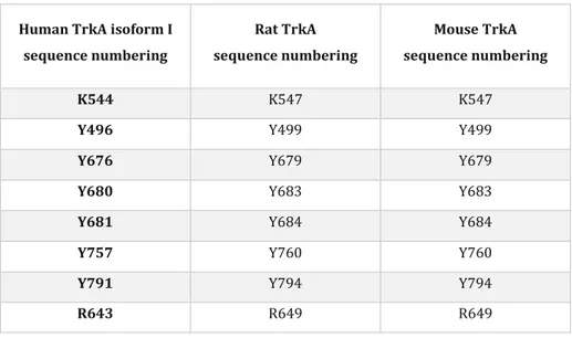
In human, the gene encoding for the TrkA protein is Neurotrophic Tyrosine Kinase receptor 1, ntrk1, located on the chromosome 1 q21-q22 [84]. The length of the gene is 23 Kb, divided into 17 exons and 16 introns; the gene presents an high homology of sequence with the Rattus Norvegicus and Mus Musculus species (86% identity in the entire protein sequence, 94.6 % identity in the TKD; see also Table 1.1). Three isoforms of TrkA protein have been identified [85]: isoform I is the most abundant form, expressed mostly in non-neuronal tissues, consisting in 790 aa and activated by both NGF and NT-3; isoform II is expressed in neuronal tissues, composed by 796 aa and activated only by NGF and finally isoform III, resulting from the splicing of exons 6, 7 and 9, is mainly present in pluripotent neuronal stem and neuronal crest progenitors and constitutively active in a ligand independent manner [86]. As aforementioned, TrkA isoform II generates a protein of 796 amino acid, with a weight of 87497 Da. The mature protein goes from amino acid 33 to amino acid 796 because the first 32 amino acid constitute the signal peptide (SP), necessary for the correct translocation of the protein in the ER; here the SP is cleaved, the protein is N-glycosylated and finally translocated at the plasma membrane. This glycosylation process can produce two alternative forms of TrkA with molecular weight of 110 KDa and 140 KDa [87]; only the latter is finally translocated at plasma membrane.
27
Human TrkA isoform I sequence numbering Rat TrkA sequence numbering Mouse TrkA sequence numbering K544 K547 K547
Y496 Y499 Y499
Y676 Y679 Y679
Y680 Y683 Y683
Y681 Y684 Y684
Y757 Y760 Y760
Y791 Y794 Y794
R643 R649 R649
Table 1.1. List of corresponding TrkA residues mentioned in the human, rat and mouse sequences. For our studies we used mainly
constructs of the rat TrkA sequence. However, some of the experiments and the molecular dynamics simulations used the human TrkA sequence. When needed, we shall use hTrkA to specify the human sequence. The two sequences (rat and human) share a very high homology.
As the majority of RTKs, TrkA receptor is organized in ECD, TMD and ICD.
The ECD constitutes the N-term of the protein, has a length of 391 amino acid and is organized in two IgL-like domains, IgL-1 and IgL-2, two CRD (CRD1 and CRD2); between CRDs there is a repetition of three 24 residues leucine-rich motifs (LRR 1-3). TrkA receptor interacts with its ligands mainly through the IgL-2 domain, but other regions are also reported to be involved in NGF binding: Arevalo et al. demonstrated that the mutation of a Cys residue in the IgL-1 domain is able to abolish NGF binding [88].
The TMD is composed by a single hydrophobic transmembrane -helix composed by 16 residues that structurally connects the ECD with the ICD of the receptor; this region plays important roles in receptor internalization and recycling [89] and in the dynamic equilibrium between pre-formed inactive dimers and NGF-induced dimer form [90].
TrkA ICD, composed by 357 aa residues, is divided like the other RTKs into JM, TKD and C-tail, with precise functional roles explained in the following; in particular this region contains 11 Tyr residues, six of which can be phosphorylated [91].
The JM region plays important roles in neurite outgrowth and differentiation [92]. Here, the Y496 residue ensures the activation of phosphoinoside-3-kinase (PI3K) signaling pathway, which starts with the conversion of Phosphatidylinositol 4,5-bisphosphate (PIP2) in Phosphatidylinositol 4,5-triphosphate (PIP3) at membrane level, and ended with the activation of Akt. Y496 is fundamental also to amplify the signaling pathway mediated by
28
Shc-binding proteins: when Shc binds the phosphorylated Y496 residue with its PTB domain, its SH2 domain is free to interact with other intracellular effectors, leading to signaling amplification. More into details, when phosphorylated, the Y496 residue can bind the adaptor proteins GAB1 (GRB2-Associated Binding Protein-1) and SHC (SH2 Containing Protein), enhancing their association with GBR2-SOS (Growth Factor Receptor-Bound
Protein-2-SOS), responsible of the GDP-GTP switch on Ras protein. The activated Ras binds Raf, which
phosphorylates and activates MEK (MAP/ERK kinase), inducing the activation of MAPKs-ERK1/2
(Mitogen-Activated Proteins-Extracellular signal-regulated Kinase), with the final effect of activating transcription factors
CREB and c-Fos, responsible of neurite growth regulation.
The structural organization of TrkA TKD resumes the same previously reported for all RTKs (see Paragraph 1.1.2), where the presence of conserved motifs ensures ATP binding, catalytic function and substrate recognition. There are three Tyr in the A-loop that are fundamental for the phospho-transfer reaction; these residues are always phosphorylated in the same order: Y680, Y676, Y681; the same was reported for IGF-1 receptor, where the corresponding Tyr involved are Y1162, Y1158 and Y1163 [93]. The Y757 residue is also an important regulatory element involved in neuronal survival, neurite outgrowth and differentiation, through the activation of PI3K pathway.
Finally, the C-term tail is characterized by the presence of Tyr Y791, fundamental for neurite outgrowth and differentiation. Phosphorylated Y791 is the binding site for PLC-; PLC-signaling starts from the conversion of PIP2 to dyacilglycerol (DAG) and inositol triphosphate (IP3), promoting the release of Ca2+. The C-tail also contains
the PPXY motif, where a proline residue (P791 in rat sequence corresponding to P788 in human sequence) is located, fundamental for TrkA ubiquitination mediated by the ubiquitin ligases E3 Nedd 4-2 [94].
29
Figure 1.9. Signal-transduction pathways induced by the binding of NGF to TrkA receptor. After NGF-binding, the ICD leads to TrkA
auto-phosphorylation and activation of signaling cascades. Proteins that interact directly with the ICD are SHC, SOS, SH2B and IAPs, some of which are shown here. Ligand binding can also trigger the RAS signaling pathway, leading to survival and differentiation, and an alternative survival-signaling pathway through PI3K. Image taken from [95].
1.2.2 TrkA mechanism of activation
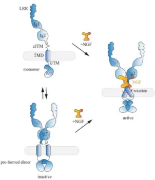
The mechanism of activation of TrkA receptor is poorly understood with respect to other components of RTKs family [96]. As mentioned in paragraph 1.2.3, the study of the crystal structure of TrkA complexed with NGF [41] corroborated, during last years, the theory that TrkA activation is ligand mediated, with NGF promoting TrkA dimerization and trans-phosphorylation in the TKD [70], [97]. Lately several works demonstrated that TrkA, expressed in different cell lines and in absence of ligand stimulation, was present as pre-formed inactive dimers in equilibrium with the monomeric population [98], [99]. This equilibrium between different oligomeric states can be explained by the rotational model of Maruyama, who proposed the existence of a mechanism of conformational switch between inactive and active TrkA dimeric states, occurring at TM and JM domains [91]; moreover the same mechanism was found also in EGFR/HER, VEGFRs and FGFRs [96], [100]. It was recently discovered the existence of two different motifs, L424xxF427A428xxF431 and S419xxxG423, located on two opposite
sites of TrkA TMD and probably responsible for the co-existence of active and inactive dimers (Fig 1.10), [90]. This work reported that in case of receptor overexpression and in absence of ligand, TrkA can be organized in
30
pre-formed inactive dimers thanks to the L424xxF427A428xxF431 motif. Instead, administration of NGF has the effect
to stabilize the active dimer conformation, allowing kinase domain activation [90]. More into details, NGF binding places the JM region in a specific configuration that promotes the rotation of the transmembrane helix from one to the other interface of the TMD [90]. In this way NGF binding induces the release of the auto-inhibition mechanism occurring at the ICD, ensuring the correct allocation of ATP, the cis-auto phosphorylation of Tyr residues of the A-loop and the trans-phosphorylation of additional Tyr residues acting as docking sites for intracellular adapter proteins, as PLC- and Shc.
Figure 1.10. Model proposed for TrkA activation. In absence of NGF, TrkA is present at the plasma membrane in equilibrium between
inactive monomer (upper panel) and pre-formed inactive dimer (lower panel). In both cases the dimeric ligand NGF binds TrkA induces the rotation of the TMD of the dimer enhancing the releasing of the auto-inhibition mechanisms and the receptor activation. Image taken from [90].
1.2.3 TrkA trafficking
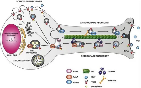
The membrane and subcellular trafficking of TrkA receptor is pivotal for the activation of the canonical RTKs signaling pathways, responsible for the neuronal growth and survival (Fig. 1.11). Following NGF binding and activation of PI3K, Ras/ERK and PLC-signaling pathways, NGF/TrkA complexes are internalized via clathrin coated pits [101], [102] or with a Pincher mediated endocytosis pathway [103] that generates multivesicular bodies (MVBs) containing multiple vesicles with complexes of NGF-TrkA. It was reported that the membrane pool of TrkA receptors starts to decrease after 5 minutes of NGF stimulation, while only after 15 minutes the receptor reached the maximum level of its phosphorylation [104]. NGF/TrkA complexes are internalized in early endosomes with different destinations [105]: receptors can be in recycling endosomes to be recycled to the cell
31
surface, sorted into late endosomes to be degraded in lysosomes [106]–[108], or transported retrogradely or anterogradely towards the soma or the neurite tip in signaling or recycling endosomes, respectively [101], [104], [109], [110].Retrograde transport was a mechanism discovered by Campenot, who demonstrated for the first time that NGF administrated at the axon tip of neurons is sufficient for the survival of neuronal cell bodies [111]. The theory of the signaling endosomes (Fig. 1.11, lower panel) claims that vesicles of TrkA-NGF are transported by dynein motor proteins retrogradely towards the soma, in complexes with signaling effectors as PI3K, MAPK and PLC-γ. When at the cell body, the signaling endosomes enhance the activation of CREB, promoting neuronal survival. However, also another model of signal transduction between axon tip and cell body was proposed, named “wave propagation model”: here the binding of NGF to TrkA generates a cascades of phosphorylation events that move retrogradely within the plasma membrane. MacInnis et al. demonstrated that after treatments at sympathetic axon tips with NGF-coupled beads which prevents NGF-TrkA complexes internalization, the survival signal at the cell body persisted, suggesting that the phosphorylated receptors alone are able to transduce the survival signaling [112]. On the other hand, TrkA anterograde trafficking was reported to be necessary for the signaling of receptors: once produced at the cell body, most Trk receptors are indeed carried towards the axonal tip, where after their exposure at plasma membrane, they are bound and activated by NTs [113].
Figure 1.11. RTK membrane trafficking in neuronal axons. Schematic representation
of signaling endosomes of TrkA and NGF; after NGF binding the activated TrkA receptors can be internalized at the axon tip and retrogradely transported to the cell soma via dynein-motor complexes in signaling endosomes (in gray). When at the cell soma, signaling endosomes are sorted and the receptors can enter a pathway of degradation, recycling to the plasma membrane, transcytosis or autophagic pathway. Image taken from [115].
32
1.2.4 TrkA ubiquitination
Ubiquitination is a post-translational modification consisting in the attachment of a 76 amino acid long poly-peptide, called ubiquitin (Ub), to the target protein. Historically, the ubiquitination process was related to misfolded proteins destined to proteasomal degradation [116], but in the last years several works demonstrated an alternative role for ubiquitination in protein sorting and signaling [117], [118].
The ubiquitination process occurs through three sequential enzymatic steps: the first ATP-dependent step comprises the activation of Ub by an E1 Ub-activating enzyme. Then the activated Ub molecule is transferred to the cysteinyl group of the E2 Ub-conjugating enzyme, which forms a complex with the E3 Ub-ligase, and the last finally transfers the ubiquitin to Lys (K) residues of the target protein. The E3 family are divided in two different groups, one consisting of a homologous to the HECT domain and the other containing RING or RING-like domain [119]. Different types of ubiquitination correspond to a different final destiny for the target protein [120]: mono-ubiquitination occurs when one Ub molecule is attached to the protein of interest, targeting it to endocytic pathway and membrane trafficking. Multi-monoubiquitination, which consists into the attachment of several Ub molecules to different Lys residues of the target protein, causes endocytosis of receptor. Finally, in the poly-ubiquitination process, one or more ubiquitin-chains are attached to the protein, determining its endocytosis and proteasomal degradation. More into details, the most frequent Lys-48 poly-ubiquitination chains is related to the degradation mediated by the 26S proteasome, while Lys-63 chains is usually associated to non-proteolytic degradation, as in the case of HectH9, Mdm2, tumor necrosis factor receptor-associated factor 6 (TRAF6), cellular inhibitor of apoptosis protein 1/2 (c-IAP1/2) and ring finger protein 8 (RNF8) [121]–[123].
Ubiquitination of TrkA receptor has an important role in regulating its degradation, trafficking and function, instead deregulations of the ubiquitination machinery are usually related to cancer [124], [125]. It was reported that TrkA is ubiquitinated by four different E3 ubiquitin ligases, namely TRAF-6 E3 RING Ub-ligase [126], Nedd 4-2 E3 HECT Ub-ligase [94], [14-27], TRAF-4 E3 RING Ub-ligase [14-28], and Cbl [14-29]. TRAF-6 poly-ubiquitinates TrkA at the lysine K485, located in the JM region [126], [130]; it interacts with p75NTR, promoting K63-linked
poly-ubiquitination, which regulates the internalization and signaling of TrkA [126], suggesting an important involvement of P75NTR in TrkA ubiquitination. The involvement of P75NTR in the TrkA subcellular trafficking is today
controversial; Kuruvilla et al. demonstrated that TrkA receptor is able to internalize after NGF stimulation without the presence of P75NTR [131]. Another work reported that P75NTR regulates negatively the ubiquitination of TrkA
33
receptors, causing a delay of its internalization in PC12 cells and protecting it from degradation [132]. Furthermore, the study of Gheeta et al. reported that TRAF6, after binding with P75NTR, needs the adaptor protein
p62 to poly-ubiquitinate TrkA receptors, enhancing its shuttling towards the proteasome [133].
The Cbl ubiquitin ligases (c-Cbl and Cbl-b) were instead reported to be negative regulators for TrkA, involved in ligand dependent ubiquitination of the receptor and targeting it to lysosomes for degradation [134],[129]. Nedd 4-2 is the unique Ub-ligase that binds TrkA but not TrkB and TrkC receptors, because it recognizes a PPXY motif present only in the TrkA sequence. Nedd 4-2 is responsible for the NGF dependent multi-mono-ubiquitination of TrkA [94], [135], responsible for degradation and NGF-mediated signaling [127]. The first evidence of the specificity of Nedd 4-2 for TrkA receptors happened with the discovery that overexpression of Nedd 4-2 induced neuronal apoptosis only in NGF-dependent DRG neurons, but not in BDNF-dependent DRG neurons [94]. Arevalo et al. demonstrated the importance of this specific motif, by generating a mouse model with a mutation in the first Pro residue of the PPXY sequence (hTrkA-P782S). After NGF stimulation, the mouse model with TrkA-P782S showed impaired ubiquitination and defects in both trafficking and degradation [127]. Moreover TrkA-P782S models showed an increased number of sensory neurons, and also increased TrkA signaling [127]. The same mouse model was used also to demonstrate that the lack of ubiquitination was related to an enhanced thermal sensitivity and inflammatory pain [136]. Finally, TRAF4 promotes TrkA ubiquitination through Lys-27 and Lys-29 ubiquitin linkages, leading to the hyper-activation of kinase activity and inducing the alteration of its phosphorylation status [128].
1.2.5 The involvement of TrkA receptor in HSAN IV disease
Pain is a protective mechanism adopted by multicellular organisms to prevent the contact with noxious stimuli [137]. Few years ago it was discovered that alterations of the signaling mediated by NGF/TrkA complexes were responsible for a deficit of sensory neurons specialized to detect pain sensation [138]. It was already known that the NGF pathway ensured the survival of both sympathetic ganglion neurons and nociceptive sensory neurons in DRG, and of ascending neurons in the basal forebrain [71], [139]. Indeed, initially it was proposed that the absence of NGF during the fetal period was the only cause for the loss of growth fibers, finally responsible for the insensibility to feel pain. However, the real turning point was the discovery that mice without nociceptive DRG neurons had lost the orthologous gene of human TrkA, promoting TrkA as a candidate for the onset of the Hereditary Sensory and Autonomic Neuropathies (HSANs) type IV (OMIM# 256800). Subsequently, genetic analysis in four patients affected by HSAN IV confirmed mutations in the coding gene for TrkA [140]. Currently, it is well known that the TrkA pathway is fundamental for innervating skin with sensory axons and for the survival of pain receptors [141]; indeed, HSAN IV (also called CIPA, Congenital Insensitivity to Pain and Anhidrosis) is
34
caused by the lack of nociceptive and sympathetic nerves and characterized by loss of feeling especially in hands and feet. In particular, HSAN IV is a rare genetic disorder inherited in an autosomal recessive manner, which usually affects female and male childrenin equal number. It is characterized by insensibility to feel pain, due to the absence of afferent neurons essential for the detection of noxious and thermal stimuli in multicellular organism [137], and by anhidrosis, caused by the altered innervation of eccrine sweat gland. Because of the loss of thermal and noxious sensitivity, children affected by this disease have usually unintentional self-mutilation, repeated fractures, and joint damages; furthermore, the inability to sweat (anhidrosis) causes recurrent episodes of fever, usually related to hyperpyrexia conditions. It was reported that patients affected by HSAN IV have usually developmental delays and learning disabilities. From a molecular point of view, HSAN IV disease is characterized by more than 105 [142] mutations inserted in the TrkA coding sequence: in most cases patients show nonsense and missense mutations and less frequently small insertions or deletions [143]. The comparison among missense mutations associated with HSAN IV showed that all are closer to the TKD and thus fundamental for the protein kinase activity [54], [144]. The group of Mardy et al. identified nine frameshift, seven nonsense, seven splice and 14 missense mutations in HSAN IV families coming from different countries; they found in one Ecuadorian and three Japanese families three mutations inside the region coding for TrkA ICD: R548fs, G571R and IVS15+3A-C [139]. 11 HSAN IV mutations were identified in families with different ethnic groups (Fig. 1.12): six missense mutations (Arg85Ser, Leu231Pro, His598Tyr, Gly607Val, Arg643Trp, Gly708Ser), two frameshift (Asn67fs, Gln308fs), one nonsense mutation (Gln9X) and finally two variants of splicing (IVS4-1G-C, IVS7+1G-A). From the comparison among 100 normal chromosomes only Leu231Pro, Arg643Trp, Gly708Ser resulted really exclusive and so responsible for HSAN IV disease. In particular, Arg643Trp and Gly704Ser, which play a fundamental role in enzymatic activity, are located in the TKD and conserved among different RTKs [87]; His598Tyr placed in the ICD is instead conserved in all members of the Trk family. Arg85Ser (in the Leu-rich domain) and Gly607Val (in the ICD) are not conserved. However, the frameshift mutations, Asn67fs and Gln308fs, which are located in the ECD, generate truncated forms of the TrkA protein.
Figure 1.12. Location of mutations in the human TrkA sequence associated to CIPA. The human TrkA gene is divided into 17 exons and 16
introns. The entire sequence was estimate to span at least 23 kb, coding for a protein of 790 or 796 aa. The asterisk denotes the common Japanese founder mutation (R548fs); three mutations in brackets are probably polymorphisms in a particular ethnic background. Image taken by [14].
35
More recently, the group of Shaikh have characterized the membrane expression, glycosylation, auto-phosphorylation, Y496 auto-phosphorylation, PLC- and neurite outgrowth of seven novel missense mutations found in patients affected by HSAN IV [143]. From the results, the mutation p.G517E showed similar glycosylation, membrane localization, auto-phosphorylation and Y496 phosphorylation of TrkA-wt, while the PLC- signaling and the neurite outgrowth are impaired. This mutation (p.G517E) does not impair kinase activity, highlighting that the evaluation of Y496 phosphorylation was not sufficient to assess the pathogenicity of HSAN mutations. On the other hand, p.G522E mutation, generating a glycosylated and membrane-translocated receptor, is not phosphorylated upon NGF stimulation, causing an impairment of the PLC- pathway and of neurite outgrowth. All the mutations p.L657P, p.I699T and p.R771C display less glycosylation and reduced membrane expression of the 140 kDa protein than the TrkA-wt. Furthermore, structural modeling demonstrated that these mutations, located in TKD, cause a rearrangement of the 3-dimentional protein structure, probably responsible for the abolishment of kinase activity [143]. Finally, p.C763S shows reduced auto-phosphorylation and Y496 phosphorylation, while C752S is considered a polymorphism because no defects in glycosylation, membrane expression, phosphorylation or neurite outgrowth are found. With an exome sequencing study, Altassan et al. discovered three missense and two non-sense novel HSAN IV mutations [144]: in particular p.Arg110Asp and p.Ser142Ter localized in the ECD, p.Lys476Ser in the JMD while p.Arg643Gln and p.Leu694Pro located in the TKD of the protein [144]. With a computational approach they analyzed the predicted effect that some of these mutations had on the TrkA structure; in particular they found that p.Arg110Asp induces the loss of two hydrogen bonds, while p.Leu694Pro and Arg643Gln only loose one, causing possible structural rearrangements that can compromise the protein function. In particular, in the case of Arg643Gln mutation (which is the human residue corresponding to the R649 we analyze in Chapter 3, see table 1.1), the substitution of an Arg with a Gln causes the loss of the positive charge and alters the local stability of the protein, because of the small size of the substituted residue [144]; the consequent lack of the hydrogen bond in this position disturbs the TKD, abolishing its function [144].
The group of Franco et al. has instead characterized the subcellular localization, kinetics of degradation, misfolding and cellular toxicity in primary neurons of three novel mutations found in children affected by HSAN IV disease: L213P and C300stop positioned in the ECD and Δ736 in the TKD of TrkA receptor [146]. They found that C300stop is rapidly disposed to autophagy, Δ736 is probably degraded by the proteasome system while L213P is a long-lived protein with a poor trend to protein degradation [146]. In particular, the latter mutation induces misfolding, retention in ER and delayed degradation, together with a marked increase of the number of autophagosomal vesicles, responsible for the induction of swollen regions in neurons and sensitization of PC12nnr5 cells to cell death.