A new hemodynamic model for the study of cerebral venous outflow
G. Gadda,1A. Taibi,1F. Sisini,1M. Gambaccini,1P. Zamboni,2and M. Ursino3
1Department of Physics and Earth Sciences, University of Ferrara, Ferrara, Italy;2Vascular Diseases Center, University of Ferrara, Ferrara, Italy; and3Department of Electrical, Electronic and Information Engineering, University of Bologna, Bologna, Italy
Submitted 8 July 2014; accepted in final form 10 November 2014
Gadda G, Taibi A, Sisini F, Gambaccini M, Zamboni P, Ursino M.
A new hemodynamic model for the study of cerebral venous outflow. Am
J Physiol Heart Circ Physiol 308: H217–H231, 2015. First published
November 14, 2014; doi:10.1152/ajpheart.00469.2014.—We developed a mathematical model of the cerebral venous outflow for the simula-tion of the average blood flows and pressures in the main drainage vessels of the brain. The main features of the model are that it includes a validated model for the simulation of the intracranial circulation and it accounts for the dependence of the hydraulic properties of the jugular veins with respect to the gravity field, which makes it an useful tool for the study of the correlations between extracranial blood redistributions and changes in the intracranial environment. The model is able to simulate the average pressures and flows in different points of the jugular ducts, taking into account the amount of blood coming from the anastomotic connections; simulate how the blood redistribution due to change of posture affects flows and pressures in specific points of the system; and simulate redistributions due to stenotic patterns. Sensitivity analysis to check the robustness of the model was performed. The model reproduces average physiologic behavior of the jugular, vertebral, and cerebral ducts in terms of pressures and flows. In fact, jugular flow drops from⬃11.7 to ⬃1.4 ml/s in the passage from supine to standing. At the same time, vertebral flow increases from 0.8 to 3.4 ml/s, while cerebral blood flow, venous sinuses pressure, and intracranial pressure are constant around the average value of 12.5 ml/s, 6 mmHg, and 10 mmHg, respectively. All these values are in agreement with literature data. mathematical modeling; cerebral outflow; posture dependence; jugu-lar veins collapse; collateral routes
THE EXTRACRANIAL VENOUS SYSTEM represents an important
de-terminant of the brain circulation, but its role in the pathology of the central nervous system is not fully understood yet (4, 26). It is recognized that, in supine position, the jugular veins represent the main outflow route for the cerebral circulation (2, 6, 25, 36, 38), being able to carry most of blood flow from the
brain and from other extracerebral territories (⬃700–720 ml/
min; Refs. 27, 33) with respect to a cerebral blood inflow of ⬃750 ml/min (45). However, the jugular venous system ex-hibits important flow limitation during upright posture changes, because the jugular veins tend to collapse as a consequence of the decrease of transmural pressure due to the gravitational field, causing a significant increase in resistance (3, 7, 12, 14, 26). In the absence of other routes for extracranial outflow, this flow limitation would have dramatic effects on the cerebral circulation, since, apart from brief transient time intervals, the average cranial arterial inflow is expected to be equal to the cranial venous outflow for the mass preservation.
As a consequence, large attention has been devoted to the venous circulation in the upright state, in an effort to under-stand which alternative routes can carry the brain venous outflow. It has long been postulated that the vertebral venous system may provide an important alternative route for venous outflow when standing or sitting; this was first demonstrated with the use of contrast media in rhesus monkeys (9) and subsequently mea-sured in humans with the Doppler and magnetic resonance imaging technique (2, 6, 7, 25, 38). Valdueza et al. (33) observed that blood flow in the jugular veins decreases from 700 ml/min in supine position down to 70 ml/min at 90° elevation, while blood flow in the vertebral veins raises from 40 to 210 ml/min.
Accordingly, a classical model of the cerebral venous out-flow assumes the existence of two main alternative routes: a route through the jugular veins, with smaller resistance in supine conditions, and a parallel vertebral route with higher resistance. In upright conditions, when the first route collapses, blood flow is diverted to the second one. Based on this idea, Gisolf et al. (14) developed a mathematical model of cerebral venous outflow: the model consists of two jugular veins (each composed of a chain of 10 units with resistances and capaci-tances) and a vertebral plexus (described with a single resis-tance). The elements in the jugular veins collapse according to the “tube law” (3, 12) during a posture change but can reopen during a Valsalva maneuver. With this model, the authors studied how venous blood flow changes from supine to upright position and confirmed that the cerebral venous distribution depends on posture. Despite the previous pivotal studies, many aspects of the cerebral venous outflow system are still prob-lematic.
First, it is well known that cerebral autoregulation maintains blood flow to the brain quite constant, despite pressure changes (1, 21): this signifies that an amount of blood flow much greater than that measured in the vertebral system must be carried out in the upright state. This discrepancy was clearly recognized by Valdueza et al. (33) who observed that “a mean difference of about 450 ml/min remained.” These authors hypothesized that other routes, such as the epidural veins, significantly contribute to the orthostatic venous outflow.
Second, some authors, using the color Doppler technique, recently observed that blood flow along the jugular veins in upright conditions is not longitudinally constant but increases progressively when the measurement site is moved from the upright sections (close to the jugular foramen into the skull) to the downstream sections (close to the subclavian vein) (6, 28, 41, 44). This observation supports the idea that additional anastomotical routes carry part of the cranial blood flow to the jugular veins even in upright position, bypassing the upstream more collapsed sections. In fact, as a consequence of the different effect of gravity, only the higher portions of the
Address for reprint requests and other correspondence: G. Gadda, Dept. of Physics and Earth Sciences, Univ. of Ferrara, Via Saragat 1, 44122 Ferrara, Italy (e-mail: [email protected]).
jugular veins are probably fully collapsed, whereas the down-stream sections are opened. A comprehensive model of the cerebral venous outflow should also include these further collateral routes.
Due to the complexity of the relationships involved, and the large variability in the anatomical parameters, it is extremely difficult to understand the effect of alterations in the extracra-nial venous circulation in simple qualitative terms. The study of the cerebral venous outflow, and of its implications in healthy and diseased conditions, can largely benefit from the use of computational models.
So far, most models of the cerebral circulation focused on the intracranial circulation and on its control mechanisms, by providing just a very simplified description of extracranial venous return. A notable exception is the model by Gisolf et al. (14) mentioned above, which, however, includes only the vertebral plexus as an alternative drainage pathway.
The aim of the present study is to develop and validate a comprehensive original lumped parameter model of the cere-bral venous outflow system, which overcomes some of the limitations noticed above. In particular, compared with the model by Gisolf et al. (14) the present model also includes:
1) An accurate model of the intracranial circulation
devel-oped in past years, which incorporates the autoregulation of cerebral blood flow (13, 30, 32). This model provides the correct values of cerebral blood flow to the venous return model. Moreover, it allows a quantitative analysis of the effect of alterations in the venous pathways on intracranial quantities, such as the effect on intracranial pressure, venous sinuses pressure, capillary pressure, and cerebrospinal fluid circulation.
2) A more sophisticated description of the collateral
path-ways, including not only the vertebral plexus but also other anastomoses leading blood to the downstream sections of the jugular veins (6, 28, 41, 44).
In the following, the model is described qualitatively, with emphasis on the new aspects, and parameters are given in accordance with the physiology. All equations are presented in the APPENDIX. The model is validated against results in the
literature, concerning the effect of a posture change in healthy subjects; a sensitivity analysis is also performed to clarify the role of the alterations in some parameters.
The model may represent an useful tool for the study of the correlation among posture variations, vessel conductances (normal or abnormal), and the consequent pressure and flow changes. This may have a great impact toward a deeper understanding of pathological disorders involving abnormali-ties of the cerebral venous outflow. In perspective, it may be used to assess which alterations in the extracranial venous outflow may be in relation with central nervous system disor-ders and aging (6, 10, 24, 40, 42).
MATERIALS AND METHODS
Model Description
Our mathematical model for the simulation of the head and cerebral circulation and drainage system includes two submodels built using lumped parameter method. The model is represented in Fig. 1.
Mathematical models may represent an useful tool for improving comprehension of complex systems like this, describing the behavior of a real process using a system of equations (14, 20, 22). What we are developing is a lumped parameter model, i.e., a model in which the continuous variation in space of the state variables of the system is
lumped parameter model allows the description of spatially distrib-uted physical systems to be simplified using a network of discrete entities that approximate the real system. The use of a lumped parameter model presents a significant advantage since it exhibits a small number of parameters able to account for an entire physiological or clinical phenomenon in a concise way. This fact improves the clinical meaning of the results obtained: the behavior of the model can be studied more easily than the system that it represents.
Every segment x of Fig. 1 consists of a capacitor Cxthat simulates
the property of the segment to accommodate volume and of a conductance Gxthat simulates the property of the segment to drain
blood.
All the model equations are presented in theAPPENDIX.
The mathematical model of the cerebral hemodynamics, enclosed in the “BRAIN” dashed rectangle of Fig. 1, has been fully developed and validated by Ursino and colleagues in previous works (13, 30, 32). This model simulates the hemodynamics of the arterial-arteriolar and venous cerebrovascular bed, the cerebral arterioles regulation mech-anisms, the cerebrospinal fluid production and reabsorption processes, the Starling resistor mechanism for the cerebral veins, and the non-linear pressure-volume relationship of the craniospinal compartment. The structure of the cerebral venous outflow model (shown in Fig. 1 out of the “BRAIN” dashed rectangle) has been developed starting from a recent work of Zamboni et al. (44). The main part of the model is composed of four venous ducts: two internal jugular veins (IJVs; left and right) and two vertebral veins (VVs; left and right). The internal jugular veins are modeled by dividing them into three seg-ments with different conductive and capacitive values, to better implement the different biomechanical properties of these vessels along their length and to better simulate the effect of hydrostatic pressure gradient in upright position.
Indeed, jugular veins are collapsible blood vessels characterized by marked changes in their cross-sectional configuration depending on transmural pressure Pxint ⫺ Pxext (26). The latter is affected by
hydrostatic pressure gradient during a posture change (7). On the other side, the hydrostatic pressure gradient does not significantly affect the lumen of the vertebral veins.
Anatomically speaking, left and right lower jugular segments (J1) correspond to the segments close to the junction of the internal jugular veins with the subclavian vein, at the confluence with the brachio-cephalic vein trunk. The middle segments (J2) correspond to the point where the veins are in an anatomical relationship with the more lateral contour of the thyroid gland. The upper segments (J3) correspond to the point before the passage through the jugular foramen into the skull (43).
To account for the growth of blood flow from J3 to J1 (6, 8, 10, 28, 33, 41, 44), we must consider that a quota of the head inflow is conveyed into the internal jugular veins more caudally with respect to the J3 position, through intra- and extracranial anastomosis. To account for this experimental evidence, the model is developed so that the two jugular veins are linked by a network of eight constant conductances and two constant capacitors that simulate the presence of collaterals and anastomotic connections. This collateral network also allows the drainage of the extracranial venous blood, i.e., of that part of blood coming from the two external arteries to serve the head organs and tissues out of the braincase (44).
In the model, the conductance of the external arteries (i.e., the conductance of the tract with blood flow Qex in Fig. 1) has been
maintained constant. Conversely, as described in previous works (30) and explained through Eqs. 7–11 in theAPPENDIX, the conductance of the intracerebral arterioles is subjected to autoregulatory mechanisms, which work to maintain quite a constant cerebral blood flow despite moderate changes in cerebral perfusion pressure. The choice of a constant extracranial conductance can be justified by the observation that autoregulation in extracranial vessels is generally weak and slow. Moreover, during hypotension a vasoconstriction of baroreflex origin
may counteract this weak vasodilatory autoregulation, thus resulting in negligible conductance changes.
We assume that, in the basal supine condition, the anastomotic upper connection c3 is latent: blood flows in it only when the posture is upright or when there is a lack of drainage in one vessel tract, or more than one.
Moreover, a basic submodel of six constant conductances and three constant capacitors is implemented in the main model to describe the lumbo-azygos system (AZY), which links jugular and vertebral path-ways at the level of superior vena cava (36).
The model is made up of several differential equations. Every equation links together the elements representative of a specific point
x (i.e., pressure Px, conductance Gx, and capacity Cx) with the
elements representative of the other segments of the system to account for mass preservation and to include effects due to posture changes. We report here (Eqs. 1 and 2) examples of the two types of equations used to built the cerebral venous outflow model.
Equation 1, one of the state equations of the model listed in the
APPENDIX, implements the mass preservation. We can see how the pressure at a given point (jr3 in this example) is related to capacity, conductances, and drops of pressure.
dPjr3
dt ⫽
1
Cjr3
关共
Pvs⫺ Pjr3兲
Gjr3⫺共
Pjr3⫺ Pc3兲
Gcjr3⫺共
Pjr3⫺ Pjr2兲
Gjr2兴
(1) To include dynamics due to posture changes from supine to upright in the gravity field, conductances in the jugular veins are modeled using a nonlinear relation (Eq. 2) with switch-like properties so that for negative or low transmural pressure Pxint⫺ Pxextat a given point
x, the related vessel conductance Gxis low, while for high transmural
pressure, vessel conductance approaches a maximum value (14).
Gx⫽ kx
冋
1⫹冉
2 冊
arctan冉
Pxint⫺ Pxext A冊册
2 (2) The effect of hydrostatic pressure gradient is hidden in the internal pressure Pxint. The value of this pressure decreases by a factorghduring the transition from supine to standing position. Obviously, the collapse of the upper segment of the jugular veins (J3) is more pronounced with respect to the lower segments (J1), due to the presence of the height h. We use the superior vena cava as the zero level for the hydrostatic pressure.
Qtot Qex Q BRAIN Qf QO hbf Qjr3 Qc3 Qjl3 Qcjr3 Qcjl3 Qvvr Qvvl Qjr2 Qc2 Qjl2 Qcjr2 Qcjl2 Qjr1 Qc1 Qjl1 Qsvc1 Qsvc2 Qazy2 Qrv Qazy1 Qlv VERTEBRAL JUGULAR AZYGOS NOTATION Capacitor:
Conductance: Fig. 1. Scheme of the lumped parameter model for thestudy of the cerebral venous outflow. The “BRAIN”
box represents the intracranial part of the model, already developed in the work of Ursino et al. (13, 30, 32). Outside the “BRAIN” box there is the extracra-nial part of the circuit that has been developed starting from the work of Zamboni et al. (44).
To solve the whole system of equations we resort to an algorithmic approach. We used the software package Berkeley Madonna (free available version beta 9.0.111; Ref. 17a), developed on the Berkeley campus under the sponsorship of National Science Foundation and National Institutes of Health and widely used in biology and biolog-ical engineering (11, 15). The package provides three fixed-stepsize integration methods and two variable-stepsize integration methods. We chose the iterative method Runge-Kutta 4 to perform all the computations. The algorithms used by Berkeley Madonna are derived from the routine methods published in the series of books Numerical
Recipes (23).
Assignment of Basal Parameters
We adjusted every parameter value in search of agreement between model simulations and measurements that can be obtained from literature results. Starting from the work on the intracranial circuit already developed by Ursino and Lodi (30), we moved around several literature data to choose the right inputs for the jugular-vertebral network, adopting reasonable criteria to determine parameters not available from literature.
Parameters were assigned on the basis of the following criteria:
1) All parameters concerning the intracranial circulation were
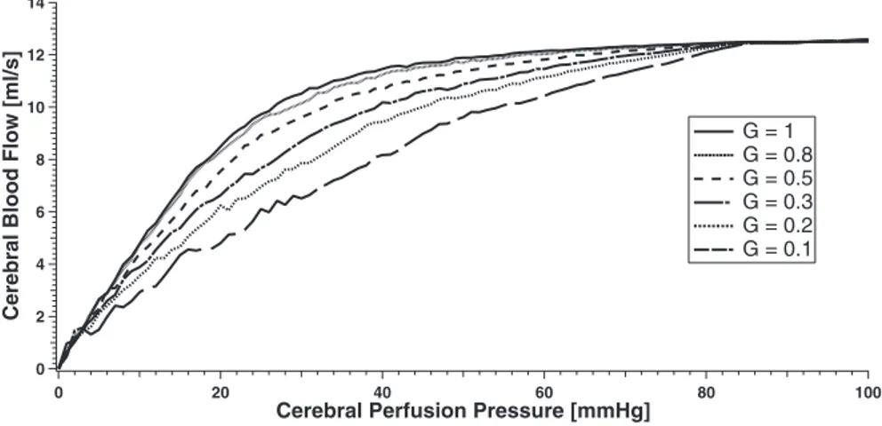
given the same values as in the previous works (30, 32), where a thorough justification can be found. Briefly, intracranial vascular resistances and capacities were assigned on the basis of physiological and anatomical data on brain hemodynamics in a normal subject. The autoregulation gain and time constants were given to simulate the typical autoregulation response of an healthy individual who exhibits mild cerebral blood flow changes despite cerebral perfusion pressure changes (see Fig. 2). Parameters summarizing the cerebrospinal fluid circulation and intracranial compliance were set to establish a normal hydrodynamics, as derived from infusion or PVI tests in the neuro-logical literature. All the intracranial basal parameters are summarized in Table 1.
2) The values of all conductances in the extracranial circulation
were assigned starting from physiological values of pressures and flows (see Tables 2 and 3, respectively). In particular, as shown in Table 2, we assumed a progressive pressure reduction from the venous sinuses to the right atrium (i.e., the central venous pressure), assuming normal values as large as 6 and 5 mmHg, respectively. Typical average values reported in Table 2 are assigned as basal values to the pressure of the superior tract of collateral Pc3, of venous sinuses
pressure Pvs(30), and of central venous pressure Pcv(14).
Interme-diate basal pressure values (Pjr3, Pjr2, etcѧ) are assigned to simulate a
homogeneous pressure drop along the whole circuit from the exit of venous sinuses to the vena cava. External pressure Pxextto the upper
sections of the jugular veins J3 and J2 is set to zero, while at the lower segments J1 is set to the average thoracic pressure during a complete
respiratory cycle (18). Looking at the basal flows shown in Table 3, cerebral blood flow Q is the total blood volume entering the cranial cavity per unit time (45). At the exit from the skull, it is drained both by jugular and vertebral veins, with the two jugular veins that contribute to drain the main part of blood only in supine position. The flow through left and right external carotid arteries Qexis set
accord-ing to the data of Yazici et al. (37) and takaccord-ing into account the lower cerebral flow reported by these authors (i.e., we assumed the same ratio between the extracranial carotid blood flow and blood flow in the common carotid artery as in Ref. 37). It is also assumed that 40% of this flow is directed to every jugular via the anastomotic connections, while the remaining blood is drained down through the middle collateral. Finally, half of the vertebral flow enters in the azygos vein, while the other half is divided between lumbar vein duct (1/3 of the total) and renal vein duct (2/3 of the total).
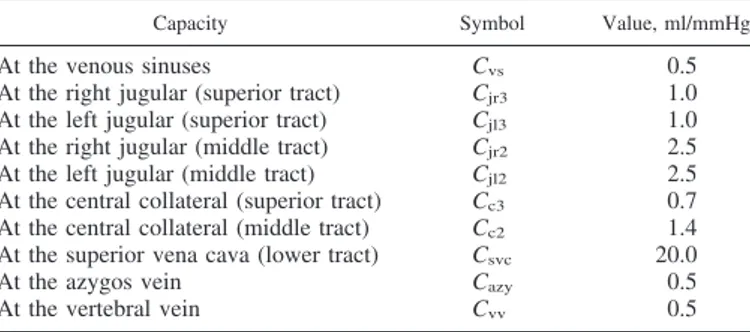
3) The values of the capacities in the extracranial circulation (Table
4) have been determined using the following considerations:
i) Capacities in the jugular tracts and in the vertebral veins have
been computed from the values of transmural pressures, lengths, and areas reported in the literature (33) and assuming a negligible un-stressed volume (i.e., we consider an average capacity, quite indepen-dent of transmural pressure).
12 10 8 6 4 2 0
Cerebral Blood Flow [ml/s]
100 80 60 40 20 0
Cerebral Perfusion Pressure [mmHg]
G = 1 G = 0.8 G = 0.5 G = 0.3 G = 0.2 G = 0.1 Fig. 2. Curves of the cerebral blood flow vs. cerebral
perfusion pressure for maximum (G ⫽ 1) and pro-gressive impaired autoregulation gain.
Table 1. Basal values of quantities related to the
intracranial circuit (supine condition)
Quantity Value Cpan 0.205 ml/mmHg ⌬Cpa1 2.87 ml/mmHg ⌬Cpa2 0.164 ml/mmHg Gaut 3 kE 0.077 ml⫺1 kR 13.1⫻ 103mmHg3·s ·ml⫺1 kven 0.155 ml⫺1 Pa 100 mmHg Picn 9.5 mmHg Ppa 58.9 mmHg Pv 14.1 mmHg Pv1 ⫺2.5 mmHg Pvs 6 mmHg Qn 12.5 ml/s R0 526.3 mmHg·s·ml⫺1 Rf 2.38⫻ 103mmHg·s·ml⫺1 Rla 0.6 mmHg·s·ml⫺1 Rpv 0.880 mmHg·s·ml⫺1 Rvs1 0.366 mmHg·s·ml⫺1 aut 20 s xaut 2.16 10⫺4
ii) Capacity of the azygos veins are assumed comparable to the
capacity of the vertebral veins.
iii) Capacities of the collateral tracts have been computed, in the
absence of clear geometrical data, by assuming that they are approx-imately proportional to the square root of conductances and using the capacities of the jugular tracts as a reference.
iv) A small value has been given to the capacity of the venous
sinuses, since most of these vessels are surrounded by the dura mater.
v) We checked that, with the previous choices, the overall capacity
of the venous outflow circulation is close to the value used in Ursino and Magosso (31) for the capacity of the cerebral venous vascular bed (10.7 ml/mmHg).
4) To complete the determination of model parameters, we need to
assign a value to A and kxin the equations describing the conductance
of the jugular veins (Eq. 2). These values have been given to address two major requirements:
i) In supine position, conductances must assume a constant value in
accordance with the linear relationship between drops of pressure and flows and in accordance with the values reported in Tables 2 and 3.
ii) In upright position, the conductance changes induced by venous
collapse (consequence of the hydrostatic pressure gradient) determine a redistribution of blood flow from the jugular veins to the vertebral-azygos complex and the collateral route. These changes have been assigned to simulate typical values of jugular blood flow and vertebral blood flow reported in Ref. 33.
It is worth noting that the last criterion is the only a posteriori information that we used in assigning parameters: all other informa-tion was set a priori, i.e., was independent of the results obtainable during posture changes. However, we did not use an automatic procedure to assign these parameters but just a manual adjustment to verify reasonable agreement between model blood flow changes and literature data. Since A and kx were assigned using a posteriori
information, we subsequently checked that the conductances vs. transmural pressure curves so obtained for each jugular tracts are in
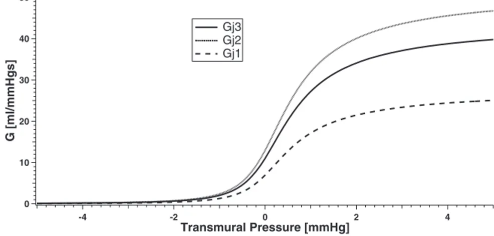
reasonable agreement with our knowledge on the biomechanics of compressible vessels. These curves, reported in Fig. 3, show that the collapse initiates at a transmural pressure approximately as high as 1–2 mmHg and is almost completed at moderate negative trans-mural pressure values in agreement with the literature information (5, 14, 16).
In the current model the value of the parameter A is 0.8, while we assigned to kjr3, kjr2, and kjr1 the value of 11.0, 13.0, and 6.9,
respectively (the same values are assigned to the left coefficients kjl3, kjl2, and kjl1). By choosing these parameters, Fig. 3 shows the relation
between conductance of the jugular segments and the transmural pressure.
RESULTS AND DISCUSSION
Once we verified that the model, with basal parameter values, can simulate the main blood flow changes from supine to upright position, we performed a sensitivity analysis on some model parameters. We focused our attention on changes in conductances in the venous pathways. Indeed, analysis of the correlation between conductances and posture variation and the subsequent pressures and flows changes might give infor-mation about the parameters that have greater impact on intracranial hemodynamics (and then on related disorders). From our particular point of view, analysis of the results before and after the variation of a conductance is meaningful, since it provides information on how the closure of a particular drain-age tract affects important physiological parameters, such as
venous sinuses pressure Pvs (and so intracranial pressure Pic
and cerebral circulation) or flows in the jugular and vertebral veins (Qsvc1and Qvv, respectively). For simplicity, we focused only on the right conductance variations, since analysis on left conductance variations will be symmetrical.
Each conductance under examination was varied from the baseline value, that is representative of physiological condi-tion, to zero value that simulates total absence of drainage from a section of the network; simulations of posture variation were performed in both situations.
Once we performed the sensitivity analysis by setting one conductance to zero, we chose to simulate total and halved occlusions of more than one drainage tract at the same time, following the typical patterns reported in Zamboni et al. (39). This work is one of the recent studies concerning the relations among the main cerebral venous outflow routes, their disorders, and the occurrence of neurological diseases (10, 17, 19, 28, 29, 34).
Simulation results of particular interest are shown in the following.
Table 2. Basal values of pressures related to the
jugular-vertebral circuit (supine condition)
Pressure Symbol Value, mmHg
At the collateral (superior tract) Pc3 6.00
At the right jugular (superior tract) Pjr3 5.85
At the left jugular (superior tract) Pjl3 5.85
At the collateral (middle tract) Pc2 5.85
At the vertebral vein Pvv 5.80
At the right jugular (middle tract) Pjr2 5.70
At the left jugular (middle tract) Pjl2 5.70
At the azygos vein Pazy 5.50
At the superior vena cava (superior tract) Psvc1 5.40
At the superior vena cava (inferior tract) Psvc 5.20
At the right atrium (central venous pressure) Pcv 5.00
External at J3 and J2 Pxext 0
External at J1 Pxext ⫺6.50
Table 3. Basal values of flows related to the
jugular-vertebral circuit (supine condition)
Flow Symbol Value, ml/s
At the brain (cerebral blood flow) Q 12.50 At every jugular vein Qjr3and Qjl3 5.85
At every vertebral vein Qvvrand Qvvl 0.40
At the external duct (face and neck) Qex 5.00
At every anastomotic connection Qcjr3, Qcjl3, Qcjr2, Qcjl2 1.00
At the middle collateral Qc2 3.00
At the azygos vein Qazy1 0.40
At the lumbar vein Qlv 0.13
At the renal vein Qrv 0.27
Table 4. Capacities related to the jugular-vertebral circuit
(supine condition)
Capacity Symbol Value, ml/mmHg
At the venous sinuses Cvs 0.5
At the right jugular (superior tract) Cjr3 1.0
At the left jugular (superior tract) Cjl3 1.0
At the right jugular (middle tract) Cjr2 2.5
At the left jugular (middle tract) Cjl2 2.5
At the central collateral (superior tract) Cc3 0.7
At the central collateral (middle tract) Cc2 1.4
At the superior vena cava (lower tract) Csvc 20.0
At the azygos vein Cazy 0.5
Simulation with Basal Parameter Values
Figure 4 shows how the model simulates the total amount of
jugular flow Qj3and vertebral flow Qvv at the equilibrium in
both supine and upright conditions. Results are also compared with the experimental mean flows reported in Valdueza et al. (33). The very good agreement between simulated supine and upright flows and experimental results means that parameters of the model are well assigned.
In Fig. 5, we show the behavior of the simulated venous
sinuses pressure Pvs over time when passing from supine to
upright condition. When basal parameters are used, our model predicts an increase of 2.68 mmHg (from 6.00 to 8.68 mmHg)
with change of posture. It also predicts that Pvsneeds⬃3 s to
reach the upright equilibrium.
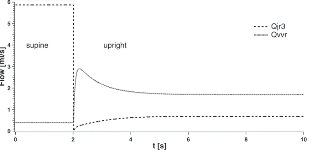
In Fig. 6, the behavior of the simulated right jugular flow Qjr3 and right vertebral flow Qvvrover time is reported in the same conditions of Fig. 5. For simplicity, we focus the atten-tion only on the right jugular and vertebral flows, since results on left vessels are symmetrical. Figure 6 shows that our model
predicts a strong decrease of the right jugular flow Qjr3 when
changing from supine to upright (from 5.87 to 0.69 ml/s). Conversely, flow at the right vertebral vein Qvvrrises from 0.40 to 1.70 ml/s during the change from the supine to upright condition. Figure 6 also shows that upright Qjr3and Qvvrflows reach their equilibrium with different behaviors.
In these conditions the flow through jugular and vertebral veins is equally distributed between left and right side. The collateral tract c3 is latent in supine position, while in upright
it drains the amount of blood that does not flow through jugular or vertebral veins (⬃7.7 ml/s).
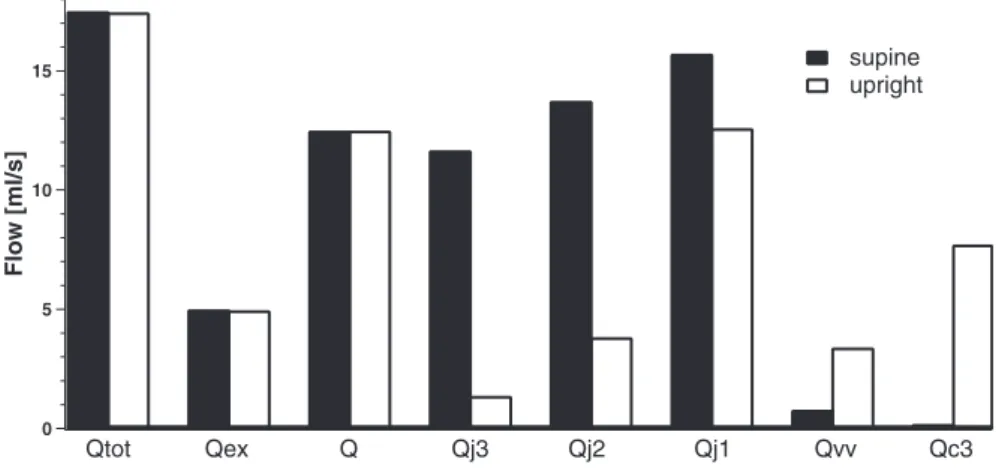
Figure 7 summarizes amounts of simulated total inflow Qtot,
flow to face and neck Qex, cerebral blood flow Q, and total
jugular, vertebral, and collateral flows at equilibrium (Qj3, Qj2, Qj1, Qvv, and Qc3) in supine and upright conditions.
We see that Qtot is the sum of Qex and Q. Moreover, the
histogram shows the different type of drainage the cerebral blood flow Q undergoes in supine and upright position. These different blood distributions are due to the changes in jugular conductances that occur when upright position is simulated (Eq. 2).
Results can be summarized as follow. Cerebral blood flow Q remains substantially constant despite the posture change, as a consequence of the action of autoregulatory mechanisms. In fact, the moderate increase in venous sinuses pressure lies
well inside the autoregulatory range (30). Blood flow Qj3in
the upper portion of the jugular vein exhibits a dramatic fall in the upright state: most of the cerebral blood flow passes through the collateral route c3. Moreover, blood flow in the
vertebral veins Qvv increases by about four times. These
results agree with those reported in literature (33). Further-more, blood flow in the jugular veins progressively in-creases from J3 to J1, since part of blood flow is drawn from the collateral route to the jugular tract via the anastomoses cj3 and cj2 (see Fig. 1). The reason is that the last portion of the jugular veins exhibits a less pronounced collapse in upright condition compared with the first tract, due to a
40 30 20 10 0 G [ml/mmHgs] -4 -2 0 2 4 Transmural Pressure [mmHg] Gj3 Gj2 Gj1
Fig. 3. Characteristic curves of the conductance (G) vs. transmural pressure in each jugular segment.
12 10 8 6 4 2 0 Flow [ml/s] Valdueza et al. Model Qj3 (upright)
Qj3 (supine) Qvv (supine) Qvv (upright)
Fig. 4. Comparison between literature flow data (33) and model outcome.
smaller gravitational pressure gradient and due to the effect of the negative intrathoracic pressure.
Sensitivity Analysis
To clarify the role of the main routes involved in cerebral venous outflow, and a possible effect of a pathological altera-tion, we performed a thorough sensitivity analysis. It consists of two steps:
1) We first interrupted blood flow in a single route by
assigning a zero value to the corresponding conductance and checked the effect in steady-state conditions.
2) Then, we simulated four typical pathological alterations
already reported in the clinical literature (39) and characterized by a conductance reduction in multiple venous paths.
Effect of a single closure. Results are summarized in Figs. 8,
9, and 10 with regards to the effect of a single closure on venous sinuses pressure Pvs, outflow from the confluence of the two jugular veins Qsvc1and vertebral blood flow Qvv, respec-tively. Results show that the cerebral venous outflow system is quite robust in response to a single vessel closure, both in supine and upright conditions. This signifies that interruption of a single path can be quite easily replaced by an alternative
route. Pressure Pvs at the venous sinuses, the link between
intracranial and jugular-vertebral circuit, increases with change
of posture from supine to upright in basal conditions (⫹2.68
mmHg) as shown in Fig. 8. In supine posture, total occlusion
of right jugular vein (Gjr3 ⫽ 0, Gjr2 ⫽ 0, and Gjr1 ⫽ 0)
produces little increases of the value of Pvs. Also, changes due
to occlusions of collateral network and vertebral veins are not appreciable. Conversely, looking at the simulation of upright condition, it is evident that Pvsis more influenced by lack of drainage of the collateral network (Gc3⫽ 0), while all the other kinds of occlusion only affect Pvswith little or not appreciable increases.
Figure 9 shows that output flow from the confluence of
jugular veins Qsvc1 decreases of about ⫺3.1 ml/s from the
supine to upright condition. In supine condition, every kind of occlusion evokes little or negligible changes of this flow. The same situation also occurs in upright condition.
Output flow from the vertebral veins Qvv rises of 2.6 ml/s
during change from supine to upright conditions as reported in Fig. 10. Little variations from the basal supine value occur when a jugular vein is occluded. Conversely, basal upright flow is quite increased by occlusion of the collateral network (Gc3⫽
0) and lowered by occlusion of right vertebral vein (Gvvr⫽ 0).
The most influential closure is found in the collateral circu-lation: in upright conditions it provokes a further increase in
venous sinuses pressure up to⬃10 mmHg and also a
redistri-bution of blood flow toward the vertebral-azygos complex. Obstruction of a jugular vein is relevant especially when it occurs close to its terminal part, causing a reduction of jugular outflow down to 11.4 ml/s. Naturally, an obstruction in a vertebral vein causes a significant decline in vertebral blood flow, with a redistribution toward the jugular and collateral circulations. 10 8 6 4 2 0 Pvs [mmHg] 10 8 6 4 2 0 t [s] supine upright
Fig. 5. Venous sinuses pressure (Pvs) behavior over
time in supine and upright simulations.
6 5 4 3 2 1 0 Flow [ml/s] 10 8 6 4 2 0 t [s] supine upright Qjr3 Qvvr
Fig. 6. Right jugular (Qjr3) and vertebral (Qvvr) flow
Finally, we tested the behavior of the external flow Qextfor
all the conditions described above. In the present model Qext
diminishes by 0.05 ml/s when venous sinuses pressure in-creases (for example, when moving from supine to upright condition both in a healthy and a pathological subject). This external flow is not significantly affected by any kind of occlusion we tested, apart for a little decrease during the occlusion of the lower jugular segment and a little increase
during the occlusion of the collateral pathway c3 (⫺0.03 and
⫹0.02 ml/s, respectively). We also performed the whole sen-sitivity analysis in the conditions of weak autoregulation re-ported in Fig. 2. Results show that for reduced cerebral auto-regulation (i.e., by changing the value of Gauttill 1/10 of its initial value) every value of pressure and flow reported in the sensitivity analysis does not change significantly. The reason is that, in our simulations, venous sinuses pressure always
exhib-its a mild change (⫹ 1 or ⫹2 mmHg), which is a minimal
fraction of cerebral perfusion pressure. Since cerebral blood flow is subjected to regulation mechanisms, the final change in
cerebral blood flow is always⬍1% of basal (even when using
a moderate autoregulation gain), which has negligible effects on the final results.
Multiple occlusions. The effect of multiple occlusions may
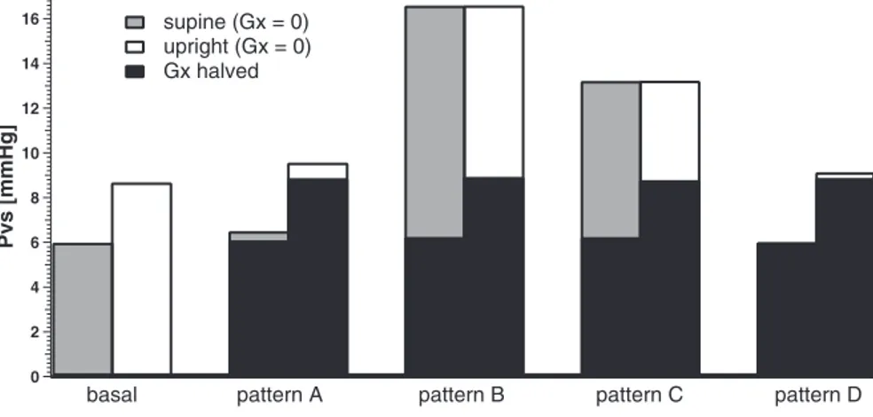
be much more dramatic. In the following, we will especially analyze the changes in venous sinuses pressure, since this quantity represents a link between the cerebral circulation and the extracranial venous outflow system. We report in Fig. 11 a
histogram to show how the basal venous sinuses pressure Pvs
varies when stenotic patterns occur. Together with the
simu-lations of null conductance, we reported also the simusimu-lations of the same patterns but with halved conductances.
A more complete description can be found in Tables 5 and 6, which report results concerning blood flows and pressures redistribution due to the different stenotic patterns described in Ref. 39, compared with the simulation of a healthy distribution. Pattern A refers to simulation of obstruction of the proximal azygos azy2, associated with a closed stenosis of the left internal jugular vein. Pattern B refers to simulation of obstruc-tions of both the internal jugular veins and the proximal azygos. Pattern C refers to simulation of obstructions of both the internal jugulars but without stenoses in the azygos system. Finally, pattern D refers to simulation of obstructions in dif-ferent tracts of the azygos vein (azy1 and azy2) associated with occlusion of the lumbar vein.
Results show that two particular pathological patterns (i.e., patterns B and C) may have a strong effect on venous sinuses pressure, which reaches values as high as 13–16 mmHg both in supine and upright positions. Such value may have conse-quences on intracranial pressure, cerebrospinal fluid circula-tion, and cerebral tissue. However, this pressure increase oc-curs only if the stenotic lesions are very severe (conductances close to zero). Moderate levels of conductance changes, al-though multiple, cause more acceptable pressure rises.
For what concerns model validation, we cannot use animal data taken from the literature, as usually done, since the extracranial venous drainage pathways are significantly differ-ent in animals compared with humans. In particular, the ex-tracranial venous circulation in humans is specifically adapted
15 10 5 0 Flow [ml/s] supine upright Qtot Qex Q Qj3 Qj2 Qj1 Qvv Qc3
Fig. 7. Basal flows in supine and upright simulations.
10 8 6 4 2 0 Pvs [mmHg] supine upright basal Gjr3 = 0 Gjr2 = 0 Gjr1 = 0 Gvvr = 0 Gc3 = 0 Fig. 8. Sensitivity analysis of venous sinuses
to the maintenance of an upright posture, which is the peculiar subject of the present work. For this reason, we are designing some ad hoc measurements on volunteers, using the Doppler technique, to assess blood flow changes and cross-sectional areas in the different portions of the jugular veins and to provide a complete quantitative set for model validation (or for discovering aspects requiring improvement). This will be the subject of a future work. When imaging with the current diagnostic methods multiple stenotic patterns or flow abnor-malities in the major extracranial and extraspinal veins, which are typical of chronic cerebrospinal venous insufficiency (CCSVI), it is difficult to confidently assign their hemody-namic significance for the intracranial circulation. It has been recently emphasized that the main issue to be investigated in this field is the definition of the hemodynamic impact in the intracranial venous system of obstruction/narrowing of the extracranial veins (46). Since, at this time, there is no estab-lished invasive or noninvasive diagnostic imaging modality capable to assess intracranial and/or parenchymal circulatory parameters in relation to extracranial brain outflow (47), the clinical application of the present model seems highly desir-able. For instance, in the present study we tested our model on four clinical patterns of extracranial venous obstruction, clin-ically detected in patients by means of catheter venography (39). In Fig. 11, we report the estimated pressure values in the cerebral dural sinuses in relation to the different CCSVI pat-terns. The application of the model demonstrates a significant increase of venous sinuses pressure especially for type B and C
but not in patterns A and D. Such a result appears to be coherent with the clinical severity and/or the topography of multiple sclerosis, the neurological disease associated with the observed patterns of venous obstruction in this cohort of patients. As far as the severity is concerned, patients with type A pattern (characterized by reduced sinusal pressure) demon-strated, with respect to B and C patterns, a significantly reduced probability to worsen to the secondary progressive clinical stage (39). Regarding the topography of the lesions in the central nervous system, type D pattern exhibits few cere-bral lesions and prevalent MRI plaque dissemination in the spinal cord (39). Speculatively, this result suggests that in-creased pressure in the cerebral sinuses may clinically influ-ence either the disease progression or the topography of mul-tiple sclerosis plaques and warrants further studies in this direction.
Conclusions
We developed a lumped mathematical model for the study of cerebral venous outflow, able to simulate the average blood flows in the main drainage vessels of the brain. The two main features of the model are that it accounts for the dependence of the hydraulic properties of the jugular veins with respect to the gravity field and that it includes a validated model for the simulation of the intracranial circulation. That makes it an useful tool for the study of the correlations between
extracra-16 14 12 10 8 6 4 2 0 Qsvc1 [ml/s] supine upright basal Gjr3 = 0 Gjr2 = 0 Gjr1 = 0 Gvvr = 0 Gc3 = 0
Fig. 9. Sensitivity analysis of jugular veins outflow (Qsvc1). 5 4 3 2 1 0 Qvv [ml/s] supine upright basal Gjr3 = 0 Gjr2 = 0 Gjr1 = 0 Gvvr = 0 Gc3 = 0
Fig. 10. Sensitivity analysis of vertebral veins out-flow (Qvv).
nial blood redistributions and changes in the intracranial envi-ronment.
We performed simulations of the effect of posture change from supine to upright on pressures and blood flows, first with basal drainage and then assuming a lack of conductance of some particular vessel tract.
Results show that the model is able to reproduce the average physiologic behavior of the jugular, vertebral, and cerebral ducts, with values in good agreement with literature data. In addition to that, the model is able to give information about the average flows in different points of the jugular ducts, so taking into account the amount of blood coming from the anastomotic connections.
Moreover, we can easily use it to verify how the blood redistribution due to change of posture affects the pressures in specific points of the system.
We are aware only of a previous model that investigates cerebral venous return in accurate quantitative terms (14). The present model shows some similarities with that model, but it also exhibits many important new aspects.
First, the description of the cerebral venous return now includes a larger number of drainage pathways encompassing not only the vertebral one but also additional collateral and anastomoses to the downstream jugular sections. Inclusion of such a more complex network of collaterals is important to simulate several experimental observations. Basically, blood flow measurements show that the sum of the jugular and vertebral blood flows decrease from supine to upright position (33). This result is in contradiction with the existence of strong autoregulation mechanisms in the brain, which maintain quite a constant cerebral blood flow despite moderate pressure changes, and reveals the existence of further collateral routes beside the vertebral veins, able to drain cerebral blood flow in
the erect position. Furthermore, measurements with the Dopp-ler technique show that the amount of blood flow in the jugular veins increases from the upstream to the downstream sections (6, 8, 10, 28, 33, 41, 44), a result that requires the presence of anastomoses, which allow a progressive reentry of blood flow. The second new aspect of the model consists in the strict link between the cerebral circulation (with its regulatory mecha-nisms), the cerebrospinal fluid circulation, and the cerebral venous return system.
Indeed, previous models were just devoted to study the cerebral venous return without paying attention to the cerebral circulation [Gisolf et al. (14)]. Other models were especially devoted to brain circulation and cerebral blood flow control with just an approximate description of the venous return, as for example in Ursino and Lodi (30). We claim that a model that incorporates both aspects is necessary, since the intercon-nections between the cerebral circulation and venous return systems may have important hemodynamics effects, especially in pathological conditions. In particular, the presence of regu-latory mechanisms able to maintain a constant cerebral blood flow even when cerebral perfusion pressure falls down to
16 14 12 10 8 6 4 2 0 Pvs [mmHg] supine (Gx = 0) upright (Gx = 0) Gx halved
basal pattern A pattern B pattern C pattern D
Fig. 11. Venous sinuses pressure Pvsin healthy and
stenotic patterns simulation. The black columns rep-resent the simulation of stenotic patterns with halved conductances with respect to the basal values.
Table 5. Venous sinuses pressure and intracranial pressure
at equilibrium in simulation of basal conditions and typical stenotic patterns
Pressure, mmHg Basal Pattern A Pattern B Pattern C Pattern D
Supine Pvs 5.98 6.51 16.60 13.22 6.01 Pic 9.44 9.51 11.09 9.95 9.44 Upright Pvs 8.68 9.57 16.55 13.23 9.14 Pic 9.81 9.93 11.12 9.95 9.87
Table 6. Flows at equilibrium in simulation of basal
conditions and typical stenotic patterns
Flow, ml/s Basal Pattern A Pattern B Pattern C Pattern D
Supine Qvv 0.79 0.51 3.86 7.86 0.33 Qjr3 5.87 11.14 3.04 2.13 6.08 Qjl3 5.87 0.81 3.04 2.13 6.08 Qjr2 6.87 12.90 2.98 1.86 7.08 Qjl2 6.87 0.88 2.98 1.86 7.08 Qjr1 7.86 15.39 0.00 0.00 8.08 Qjl1 7.86 0.00 0.00 0.00 8.08 Qsvc1 15.72 15.39 0.00 0.00 16.16 Qc1 1.00 1.57 12.98 9.24 1.01 Qtotout 17.50 17.47 16.91 17.10 17.50 Upright Qvv 3.39 1.54 3.64 7.87 1.37 Qjr3 0.69 1.62 1.14 2.10 1.10 Qjl3 0.69 1.32 1.14 2.10 1.10 Qjr2 1.92 4.99 2.95 1.84 2.48 Qjl2 1.92 1.62 2.95 1.84 2.48 Qjr1 6.30 13.38 0.00 0.00 7.24 Qjl1 6.30 0.00 0.00 0.00 7.24 Qsvc1 12.59 13.38 0.00 0.00 14.48 Qc1 1.46 2.48 12.97 9.23 1.59 Qtotout 17.45 17.40 16.91 17.10 17.44
50 – 60 mmHg might produce large increases in venous sinuses pressure, if the venous return system becomes unable to drain all the required amount of blood flow.
The present simulations suggest two important consider-ations. The cerebral venous return system is quite robust: a single occlusion, or even multiple occlusions of moderate entity, can induce only mild changes in venous sinuses pres-sure and in total blood flow. Indeed, this is the fundamental role of the strong anastomotical connections incorporated in the model. However, we have also shown that pathological states, characterized by multiple severe obstructions, can lead to significant pressure changes in the venous sinuses, hence, in possible alteration in cerebrospinal fluid circulation and brain tissue pressure. Which adjustments may be produced in these cases (either the opening of new collaterals, a reset of the autoregulation set point, or a permanent pressure increase) remains to be investigated.
A final important aspect of the model consists in the possi-bility to simulate the progressive increase in jugular blood flow when moving from the upstream sections close to the skull to the downstream sections in the thoracic portion. These changes are amplified by a posture change, when the different sections of the jugular veins experience a different amount of collapse and are significantly affected by any individual or pathological alteration in the collateral pathways.
Hence, a study of these blood flow variations, for instance via quantitative Doppler measurements, may represent a strong test to validate the model and to adapt its parameters to individual characteristics.
APPENDIX
Intracranial Circuit
Mathematical equations for intracranial blood dynamics and cere-brospinal fluid circulation have been written by imposing the mass preservation principle at all the circuit nodes.
In the following, we refer to resistance of a given tract Rxas the
inverse of the conductance Gxof the same tract. Table A1 provides a
glossary of terms.
Mass preservation at the node of pial arterioles pa implies the following equation: d
共
Ppa⫺ Pic兲
dt ⫽ 1 Cpa冋
Pa⫺ Ppa Rla⫹ Rpa⁄ 2 ⫺Ppa⫺ Pc Rpa⁄ 2 ⫺dCpa dt共
Ppa⫺ Pic兲
册
(3) Capillary pressure Pcis given by:Pc⫽
冉
Pv Rpv ⫹ Ppa Rpa⁄ 2 ⫹Pic Rf冊
⁄冉
1 Rpv ⫹ 1 Rpa⁄ 2 ⫹ 1 Rf冊
(4) The left term of Eq. 3 is the variation of transmural pressure in time at the node of pial arterioles. It depends on the blood flow entering and leaving the node (the first 2 terms in the brackets at the right side) and on the active changes in arterial capacity Cpaover time (the last termin brackets). The value of Cpaat the denominator accounts for the
ability of the duct to store blood without variations of transmural pressure: the higher the value of Cpathe lower the change in pressure
over time. All other mass preservation equations have a similar meaning.
Mass preservation at the node of cerebral veins vi implies the following equation: d
共
Pv⫺ Pic兲
dt ⫽ 1 Cvi冋
Pc⫺ Pv Rpv ⫺Pv⫺ Pvs Rvs册
(5)The relationship between Cviand pressure is given by the following
equation:
Cvi⫽ 1
kvcn
共
Pv⫺ Pic⫺ Pv1兲
(6)Control mechanisms work at the level of the arteriolar cerebrovas-cular bed by modifying Rpa and Cpa. Autoregulation activated by
relative changes in cerebral blood flow Q is given by the following equation:
dxaut
dt ⫽
冉
1
aut
冊冋
⫺xaut⫹ Gaut冉
Q⫺ Qn
Qn
冊册
(7) where the minus sign of xautsimulates the fact that a fall in blood flow
causes a rapid dilatation of resistance vessels, whereas a rise in blood pressure causes vasoconstriction.
The existence of maximal limits for the vascular response (total vasodilation and maximal vasoconstriction) is simulated by a sigmoi-dal relationship with upper and lower saturation levels acting on pial arteries capacity Cpa, so that:
Cpa⫽
冉
Cpa n⫺ ⌬Cpa 2冊
⫹冉
Cpan⫹ ⌬Cpa 2冊
exp冋
⫺xaut kCpa册
1⫹exp冋
⫺xaut kCpa册
(8)The sigmoidal curve cannot be symmetrical because the increase in blood volume induced by vasodilation is higher than the blood volume decrease induced by vasoconstriction. Hence, two different values must be chosen for the parameter ⌬Cpa, depending on whether
vasodilation or vasoconstriction is considered. We have
if xaut⬍ 0 then ⌬Cpa⫽ ⌬Cpa1and kCpa⫽ ⌬Cpa1⁄ 4 (9) for the vasodilation simulation, and
if xaut⬎ 0 then ⌬Cpa⫽ ⌬Cpa2and kCpa⫽ ⌬Cpa2⁄ 4 (10) for the vasoconstriction simulation.
The value of pial arterioral resistance is given by the formula:
Rpa⫽ kRCpan
2
关共
Ppa⫺ Pic兲
Cpa兴
2 (11)The following equations account for cerebrospinal fluid formation rate Qfand outflow rate Q0
Qf⫽Pc⫺ Pic
Rf if Pc⬎ Pic, else Qf⫽ 0 (12)
Q0⫽Pic⫺ Pvs
R0 if Pic⬎ Pvs, else Q0⫽ 0 (13)
An expression for the resistance of the terminal intracranial veins
Rvsis computed as follows:
Rvs⫽ Pv⫺ Pvs Pv⫺ Pic
Rvs1if Pv⬎ Pvs, else Rvs⫽ Rvs1 (14) Application of mass preservation at the intracranial volume leads to the following equations:
dPic dt ⫽ 1 Cic
冋
d共
Ppa⫺ Pic兲
dt Cpa⫹ d共
Pv⫺ Pic兲
dt Cvi⫹ dCpa dt共
Ppa⫺ Pic兲
⫹ Qf⫺ Q0⫹ hbf册
(15) andTable A1. Glossary of terms
Symbol Quantity Symbol Quantity Symbol Quantity Symbol Quantity
A Parameter related to the resistance of the jugular segments to collapse
Cazy Capacity of the azygos system Cpa Capacity of the pial arterioles ⌬Cpa Amplitude of the curve of the
pial arterioles capacity AZY Lumbo-azygos system Cc2 Capacity of the middle segment
of the collateral network
Cpan Basal capacity of the pial
arterioles
⌬Cpa1 Value of the capacity of the
pial arterioles during vasodilation simulation azy1 Distal azygos Cc3 Capacity of the upper segment
of the collateral network
CSF Cerebrospinal fluid ⌬Cpa2 Value of the capacity of the
pial arterioles during vasoconstriction simulation azy2 Proximal azygos Cic Intracranial capacity Csvc Capacity of the superior vena
cava
g Gravity acceleration c3 Upper segment of the
collateral network
Cjl2 Capacity of the middle segment
of the left jugular vein
Cvi Capacity of the intracranial
veins
G0 Conductance of the cerebrospinal
fluid outflow tract CBF Cerebral blood flow Cjl3 Capacity of the upper segment
of the left jugular vein
Cvs Capacity of the terminal
intracranial veins
Gaut Gain of the autoregulation
mechanism related to CBF variations
cj2 Lower anastomoses Cjr2 Capacity of the middle segment
of the right jugular vein
Cvv Capacity of the vertebral
veins
Gazy1 Conductance of the distal
azygos cj3 Upper anastomoses Cjr3 Capacity of the upper segment
of the right jugular vein
Cx Capacity of the generic
segment x of the circulatory system
Gazy2 Conductance of the proximal
azygos
Gc1 Conductance of the
lower segment of the collateral network
Gex Conductance of the external
carotid arteries
Glv Conductance of the lumbar
vein
Gx Conductance of the generic
segment x of the circulatory system
Gc2 Conductance of the
middle segment of the collateral network
Gjl1 Conductance of the lower
segment of the left jugular vein
Gsvc1 Conductance of the upper
segment of the superior vena cava (jugular confluence)
h Length of a jugular segment
Gc3 Conductance of the
upper segment of the collateral network
Gjl2 Conductance of the middle
segment of the left jugular vein
Gsvc2 Conductance of the lower
segment of the superior vena cava
hbf Mock cerebrospinal fluid possibly injected into or subtracted from the cranial cavity
Gcjl2 Conductance of the
lower anastomotic connection (left side)
Gjl3 Conductance of the upper
segment of the left jugular vein
Gvs Conductance of the terminal
intracranial veins
IJV Internal jugular vein
Gcjl3 Conductance of the
upper anastomotic connection (left side)
Gjr1 Conductance of the lower
segment of the right jugular vein
Gvv2 Conductance of the lower
part of the vertebral vein
J1 Lower segment of the internal jugular veins
Gcjr2 Conductance of the
lower anastomotic connection (right side)
Gjr2 Conductance of the middle
segment of the right jugular vein
Gvvl Conductance of the left
vertebral vein
J2 Middle segment of the internal jugular veins
Gcjr3 Conductance of the
upper anastomotic connection (right side)
Gjr3 Conductance of the upper
segment of the right jugular vein
Gvvr Conductance of the right
vertebral vein
J3 Upper segment of the internal jugular veins
jr3 Upper segment of the right jugular vein
kjr2 Parameter for the basal
conductance of the middle segment of the right jugular vein
Pazy Pressure in the azygos
system
Pj1ext Pressure outside the lower
segment of the jugular veins
kCpa Parameter for the
capacity of the pial arterioles
kjr3 Parameter for the basal
conductance of the upper segment of the right jugular vein
Pc Pressure in the intracranial
capillaries
Pj2ext Pressure outside the middle
segment of the jugular veins
kE Intracranial elastance
coefficient
kR Parameter for the resistance of
pial arterioles
Pc2 Pressure in the middle
segment of the collateral network
Pj3ext Pressure outside the upper
segment of the jugular veins
kjl1 Parameter for the basal
conductance of the lower segment of the left jugular vein
kven Parameter for the intracranial
venous capacity
Pc3 Pressure in the upper
segment of the collateral network
Pjl2 Pressure in the middle
segment of the left jugular vein
kjl2 Parameter for the basal
conductance of the middle segment of the left jugular vein
kx Parameter for the basal
conductance of jugular and vertebral veins
Pcv Pressure in the vena cava Pjl3 Pressure in the upper segment
of the left jugular vein
kjl3 Parameter for the basal
conductance of the upper segment of the left jugular vein
pa Pial arterioles Pic Intracranial pressure Pjr2 Pressure in the middle
segment of the right jugular vein
Cic⫽ 1 kEPic
(16) This formula states that the variation in time of the intracranial pressure is the result of several factors. The first and the second term in brackets at the right side of Eq. 15 refer to changes in transmural pressure at the level of arterioles and cerebral veins, the third term refers to change on pial artery capacity, while the other terms refer to cerebrospinal fluid inflow or outflow. Intracranial capacity Cicat the
denominator accounts for the ability of the skull to store volume.
Jugular-Vertebral Circuit
Mathematical equations for cerebral venous outflow simulation have been written by imposing the mass preservation principle at all the circuit nodes.
The state equations used to build the jugular-vertebral circuit are the following: dPvs dt ⫽ 1 Cvs
关共
Pv⫺ Pvs兲
Gvs⫺共
Pvs⫺ Pic兲
G0⫺共
Pvs⫺ Pjr3兲
Gjr3 ⫺共
Pvs⫺ Pjl3兲
Gjl3⫺共
Pvs⫺ Pc3兲
Gc3⫺共
Pvs⫺ Pvv兲
Gvvl ⫺共
Pvs⫺ Pvv兲
Gvvr兴
(17) dPjr3 dt ⫽ 1 Cjr3关共
Pvs⫺ Pjr3兲
Gjr3⫺共
Pjr3⫺ Pc3兲
Gcjr3⫺共
Pjr3⫺ Pjr2兲
Gjr2兴
(18) dPjr2 dt ⫽ 1 Cjr2关共
Pjr3⫺ Pjr2兲
Gjr2⫺共
Pjr2⫺ Pc2兲
Gcjr2 ⫺共
Pjr2⫺ Psvc1兲
Gjr1兴
(19) dPjl3 dt ⫽ 1 Cjl3关共
Pvs⫺ Pjl3兲
Gjl3⫺共
Pjl3⫺ Pc3兲
Gcjl3⫺共
Pjl3⫺ Pjl2兲
Gjl2兴
(20) Table 1.—ContinuedSymbol Quantity Symbol Quantity Symbol Quantity Symbol Quantity
kjr1 Parameter for the basal
conductance of the lower segment of the right jugular vein
Pa Arterial pressure Picn Basal intracranial pressure Pjr3 Pressure in the upper segment
of the right jugular vein
Plv Pressure in the lumbar
vein
Pvv Pressure in the vertebral veins Qc1 Flow in the lower segment of
the collateral network
Qex Flow in the external carotid
arteries (flow to face and neck)
Ppa Pressure in the pial
arterioles
Px Pressure in the generic segment x of the internal jugular veins
Qc2 Flow in the middle segment
of the collateral network
Qf Cerebrospinal fluid formation
rate Psvc Pressure in the lower
segment of the superior vena cava
Pxext External pressure of the x
segment of the internal jugular veins
Qc3 Flow in the upper segment of
the collateral network
Qj1 Total flow in the lower
segments of the jugular veins
Psvc1 Pressure in the upper
segment of the superior vena cava (jugular confluence)
Pxint Internal pressure of the x
segment of the internal jugular veins
Qcjl2 Flow in the lower
anastomotic connection (left side)
Qj2 Total flow in the middle
segments of the jugular veins
Pv Pressure in the cerebral
veins
Q Cerebral blood flow Qcjl3 Flow in the upper
anastomotic connection (left side)
Qj3 Total flow in the upper
segments of the jugular veins
Pv1 Transmural pressure
value at which cerebral veins collapse
Q0 Cerebrospinal fluid outflow rate Qcjr2 Flow in the lower
anastomotic connection (right side)
Qjl1 Flow in the lower segment of
the left jugular vein
Pvs Pressure in the venous
sinuses
Qazy1 Flow in the distal azygos Qcjr3 Flow in the upper
anastomotic connection (right side)
Qjl2 Flow in the middle segment
of the left jugular vein
Qjl3 Flow in the upper
segment of the left jugular vein
Qsvc1 Flow in the upper segment of
the superior vena cava (jugular confluence)
Rf Resistance to the
cerebrospinal fluid formation
Rvs1 Terminal intracranial vein
resistance when Picis equal
to Pvs
Qjr1 Flow in the lower
segment of the right jugular vein
Qtot Total inflow Rx Resistance of the generic
segment x of the circulatory system
t Time
Qjr2 Flow in the middle
segment of the right jugular vein
Qtotout Total outflow Blood density aut Time constant of the
autoregulation mechanism related to cerebral flow variations
Qjr3 Flow in the upper
segment of the right jugular vein
Qvv Total flow in the vertebral
veins
Rla Resistance of the basal
intracranial arteries
vi Cerebral veins
Qlv Flow in the lumbar
vein
Qvvl Flow in the left vertebral vein Rpa Resistance of the pial
arterioles
VV Vertebral vein Qn Basal cerebral blood
flow
Qvvr Flow in the right vertebral vein Rpv Resistance of the proximal
intracranial veins
x Generic segment of the circuit
Qrv Flow in the renal vein R0 Resistance to the cerebrospinal
fluid outflow
Rvs Resistance of the terminal
intracranial veins
xaut State variable of the
autoregulation mechanism related to cerebral flow variations
![Fig. 9. Sensitivity analysis of jugular veins outflow (Q svc1 ). 5 4 3 2 1 0Qvv [ml/s] supine upright basal Gjr3 = 0 Gjr2 = 0 Gjr1 = 0 Gvvr = 0 Gc3 = 0](https://thumb-eu.123doks.com/thumbv2/123dokorg/4733136.46166/9.904.68.824.107.350/sensitivity-analysis-jugular-veins-outflow-supine-upright-gvvr.webp)