UNIVERSITÀ DEGLI STUDI DI CAMERINO
School of Advanced Studies
Doctoral Course in
Chemical and Pharmaceutical Sciences and Biotechnology –
Pharmaceutical SciencesCycle XXXI
SYNTHESIS AND BIOLOGICAL EVALUATION
OF NEW LIGANDS FOR THE
ADENOSINE RECEPTORS
Medicinal Chemistry: CHIM/08PhD Candidate
Supervisors
Michael Alliance Ngouadjeu Ngnintedem
Prof.ssa Rosaria Volpini
Prof.ssa Catia Lambertucci
Coordinator
Prof. Sauro Vittori
2
V: Dedication
I dedicate this thesis to the Lord Jesus Christ, my Lord and my personal savior. My faithful friend and my everlasting Father.
VI: Acknowledgements
I would like to thank the Lord Jesus Christ for his strength, his love, his mercy, his health and his patience. I thank God for my family and for my life.
The institutions
I would like to thank the University of Camerino and in particular the School of Pharmacy and Health Products for its welcoming environment and facilities for international students.
I would like to thank the School of Advanced Studies (SAS) of the University of Camerino for its support, guidance and training during these three years.
I would like to thank the Volpini et al laboratory for the training and the knowledge acquired during these years.
I would like to thank the Italian government for financing my tuition and my cost of living through the ERSU grant for my entire PhD program.
I would like to thank the URCA-DSCHANG project which allowed me to move from my country to Italy thanks to a mobility grant between the University of Dschang (Cameroon) and the University of Camerino (Italy).
I would like to thank the University of Dschang where I started my university studies and the partnership they established with the University of Camerino and which allowed me to move from Cameroon to Italy.
Mentors
I would like to thank my supervisors: Professors Volpini Rosaria, Gabriella Marucci and Lambertucci Catia for their invaluable support in my studies, research training, research career and life outside the laboratory. I would also like to thank them for their patience and understanding during these three years.
I would like to thank Professor Vittori Sauro for his support and for the opportunity he has given me since he led the URCA-DSCHANG project.
I would like to thank Dr. Ajiroghene Thomas for his full support during the first year of my PhD program
3
I would like to thank Dr. Kenfack Bruno, who has been in charge of the university career since the beginning, even today.
Contributions
I would like to thank Professors Volpini Rosaria and Lambertucci Catia for being my supervisors during my PhD training.
I would like to thank my colleague, Dr. Andrea Spinaci, for his assistance and guidance which allowed me to grow in the field of nucleoside chemistry.
I would like to thank Professor Marucci's laboratory, especially Professor Buccioni Michella and Dr. Aleix, for providing the biological data of the synthesized compounds.
I would like to thank Professor Diego dal Ben for the docking studies of synthesized compounds.
Family
I would like to thank all my family in Cameroon for their advice, availability, encouragement and psychological support.
I would like to thank my fiancee, Dr. Marielle Azangue, for her love, kindness and understanding.
I would like to thank my cousin Dr.Yonkoua Esther and her husband for being my family in Italy and for their kindness, support and availability whenever I needed help.
I would especially like to thank my elder brother, Joel Ngouadjeu, who lives Germany for his financial support and encouragement.
Friends
I would like to thank all my friends for their daily support and encouragement. Special thanks to Manuella Lesly, Larissa, Godsglory, Dr. Roger Michel, Dr. Julienne Dejolie, Dr. Misfalline Badawe, Dr.khawla, Dr.Brice Fokom, Dr. Franks Zekoue, Dr. Nkuimie Joice, Dr. yannick Fotio, Dr. Alain Tenoh, Dr.Azebaze Mureille, Dr.Temgue Gael, Dr.Ngahang Stephane.
4
VII: Dédicace
Je dédie cette thèse au Seigneur Jésus-Christ, mon Seigneur et Sauveur personnel. Mon fidèle ami et mon père éternel.
VIII : Remerciements
Je remercie le Seigneur Jésus-Christ pour sa force, son amour, sa miséricorde, sa santé et sa patience. Je remercie Dieu pour ma famille et pour ma vie.
Les institutions
Je voudrais remercier l'Université de Camerino et en particulier l'école de pharmacie et de produits de santé pour son environnement accueillant et ses installations pour les étudiants internationaux.
Je voudrais remercier l’école d’études avancées (SAS) de l’université de Camerino pour son soutien, son accompagnement et sa formation au cours de ces trois années.
Je voudrais remercier le laboratoire Volpini et al pour la formation et les connaissances acquises au cours de ces années.
Je voudrais remercier le gouvernement italien qui a financé mes frais de scolarité et mon coût de la vie à travers la bourse ERSU pour l'ensemble de mon programme de doctorat.
Je voudrais remercier le projet URCA-DSCHANG qui m'a permis de passer de mon pays en Italie grâce à une bourse de mobilité entre l'Université de Dschang (Cameroun) et l'université de Camerino (Italie).
Je voudrais remercier l’Université de Dschang où j’ai commencé mes études universitaires ainsi que le partenariat qu’ils ont établi avec l’Université de Camerino et qui m’a permis de quitter le Cameroun pour l’Italie.
Mentors
Je tiens à remercier mes supérieurs hiérarchiques: le Professeur Volpini Rosaria, le Professeur Gabriella Marucci et le Professeur Lambertucci Catia pour leur grand soutien, dans le cadre de mes études, de ma formation à la recherche, de la construction de ma carrière de chercheur, de ma vie en dehors du laboratoire. Je tiens également à les remercier pour leur patience et leur compréhension au cours de ces trois années.
Je voudrais remercier le Professeur Vittori Sauro pour le soutien, l’accompagnement et l’opportunité qu’il m’a donnée en sa qualité de responsable du projet URCA-DSCHANG.
Je voudrais remercier le Dr Ajiroghene Thomas pour son soutien total au cours de ma première année de thèse.
5
Je tiens à remercier le Dr Kenfack Bruno, qui s’occupe de ma carrière universitaire depuis le début jusqu’à aujourd'hui.
Contributions
Je voudrais remercier les professeurs Volpini Rosaria et Lambertucci Catia d’avoir été mes superviseurs lors de ma formation de doctorat.
J'aimerais remercier mon collègue, le Dr Andrea Spinaci, pour son aide et ses conseils qui m'ont permis de progresser dans le domaine de la chimie des nucléosides.
Je voudrais remercier le laboratoire du professeur Marucci, en particulier le Professeur Buccioni Michella et Dr.Aleix, pour avoir fourni les données biologiques des composés synthétisés.
Le Professeur Diego dal Ben pour les études de docking des composés synthétisés.
Famille
Je voudrais remercier toute ma famille au Cameroun pour leurs conseils, leur disponibilité, leurs encouragements et leur soutien psychologique.
J'aimerais remercier ma fiancée, le Docteur Marielle Azangue, pour son amour, sa gentillesse et sa compréhension.
Je voudrais remercier ma cousine Dr.Yonkoua Esther et son mari d’être ma famille en Italie et de leur gentillesse, leur soutien et leur disponibilité chaque fois que j’étais dans le besoin.
Je voudrais remercier tout particulièrement mon frère aîné, Joel Ngouadjeu, qui réside en Allemagne pour son soutien financier et pour ses encouragements.
Amis
Je voudrais remercier tous mes amis pour leur soutien et leurs encouragements quotidiens. Un merci particulier à Manuella Lesly, Larissa, Godsglory, Dr. Roger Michel, Dr. Julienne Dejolie, Dr. Misfalline Badawe, Dr. Khawla, Dr. Brice Fokom, Dr. Franks Zekoue, Dr. Nkuimie Joice, Dr. Yannick Fotio, Dr. Alain Tenoh, Dr. Azebaze Mureille, Dr. Temgue Gael, Dr. Ngahang Stephane.
6
Summary
Adenosine (Ado) is an endogenous nucleoside ubiquitous in mammals promoting protection and cells repair during metabolic stress conditions. Through interaction with the four Ado receptor subtypes (ARs), AR ligands have shown potential therapeutic interest for many disorders. In this work both new A2AAR agonists and A3AR antagonists were designed,
synthesized and tested in vitro.
Although Ado 5’-N-ethylcarboxamide derivatives like VT 7 and GCS21680 display a good affinity and selectivity for A2AAR, the development of new agonists for this receptor subtype
is still a big challenge in nucleoside chemistry. In this current decade, some papers have reported that a tetrazolyl residue in 4’-position of Ado derivatives led to compounds endowed with good A2AAR affinity. Hence, in this work, compounds bearing the
N-ethyltetrazoyl moiety in 4’- position of the Ado ribose portion together with different arylalkylthio and arylalkylamino chains in C2-position were designed and synthesized. The new compounds were prepared using a convergent approach. To this purpose, 2,6- dichloropurine was coupled with the suitable modified sugar to afford a nucleoside which was further modified by introducing an amino group at the C6- and the suitable side chain at the C2- position. The modified sugar used in the coupling reaction was synthesized starting from the commercially available D-ribose in seven steps. The binding assay and functional study performed with the new compounds at all AR subtypes transfected on Chinese hamster ovary (CHO) cells revealed that the 2-phenylethylthio derivative was the compound endowed with the better affinity for the A2AAR/A3AR subtypes (17: Ki hA2AR = 5.8 nM; Ki
hA3R = 1.2 nM). It is worthwhile to note that the presence of the ethyltetrazolyl substituent
in the sugar moiety favors the interaction with the receptor respect to the ethylcarboxamido group. As expected, the new derivatives show a dual behavior at ARs, resulting A2AAR
agonists and A3AR antagonists (17: IC50 at hA3AR of 8.4 nM). Furthermore, the wound
healing potential of the news nucleosides was evaluated respect to VT 7, CGS21680 and epidermal growth factor (EGF, used as positive control). The compounds, 17, 19, 20, 21 and
22 showed all better wound healing potential respect to VT 7, CGS21680 and EGF.
Therefore such compounds are good candidates for further investigation in in vivo model of wound healing.
New A3AR antagonists were also prepared based on the observations that, the substitution of
the 8-bromine atom of 8-bromo-9-ethyladenine (Ki hA2AAR = 52 nM, Ki hA3AR = 2,800
7 nM, hA2AAR= 600 nM) from A2AAR to A3AR. Hence, from these facts, three series of
compounds were prepared. The first one was 8-phenylethynyladenine derivatives substituted at N-9 position with different alkyl/arylalkyl chains. The second one combines substitution on the phenyl ring of 8- phenylethynyladenine with either 9 cyclopentyl or N-9 phenethyl since they resulted being the best N-N-9 substituents of the first series. The third series combines a fixed para-methoxy- phenylacetylene in C-8 with either N-9 cyclopentyl or N-9 phenethyl, 2-chloro, and with different N6 substituents. The 8-arylethynyladenine derivatives substituted at 9 position with different alkyl/arylalkyl chains, and the corresponding compounds further substituted at the 2 and N6 position, were synthesized starting from commercially available adenine or 2,6- dichlorpurine in three/five steps, respectively. The results of the in vitro test reported that: N-9 cyclopentyl improved affinity while N-9 phenylethyl improved selectivity, the chlorine atom is well tolerated especially when combined with C-8 para-methoxy-phenylacetylene, the N6 substitution gave compounds with maintained selectivity in the same range of the non- substituted derivatives but with a slight decrease of the affinity. Most of the new compounds are endowed with high affinity and different degree of selectivity for the A3AR subtype. In particular, the
tetra-substituted adenine derivative 38 (Ki A3R = 8.4 nM; Ki A1R and Ki A2AR >30,000 nM)
resulting the most active and selective ligand and it represents a very good ligand to study the A3AR subtype and its function.
8
TABLE OF CONTENTS
I: List of figures ... 10
II: List of tables... 11
III: List of schemes ... 12
IV: Abbreviations ... 13
CHAPTER 1: INTRODUCTION ... 15
1. Overview ... 16
1.1 Purinergic receptors ... 16
1.2 Purinergic receptors family ... 17
1.3 Different P2 receptors sub-family ... 18
2. Production, transport, metabolism and physiological role of ATP ... 20
2.1 Production ... 20
2.2 Transport and Metabolism ... 21
2.3 Physiological role of ATP ... 21
3. From ATP to Adenosine ... 24
3.1 P1 Receptors ... 24
3.2 Synthesis, transport and metabolism of Ado ... 25
3.3 Adenosine receptors ... 27
3.4 Structure of the adenosine receptors ... 28
3.4 Signal transduction ... 31
3.5 Adenosine receptor subtypes ... 33
4.0 Adenosine ... 39
4.1 Overview ... 39
Introduction ... 39
4.2 Production, metabolism and physiological role of adenosine ... 39
9
6. Adenosine derivatives: agonists, antagonists and allosteric modulators. ... 43
6.3 Allosteric modulators for adenosine receptors ... 46
7. Therapeutic potential of Ado and adenosine receptor agonists. ... 50
7.1 Uses of Ado receptor agonists in diagnosis and in therapy ... 50
7.2 Uses of Ado receptor antagonists in diagnostic and in therapy ... 51
CHAPTER 2: OBJECTIVES ... 55
CHAPTER 3: Synthesis of new 4’-tetrazolyl adenosine derivatives as potential agents for wound healing ... 57
3.1 Aim of the research ... 58
3.2 Chemistry ... 62
3.2.1 Synthesis of the modified sugar ... 62
3.2.2 Glycosylation reaction ... 63
3.2.3 Final compounds ... 65
3.3 Results and discussion ... 67
3.4 Wound healing assay ... 73
3.5 Conclusion ... 79
3.6 Experimental section ... 81
3.6.1 Materials and methods ... 81
3.6.2 Chemistry ... 82
3.7 Biological assay ... 89
3.7.1 Cell culture ... 89
3.7.2 Preparation of membranes ... 90
3.7.3 Binding assays ... 90
3.7.4 GloSensor cAMP Assay ... 91
3.7.5 Wound healing-migration assay ... 91
CHAPTER 4: SINTHESYS OF NEW ADENINE DERIVATIVES AS POTENT ANTAGONIST OF THE HUMAN A3AR ... 93
10
4.1 Aim of the work ... 94
4.2 Chemistry ... 96
4.2.1 First series of compounds ... 96
4.2.2 Second series of compounds ... 97
4.2.3 Third series of compounds ... 99
4.3 Results and discussion ... 101
4.4: Experimental section ... 107
4.4.1: Materials and methods ... 107
4.4.2: Chemistry ... 107
4.5: Biological assay ... 114
I: List of figures Figure1.1:Diagram of three stages of cellular metabolism that leads to ATP production from food………..16
Figure 1.2:ATP hydrolysis coupled with ATP synthesis………..17
Figure 1.3 : Chemical structure of Adenosine………...24
Figure 1.4: Ado synthesis, metabolism, and transport……….26
Figure 1.5: Classification of purinergic receptors………..27
Figure 1.6Structure of GPCRs………...29
Figure 1.7: Structure of the antagonist ZM241385-bound A2AAR………....30
Figure 1.8: Interactions between the human A2AAR and a) ZM 241385 and b) Ado…...31
Figure 1.9: Schematic representation of the signal transduction pathways associated with ARs……….…...32
Figure 1.10: Activation mechanism of G protein ………...,.33
Figure 1.11: Interactions between A2AAR and D2 receptors………36
Figure 1.12: Structure of Adenosine ………....40
Figure 1.13: Production and Metabolism of Adenosine………41
Figure 1.14: example of Ado receptors agonists………44
Figure 1.15: example of Adenosine receptors antagonists………45
11
Figure 1.17: structure of ARs agonists having a very good therapeutic potential………….51
Figure 1.18: structure of AR antagonists having a very good therapeutic potential………..53
Figure 3.1: Some promising A2A agonist ………...59
Figure 3.2: Lead compounds used for the design of the AR ligands in this project……….60
Figure 3.3: Plan of the designed research project ………...61
Figure 3.4: NECA and 4’-N-ethyltetrazolyl Ado derivative in the A2AAR binding pocket...72
Figure 3.5: Example of the scratched wound assay………...74
Figure 3.6: Effect of CGS21680 on wound healing assay………...75
Figure 3.7: Effect of VT 7 on wound healing assay……….76
Figure 4.1: previously synthesized compounds and their AR binding……….94
Figure 4.2: structure of designed compounds ……….95
II: List of tables Table 1.1: reports some key information about distribution and physiological role of P2X receptors sub-family... 19
Table 1.2: reports some key information about distribution and physiological of P2Y receptors Sub-family... 20
Table 1.3, 1.4 and 1.5: information’s about the implication of ATP in various pathological and physiological states ... 22-23 Table 1.6.1: ARs receptor subtypes. ... 42
Table 1.7: Description of AR antagonists with very good therapeutic potential. ... 52
Table 3.1: Binding studies at human A1, A2A, and A3 ARs cloned and transfected in CHO cells and functional activity at A2BAR cloned and transfected in CHO cells. ... 68
Table 3.2: Functional studies on chosen candidates at human A1, A2A, A2B, and A3 ARs cloned and transfected in CHO cells. ... 71
Table 3.3: Activity of tested compounds 12, 16-22 in the wound-healing assay; CGS and VT 7 were used as reference compounds. ... 77
Table 4.1: Biological profile of 8-phenylethyladenine derivatives 8–10... ……….102
Table 4.2: Biological profile of synthesized compounds 12–20. ... 103
Table 4.3: Biological profile of synthesized compounds 24–38 ... 104
Table 4.4: Functional data, as IC50 value (nM), at human A3ARs in comparison with binding affinity data (Ki, nM) of compounds 16, 18, and 38………..105
12
III: List of schemes
Scheme 3.1 : Synthesis of the modified sugar ………..63
Scheme 3.2 : N2Glycosylation reaction……….64
Scheme 3.3: N1Glycosylation reaction………..65
Scheme 3.4: Final compounds………..66
Scheme 4.1 : First series of compounds ………97
Scheme 4.2 : Second series of compounds………...98
13
IV: Abbreviations
AC: Adenylyl cyclase Ado: Adenosine ADA: Ado deaminase
ADP: Adenosine diphosphate AK: Ado kinase
AMP: Adenosine monophosphate AR: Adenosine receptor
ARs: Adenosine receptor subtypes
ATP: Adenosine triphosphate
cAMP: cyclic AMP CB: Cannabinoid receptor cDNA : complementary DNA
CHO Cell: Chinese Hamster ovary cell CNS: Central nervous system
CNTs: concentrative nucleoside transporters CREB: cAMP responsive element binding protein CoA: Coenzyme A
COPD: Chronic Obstructive Pulmonary Disease CO2: carbon dioxide
CTP: Cytosine triphosphate DAG: diacylglycerol
DARPP-32: dopamine- and cAMP-regulated neuronal phosphoprotein EGF: Epidermal growth factor
EL: Extracellular loops
ENTs: equilibrative nucleoside transporters GFR: glomerular filtration rate
GDP: Guanosine diphosphate GMP: Guanosine monophosphate GPCR: G proteins coupled receptors His: Histidine
IL1 : intracellular loops
14
IP3: Inositol triphosphate KDa: Kilo daltons
µM: micromolar nM: nanomolar
NECA: 5'- N-ethylcarboxamidoAdo NOS: nitric oxide synthase
5’-NT: ecto-5’-nucleotidase
NTPDase1: ectonucleoside triphosphate diphosphohydrolase1 O2: molecular oxygen
PCR: polymerase chain reaction PD: Parkinson’s disease
Pi: Inorganic phosphate PKA: protein kinase A PLC: Phospholipase C PLA2: Phospholipase A2
PLD: Phospholipase D P: Purinergic
PX24R: Purinergic receptor subtype P2X4 PX27R: Purinergic receptor subtype P2X7 P2Y12: Purinergic receptor subtype P2Y12 RNA: Ribonucleic acid
SAH: S-adenosylhomocysteine hydrolase Ser: Serine
TM: Transmembrane TNF: Tumor necrotic factor Trp: Tryptophane
UTP: Uridine triphosphate Val: Valine
15
CHAPTER 1: INTRODUCTION
16
1. Overview
1.1 Purinergic receptors
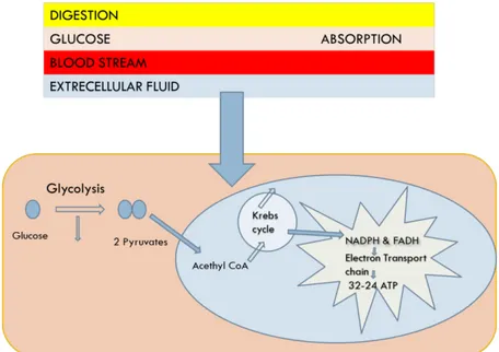
Adenosine triphosphate (ATP) is the main energy source of the cell. ATP is produced through a complex pathway involving both physiological and biochemical processes which convert the food we eat to the useful energy source for the cell. Figure 1.1 summarizes the ATP production, which starts with food digestion followed by glycolysis and the citric acid cycle.
Figure 1.1: Diagram of three stages of cellular metabolism that leads to ATP production from food. Stage 1: occurs
outside of the cell. Stage 2: occurs mainly in the cytosol, except for the final step of conversion of pyruvate to acetyl groups on acetyl CoA, which occurs in mitochondria. Stage 3: occurs in mitochondria1.
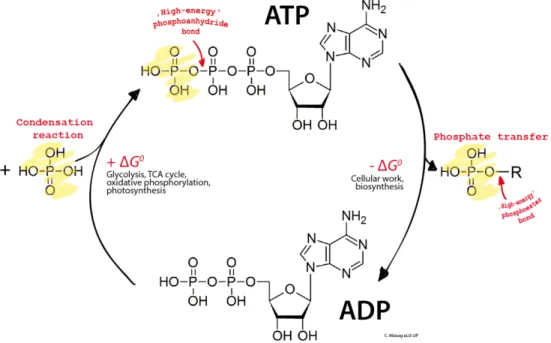
ATP produced is used as a fuel for energy-needed processes in living cells. Such processes include movement of substances across the cell membranes, muscle contraction and synthesis of many macromolecules essential for the cell existence. The energy stored in ATP is released by hydrolysis, which is a breaking down of ATP in ADP and Pi. ADP and Pi production is coupled with ATP synthesis, which guarantees ATP regeneration. Figure 1.2 reports the ATP hydrolysis and ATP synthesis.
17
Figure 1.2: ATP hydrolysis coupled with ATP synthesis.
Concerning the storage, ATP usually reaches high concentrations within cells. However, ATP content is quickly depleted due to ATP-dependent processes together with its low stability in water. Hence, storage and compensative processes are needed to maintain the high ATP concentration within the cell. ATP itself cannot be stored easily within cells, hence ATP is stored as carbon sources (such as triglycerides or glycogen), ADP and AMP, which are converted to ATP through glycolysis and with the help of ATP synthase2.
Surprisingly, Dowdall and co-workers found a noticeable amount of ATP together with acetylcholine in cholinergic vesicles from the electric organ of Torpedo marmorata3. Considerable similar findings (co-storage with noradrenaline, serotonin, dopamine, etc.) were observed in subsequent years in other mammalian and non-mammalian species. Hence, the evidence of co-storage of ATP with neurotransmitters has strongly supported the idea that ATP is a fundamental mediator of purinergic neurotransmission in sympathetic and parasympathetic nerves, where it can induce several purinergic responses (i.e., control of autonomic functions, neural glial interactions, pain and vessel tone control). The purinergic signaling and purinergic receptors sub-family will be discussed in the following section.
1.2 Purinergic receptors family
For years, interest in purines (ATP, ADP, AMP, GTP, GDP, GMP and IMP) and pyrimidines (CTP and UTP) nucleotides was focused on the involvement of ATP in cell metabolism and its role as an energy source 4. Nonetheless, in 1972, Burnstock raised the possibility that ATP was a
18 neurotransmitter5 and in 1978, he proposed that specific extracellular receptors, known as P1
(will be discussed later) and P2, mediate the physiological effects of adenosine (Ado) and ATP, respectively. Subsequently, ATP was known as a co-transmitter5. Hence ATP is now considered either as an exclusive transmitter or as a co-transmitter in both peripheral and central nervous system (CNS)6. The evidence of ATP as neurotransmitter has driven a great interest in purinergic signaling and nowadays a lot has been discovered and published around P2 receptor purinergic transmission, especially, the different P2 receptors sub-family which have been well documented and are discussed in the following section.
1.3 Different P2 receptors sub-family
The work of Burnstock et al in 1985 allowed the classification of P2 purinoreceptors in two major sub-families known as P2X and P2Y7. Subsequent research always carried out by Burnstock allowed to clone and functionally characterized seven subtypes of the human P2X sub-family and eight subtypes of the human P2Y sub-family8.
1.3.1 P2X receptor sub-family
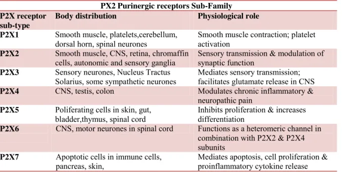
Members of this sub-family are seven ionotropic receptors subtypes most of which are assembled as trimers. The monomers show the following common characteristic: an intracellular N- and C-termini; two trans-membrane spanning regions (TM1 and TM2), a large extracellular loop, with 10 conserved cysteine residues forming a series of disulfide bridges and an ATP-binding site9. Additional details are given in Table 1.1.
19
Table 1.1: some key information about distribution and physiological role of P2X receptor
sub-family10.
PX2 Purinergic receptors Sub-Family P2X receptor
sub-type
Body distribution Physiological role P2X1 Smooth muscle, platelets,cerebellum,
dorsal horn, spinal neurones
Smooth muscle contraction; platelet activation
P2X2 Smooth muscle, CNS, retina, chromaffin cells, autonomic and sensory ganglia
Sensory transmission & modulation of synaptic function
P2X3 Sensory neurones, Nucleus Tractus Solarius, some sympathetic neurones
Mediates sensory transmission; facilitates glutamate release in CNS P2X4 CNS, testis, colon Modulates chronic inflammatory &
neuropathic pain P2X5 Poliferating cells in skin, gut,
bladder,thymus, spinal cord
Inhibits proliferation & increases differentiation
P2X6 CNS, motor neurones in spinal cord Functions as a heteromeric channel in combination with P2X2 & P2X4 subunits
P2X7 Apoptotic cells in immune cells, pancreas, skin,
Mediates apoptosis, cell proliferation & proinflammatory cytokine release
1.3.2 P2Y Receptors sub-family
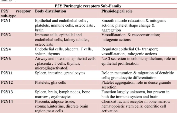
P2Y receptor sub-family comprises eight metabotropic receptor-subtypes characterized by: an extracellular N-terminus, an intracellular C-terminus, and seven trans-membrane spanning regions. Each P2Y receptor subtype binds to a single heterotrimeric G protein (typically Gq/11). However, P2Y12 couples to Gi while P2Y11 can couple to both Gq/11 and Gs. Both purine and
pyrimidine nucleotides activate P2Y2, P2Y4 and P2Y6 receptors, whereas P2Y1 and P2Y11-13 are
activated by purine nucleotides alone. The P2Y14 is dually activated by UDP-sugars, such as
UDP-glucose, and also by UDP. Activation of recombinant P2Y receptors subtype leads either to intracellular calcium release following phospholipase C activation or to cyclic AMP (cAMP) levels alteration due to interaction with adenylyl cyclase (AC)9. Additional details are given in Table 1.2.
20
Table 1.2: some key information about distribution and physiological role of P2Y receptor
sub-family11.
P2Y Purinergic receptors Sub-Family P2Y receptor
sub-type
Body distribution Physiological role P2Y1 Epithelial and endothelial cells ,
platelets, immune cells, osteoclasts , brain
Smooth muscle relaxation & mitogenic actions; platelet shape change & aggregation
P2Y2 Immune cells, epithelial and endothelial cells, kidney tubules, osteoclasts
Vasodilatation & vasoconstriction; mitogenic actions
P2Y4 Endothelial cells, placenta, T cells, spleen, thymus.
Regulates epithelial Cl– transport; vasodilatation, mitogenic actions P2Y6 Airway and intestinal epithelial cells
, placenta , T cells, thymus, microglia(activated)
NaCl secretion in colonic epithelium; role in epithelial proliferation
P2Y11 Spleen, intestine, granalocytes Role in maturation & migration of dendritic cells; granulocytic differentiation
P2Y12 Platelets, glia cells Platelet aggregation; role in dense granule secretion
P2Y13 Spleen, brain, lymph nodes, bone marrow , erythrocytes
Function largely unknown, but present in both the immune system and brain P2Y14 Placenta, adipose tissue,
stomach,intestine, discrete brain region,mast cells
Chemoattractant receptor in bone marrow hematopoietic stem cells; dendritic cell activation
2. Production, transport, metabolism and physiological role of ATP
2.1 Production
ATP is mainly produced from food we eat, from stored macromolecules as glycogen or triglycerides, from ADP and AMP (discussed already in more detail in the section above). Briefly, glucose released either from digestion of sugars or from breaking down of glycogen is transported into the intracellular compartment where it is subsequently converted in pyruvate through glycolysis. At this stage, small amount of ATP and NADH is produced. Then the pyruvate obtained from glycolysis is converted to acetyl coenzyme A (Acetyl-CoA). Finally, Acetyl-CoA moves from cytosol to mitochondria where it enters in Krebs cycle to produce a considerable amount of ATP. In fact, acetyl group is oxidized to carbon dioxide (CO2),
generating a considerable amount of the electron carrier NADH. Then the NADH enter in the electron-transport chain within the mitochondrial inner membrane; during that process of transfer there is a release of energy which is used to drive a process that produces ATP and consumes molecular oxygen (O2)1.
21
2.2 Transport and Metabolism
The synthesized ATP is transported from mitochondria to cytosol by the help of the mitochondrial ADP/ATP carrier. Once in cytosol, ATP is used as energy source for vital cell processes such as cell membrane transport and synthesis of macromolecules12. The mitochondrial ADP/ATP carrier is one of the most abundant carrier proteins of the mitochondrial inner membrane13. The essential activity of the carrier is to import ADP back from cytosol into the mitochondrion for ATP synthesis and to export the synthesized ATP out of the mitochondrion for use in the cytosol. The activity of mitochondrial ADP/ATP carrier allows ATP to reach high intracellular concentration. Even though ATP concentration reaches millimolar range within the cell, it is not stored. In fact, storage is mainly impeded by the high demand of energy needed processes but also partly by the low water stability of ATP. So, to maintain the high concentration of ATP necessary for the cell survival, the depletion of ATP is coupled with a compensative process which converts back ADP generated during ATP hydrolysis to ATP. The compensative process resides on the mitochondrial ADP/ATP carrier, which transports back ADP from cytosol to mitochondria allowing ADP to be reconverted in ATP. Summarizing, ATP synthesized in the mitochondria is transported in cytosol where it is used as energy source for cellular metabolism. ATP is not stored and its high concentration of ATP within the cell is maintained by the reconversion of ADP to ATP.
However, Dowdall and co-workers has brought additional knowledge about the storage of ATP showing that, even if ATP used as energy source is not stored alone, considerable amount of ATP is stored together with neurotransmitters3. An example is the case of co-storage of ATP with acetylcholine in the cholinergic vesicles. This co-storage has brought evidence that ATP, beside its energetic role, is also released in the extracellular space where it behaves like any neurotransmitter. Specifically, neurotransmitter properties of ATP have been proved in purinergic signalling where ATP is the signal molecule mediating the purinergic neurotransmission both in sympathetic and parasympathetic nerves2. More details about ATP as a fundamental mediator of purinergic signalling are given in the next section.
2.3 Physiological role of ATP
Although evidences of ATP as signal molecule were already reported before, the concept of ATP as a signal molecule was accepted in 1998 after P2X and P2Y receptors sub-family were cloned and characterized as receptors for purines and pyrimidines (ATP, ADP and UTP)14. From these preliminary findings, subsequent studies (physiological, pharmacological and biochemical)
22 allowed to expand knowledge about the concept of ATP as signal molecule. Hence, it is now known that, beside the energetic role, ATP mediates neurotransmission in both peripheral and CNS. In addition, ATP is also known as a powerful extracellular messenger modulating the physiology of non-neuronal cells including secretory, exocrine and endocrine, endothelial, immune, musculo-skeletal and inflammatory cells6, 15.
The demonstration of ATP as a neurotransmitter, but also as an extracellular signal molecule in non-neuronal cells, has driven a great interest about the implication of ATP in both physiological and pathological conditions. In the following section it will be discussed briefly some disorders focusing our interest in the implication of ATP either as the cause of the disorder or as potential repair pathway. All informations are gathered in Tables 1.3, 1.4 and 1.5.
The Tables 1.3, 1.4 and 1.5: content information is about the implication of ATP in various pathological and physiological states.
Table 1.3
Disorders Example of disorders
Central nervous system disorder Neurodegenerative diseases Alzheimer’s Diseases (AD) Parkinson’s Disease (PD) Implication of ATP in the disorder P2X7R antagonists are potential therapeutic candidates16
Both P2X7R and P2Y4R antagonists are potential therapeutic17,18
A P2X7R antagonists, brilliant blue G, was recently shown to be protective in an animal model of PD19 Brain injury, Neuroprotection, Neuroregeneration
Brain injury Neuroprotection Neuroregeration
Implication of ATP in the disorder
P2X7R antagonists are target to prevent secondary
neurological injury after traumatic brain injury e and after spinal cord injury20
P2X4R are required for neuroprotection via ischemic preconditioning. Neuroprotection mediated by microglia is associated with P2X7R activation and release of tumor necrosis factor-α21 Activation of P2Y2R evokes regeneration of gial cells and nerves. Neural stem cell activation leads to
neuroregeneration, probably via P2X4R and P2X7R17
23
Table 1.4
Table 1.5
Disorders Example of disorders
Cardiovascular diseases
Heart Failure Hypertension Thrombosis, Inflammation, and Stroke Implication of ATP in the disorder Application of ATP, prior to or just after cardiac ischaemia is cardioprotective22 ATP released as a cotransmitter from sympathetic nerves together with noradrenaline (NA) potentializes, via P2X1R vasoconstriction in hypertension23
Nucleotides are mediators of vascular inflammation and thrombosis. Also P2Y12 antagonists, inhibits platelets aggregation and are widely prescribed for thrombosis and stroke24,25
Diseases of the airways
Asthma Chronic Obstructive Pulmonary Disease (COPD) Airway Infections Implication of ATP in the disorder In human lung mast cells, ATP is an important modulator of histamine release25 COPD is characterized by up-regulation of ATP in bronchoalveolar lavage fluid, which promotes inflammation and tissue degradation. ATP-induced pulmonary vasodilation occurs in patients with COPD 26
Antibiotics, including erythromycin, are used widely for the treatment of lower and upper respiratory tract infections. Erythromycin blocks the P2XR-mediated Ca2+ influx and could
represent one mechanism by which it exerts its effects stroke 27,23
Example of disorders
Implication of ATP in the disorder
Neuropathic pain Brain tumor Lung injury
Antagonists to P2X7R and P2Y12R on microglia reduce neuropathic pain27,28 Neuroblastoma, expresses P2X7Rwhich may be target for treatment since it regulates metabolic activity, angiogenesis and mediates
proliferation29,30
The initial inflammatory cells recruited during lung injury are pulmonary neutrophils and P2X7R antagonists reduced neutrophil infiltration and proinflammatory cytokine level31
24
3. From ATP to Adenosine
The binding of extracellular ATP either on P2X or P2Y receptors subtypes is a key step in the purinergic transmission. In fact, as previously discussed, the interaction of ATP with P2 receptors sub-family is implicated is many disorders and diseases (details are shown in tables 1.3, 1.4 and 1.5). Extracellular ATP is also susceptible to ectonucleotidases, which break down ATP in adenosine (Ado)32. This enzymatic degradation of ATP is one of the main ways of Ado production but represents also the tight link between P2 and P1 purinergic transmission since adenosine is the endogenous ligands of P1 receptors sub-family. P1 purinergic transmission and P1 receptor sub-family are discussed in more detail in the following section.
3.1 P1 Receptors
6-Amino-9-β-D-ribofuranosyl-9H-purine (Ado Fig.1.3) is an endogenous nucleoside, widespread in mammals. Ado is made up of a purine ring substituted in 6 position by a primary amino group and in 9 position by a D-ribosyl ring.
Figure 1.3: Chemical structure of Ado
Drury and Szent-Györgyi in the beginning of years 1900 were the first to outline the role of Ado as an extracellular messenger. In fact, they depicted the considerable vasodilatory potential of Ado33. Consequently during the years 70s, Sattin and Rall exhibited a particular role of Ado in the central nervous system (CNS), showing that it was found to be implicated in the increase of 3',5'-cyclic Ado monophosphate (cAMP) in mammalian brain cuts. They also found that the cAMP formation was hindered by methylxanthines (for example caffeine and theophylline)34. Later on, an inhibitory role of Ado was displayed in cortical neuron35, cerebellar36 and potential
25 synaptic excitatory cortical cuts and hippocampus37. The inhibitory role of Ado on the discharge of acetylcholine38, norepinephrine39, amino acids, and excitatory serotonin40,41 was additionally demonstrated in different brain zones. Each single one of these effects was hindered by methylxanthines while expanded by Ado reuptake inhibitors, and related with an adjustment in cAMP levels. The experimental investigations that pursued these basic informations demonstrated the role of Ado in the homeostasis of the CNS as well as in the homeostasis of peripheral tissues.
Ado operates like a cytoprotective modulator under physiological and pathological conditions in response to oxidative stress in organs and tissues42. This defensive reaction may be expressed in the four following manner: by expanding the oxygen supply whenever required, securing against ischemic damage, activating inflammatory reactions, and stimulating angiogenesis43. Therefore, it is conceivable to expect the presence of a guard system that permits the activation of different reactions important for the recovery of the cell work and an ordinary oxygen homeostasis. Ado has characteristics very close to those of neurotransmitter. In fact, it exerts its function by binding to specific receptors known as adenosine receptors (ARs), it is metabolized in the synapses by the help of specific enzymes, and the fact that its functions can be obstructed by selective antagonists, by the reuptake system and its catabolism in the cytoplasm44. Moreover,
studies have demonstrated that Ado additionally is stored in synaptic vesicles and discharged by neurons in rat45.
3.2 Synthesis, transport and metabolism of Ado
Ado is produced in physiological conditions at the intracellular and extracellular level. It is present in the cytoplasm mostly in its phosphorylated forms. For example Ado monophosphate (AMP), Ado diphosphate (ADP), and Ado triphosphate (ATP). Ado is present in low amount in the extracellular space in the range from 30 to 200 nanomolar (nM)46. During ischemia or
subsequently to an enormous tissue injury that induces cell necrosis, levels of extracellular Ado can achieve an amount of 30 micromolar (µM)16.
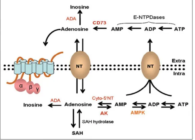
Generation of cellular Ado (Fig. 1.4) relies upon the hydrolysis of AMP. Such hydrolysis is carried out by intracellular 5'- nucleotidase (cN-1), or by the activity of S-adenosylhomocysteine hydrolase on its substrate S-adenosylhomocysteine (SAH)47. The intracellular Ado can be
released into the extracellular space by the help of specific bidirectional transporters (equilibrative nucleoside transporters or ENTs). This movement of Ado out of the cell is crucial since it allows the balancing of the intra- and extracellular level of Ado (Fig. 1.4). ENT1 and
26 ENT2 are the two well documented among the four ENT isoforms which have been found to be encoded by the human genome (ENT1-4). The classification from 1-4 is determined by their sensibility to inhibition carried out by nitrobenzylthioinosine48. These transporters, specifically
ENT1, are widely expressed in the CNS49.
Figure 1.4:Ado synthesis, metabolism, and transport.
In addition to equilibrative transporter-proteins, a few tissues present Na+-dependent concentrative nucleoside transporters (CNTs), able to maintain high concentration of Ado by exchanging against gradient.50 These transporter-proteins have been recognized in brain
macrophages (microglia), thymocytes, liver, lung, choroid plexus, kidneys, and intestine.51Their
activity may vary upon interaction with medications or with decrease of body temperature52.
At the extracellular level, Ado is produced by consecutive dephosphorylation of adenine nucleotides, for example, ATP, mediated by a number of ectoenzymes, such as the apyrase (E-NTPDase1 or CD39) and the 5'- nucleotidase (CD73), found on the cell surface of numerous tissues. The CD73-mediated dephosphorylation which converts extracellular AMP to Ado is a rate restricting step of the nucleoside production53. The whole catalytic pathway is achieved
27 The metabolism of Ado is mostly controlled by two enzymes: Ado deaminase (ADA) and Ado kinase (AK). ADA works at high amount of substrate by converting Ado to inosine. ADA is found both in intra and extra cellular level where it is attached to the layer and takes part in the degradation mechanism of extracellular Ado54. AK, at contrary, works at low amount of substrate
changing Ado to AMP.
Because endogenous amount of Ado , as referenced above, is in the order of nanomolar, it is reasonable to access that certainly, under physiological conditions the fundamental catabolic pathway of Ado is the phosphorylation lead by AK, while the activity of ADA rise up in conditions inducing an increase of Ado, for example, during an ischemic episode55.
3.3 Adenosine receptors
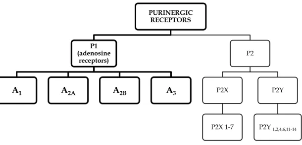
Ado achieves its functions by interfacing with specific membrane receptors coupled to G proteins (GPCR), known as ARs. ARs are classified as P1 purinergic receptor family. Up until now, four diverse receptor subtypes have been identified. According to their chronological discovery they are named A1, A2A, A2B, and A3 (Fig. 1.5).
Figure 1.5: Classification of purinergic receptors.
P1 receptors were firstly divided only into A1 and A2 subtypes. This division was based on their
capacity to repress or to activate the adenylate cyclase (AC) 56. Later on, the A
2 subtype was
additionally studied and further characterized by Daly et al57 describing them, on the base of
PURINERGIC RECEPTORS P1 (adenosine receptors) A1 A2A A2B A3 P2 P2X P2X 1-7 P2Y P2Y 1,2,4,6,11-14
28 higher or lower affinity for Ado, in two subtypes: A2A, which has higher affinity for the Ado
(0.1-1.0 µM), and A2B, having low affinity for the Ado (> 10 µM). A3AR was the latest to be
discovered in 1991 by recognizable proof of the rat cDNA sequences encoding a receptor coupled to regulatory proteins (G proteins) utilizing a polymerase chain reaction (PCR). Subsequently, Zhou and et al58, reported that one of these sequence had high homology with AR
subtypes A1 and A2A. However, unlike to others AR subtypes which have present high homology
among the different species, the "newfound" receptor demonstrated a moderately low sequence homology between the rat and human subtypes59. Furthermore, the "newfound" receptor
demonstrated a difference binding with antagonists60. The classification of P1 receptors has been
affirmed with molecular cloning studies allowing the expression of the four receptor subtypes.42
ARs are found on the membrane of various cell types in the CNS, for example, neurons and glial cells61. At peripheral level they are found on the cells of the vascular smooth musculature, in
platelets, lymphocytes, monocytes, macrophages, neutrophils, basophils, eosinophils, mast cells, lungs, heart, bladder and in immune tissues62.
3.4 Structure of the adenosine receptors
ARs are metabotropic receptors coupled to G proteins (Fig. 1.6). GPCRs are integral membrane proteins fundamentally made up of a single polypeptide chain that crosses 7 times the plasma membrane and having an extracellular N-terminal domain and an intracellular C-terminal domain. The seven transmembrane domains (TM1-7) are sorted out in an α-helix structures, each comprising from 20-27 hydrophobic amino acids, associated together by three intracellular loops (IL1-3) and three extracellular loops (EL1-3). Two cysteine amino acid residues (one interface TM3/EL1, and the other in EL2), forming a disulfide connect, are essential for the right folding of the protein63.
ARs vary essentially in the domain length of the N-terminus, the C-terminus domain, as well as in the intra and extra cellular loops. Every one of these domain gives explicit properties to every AR subtype deciding explicit ligand selectivity profiles. The terminal area contains N-glycosylation sites having a key role in the right distribution of the receptor in the cell, whereas the C-terminal area has serine and threonine residues, phosphorylation sites for protein kinase implicated in the receptor desensitization64.
29
Figure 1.6: Structure of GPCRs.
The C-terminal tail of A1, A2B, and A3 ARs has a conserved cysteine residue which might be a
palmitoylation site, permitting the arrangement of a fourth IL. ARs are among the littlest members of the GPCR family. Human A1, A2B, and A3 are made up of 326, 328, and 318 amino
acids, respectively, while A2A subtype with its 410 amino acids has a more extended C-terminal
tail. The additional properties of this more extended C-terminal tail is that it contains the binding site of accessory proteins65.
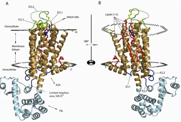
Additional information about the structure of A2A subtype was provided in 2008 thanks to the
crystal structure of the A2A antagonist ZM241385 bound with A2AAR66 (Fig 1.7). In the following
years A2A subtype was also crystallized with A2AAR agonists UK 432,09767, 5'-
30
Figure 1.7: Structure of the antagonist ZM241385-bound A2AAR.
Prior to the discovery of the crystal structure, the primary prediction strategy used for ARs was the homology modelling in which the receptor model was made according to the crystallographic structure of rhodopsin and mutagenesis studies.
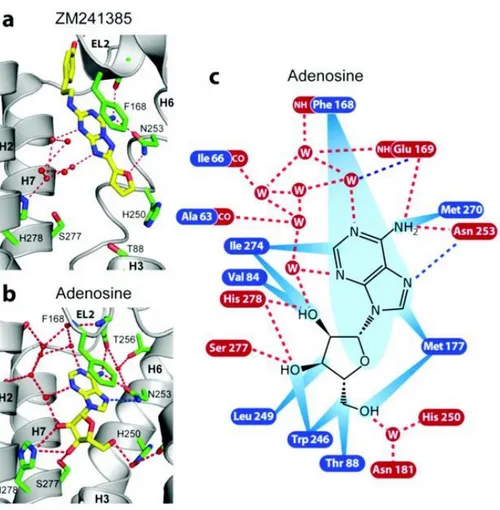
The principle distinction of binding mode between AR agonists and AR antagonists reside on the ability of the agonist to establish hydrogen bonds between its hydroxyl groups of the ribosyl moiety with Ser277 (7.42) and His278 (7.43) residue of the AR (Fig. 1.8). In fact those hydrogen bonds allow the pulling of the extracellular end of TM3, TM5, and TM7, and this pulling has been suggested to be essential for the activation of the receptor. Moreover, because of the existence of hydrogen bond donors in the ribose moiety, amino acids Val84 (3.32) and Trp246 (6.48) could considerably move from their positions inducing like this a change that seems, critical for the accomplishment of the conformation vital for receptor activation.
31
Figure 1.8: Interactions between the human A2AAR and a) ZM 241385 and b) Ado. The interactions between the
receptor and Ado c) are shown as red dotted lines (hydrogen bonds), blue dotted lines (polar interactions), and blue rays (van der Waals interactions).
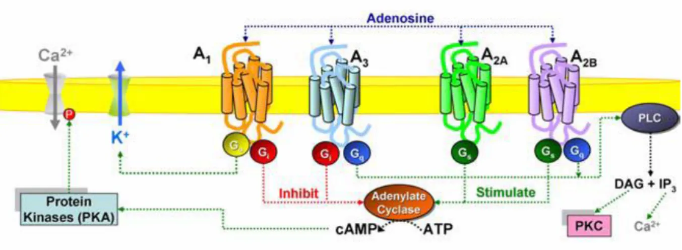
3.4 Signal transduction
The presence of GPCRs on the outer part of the cell membrane, allows them to transduce many extracellular signals into the cells. This happens through the activation of at least one heterotrimeric G proteins situated on the cytoplasmic side of the membrane when an agonist binds to the extracellular domain of receptor followed by the interaction with different effector systems, for example, ion channels, phospholipase, and adenylyl cyclase (AC).
G proteins are made up of three subunits: α, β, and γ. At least 20 subtypes of the α subunits, 7 subtypes of the of β subunits and 12 subtypes of the subunits are known in humans. According to actions of the functional subunit α, we can recognize the following diverse G proteins: Gs
(stimulating the AC), Gi (repressing AC), Gq (activating phospholipase C, PLC),.
A1 and A3 ARs are generally coupled to inhibitory G proteins (Gi) whereas A2A and A2B subtypes
32
Figure 1.9: Schematic representation of the signal transduction pathways associated with ARs.
The G protein in the latent state is a trimer, with the α-subunit bound to guanosine diphosphate (GDP- αβ). When an agonist come to interact and activates the receptor, there is a conformational change allowing the receptor to interact with the G protein. The conformational change is followed by the separation of GDP from αβ allowing the formation of a vacuum transition state (αβ). Then transition state (αβ) interacts with guanosine triphosphate (GTP) to build up GTP- αβ. The complex GTP- αβ triggers a conformational change which induces the activation and separation of the protein from the receptor. At this point the activated G protein separates into two components: the complex GTP-α and the dimer β, which activate the effectors of the signal transduction. The endogenous GTP-ase activity of the α-subunit, in the complex GTP-α, hydrolyses GTP to GDP. The hydrolysis results to reconstitution of the initial GDP- αβ complex which can therefore interact with another GPCR when activated by an agonist (Fig. 1.10). Antagonists prevent the separation of GDP from the GDP- αβ complex and by consequent they hamper the G protein activation.
The primary effectors in charge of the generation of second messengers are the two following enzymes: AC, in charge for the production of cAMP and PLC, the enzyme in charge of the production of inositol triphosphate (IP3) and diacylglycerol (DAG).
33
Figure 1.10: The activation mechanism of G proteins.
Even though the fundamental way of signal transduction for all ARs is through the interaction with G proteins, there are noticeable differences in signal transduction among the four ARs.
3.5 Adenosine receptor subtypes 3.5.1 A1AR
The A1AR subtype was purified, cloned and sequenced in various species including human
species. Although there were featured contrasts in G protein-coupling as well as a species- dependent tissue distribution, a considerable homology has been found among the A1AR
subtypes of different species. The gene which guarantees the existence of the A1AR subtype in
human species is found on chromosome 1q32 and encodes a protein of 326 amino acids, with a molecular weight of about 36.7 kDa63.
A1AR subtype is generally distributed but mainly expressed in the CNS in pre-and postsynaptic
34 hippocampus, cerebellum, thalamus, the spinal line and fat tissue. Moderate expression is found in skeletal muscle, liver, kidney, salivary organs, throat, colon, eyes, in the sinoatrial and atrioventricular hub of the heart, in the cave of the stomach and in the testicles. Relatively lower expression is notice in the lungs, ventricles and pancreas.70
About the signal transduction, A1ARs are essentially coupled to G proteins which belong to Gi/q
family71 and many are the signal transduction pathways which have been attributed to this
receptor subtype. Some of them are listed below:
inhibition of AC with a subsequent decline of cAMP72levels followed by phosphorylation of various target proteins by cAMP-dependent protein kinase (PKA)
activation of PLC with a subsequent increment in the generation of IP3, DAG, and
accumulation of Ca2+ from intracellular stores contributing to the activation of protein kinase C (PKC), phospholipase A2 (PLA2), and nitric oxide synthase (NOS)73 ;
activation of various K+ channels through coupling with G proteins having a place in G o
family.
Through those signal transduction pathways, A1ARs modulates many responses in various
systems of the organism. Their stimulation causes:
in the CNS: a decrease of transmitters release, sedation, anticonvulsant impacts, anxiety, locomotor-depressants74;
in metabolic systems: there is an antilipolytic effect and an increase insulin sensitivity75;
in the gastrointestinal tract: there is an inhibition of chloridiric acid production;
in the renal system: a decrease in glomerular filtration rate (GFR), an inhibition of renin release, an antidiuretic effect and a supply route vasoconstriction;
in the cardiovascular system: a negative inotropic effect, negative chronotropic effects, negative dromotropic effects, negative bathmotropic effects and cardioprotection76.
3.5.2. A2AAR
The first in all crystallographic structure of A2AAR was resolved in 2008.66 The gene which
guarantees the existence of this subtype in human species is found on chromosome 22 and encodes a protein of 410 amino acids77 with a molecular weight of 45 kDa78. The A
2AAR is found at
central level in dopamine-rich regions, for example, in the striatum79, nucleus accumbens,
olfactory tubercle, and in the Purkinje cells of the cerebellum. At peripheral level they are plenty in endothelial cells, platelets, lymphocytes, monocytes, macrophages, neutrophils, basophils, eosinophils, pole cells, lungs, heart, bladder, and immune tissues33,80.
35 A2AAR is linked by the II and III intracytoplasmic loops to Gs proteins at the peripheral level
and to Golf at the CNS level, both stimulating AC. The stimulation of AC induces an increase of intracellular amount of cAMP81. Then the high amount of intracellular cAMP induces the
activation of PKA. The activated PKA can then activate different receptors, ion channels, phosphodiesterases and cAMP-managed phosphoprotein (CREB, cAMP responsive element binding protein), critical for numerous neuronal capacities. The activities of the A2AAR are very
huge because it is sometime co-localized or being sometime physically connected with orther GPCRs, for example, the formation of heterodimers with the D2 dopaminergic receptor
(D2/A2AAR)82, with CB1 cannabinoid receptor (CB1/A2AAR)83, and with mGluR5 glutamate
receptor (mGluR5/A2AAR)84, and heterotrimers (CB1/A2AAR/D2)85.
The A2AAR is particularly implicated in the modulation of vasodilation; it stimulates the
synthesis of new blood vessels, and preserves tissues from collateral damage of inflammatory process. In the brain, A2AAR indirectly impact the activity of the basal ganglia.
Additional mechanism mediated by A2AAR includes:
• in the CNS: stimulation of the sensory nerve and synergistic repression of D2;
•in the immune system: repression of polymorphonuclear leukocytes, repression of the discharge pro-inflammatory cytokines (TNF, IL-6, IL-8, and IL-12)86, and activation of the discharge of
inflammatory cytokines (IL-10);
•in the cardiovascular system: repression of platelet aggregation.
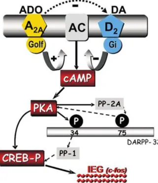
3.5.2.1 Interaction between A2AAR and the dopamine D2 receptor
Strong experimental proof shows that the A2AAR subtypes interacts in an extremely broad way
with dopaminergic receptors at the level of the basal ganglia. A2AAR and D2 receptors are
co-localized in the membrane of the GABAergic striatopallidal neurons. This co-localization provides anatomical basis for the existence of functional association between these two receptors, moreover it has been broadly proved that activation of A2AARs enhance the
functionality of striatopallidal neurons, due to the decrease of D2 receptor activity87. This
co-expression of A2AAR and D2 receptors on neurons of the striatum and pallidum gives the
anatomical premise to the presence of a practical association between these receptors. Furthermore, the impact of excitatory neurons in the striatum and pallidum applied by Ado upon A2AAR is due somehow to their antagonistic effect on the activation of D2 receptors by
dopamin88. Additional biochemical studies performed in rat striatal tissue, in human, and in
36 has been shown that activation of A2AARs diminishes the affinity of D2 receptors for dopamine
binding.89These impacts are related with the formation of heterodimeric complexes between the
two receptors90. The interaction existing between the A
2AAR and D2 receptors is extended even
at the second messenger level because, whereas the activation of D2 receptors represses AC, the
activation of A2A ARs stimulates AC (Fig. 1.11)73.
Figure 1.11: Interactions between A2AAR and D2 receptors. The two receptor subtypes exert opposite regulatory
activity on the AC, which is inhibited by D2 receptors and stimulated by A2AAR.
Moreover, it has been demonstrated that the activation of A2AAR can repress the Ca2+-dependent
reactions mediated by the D2 receptor74, 90.
It has been demonstrated also that A2AARs can influence the function of the D1 receptors.
Hence, it has been seen that administration of A2AAR antagonists can enhance both the
behavioral and neurochemical effects induced by iactivation D1 receptor in the rat.91 Because
A2AAR and D1 receptors are located on various neuronal populations,92 it is reasonable to suggest
that, the interaction between these receptors happens via the side fibers of the striatum or via circuits that incorporate subthalamic-nigro-cortico striatal projections. In mice missing the dopamine D2 receptors, activation or blockade of A2AARs generates behavioral as well as
biochemical effects, supporting the theory that the activity of A2AARs might be autonomous of
37 3.5.3 A2BAR
The gene which guarantees the existence of the human A2BAR is found on chromosome
17p11.2-p12 and encodes a protein of 328 amino acids, with molecular weight of about 37.0 kDa63. A
2BAR signal transduction initiates with activation of AC via a direct coupling with Gs
protein resulting to an increment of cAMP levels; then the increase of cAMP levels induces a stimulation of P type Ca2+ channels as well as the activation of PKA which phosphorylates protein residues, treonine and serine, and stimulates gene transcription. Moreover, A2BAR is
known to be couple through Gq protein family, to a phosphatidylinositol-lipase protein C (PI-PLC) system promoting an increase of DAG, which stimulates PKC, and IP3, assembling intracellular Ca2+.
At first, it was believed that the expression of the A2BAR was limited to organs like the bladder,
bowel, lung, epididymis, vas deferens, spine, and brain. However, they were found later to be also present in fibroblasts94, hematopoietic cells95, mast cells96, cells of the myocardium97, muscle cells98, and endothelium99.
Large amounts of the A2BAR expression were found in various parts of the intestinal tract and
their activity on gastric mucosal cells has been investigated100. There are also proofs supporting
that the presence of A2BAR on mast cells is implicated in inflammatory processes, for example,
asthma101. These A
2BARs on mast cells activate the human mast cells MHC-I and stimulate the
release of IL-8.
Numerous investigations associate A2BARs with cardiovascular effects since their activation can
regulate fibroblast proliferation102, heart recoveries, and illness such as hypertension or
myocardial infarction103.
Among different effects mediated by A2BAR there are:
• in CNS: inhibition of nerve transmission, stimulation of IL-6 production in astrocytes; • in gastrointestinal system: stimulation of chloridric acid production into the intestinal lumen.
3.5.4 A3AR
The gene which guarantee the existence of the human A3AR is found on chromosome 1p13.3
and encodes for a protein of about 318 amino acids, with a molecular weight between 36.0 to 37.0 kDa63. In contrast with other ARs, the A
3AR has huge differences in structure, function and
tissues distribution among species104. These differences make especially complex the A 3AR
signalling physiology specifically among rodents and primates. The activation of A3AR induces
38 cAMP. This activation stimulate also PLC and phospholipase D (PLD), via the coupling with the Gq protein105, and finally stimulate the release inflammatory mediators (for example histamine
from mast cells)106.
A3ARs are found with high density in the lungs, liver, and immune system cells like neutrophils,
eosinophils, and T lymphocytes, while it is present in low density in order tissues like brain, heart, testes, and many others.
Via interaction with A3ARs, Ado has a cerebral protective role. Elevated amounts of A3ARs
were found in eosinophil cells in the lungs of rat, where they promote the inhibition of the degranulation and the discharge of free superoxide anion radicals107.
Anticancer properties have been also assigned to A3ARs: in fact, their activation seems to repress
the tumor development by modulating the Wnt pathway and by regulating NF-κB108. In addition,
the ability of A3AR antagonists to decrease intraocular pressure is under study as potential
therapeutic agents in the treatment of glaucoma109. Other effects promoted by A
3ARs are:
• in the CNS: neuroprotection, sedative-hypnotic, anticonvulsant and anxiolytic effects, inhibition of exocytosis;
39
4. Adenosine 4.1 Overview
In chapter 1, the discussion was made around ATP as cell energy source and ATP as signal molecule for P2 purinergic signalling. In this chapter, the attention is focused on the Ado, P1 receptors sub-family and therapeutic potential of Ado receptor ligands.
Introduction
Previously, the two major metabolic pathways of extracellular ATP were described. Until recently, damaged or dying cells were considered as the only source of extracellular ATP. However, the release of ATP induced mechanically from healthy cells is now also accepted as a physiological mechanism110. A well-known example of physiological release of extracellular ATP is the co-release of ATP and noradrenaline found in sympathetic nerves111. Once in the extracellular compartment, ATP follows two main pathways, which are the modulation of P2 purinergic signalling (details in the chapter one) and the production of Ado through cascade of enzymatic processes catalyzed by ectonucleotidases. In fact, both ATP and ADP are hydrolyzed to AMP by a subtype of ectonucleotidases known as ectonucleoside triphosphate diphosphohydrolase1 (NTPDase1), which is abbreviated CD39 for simplicity. The obtained AMP is further hydrolyzed to Ado by another subtype of ectonucleotidase known as ecto-5’-nucleotidase (5’NT) generally known as CD7332. Another important source of extracellular Ado is cAMP, which can be released from neurons and then converted into Ado in two steps: that is conversion of cAMP to AMP by extracellular phosphodiesterases followed by the conversion of AMP to Ado by CD7342. Extracellular Ado produced either from ATP or cAMP or constitutes the signal molecule for the P1 purinergic receptor signalling, which modulates several physiological and pathological conditions. In order to address the therapeutic potential of Ado, we will discuss in more details Ado, P1 purinergic receptors sub-family, and ligands for adenosine receptors (ARs) that are agonists, antagonists and allosteric modulators.
4.2 Production, metabolism and physiological role of adenosine
Ado (6-amino-9-β-D-ribofuranosyl-9H-purine) is an endogenous nucleoside ubiquitous in humans and other species. The structure of Ado is constituted from adenine and D-ribose, which
are linked together with a N9 β-glycosidic bond (shown in figure 1.12). Ado plays a vital role in the body since it regulates the function of almost all tissues and constitutes a key component in the composition of many biomolecules such as ATP, RNA, NADH, etc112.