Prof. Pietro Cortelli
Prof.ssa Maria Giovanna Gandolfi
I
n
s
e
ri
s
c
i
il
t
e
s
t
o
q
u
i
Prof. Carlo Prati
Summary
Peri implant and periapical bone defects are widespread in the world population. Considering the
high number of implant placement and root canal treatment performed every year, resolution of
these bone defects will be of predominant interest in the next future.
The present project has several aims:
The first part of the project aimed to investigate how and what factors affect peri implant bone
remodeling process. Microchemical analysis of the peri implant bone interface of retrieved human
dental implants was performed. In addition, prospective clinical studies were carried out to evaluate
the factors mostly related to peri implant bone loss.
The second part of the project aimed to evaluate chemical physical and micromorphological
properties of hydraulic calcium silicates based materials (CaSi), which gained a particular attention
in the endodontic field. Use of these materials claimed to resolve several complex endodontic
complications, however few information is present in literature. These materials were compared
with different “gold standard” bioinert materials.
Given the attractive biointeractive properties of CaSi based materials, the last part of the study will
focus on the design and characterization of new mineral based scaffolds, aimed to be applied in
future bone regeneration procedures. Scaffolds, composed of a polymeric matrix were doped with
CaSi and calcium phosphates, in order to increase the materials biointeractive properties. A
complete characterization of their chemical-physical-mechanical-thermal properties was performed,
as well as the evaluation of apatite forming ability (bioactivity) and biocompatibility of these
mineral based scaffolds. The combination of Human Periapical-cysts Mesenchymal Stem Cells
(hPC-MSC) as a potential strategy to achieve periapical bone regeneration was also evaluated.
Finally, the angiogenesis potential of these scaffolds was investigated through the growth and
proliferation of porcine vascular wall mesenchymal stem cells (pVW-MSCs) was performed.
1. Peri-implant bone remodeling and clinical strategies to
achieve soft and hard tissue stability
1.1 BONE REMODELLING AT THE PERI-IMPLANT BONE INTERFACE
1.1.1 Microchemical and micromorphologic ESEM-EDX analysis of bone mineralization at the thread interface in human dental implants retrieved for mechanical complications after 2 months to 17 years 1.1.2 Esem-Edx Microanalysis Of The Bone-Implant Interface Around Loaded And Unloaded Retrieved Dental Implants.
1.2 CLINICAL STRATEGIES TO ACHIEVE (ALVEOLAR PERI IMPLANT) BONE STABILITY
1.2.1 Factors affecting soft and hard tissues around over tissue level hyperbolic neck implants. A three-year prospective cohort study
1.2.2 A multi-level analysis of platform-switching flapless implants placed tissue-level: 4-year prospective cohort study
2. Endodontic-related periapical bone defects and new
biomaterials for periapical bone regeneration
2.1 ENDODONTIC-RELATED PERIAPICAL BONE DEFECTS
2.1.1 The fate of root canals obturated with Thermafil: 10-year data for patients treated in a master's program
2.1.2 A 20-year historical prospective cohort study of root canal treatments. A Multilevel analysis 2.2 NEW BIOMATERIALS FOR PERIAPICAL BONE REGENERATION
2.2.1 Properties of a novel polydimethylsiloxane endodontic sealers
2.2.2 Properties of bioroot RCS, a tricalcium silicate endodontic sealer modified with povidone and polycarboxylate
2.2.3 Properties of calcium silicate-monobasic calcium phosphate materials for endodontics containing tantalum pentoxide and zirconium oxide
3. Tissue engineering approaches for experimental bone
regeneration procedures
3.1 DEVELOPMENT OF EXPERIMENTAL MINERAL DOPED POLYMERIC SCAFFOLDS
3.1.1 Polylactic Acid-Based Porous Scaffolds Doped With Calcium Silicate And Dicalcium Phosphate Dihydrate Designed For Biomedical Application
3.1.2 Highly Porous Polycaprolactone Scaffolds Doped With Calcium Silicate And Dicalcium Phosphate Dihydrate Designed For Bone Regeneration
3.2 CHARACTERIZATION OF EXPERIMENTAL MINERAL DOPED POLYMERIC
SCAFFOLDS
3.2.1 Pla-Based Mineral-Doped Scaffolds Seeded With Human Periapical Cyst-Derived Mscs: A Promising Tool For Regenerative Healing In Dentistry
3.2.2 Culture Of Porcine Vascular Wall - Mesenchymal Stem Cells On 3d Biodegradable Highly Porous Mineral Doped Poly (a-Hydroxy) Acids Scaffolds
1. Peri-implant bone remodeling and clinical strategies to
achieve soft and hard tissue stability
Dental implants have become an attractive treatment to restore edentulism allowing the preservation of natural adjacent teeth (Jivray & Chen 2006), restoring edentulous jaws, masticatory function and quality of life (Jofre et al. 2013; Hartlev et al. 2014). A high % of survival has been described in literature, with values over 90% (Lang & Berghlund 2011, Buser et al. 2012, Degidi et al. 2012, Gottfredsen et al. 2012, Jemt 2016).
A substantial increase in the world proportion of individuals receiving implants has been observed. Every year, more than 1.8 million implant surgeries are performed in the European Union (Dental Implants Market to 2027 - Global Analysis and Forecasts by Product; Material; End User and Geography); this number is expected to increase due to the significative increase of the elderly population. A quarter of the whole European population will be over 60 years of age by 2020. (Market research report 2018).
Annually, more than 800,000 individual implants are placed in the United States. This country already faced a significative increase of implant placement in the last years, from 0.7% of the whole population in 1999 to 5.7% to 2016. Considering that the greater increase was observed in elderly patients, population with at least one dental implant rehabilitation is expected to jump over 17 % by 2026 (Elani et al. 2018).
It is important to investigate and monitor the bone levels in the first year from implant insertion. Several biological processes start immediately after implant insertion; these events bring to the formation of new bone tissue close to the implant surface (osseointegration) (Albrektsson & Johansson 2001). This process follows the traditional intrabony wound healing phases: hemostasis (minutes to first hours), inflammatory phase (first hours to days), proliferative days (days to 3 weeks) and remodeling phase (from 3 weeks to years) (Terheyden et al. 2012).
Different circumstances may occur in this period and are critical for long-term success, as may trigger bone level alterations (Galindo Moreno et al. 2014). Indeed, early implant failures have been described in literature in approx. 3-6 % of the cases (Berghlund et al. 2002; Carr et al. 2019). Initial breakdown of the implant-tissue interface generally begins at the crestal region in successfully osseointegrated endosteal implants. In these cases, bone loss beyond the first thread of dental implants may be detected radiographically during the first year of function. (Oh et al. 2002).
The present chapter will first focus on the bone remodeling processes at the peri implant bone interface. Then, several clinical strategies will be explored to maintain bone level stability during the first months from implant placement.
REFERENCES
Albrektsson T, Johansson C. Osteoinduction, osteoconduction and osseointegration. Eur Spine J. 2001 Oct;10 Suppl 2:S96-101.
Berglundh T, Persson L, Klinge B. A systematic review of the incidence of biological and technical complications in implant dentistry reported in prospective longitudinal studies of at least 5 years. J Clin Periodontol 2002, 29, 197-212.
Buser D, Janner SF, Wittneben JG, Bragger U, Ramseier CA, Salvi GA. 10-year survival and success rates of 511 titanium implants with a sandblasted and acid-etched surface: a retrospective study in 303 partially edentulous patients. Clin Implant Dent Relat Res 2012; 14: 839–851.
Carr AB, Sinha N, Lohse CM, Muller OM, Salinas TJ. Association Between Early Implant Failure and Prosthodontic Characteristics. J Prosthodont. 2019 Jan;28(1):30-35.
Degidi M, Nardi D, Piattelli A. 10-year follow-up of immediately loaded implants with TiUnite porous anodized surface. Clin Implant Dent Relat Res 2012; 14: 828–838.
Dental Implants Market to 2027 - Global Analysis and Forecasts by Product; Material; End User and Geography.
Elani HW, Starr JR, Da Silva JD, Gallucci GO. Trends in Dental Implant Use in the U.S., 1999-2016, and Projections to 2026. J Dent Res. 2018 Dec;97(13):1424-1430.
Fischer K, Stenberg T. Prospective 10-year cohort study based on a randomized controlled trial (RCT) on implantsupported full-arch maxillary prostheses. Part 1: sandblasted and acid-etched implants and mucosal tissue. Clin Implant Dent Relat Res 2012; 14: 808–815.
Galindo-Moreno P, León-Cano A, Ortega-Oller I, Monje A, O Valle F, Catena A. Marginal bone loss as success criterion in implant dentistry: beyond 2 mm. Clin Oral Implants Res. 2015 Apr;26(4):e28-e34.
Gotfredsen K. A 10-year prospective study of single tooth implants placed in the anterior maxilla. Clin Implant Dent Relat Res 2012; 14: 80–87.
Hartlev, J, Kohberg, P, Ahlmann, S, Andersen, NT, Schou, S, Isidor, F. 2014. Patient satisfaction and esthetic outcome after immediate placement and provisionalization of single-tooth implants involving a definitive individual abutment. Clin Oral Implants Res. 25(11):1245–1250.
Jemt T. Single-implant survival: more than 30 years of clinical experience. Int J Prosthodont 2016; 30: 551–558.
Jepsen S, Berglundh T, Genco R, et al. Primary prevention of peri‐im‐plantitis: managing peri‐implant mucositis. J Clin Periodontol. 2015;42 Suppl. 16:S152–157.
Jivraj S, Chee W. Rationale for dental implants. Br Dent J. 2006 200(12):661–665.
Jofre J, Castiglioni X, Lobos CA. Influence of minimally invasive implant-retained overdenture on patients’ quality of life: a randomized clinical trial. Clin Oral Implants Res. 2013 24(10):1173–1177.
Lang NP, Berglundh T, Working Group 4 of Seventh European Workshop on P. Periimplant diseases: where are we now?–Consensus of the Seventh European Workshop on Periodontology. J Clin Periodontol. 2011;38 Suppl. 11:178– 181.
Market research report. Dental Implants Market Size, Share & Trends Analysis Report By Product (Titanium Implants, Zirconium Implants), By Region (North America, Europe, Asia Pacific, Latin America, MEA), And Segment Forecasts, 2018 – 2024; ID GVR-1-68038-566-3.
Sanz M, Chapple IL, Working Group 4 of the VEWoP. Clinical research on peri‐implant diseases: consensus report of Working Group 4. J Clin Periodontol. 2012;39 Suppl 12:202–206.
1.1 Bone remodelling at the peri-implant bone interface
Bone is a dynamic, highly vascularized connective tissue with the unique capacity to heal and remodel depending of physiological load (Jayakumar & Di Silvio, 2010). Modifications in response to mechanical demands lead to significative variations of mineral and organic content (Davies 2014).
Oral bone remodeling processes close to the implant threads gained a particular interest in modern implantology, as a stable peri-implant bone tissue is essential to achieve long term results (Davies et al. 2003, Qian et al. 2012). Numerous factors may affect peri-implant bone stability and during the first months from insertion, including surgical techniques (De Bruyn et al. 2014, Prati et al. 2016), implant placement timing (Lang et al. 2012, Zhao et al. 2016, Prati et al. 2017), surgical trauma, occlusal overload (Oh et al. 2002), implant insertion depth (Hermann et al. 1997, Sanz et al. 2015) and design of implant system (Araujo & Lindhe 2018). Towards all these factors, mechanical loading is considered as the most critical (Davies et al. 2003, Halldin et al. 2014, Araujo & Lindhe 2018). Interfacial stress of the implants is mainly located at the interface between the implants and the surrounding bone, thus possibly induce bone resorption and remodelling in this critical area (Chen et al. 2011).
The peri-implant bone interface has been investigated mostly on animal models (Palmquist 2009, 2010). Histological and histomorphometric studies evidenced a tight bone to implant contact with the implant and bone and high percentages of bone to implant contact were related to implant stability and success (Palmquist 2009 and 2010).
However, in-vivo peri-implant bone interface may be highly heterogeneous and may include mineralised, partially mineralised (osteoid), and unmineralised areas (Haiat et al. 2014). To date very few studies analysed the degree of mineralization at the peri-implant bone interface around human dental implants, for it is very difficult to obtain the sample histology (Shah et al. 2014 and 2019, Mangano et al. 2015, Iezzi et al. 2016).
Environmental scanning electron microscope (ESEM) is a useful nondestructive investigation able to analyse mineralized tissue micromorphology without sample manipulation or deterioration (Gandolfi et al. 2017 and 2018) and may result attractive in the investigation of the peri-implant bone interface.
Energy-dispersive x-ray spectroscopy (EDX) is useful for studying the composition of mineralized tissues or mineral (apatitic) deposits and the mineralization degree of bone by calculating the element ratios (atomic or weight) and it allows detection of elements that may have migrated from an implanted material into the bone tissue.
Calcium-to- phosphorous (Ca/P) ratio has been used in several studies to assess bone mineralization (Bigi et al. 1997, Kourkoumelis et al. 2012). Likewise, atomic calcium-to-nitrogen (Ca/N) and phosphorus-to-nitrogen (P/N) ratios have been used to investigate the degree of mineralization by evaluation of organic components in dentin and bone tissue (Engfeldt 1974, Eliades et al. 2013, Gandolfi et al. 2018).
In the following studies, the peri implant bone interface of different retrieved dental implants was investigated using a non-destructive methodology, which allowed assessing the degree of mineralization and the presence of metal contaminants in such critical area.
The second study aimed to investigate the role of loading on peri implant bone remodelling events.
REFERENCES
Araujo MG, Lindhe J. Peri-implant health. Journal of Clinical Periodontology 2018;45:S230-S236.
Bigi A, Cojazzi G, Panzavolta S, Ripamonti A, Roveri N, Romanello M, Noris Suarez K, Moro L. Chemical and structural Characterization of the Mineral Phase from Cortical and Trabecular Bone. J Inorg Biochem 1997;68:45-51.
Chen LJ, He H, Li YM, Li T, Guo XP, RF Wang. Finite element analysis of stress at implant–bone interface of dental implants with different structures. Transactions of Nonferrous Metals Society of China 2011;21: 1602-1610.
Davies JE, Mendes VC, Ko JC, Ajami E. Topographic scale-range synergy at the functional bone/implant interface. Bio materials 2014;35:25–35.
Davies JE. Understanding peri-implant endosseous healing. J Dent Educ. 2003;67:932-49.
De Bruyn H, Atashkadeh M, Cosyn J, van de Velde T. Clinical outcome and bone preservation of single TiUnite™ implants installed with flapless or flap surgery. Clin Implant Dent Relat Res 2011; 13:175–183.
Eliades G, Mantzourani M, Labella R, Mutti B, Sharma D. Interactions of dentine desensitisers with human dentine: morphology and composition. J Dent 2013;41:S28-S39.
Engfeldt B, Hjerpe A. Density gradient fractionation of dentine and bone powder. Calcif Tissue Res 1974;16:261-275.
Gandolfi MG, Iezzi G, Piattelli A, Prati C, Scarano A. Osteoinductive potential and bone-bonding ability of ProRoot MTA, MTA Plus and Biodentine in rabbit intramedullary model: Microchemical characterization and histological analysis. Dent Mater 2017;33:221-238.
Gandolfi MG, Zamparini F, Iezzi G, Degidi M, Botticelli D, Piattelli A, et al. Microchemical and micromorphologic ESEM-EDX analysis of bone mineralization at the thread interface in human dental implants retrieved for mechanical complications after 2 months to 17 years. Int J Periodontics Restorative Dent 2018;38:431-441.
Haïat G, Wang HL, Brunski J. Effects of biomechanical properties of the bone-implant interface on dental implant stability: from in silico approaches to the patient's mouth. Annu Rev Biomed Eng. 2014 Jul 11;16:187-213.
Halldin A, Jimbo R, Johansson CB, Wennerberg A, Jacobsson M, Albrektsson T, Hansson S. Implant stability and bone remodeling after 3 and 13 days of implantation with an initial static strain. Clin Implant Dent Relat Res. 2014;16:383-93.
Hermann JS, Cochran DL, Nummikoski PV, Buser D. Crestal bone changes around titanium implants. A radiographic evaluation of unloaded nonsubmerged and submerged implants in the canine mandible. J Periodontol. 1997;68:1117– 1130.
Iezzi G, Piattelli A, Mangano C, Degidi M, Testori T, Vantaggiato G, Fiera E, Frosecchi M, Floris P, Perroni R, Ravera L, Moreno GG, De Martinis E, Perrotti V. Periimplant bone response in human-retrieved, clinically stable, successful, and functioning dental implants after a long-term loading period: A report of 17 cases from 4 to 20 years. Implant Dent 2016;25:380-386.
Jayakumar P, Di Silvio L. Osteoblasts in bone tissue engineering. Proc Inst Mech Eng H. 2010 Dec;224(12):1415-40.
Kourkoumelis N, Balatsoukas I, Tzaphlidou M. Ca/P concentration ratio at different sites of normal and osteoporotic rabbit bones evaluated by Auger and energy dispersive X-ray spectroscopy. J Biol Phys 2012;38:279-291.
Lang NP, Pun L, Lau KY, Li KY, Wong MC. A systematic review on survival and success rates of implants placed immediately into fresh extraction sockets after at least 1 year. Clin Oral Implants Res 2012;23:39-66.
Mangano C, Piattelli A, Mortellaro C, Mangano F, Perrotti V, Iezzi G. Evaluation of Peri-Implant Bone Response in Implants Retrieved for Fracture After More Than 20 Years of Loading: A Case Series. J Oral Implantol. 2015 ;41:414-418.
Oh TJ, Yoon J, Misch CE, Wang HL. The causes of early implant bone loss: myth or science? J Periodontol 2002;73:322–333.
Palmquist A, Lindberg F, Emanuelsson L, Branemark R, Engqvist H, Thomsen P. Biomechanical, histological, and ultrastructural analyses of laser micro and nano-structured titanium alloy implants: a study in rabbit. J Biomed Mater Res A 2010;92:1476–1486.
Palmquist, A, Lindberg F, Emanuelsson, L, Branemark, R, Engqvist H, Thomsen P. Morphological studies on machined implants of commercially pure titanium and titanium alloy (Ti6Al4V) in the rabbit. J Biomed Mater Res B 2009 91B, 309 –319.
Prati C, Zamparini F, Scialabba VS, Gatto MR, Piattelli A, Montebugnoli L, et al. A 3-year prospective cohort study on 132 calcium phosphate-blasted implants: Flap vs flapless technique. Int J Oral Maxillofac Implants 2016;31:413-423.
Prati C, Zamparini F, Pirani C, Gatto MR, Piattelli A, Gandolfi MG. Immediate Early and Delayed Implants: A 2- Year Prospective Cohort Study of 131 Transmucosal Flapless Implants Placed in Sites With Different Pre-extractive Endodontic Infections. Impl Dent 2017; 26:654-663.
Qian J, Wennerberg A, Albrektsson T. Reasons for Marginal Bone Loss around Oral Implants. Clin Implant Dent Relat Res 2012;14:792-807.
Sanz M, Ivanoff C, Weingart D, Wiltfang J, Gahlert M, Cordaro L, et al. Clinical and radiologic outcomes after submerged and transmucosal implant placement with two-piece implants in the anterior maxilla and mandible: 3-year results of a randomized controlled clinical trial. Clin Implant Dent Relat Res 2015;17:234-246.
Shah FA, Nilson B, Brånemark R, Thomsen P, Palmquist A. The bone-implant interface nanoscale analysis of clinically retrieved dental implants. Nanomedicine 2014;10:1729-1737.
Shah FA, Thomsen P, Palmquist A. Osseointegration and current interpretations of the bone-implant interface. Acta Biomater. 2019;84:1-15.
Zhao D, Wu Y, Xu C, Zhang F. Immediate dental implant placement into infected vs. non-infected sockets: a meta-analysis. Clin Oral Implants Res 2016;27:1290-1296.
The International Journal of Periodontics & Restorative Dentistry
© 2018 BY QUINTESSENCE PUBLISHING CO, INC. PRINTING OF THIS DOCUMENT IS RESTRICTED TO PERSONAL USE ONLY. NO PART MAY BE REPRODUCED OR TRANSMITTED IN ANY FORM WITHOUT WRITTEN PERMISSION FROM THE PUBLISHER.
Volume 38, Number 3, 2018 431
©2018 by Quintessence Publishing Co Inc.
1 Professor, Laboratory of Biomaterials and Oral Pathology, School of Dentistry, Department
of Biomedical and Neuromotor Sciences, University of Bologna, Bologna, Italy.
2 PhD Student, Laboratory of Biomaterials and Oral Pathology, School of Dentistry,
Department of Biomedical and Neuromotor Sciences, University of Bologna, Bologna, Italy.
3 Professor, Department of Medical, Oral and Biotechnological Sciences,
University of Chieti-Pescara, Chieti, Italy.
4 Private Practice, Bologna, Italy. 5 ARDEC, Rimini, Italy.
6 Clinical Professor, Department of Medical, Oral and Biotechnological Sciences,
University of Chieti-Pescara, Chieti, Italy.
7 Clinical Professor, School of Dentistry, Department of Biomedical and Neuromotor Sciences,
University of Bologna, Bologna, Italy.
Correspondence to: Prof Maria Giovanna Gandolfi, Via san Vitale 59, 40125, Bologna, Italy. Phone: +39 0512088176. Fax: +39 051 225208. Email: [email protected]
Microchemical and Micromorphologic ESEM-EDX Analysis of
Bone Mineralization at the Thread Interface in Human Dental
Implants Retrieved for Mechanical Complications After
2 Months to 17 Years
The aim of this study was to analyze the degree of mineralization around nine clinically stable titanium dental implants retrieved after 2 months to 17 years for mechanical complications from five patients. The micromorphology and microchemistry of the interface bone at the coronal and apical sides of the threads were analyzed by environmental scanning electron microscope and energy-dispersive X-ray spectroscopy (EDX) on histologic samples. Mineralization was investigated by atomic calcium-to-nitrogen (Ca/N), phosphorous-to-nitrogen (P/N), and calcium-to-phosphorous (Ca/P) ratio evaluation (statistical analysis by two-way analysis of variance with Student-Newman-Keuls; P < .05). EDX showed higher Ca/N, P/N, and Ca/P values for the bone at the coronal side compared to the apical side of the threads in the long-term (≥ 14 years) samples. The two most significant findings were that (1) the interface bone located at the coronal side of the implant threads was generally more mineralized than the interface bone located at the apical side, and (2) the mineralization of the peri-implant bone at the interface increased over time. A higher degree of mineralization was found at 2 months in an immediately loaded implant when compared to the 2-month submerged unloaded control, likely related to the different remodeling events (coronal vs apical side of the implant threads) due to the direction of the loading forces. Int J Periodontics Restorative Dent 2018;38:431–441. doi: 10.11607/prd.3503
Dental implants usually appear to be osseointegrated, demonstrat-ing a mineralized interface on the metal surface.1–4 Bone is a dynamic
tissue, continuously modified in re-sponse to mechanical demands.5,6
Bone remodeling leads to consider-able variations in terms of mineral (calcium phosphates) and organic (bone matrix, mainly composed of collagen-1) content.
Environmental scanning electron microscope (ESEM) allows investiga-tion of the bone tissue morphology and the bone-implant interface.1,7–10
Energy-dispersive x-ray spectros-copy (EDX) is useful for studying the composition of mineralized tissues or mineral (apatitic) deposits11 and the
mineralization degree of bone by calculating the element ratios (atomic or weight),2,6,10,12,13 and it allows
de-tection of elements that may have migrated from an implanted material into the bone tissue.10,14
Calcium-to-phosphorous (Ca/P) ratio has been used in several studies to assess bone mineralization.6,10,13 Likewise,
atomic calcium-to-nitrogen (Ca/N) and phosphorus-to-nitrogen (P/N) ratios have been used to investi-gate the degree of mineralization by evaluation of organic components in dentin and bone tissue.10,12,15
Some histomorphometric anal-yses of bone-implant contact in a large number of dental implants retrieved for various causes have
Maria Giovanna Gandolfi, DBiol, M Biol, PhD1
Fausto Zamparini, DDS, M Endo2/Giovanna Iezzi, DDS, PhD3
Marco Degidi, MD, DDS4/Daniele Botticelli, MD, DDS, PhD5
Adriano Piattelli, MD, DDS6/Carlo Prati, MD, DDS, PhD7
© 2018 BY QUINTESSENCE PUBLISHING CO, INC. PRINTING OF THIS DOCUMENT IS RESTRICTED TO PERSONAL USE ONLY. NO PART MAY BE REPRODUCED OR TRANSMITTED IN ANY FORM WITHOUT WRITTEN PERMISSION FROM THE PUBLISHER.
The International Journal of Periodontics & Restorative Dentistry 432
been published.16–18 Few studies,
however, have investigated by scan-ning electron microscopy (SEM) the bone-implant interface of implants retrieved from humans1 or analyzed
by EDX the bone Ca/P around clini-cally stable dental implants in hu-mans because of the difficulty of gathering and processing these samples.7 Therefore, the purpose of
this study was to analyze the degree of mineralization of the interface bone at the coronal and apical sides of the threads of stable titanium dental implants retrieved for me-chanical complications at different time periods (2 months to 17 years).
Materials and Methods Nine implants were retrieved from five patients (three men and two women; mean age 59 years, range 41 to 68 years). Nine biopsy samples were obtained and analyzed. Some histologic histomorphometric data on these samples have been previ-ously published.19–23 Table 1 reports
implant location, implant type, time
of implant retrieval, and reason for retrieval. Histologic preparation was performed using the present authors’ well-recognized protocol.22
The specimens were sectioned longitudinally with a high-precision diamond-coated steel disc along the major axis of the implant at ap-proximately 150 mm and ground down to approximately 30 mm.22
ESEM-EDX microanalyses were performed following the well-recognized protocol.9,10 The
histo-logic samples were observed under ESEM in their entirety, from the coronal to the most apical portion of the implant, at ×500 magnification. Microchemical analysis was then performed in a thread where bone tissue was present on both sides. EDX was performed to evaluate the qualitative and semiquantitative (weight % and atomic %) element content on the sample area. Analy-ses were carried out at areas ap-proximately 30 × 30 μm in three different regions of interest: bone adjacent to the coronal side of the implant thread, bone adjacent to the apical side of the implant thread,
and remote bone located at about 300 to 500 μm from the implant thread. For all the acquired spectra, the atomic Ca/N, P/N, and Ca/P ra-tios were calculated.10 Punctual EDX
was also carried out on the implant section to evaluate the presence of contaminant elements (ie, Fe++). Ca/N, P/N, and Ca/P mean values were statistically analyzed using two-way analysis of variance followed by Student-Newman-Keuls test. P value was previously set at .05.
Results
Histologic Evaluation
All implants were surrounded by mineralized bone tissue, most of which was in close contact with the metal surface. In the specimens re-trieved after many years (samples 1 to 7) the bone was more mature, with small osteocyte lacunae and small marrow spaces, and no fibrous connective tissue was observed at the interface. In the specimens re-trieved after 2 months (samples 8
Table 1 Implant Distribution According to Retrieval Time, Implant Type, Implant Location, and Reason for Retrieval
Sample (code) Retrieval time Implant type Implant location Reason for retrieval
1 (6357/1) 17 y TiOblast Anterior mandible Implant fracture
2 (6357/2) 17 y TiOblast Anterior mandible Implant fracture
3 (6357/3) 14 y TiOblast Anterior mandible Implant fracture
4 (6357/4) 14 y TiOblast Anterior mandible Implant fracture
5 (6223) 14 y Unknown Posterior maxilla Implant fracture
6 (6090) 8 y Unknown Posterior mandible Implant fracture
7 (5933) 4 y Oral-Plant Posterior mandible Implant fracture
8 (3892) 2 mo (IL) Ankylos Plus Posterior mandible Part of a clinical histologic study
9 (3893) 2 mo (UL) Ankylos Plus Posterior maxilla Part of a clinical histologic study
IL = immediately loaded; UL = unloaded.
© 2018 BY QUINTESSENCE PUBLISHING CO, INC. PRINTING OF THIS DOCUMENT IS RESTRICTED TO PERSONAL USE ONLY. NO PART MAY BE REPRODUCED OR TRANSMITTED IN ANY FORM WITHOUT WRITTEN PERMISSION FROM THE PUBLISHER.
Volume 38, Number 3, 2018 433
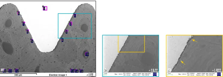
and 9), the interface was character-ized by diffuse presence of connec-tive tissue. No inflammatory infiltrate was present at the bone-implant in-terface or at the level of the marrow spaces. No foreign body reaction cells or epithelial downgrowth were present (Fig 1).
ESEM-EDX Analysis
Sample 1 (6357/1): Retrieved After 17 Years
ESEM analysis of a thread located at the middle part of the implant showed a compact and mature cor-tical bone tissue (Fig 2a, Table 2).
Osteon bone lamellae were ar-ranged concentrically in the areas between threads. Osteocyte lacu-nae closer to the implant surface were disposed near the interface and followed the thread contour. Some small, dense metal fragments, evident at higher magnifications
Fig 1 (a, b) In the 17-year samples, compact mature lamellar bone with small marrow spaces was present around the implant surface. Remodeling areas were present close to the implant surface. (c to e) In the 14-year samples, osteons were present in the proximity of the implant body. Mature old bone was in close contact with the implant surface. Trabecular mature bone could be detected at the interface of the implant. (f) In the 8-year sample, lamellar bone with a low affinity for the staining could be distinguished. (g) In the 4-year sample, bone was in tight contact with the implant surface and adapted to all its microirregularities. (h) Signs of bone formation were present inside the concavity of an implant thread in the sample retrieved after 2 months. (i) Newly formed bone was present along the perimeter of the unloaded implant retrieved after 2 months. Many blood vessels were evident in the marrow spaces. (Toluidine blue and acid fuchsin; magnification ×40 and ×100.) a d g b e h c f i 6357/1 17 y 6357/4 14 y 5933 4 y 6357/2 17 y 6223 14 y 3893 2 m 6357/3 14 y 6090 8 y 3892 2 m
© 2018 BY QUINTESSENCE PUBLISHING CO, INC. PRINTING OF THIS DOCUMENT IS RESTRICTED TO PERSONAL USE ONLY. NO PART MAY BE REPRODUCED OR TRANSMITTED IN ANY FORM WITHOUT WRITTEN PERMISSION FROM THE PUBLISHER.
The International Journal of Periodontics & Restorative Dentistry 434
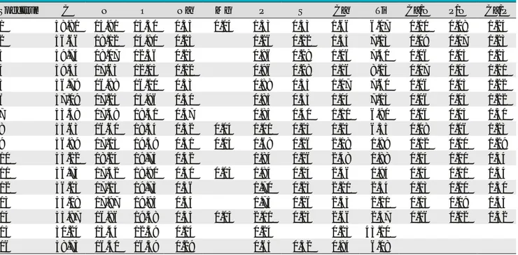
Table 2 Atomic Percentages and Ratios Obtained by EDX Analysis on Sample 1 (6357/1)
Spectrum C N O Na Mg P S Cl Ca Ti Ca/N P/N Ca/P
1 48.47 15.61 20.56 0.21 0.08 2.62 0.12 3.50 8.83 0.22 0.17 1.33 2 51.01 15.17 19.92 0.24 0.07 2.73 0.12 3.51 7.22 0.23 0.18 1.28 3 53.31 13.79 19.49 0.26 0.09 2.83 0.13 3.70 6.41 0.26 0.21 1.31 4 53.13 14.52 20.00 0.27 0.09 20.81 0.11 3.68 5.39 0.25 0.19 1.31 5 58.14 16.13 14.21 0.24 1.96 0.13 2.38 6.81 0.15 0.12 1.21 6 59.41 16.54 13.56 0.28 1.88 0.11 2.25 5.97 0.14 0.11 1.19 7 50.78 14.48 22.28 0.36 0.06 2.69 0.16 3.26 5.92 0.19 0.19 1.21 8 51.95 13.80 22.17 0.38 0.07 2.74 0.15 3.36 5.36 0.25 0.20 1.22 9 27.33 10.88 17.85 0.58 0.62 42.74 10 57.67 11.34 21.48 0.33 2.97 0.31 3.97 1.92 0.35 0.26 1.33 11 54.27 12.53 22.68 0.39 0.07 3.42 0.16 4.58 1.89 0.36 0.27 1.34 12 52.41 12.39 24.95 0.37 3.29 0.26 4.38 1.95 0.35 0.27 1.33 13 55.39 11.69 22.86 0.44 0.06 3.31 0.17 4.19 1.89 0.36 0.28 1.26 14 53.45 14.25 22.45 0.45 3.21 0.18 4.23 1.78 0.30 0.22 1.32 15 57.66 13.32 20.33 0.39 2.84 0.16 0.04 3.64 1.63 0.27 0.21 1.28 16 47.27 16.08 21.21 0.37 0.09 2.53 0.12 3.19 9.13 0.20 0.16 1.26 17 56.68 14.63 15.99 0.24 1.65 0.11 1.92 8.79 0.13 0.11 1.16 18 61.98 15.23 13.51 0.27 2.35 0.12 2.99 3.55 0.20 0.15 1.27 19 58.56 14.99 15.72 0.28 2.73 0.14 3.49 4.09 0.16 0.18 1.27 20 61.93 15.52 13.49 0.25 2.32 0.11 2.87 3.52 0.18 0.15 1.23
Table 3 Atomic Percentages and Ratios Obtained by EDX Analysis on Sample 1 (6357/1) in Focused Area (Frame 2c)
Spectrum 1 C O Na P S Ca Ti Fe
53.96 33.23 0.27 2.81 0.18 3.75 4.49 1.30
Fig 2 ESEM images of Sample 1 (6357/1). (a) Compact cortical bone showing close bone-implant contact, well evident at higher magnifications (b, c). Osteocyte lacunae closer to the implant surface were arranged following the implant thread design.
c
×3,000
b ×1,500
×500
a
© 2018 BY QUINTESSENCE PUBLISHING CO, INC. PRINTING OF THIS DOCUMENT IS RESTRICTED TO PERSONAL USE ONLY. NO PART MAY BE REPRODUCED OR TRANSMITTED IN ANY FORM WITHOUT WRITTEN PERMISSION FROM THE PUBLISHER.
Volume 38, Number 3, 2018 435
(×1,500 and ×3,000), were embed-ded in the peri-implant bone tis-sue (Figs 2b and 2c). EDX analysis showed higher Ca/N and especially Ca/P values at the coronal side (sites 1 to 4 and 16; Table 3) than at the
apical side (sites 5 to 8 and 17; Fig 3, Table 4), revealing a more mineral-ized and more mature bone at the coronal side of the thread. The high-est Ca/N and P/N values (0.33 and 0.25, respectively) were measured
on remote bone (sites 10 to 15). EDX on the implant (site 9) showed Ti (71.68 weight %) with no Al, V, or Fe. Microanalysis on a metal frag-ment showed presence of Fe (4.21 weight %) (Fig 2c).
Fig 3 Mean values of Ca/N (a), P/N (b), and Ca/P (c) ratios of the examined bone samples. Ca/N and P/N values of the interface bone at the coronal side of the thread were higher compared to the apical side, and remote bone had the highest values. Ca/P was mainly higher on the coronal side with respect to the apical side; in the long-term samples, Ca/P had the same value in the coronal side and in remote bone. IL = immediately loaded; UL = unloaded.
Table 4 Ca/P, Ca/N, and P/N Atomic Ratios (Mean ± SD) Calculated from EDX Analyses Carried Out in Bone at Coronal and Apical Sides Around Implant Thread and in Remote Bone
Sample (code)
Ca/P Ca/N P/N
Coronal Apical Remote bone Coronal Apical Remote bone Coronal Apical Remote bone
1 (6357/1) 1.30 ± 0.02aA 1.19 ± 0.02aB 1.31 ± 0.02aA 0.23 ± 0.02aA 0.17 ± 0.05aA 0.33 ± 0.04aB 0.18 ± 0.02aA 0.15 ± 0.05aA 0.25 ± 0.03aB 2 (6357/2) 1.33 ± 0.05aA 1.21 ± 0.02aB 1.33 ± 0.02aA 0.24 ± 0.02aA 0.21 ± 0.01aA 0.29 ± 0.09aB 0.18 ± 0.02aA 0.17 ± 0.08aA 0.25 ± 0.02aB 3 (6357/3) 1.28 ± 0.02aA 1.13 ± 0.04aB 1.27 ± 0.04aA 0.18 ± 0.02aA 0.06 ± 0.04bB 0.21 ± 0.05bA 0.14 ± 0.02aA 0.06 ± 0.01bB 0.20 ± 0.02aC 4 (6357/4) 1.28 ± 0.09aA 1.21 ± 0.08aA 1.36 ± 0.07aB 0.18 ± 0.06aA 0.16 ± 0.08aA 0.51 ± 0.07cB 0.15 ± 0.05abA 0.13 ± 0.06aA 0.37 ± 0.05bB 5 (6223) 1.23 ± 0.04aA 1.25 ± 0.02aA 1.37 ± 0.03aB 0.11 ± 0.05abA 0.07 ± 0.05bA 0.20 ± 0.02bB 0.10 ± 0.04bAB 0.06 ± 0.04bA 0.14 ± 0.03cB 6 (6090) 1.31 ± 0.01aA 1.38 ± 0.06bB 1.41 ± 0.02bB 0.25 ± 0.06aAB 0.20 ± 0.02aA 0.31 ± 0.08aB 0.19 ± 0.04aAB 0.15 ± 0.02aA 0.23 ± 0.05aB 7 (5933) 1.26 ± 0.08aA 1.23 ± 0.06aA 1.24 ± 0.10cA 0.03 ± 0.01bA 0.05 ± 0.03bA 0.08 ± 0.04dA 0.03 ± 0.01cA 0.04 ± 0.02bAB 0.08 ± 0.02cB 8 (3892) 1.32 ± 0.01aA 1.24 ± 0.11aA 1.30 ± 0.04aA 0.04 ± 0.01bA 0.03 ± 0.02bA 0.10 ± 0.05dB 0.04 ± 0.01bcAB 0.02 ± 0.01bA 0.08 ± 0.05cB 9 (3893) 1.23 ± 0.01aA 1.24 ± 0.04aA 1.32 ± 0.02aB 0.08 ± 0.02bA 0.07 ± 0.01bA 0.14 ± 0.01dB 0.06 ± 0.01bcA 0.05 ± 0.02bA 0.11 ± 0.01cB
Superscript letters represent statistically significant differences in the same row (capital) or in the same column (lowercase).
17 y 17 y14 y14 y Retrieval time 14 y 8 y 4 y 2 m IL 2 mUL Atomic ratio (%) 0.6 0.5 0.4 0.3 0.2 0.1 0 Coronal Apical Remote b 17 y 17 y14 y14 y Retrieval time 14 y 8 y 4 y 2 m IL 2 mUL Atomic ratio (%) 1.45 1.40 1.35 1.30 1.25 1.20 1.15 1.10 1.05 1 Coronal Apical Remote c 17 y 17 y14 y14 y Retrieval time 14 y 8 y 4 y 2 m IL 2 mUL Atomic ratio (%) 0.6 0.5 0.4 0.3 0.2 0.1 0 Coronal Apical Remote a
© 2018 BY QUINTESSENCE PUBLISHING CO, INC. PRINTING OF THIS DOCUMENT IS RESTRICTED TO PERSONAL USE ONLY. NO PART MAY BE REPRODUCED OR TRANSMITTED IN ANY FORM WITHOUT WRITTEN PERMISSION FROM THE PUBLISHER.
The International Journal of Periodontics & Restorative Dentistry 436
Sample 2 (6357/2): Retrieved After 17 Years
ESEM analysis carried out in one area located at the middle portion of the implant showed a homogeneous bone-implant interface and uniform compact bone. Osteons close to but not in contact with the implant sur-face showed lamellae concentrically arranged (Fig 4a). Osteocyte lacu-nae closer to the implant surface fol-lowed the thread contour. At ×1,500 and ×3,000 magnification (Figs 4b and 4c), the bone at the apical side appeared to be detached from the implant surface and many metal fragments were observed near the interface. EDX revealed similar Ca/N and P/N ratios at the coronal (sites 5 to 8) and apical (sites 1 to 4) sides (Table 4, Fig 3), while the highest Ca/N and P/N values were observed on remote bone (sites 10 to 15). The Ca/P value was similar on the coro-nal side (1.33) and remote bone. EDX on the implant section (site 9) revealed Ti (74.67 weight %).
Sample 3 (6357/3): Retrieved After 14 Years
A thread located in the middle part of the implant was investigated. Compact bone with a close bone-implant contact was primarily pres-ent on the coronal side of the implant thread, where osteons were evident. Bone lamellae were concentrically arranged and followed the coronal implant surface. On the apical side, only small portions of compact bone were present at the implant interface and wide remodeling areas were vis-ible. EDX showed markedly lower Ca/N, P/N, and Ca/P values at the apical side compared to the coronal side and especially to remote bone (Table 4, Fig 3). Ca/P values on the coronal side and on remote bone were similar. EDX on the implant section showed Ti (76.18 weight %).
Sample 4 (6357/4): Retrieved After 14 Years
ESEM analysis focused on one im-plant thread located at the
middle-apical portion of the implant showed mature compact bone tissue. Bone lamellae with numerous osteocyte lacunae were arranged concentrical-ly. Osteocyte lacunae closer to the implant surface followed the implant thread design. Some osteons abut-ted the implant thread at the coronal and apical sides. EDX revealed dif-ferences in mineral content among the examined areas, showing mark-edly higher Ca/N and P/N in remote bone (Fig 3). Ca/N and P/N values were similar at both thread sides, re-vealing similar organic content; how-ever, a higher atomic Ca/P ratio was detected on the coronal side com-pared to the apical side (Table 4). Mi-croanalysis on the implant revealed Ti (89.24 weight %).
Sample 5 (6223): Retrieved After 14 Years
ESEM analysis of a thread located in the middle part of the implant revealed a mature trabecular bone. Bone lamellae were arranged
fol-Fig 4 ESEM images of Sample 2 (6357/2). (a) Mature compact bone showing osteon lamellae concentrically arranged and close to the implant surface. (b,c) At higher magnifications, the interface appeared detached from bone tissue and small debris (arrows) and metal fragments were present.
c ×3,000
b ×1,500
a
×500
© 2018 BY QUINTESSENCE PUBLISHING CO, INC. PRINTING OF THIS DOCUMENT IS RESTRICTED TO PERSONAL USE ONLY. NO PART MAY BE REPRODUCED OR TRANSMITTED IN ANY FORM WITHOUT WRITTEN PERMISSION FROM THE PUBLISHER.
Volume 38, Number 3, 2018 437
lowing the contour of the implant thread, with a limited presence of osteocyte lacunae. More bone tis-sue was found on the coronal side of the implant thread, closely apposed to the metal implant surface. On the apical side, only small areas of bone tissue were in contact with the im-plant surface. EDX microanalysis re-vealed low Ca/N and P/N values on coronal and apical thread sides, re-vealing a bone tissue with a high or-ganic component (Table 4, Fig 3). On remote bone, slightly higher Ca/N and P/N values showed a more min-eralized structure, while markedly high Ca/P values revealed mature bone. EDX on the implant section revealed Ti (65.93 weight %) and traces of Fe (0.13 weight %).
Sample 6 (6090): Retrieved After 8 Years
ESEM analyses of a thread located at the coronal-middle part of the implant body showed detachment of bone tissue from the implant
sur-face, likely attributable to the sample retrieval and/or histologic prepara-tion procedures (Fig 5). A mature compact bone with few Haversian channels in the osteon structure was visible. Bone lamellae did not follow the implant thread design. Numerous osteocyte lacunae were identified but appeared not to have a uniform disposition. EDX revealed Ca/N and P/N values higher on the coronal side (sites 1–4) of the thread compared to the apical side (sites 5 to 8), revealing a more mineralized bone with a lower organic composi-tion (Table 4, Fig 3).
Similar to the majority of the samples examined, the highest Ca/N and P/N values were regis-tered in remote bone. Unlike the previously examined samples, the Ca/P values were very high, particu-larly for the apical side and remote bone (1.38 and 1.41, respectively). This indicated a mature bioapatite (Fig 3). EDX on the implant body (site 9) revealed Ti (69.81 weight %).
Sample 7 (5933): Retrieved After 4 Years
ESEM of a thread located on the middle part of the implant showed a mature compact bone with a vis-ible osteon at the apical side of the implant thread (Fig 6). Bone lamel-lae were well represented on the coronal side, which followed the margin of the implant thread, while on the apical side the structure ap-peared less organized (Fig 6). EDX analyses revealed similar Ca/P, Ca/N, and P/N values at the coronal (sites 5 to 8 and 17), apical (sites 1 to 4), and remote bone (sites 10 to 15) (Table 4, Fig 3), revealing a less min-eralized tissue with a high presence of organic components. Ca/N and P/N values were lower compared to the previous (long-term) examined samples. Traces of Al ions on all the spectra were acquired at the bone-implant interface and on remote bone (site 14).
The analysis of the implant (site 9) revealed Ti (73.46 weight %) and
Fig 5 ESEM image of Sample 6 (6090). Bone tissue appeared compact with few Haversian channels and numerous osteocyte lacunae not uniformly disposed.
Fig 6 ESEM image of Sample 7 (5933). Bone lamellae followed the implant thread contour mostly on the coronal (left) side, while the structure appeared less organized on the apical (right) side.
×500 ×500
© 2018 BY QUINTESSENCE PUBLISHING CO, INC. PRINTING OF THIS DOCUMENT IS RESTRICTED TO PERSONAL USE ONLY. NO PART MAY BE REPRODUCED OR TRANSMITTED IN ANY FORM WITHOUT WRITTEN PERMISSION FROM THE PUBLISHER.
The International Journal of Periodontics & Restorative Dentistry 438
traces of Al (0.08 weight %); punctual EDX on a dense metal fragment (site 16) revealed the same Al content.
Sample 8 (3892): Retrieved After 2 Months Immediate Loading The section, located on the middle-apical portion of the implant, showed the presence of less dense
bone tissue around the thread at apical (sites 1 to 4) and coronal (sites 5 to 8, 17, and 18) sides. This mor-phology suggested new bone for-mation (Fig 7, Table 5). Trabecular bone with many osteocyte lacunae could be found at about 50 to 100 μm from the coronal side of the thread. Small metal fragments
were observed in the bone (Fig 7) at coronal and apical sides. EDX analysis revealed a less mineralized and more immature peri-implant bone. Ca/N and P/N values were similarly low on coronal (sites 1 to 4) and apical (sites 5 to 8, 17, and 18) sides. Ca/P ratios were unex-pectedly higher on the coronal side
Table 5 Atomic Percentages and Ratios Obtained by EDX Analysis on Sample 8 (3892), IL
Spectrum C N O Na P S Ca Ti Ca/N P/N Ca/P
1 54.61 16.44 17.68 0.31 0.76 0.36 0.92 8.91 0.06 0.05 1.21 2 54.53 16.61 18.59 0.33 0.56 0.34 0.75 8.29 0.05 0.03 1.33 3 56.27 17.32 17.14 0.28 0.47 0.34 0.62 7.54 0.04 0.04 1.32 4 53.99 16.97 19.67 0.33 0.50 0.37 0.71 7.44 0.04 0.05 1.42 5 63.54 16.46 13.38 0.10 0.06 0.08 0.07 6.31 0.01 0.01 1.16 6 63.48 16.79 13.86 0.07 0.08 0.05 0.09 5.59 0.004 0.004 1.12 7 60.08 16.45 15.41 0.21 0.21 0.20 0.29 7.14 0.02 0.01 1.38 8 56.16 17.31 18.71 0.26 0.66 0.32 0.77 5.81 0.04 0.01 1.16 9 32.47 12.99 13.26 0.11 0.10 41.07 10 53.56 17.85 21.21 0.39 1.71 0.25 2.26 2.77 0.13 0.01 1.32 11 54.90 16.02 21.27 0.38 1.82 0.25 2.41 2.94 0.15 0.10 1.32 12 54.12 16.21 22.01 0.42 1.89 0.24 2.50 2.62 0.15 0.11 1.33 13 57.57 17.44 20.23 0.35 0.79 0.36 0.99 2.26 0.06 0.12 1.30 14 57.09 17.67 19.93 0.40 0.74 0.38 0.91 2.88 0.05 0.05 1.22 15 57.40 17.59 19.60 0.36 0.69 0.37 0.92 3.07 0.05 0.04 1.33 16 63.57 16.41 14.83 0.14 0.16 0.13 0.17 4.58 17 57.87 16.56 17.86 0.27 0.57 0.43 0.72 5.73 0.04 0.03 1.26 18 58.28 16.44 18.31 0.26 0.49 0.32 0.68 5.23 0.04 0.03 1.38
Fig 7 ESEM image of Sample 8 (3892), IL. An irregular bone-forming structure with few osteocyte lacunae was seen, with less dense areas around the thread at apical (right) and coronal (left) sides. A large number of small dense granules were also well evident (arrows).
×500
© 2018 BY QUINTESSENCE PUBLISHING CO, INC. PRINTING OF THIS DOCUMENT IS RESTRICTED TO PERSONAL USE ONLY. NO PART MAY BE REPRODUCED OR TRANSMITTED IN ANY FORM WITHOUT WRITTEN PERMISSION FROM THE PUBLISHER.
Volume 38, Number 3, 2018 439
(Table 4, Fig 3) compared to the apical portion. Remote bone (sites 10 to 15) showed a more mineral-ized structure with higher Ca/N and P/N values. Implant section (site 9) EDX showed Ti (71.30 weight %).
Sample 9 (3893): Retrieved After 2 Months Unloaded (Control)
ESEM microanalysis of a thread located on the middle portion of
the implant showed a low-density trabecular bone that suggested ongoing bone formation. Remote bone appeared to be more mature, with many osteocyte lacunae and some localized dense areas (Fig 8, Table 6). Some metal fragments were present in the bone tissue on both coronal and apical (sites 5 to 8) sides (Table 4, Fig 3). EDX mi-croanalysis revealed extremely low
coronal (sites 1 to 4) and apical Ca/N and P/N values, suggesting a high presence of organic materials; low Ca/P values were detected. Remote bone (sites 9 to 14) showed mark-edly higher Ca/N, P/N, and Ca/P values. EDX of the implant (site 15) showed Ti (72.74 weight %).
Table 6 Atomic Percentages and Ratios Obtained by EDX Analysis on Sample 9 (3893), UL
Spectrum C N O Na Mg P S Ca Ti Ca/N P/N Ca/P
1 58.81 15.91 15.50 0.35 0.04 1.33 0.33 1.66 6.07 0.10 0.08 1.24 2 56.66 18.12 14.80 0.25 1.26 0.22 1.55 7.13 0.09 0.07 1.23 3 59.74 18.07 12.36 0.23 0.86 0.28 1.06 7.41 0.06 0.05 1.23 4 59.54 17.65 12.05 0.22 0.96 0.28 1.16 8.15 0.07 0.05 1.21 5 56.79 16.88 16.10 0.33 0.88 0.35 1.07 7.60 0.06 0.05 1.22 6 57.09 17.25 15.96 0.30 0.85 0.35 1.04 7.15 0.06 0.05 1.22 7 53.38 17.48 19.51 0.37 0.85 0.40 1.11 6.90 0.06 0.05 1.30 8 55.63 16.60 18.54 0.32 0.04 1.01 0.25 1.25 6.35 0.08 0.06 1.23 9 56.89 17.15 19.59 0.31 0.05 1.68 0.26 2.18 1.89 0.12 0.10 1.29 10 55.22 18.24 19.75 0.32 1.84 0.26 2.48 1.88 0.14 0.10 1.34 11 56.73 17.52 18.90 0.30 0.05 1.84 0.24 2.46 1.94 0.14 0.11 1.34 12 56.25 17.15 19.73 0.36 1.70 0.25 2.21 2.34 0.13 0.10 1.30 13 55.18 17.97 19.95 0.34 1.75 0.26 2.33 2.21 0.13 0.09 1.33 14 55.87 16.86 19.58 0.35 0.05 2.01 0.25 2.66 2.37 0.16 0.12 1.32 15 30.24 13.44 12.59 0.14 0.24 0.25 43.10 16 58.73 16.50 16.59 0.29 0.65 0.32 0.84 6.08
Fig 8 ESEM image of Sample 9 (3893), UL. Bone tissue appeared less dense on apical (left) and coronal (right) sides, suggesting ongoing bone formation. Remote bone appeared more organized, with a larger number of osteocyte lacunae. Small dense granules were embedded into the bone structure.
×500
© 2018 BY QUINTESSENCE PUBLISHING CO, INC. PRINTING OF THIS DOCUMENT IS RESTRICTED TO PERSONAL USE ONLY. NO PART MAY BE REPRODUCED OR TRANSMITTED IN ANY FORM WITHOUT WRITTEN PERMISSION FROM THE PUBLISHER.
The International Journal of Periodontics & Restorative Dentistry 440
Discussion
In the present study, ESEM-EDX analysis provided new information regarding peri-implant bone struc-ture, its degree of mineralization, and its element composition, reveal-ing some important features.
In the long-term samples (re-trieved after 8 to 17 years) the Ca/N and P/N values (representing the mineral component with respect to the organic component) of the inter-face bone at the coronal side of the thread were higher with respect to the apical side, indicating more min-eralized bone (Figs 3a and 3b). Ca/P ranged from 1.06 to 1.41, which is lower than the values reported in the literature (range: 1.37 to 1.62).7
This may be related to the fact that EDX analysis was performed on histologic samples, and the resin content likely contributed to the lower Ca/P ratios. Because of this, N rather than C has been used as an index of organic content to evaluate the mineralization degree of bone.
Four of the five implants re-trieved after a long period (14 to 17 years) showed higher Ca/P ratios on the interface bone at the coronal side compared to the apical side of the thread (Fig 3c), with statistically significant differences in three of them (P < .05). One sample retrieved after 14 years (sample 5) showed a different Ca/P trend compared to the other implants. This may be re-lated to implant placement, as this implant was retrieved from the max-illa while the other implants were re-trieved from the mandible.
Implants retrieved after a short-er pshort-eriod (4 to 8 years) or during
osseointegration phases (2 months) showed a different mineralization pattern. Implants retrieved during osseointegration phases showed an increased presence of organic com-ponent, revealed by significantly low Ca/N and P/N values (P < .001) at both coronal and apical sides of the thread. The sample retrieved af-ter 8 years showed an unexpectedly higher Ca/P ratio on the apical side than on the coronal side (Fig 3c), while the sample retrieved after 4 years showed the lowest (not statis-tically significant, with high SD) Ca/P value detected on remote bone among all the samples. Active bone remodeling on coronal and apical sides, as shown by ESEM morpho-logic analysis and confirmed by the high SD values, justifies these results of the implants retrieved after short periods. These findings may appear to be in contrast with data reported in the literature. In a recent study, 29 porous zirconia implants were retrieved for increased bone loss.24
Through histologic evaluations, the authors found that the most com-pact bone was in the periapical re-gion. These data are different from the results of the present study for several reasons. First, the implants were retrieved for increased bone loss (ie, peri-implantitis)24 while in
the present study the implants were retrieved for mechanical complica-tions and not for infective process-es. Second, the authors performed a histomorphometric analysis,24 and
bone mineralization was not quali-tatively assessed as in the present study.
A recent study analyzed the soft tissue around dental implants
affected by peri-implantitis, de-scribing the presence of metals/con-taminants (Fe and Ti ions).14 Ti ionic
leakage may occur around turned and rough-surface dental implants. No correlation between increas-ing roughness and ion release was found either in vitro or in vivo.25 In
the present study, metal fragments were found in two specimens and traces of Fe ions were detected with no presence of altered bone tissue/bone remodeling. It could be hypothesized that the presence of these metal fragments did not affect implant osseointegration pro-cesses; however, this aspect needs to be clarified in further studies.
Previous human histologic stud-ies demonstrated continuous re-modeling at the interface of dental implants.26 Over time, the newly
formed woven bone located around the implant perimeter undergoes remodeling, changing its structure to a more organized lamellar bone.26
The hardness and the elastic modu-lus of the peri-implant bone tended to increase over time.26 The
pres-ent study is in accordance with a previous 12-month animal study27
showing that removal torque (corre-latable to the increased bone min-eralization) increased over time. In two previous studies from the labo-ratory of the present authors,19,21 the
number of osteocytes was found to increase in immediately loaded implants when compared with un-loaded controls, and there was a statistically significant increase in os-teocytes in the first few years after implant loading. This increase could be important for the mechanical competence of bone.19,21
© 2018 BY QUINTESSENCE PUBLISHING CO, INC. PRINTING OF THIS DOCUMENT IS RESTRICTED TO PERSONAL USE ONLY. NO PART MAY BE REPRODUCED OR TRANSMITTED IN ANY FORM WITHOUT WRITTEN PERMISSION FROM THE PUBLISHER.
Volume 38, Number 3, 2018 441
Conclusions
The two most significant findings were that the interface bone located at the coronal side of the implant threads was generally more mineral-ized than the interface bone at the apical side of the threads and that the mineralization of the peri-implant bone at the interface increased over time. These results could be related to the different remodeling events (apical versus coronal sides) due to the direction of the loading forces. Notable as a proof of principle was the unexpected finding of a high-er degree of minhigh-eralization in the 2-month immediately loaded implant when compared to the 2-month sub-merged unloaded control.
Acknowledgments
The authors reported no conflicts of interest related to this study.
References
1. Albrektsson T, Brånemark PI, Hans-son HA, Lindström J. Osseointegrated titanium implants. Requirements for ensuring a long-lasting, direct bone-to-implant anchorage in man. Acta Orthop Scand 1981;52:155–170.
2. Albrektsson T, Johansson C. Osteoin-duction, osteoconduction and osseo-integration. Eur Spine J 2001;10(suppl): s96–s101.
3. Davies JE. Mechanisms of endosseous integration. Int J Prosthodont 1998;11: 391–401.
4. Davies JE, Mendes VC, Ko JC, Ajami E. Topographic scale-range synergy at the functional bone/implant interface. Bio-materials 2014;35:25–35.
5. Feng X, McDonald JM. Disorders of bone remodeling. Annu Rev Pathol 2011;6: 121–145.
6. Kourkoumelis N, Balatsoukas I, Tzaphli-dou M. Ca/P concentration ratio at dif-ferent sites of normal and osteoporotic rabbit bones evaluated by Auger and energy dispersive x-ray spectroscopy. J Biol Phys 2012;38:279–291.
7. Shah FA, Nilson B, Brånemark R, Thomsen P, Palmquist A. The bone-implant interface—Nanoscale analysis of clinically retrieved dental implants. Nanomedicine 2014;10:1729–1737. 8. Palmquist A, Emanuelsson L, Sjövall
P. Chemical and structural analysis of the bone-implant interface by TOF-SIMS, SEM, FIB and TEM: Experimental study in animal. Appl Surf Sci 2012;258: 6485–6494.
9. Gandolfi MG, Taddei P, Siboni F, et al. Micro-topography and reactivity of implant surfaces: An in vitro study in simulated body fluid (SBF). Microsc Microanal 2015;21:190–203.
10. Gandolfi MG, Iezzi G, Piattelli A, Prati C, Scarano A. Osteoinductive potential and bone bonding ability of ProRoot MTA, MTA Plus and Biodentine in rabbit intra-medullary model: Microchemical char-acterization and histological analysis. Dent Mater 2017;33:e221–e238.
11. Gandolfi MG, Spagnuolo G, Siboni F, et al. Calcium silicate/calcium phosphate biphasic cements for vital pulp therapy: Chemical-physical properties and hu-man pulp cells response. Clin Oral In-vestig 2015;19:2075–2089.
12. Engfeldt B, Hjerpe A. Density gradient fractionation of dentine and bone pow-der. Calcif Tissue Res 1974;16:261–275. 13. Bigi A, Cojazzi G, Panzavolta S, et al.
Chemical and structural characterization of the mineral phase from cortical and trabecular bone. J Inorg Biochem 1997; 68:45–51.
14. Fretwurst T, Buzanich G, Nahles S, Woelber JP, Riesemeier H, Nelson K. Metal elements in tissue with dental peri-implantitis: A pilot study. Clin Oral Implants Res 2016;27:1178–1186. 15. Eliades G, Mantzourani M, Labella R,
Mutti B, Sharma D. Interactions of dentine desensitisers with human dentine: Mor-phology and composition. J Dent 2013; 41(suppl):s28–s39.
16. Bolind P. On 606 Retrieved Oral and Craniofacial Implants. An Analysis of Consecutively Received Human Speci-mens [thesis]. Gothenburg: University of Gothenburg, 2004.
17. Bolind P, Johansson CB, Balshi TJ, Langer B, Albrektsson T. A study of 275 retrieved Brånemark oral implants. Int J Periodontics Restorative Dent 2005; 25:425–437.
18. Bolind PK, Johansson CB, Becker W, Langer L, Sevetz EB Jr, Albrektsson TO. A descriptive study on retrieved non-threaded and non-threaded implant designs. Clin Oral Implants Res 2005;16:447–455. 19. Barros RR, Degidi M, Novaes AB, Piattel-li A, ShibPiattel-li JA, Iezzi G. Osteocyte density in the peri-implant bone of immediately loaded and submerged dental implants. J Periodontol 2009;80:499–504. 20. Degidi M, Piattelli A, Shibli JA,
Per-rotti V, Iezzi G. Bone formation around immediately loaded and submerged dental implants with a modified sand-blasted and acid-etched surface after 4 and 8 weeks: A human histologic and histomorphometric analysis. Int J Oral Maxillofac Implants 2009;24:896–901. 21. Piattelli A, Artese L, Penitente E, et al.
Osteocyte density in the peri-implant bone of implants retrieved after differ-ent time periods (4 weeks to 27 years). J Biomed Mater Res B Appl Biomater 2014;102:239–243.
22. Iezzi G, Piattelli A, Mangano C, et al. Periimplant bone response in human-re-trieved, clinically stable, successful, and functioning dental implants after a long-term loading period: A report of 17 cas-es from 4 to 20 years. Implant Dent 2016; 25:380–386.
23. Botticelli D, Perrotti V, Piattelli A, Iezzi G. Four human stable and functioning den-tal implants retrieved for fracture after 14 and 17 years from the same patient: A histological and histomorphometrical report. Int J Periodontics Restorative Dent [in press].
24. Kohal RJ, Schwindling FS, Bächle M, Spies BC. Peri-implant bone response to retrieved human zirconia oral implants after a 4-year loading period: A histo-logic and histomorphometric evaluation of 22 cases. J Biomed Mater Res Part B Appl Biomater 2016;104:1622–1631. 25. Wennerberg A, Ide-Ektessabi A,
Hat-kamata S, et al. Titanium release from implants prepared with different sur-face roughness. Clin Oral Implants Res 2004;15:505–512.
26. Gil LF, Suzuki M, Janal MN, et al. Pro-gressive plateau root form dental implant osseointegration: A human retrieval study. J Biomed Mater Res B Appl Biomater 2015;103:1328–1332. 27. Johansson C, Albrektsson T. Integration
of screw implants in the rabbit: A 1-year follow-up of removal torque of titanium implants. Int J Oral Maxillofac Implants 1987;2:69–75.
© 2018 BY QUINTESSENCE PUBLISHING CO, INC. PRINTING OF THIS DOCUMENT IS RESTRICTED TO PERSONAL USE ONLY. NO PART MAY BE REPRODUCED OR TRANSMITTED IN ANY FORM WITHOUT WRITTEN PERMISSION FROM THE PUBLISHER.
1.1.2 Esem-Edx Microanalysis Of The Bone-Implant Interface Around Loaded And Unloaded
Retrieved Dental Implants.
Prati Carlo1, Zamparini Fausto1,2, Botticelli Daniele3, Daichi Yonezawa4, Adriano Piattelli5, Gandolfi Maria Giovanna2.
1. School of Dentistry, Department of Biomedical and Neuromotor Sciences, University of Bologna, Bologna, Italy 2. Laboratory of Biomaterials and Oral Pathology, School of Dentistry, Department of Biomedical and Neuromotor Sciences, University of Bologna, Bologna, Italy
3. Ardec Academy, Rimini, Italy
4. Clinical Assistant Professor, Department of Applied Prosthodontics, Graduate School of Biomedical Sciences, Nagasaki University
5. Department of Medical Oral and Biotechnological Sciences, University of Chieti - Italy
AIM
The aim of the present study is to analyse the mineralization degree at the bone implant interface and the effect of initial loading procedures around clinically stable retrieved dental implant before (4 months from insertion) and after 2 months of loading (4 months from insertion).
MATERIALS AND METHODS
The protocol of the study was approved by Ethical Committee of the Corporación Universitária Rafael Núñez, Cartagena de Indias, Colombia. Detailed information on Material and Methods and histological data have been reported in a previous published paper (Yonezawa et al. 2018). Sixteen volunteers were included in the study and two mini implants were installed in a non-submerged fashion in the edentulous distal region of the jaws. After two months, one mini implant was randomly selected to be loaded with a single crown while the other mini implant was left unloaded. After 2 more months, both mini implants were removed with a trephine. Twenty biopsies retrieved from 10 volunteers were available for histological analysis. The biopsies were sectioned using a diamond steel disc along the major axis of the implants at approximately 150 mm and ground to about 30 microns The sections were stained with acid fuchsin and toluidine blue. Ten ground sections from 5 patients were used for OM and ESEM-EDX Microanalysis.
OM and ESEM-EDX Microanalysis
The histological samples were observed under Optical microscopy (OM) to identify the macrostructure of the sample and bone macromorphology.
Then, ESEM observation were preformed entirely, from the coronal to the most apical portion of the implant at 500x magnification. ESEM-EDX micro-analyses were performed following Gandolfi protocol (Gandolfi et al. 2017 and 2018). Briefly, micromorphological and microchemical analyses were performed at both coronal and apical sides of the upper (where bone was present) and deep threads of the implants and at 300 and 500 μm from the implant thread (distant bone as mineralization control of each sample).
EDX analyses were carried out at areas of approx. 30x30 microns (n=3 per thread side) and qualitative and semi-quantitative element (weight % and atomic %) content were investigated. For all the acquired spectra, the atomic Ca/N,
P/N and Ca/P ratios were calculated (as mean and standard deviation) to evaluate the degree of mineralization (Gandolfi et al. 2017 and 2018).
Statistical analysis
Ca/N, P/N and Ca/P mean values were statistically analyzed using two-way ANOVA followed by Student-Newman-Keuls test. P value was previously set at 0.05.
RESULTS
The investigated samples are reported in Figure 1. Out of 10 implants, 5 were retrieved after 4 months from placement (unloaded group) and 5 were retrieved after 6 months (loaded group, namely retrieved after 2 months of loading and 4 months healing period).
Figure 1: Retrieved implants histology analysed in the present study. Five were retrieved after 4 months unloaded, the other five were retrieved after 4 months (2 months of loading).
Figure 2: ESEM microanalysis of one thread located on the upper portion revealed electron dense cortical bone tissue well-evident osteons and bone niches, indicating high bone remodelling activity. EDX microanalysis revealed similar Ca/N and P/N atomic ratios at both coronal and apical side of the thread.
Loaded samples (2 months unloaded + 2 months loading)
Sample 6504
OM revealed cortical bone tissue along all the specimen (Table 1). The first bone to implant contact was identified at thread 1.
ESEM microanalysis of the upper thread revealed electron dense cortical bone tissue with numerous osteons and bone niches, which suggest high bone remodelling activity. Bone tissue appeared in strict contact with the apical side of the thread, while few bone was present at the implant thread crest.
EDX revealed similar Ca/N and P/N atomic ratios at both coronal and apical side of the upper thread (mean values were 0.140.03 and 0.140.07. High standard deviation of the apical portion also suggests bone remodelling in this area
(Tables 2,4). Distant bone revealed higher values of Ca/N and P/N, with low SD values. Ca/P values were similar at coronal and apical side of the thread, both lower than Ca/P values of remote bone (p<0.05) (Tables 6,7).
ESEM analysis on one deep thread revealed few cortical bone tissue with some osteocytes lacunae. Low-electron dense bone tissue was in strict contact with the implant surface, the bone lamellae are concentrically arranged following the implant thread design. No osteons were present close to the implant thread. Similarly low electron dense bone tissue was also identified on remote bone.
EDX microanalysis revealed significantly high Ca/N values on the coronal side with respect to the apical side (p<0.05), mean values were 0.170.001 and 0.090.02 respectively. Interestingly, P/N values followed a opposite trend, being that the apical portion revealed high P/N values when compared to the coronal side of the deep thread (mean P/N were 0.080.02 and 0.030.001). (Tables 3,5). Distant bone showed higher P/N and Ca/N values when compared to both apical and coronal side. Ca/P ratios on calculated on the apical side revealed statistically lower values compared to both coronal side and bone located at 300 and 500 micron (p<0.05) (Tables 6,7).
Sample 6508
OM analysis revealed trabecular bone tissue along all the implant section. Bone tissue starts 300 micron over the upper thread. ESEM investigation on the upper thread showed close bone implant contact only on the apical side. Electron-dense bone tissue was observed with some osteocytes lacunae and few osteons, suggesting low bone remodelling activity.
EDX microanalysis revealed significantly high Ca/N and P/N ratios at the apical side of the implant thread, when compared to coronal side (p<0.05), suggesting a more mineralized area. (Tables 2,4). No differences were observed between coronal and apical side of the thread in terms of Ca/P. (Tables 6,7).
ESEM investigation of one deep thread revealed the presence of trabecular bone tissue with numerous bone niches, and bone lamellae concentrically arranged following the thread contour and osteons located distant from the implant thread. Bone implant contact was scarce, only at the crest of the thread and in few areas of the coronal and apical side. EDX microanalysis revealed statistically (p<0.05) high Ca/N and P/N values of the apical side of the thread when compared to the coronal side, mean values were 0.26 0.01 and 0.196 0.03 for Ca/N, 0.193 0.031 and 0.149 0.008 for P/N, respectively (Tables 3,5). No differences were observed between coronal and apical side of the thread in terms of Ca/P. (Tables 6,7).
Sample 6514
At OM observation, bone tissue was present from thread 2, only around the top of the thread. Cortical bone was present along all the implant threads.
ESEM investigation on one upper thread revealed cortical bone tissue with numerous osteocyte lacunae and osteons. EDX microanalysis revealed higher Ca/N and P/N values on the apical side of the thread with respect to the coronal side, (Ca/N values were 0.23 0.02 and 0.13 0.06 respectively; P/N values were 0.16 0.02 and 0.14 0.02 respectively). Statistically significant differences were observed only on Ca/N values (p<0.05) (Tables 2,4). High SD values suggested bone remodelling on the coronal side. Bone implant contact was evidenced on the root of thread 2 and 3 with numerous bone niches creating a strict bone-implant interface. Ca/P ratio was similar on both coronal and apical sides. (Tables 6,7).
ESEM analysis on one deep thread revealed electron dense cortical bone tissue with a large number of osteons and osteocytes lacunae disposed close to the apical side of the implant interface and on remote bone.
EDX microanalysis revealed higher Ca/N and P/N values on the apical side of the implant thread, when compared to the cortical side, (Ca/N values were 0.206 0.03 and 0.18 0.07 respectively; P/N values were 0.19 0.03 and 0.10 0.05 respectively) the values were statistically significant only for P/N values (p<0.05) (Tables 3,5). Ca/P ratios were similar on coronal, apical side of the thread, statistically significant differences were observed only on remote bone (at 500 micron) (p<0.05). (Tables 6,7).