Same Donor Laparoscopic Liver and Kidney Procurement
for Sequential Living Donor Liver–Kidney Transplantation
in Primary Hyperoxaluria Type I
Roberta Angelico, MD, PhD, FEBS,1 Isabella Guzzo, MD,2Marco Pellicciaro, MD,1
Maria Cristina Saffioti, MD,1Chiara Grimaldi, MD,1Chebl Mourani, MD,3Francesco Smedile, MD,4 Rosanna Pariante, MD,4 Alessia Semprini, MD,5Lidia Monti, MD,5
Manila Candusso, MD,6 Luca Dello Strologo, MD,2and Marco Spada, MD, PhD, FEBS1
Abstract
Background: Sequential liver–kidney transplantation (SeqLKT) from the same living donor has shown excellent
results in children with primary hyperoxaluria type 1 (PH1), yet its experience is limited due to the invasiveness of
two major procedures for liver–kidney procurement in a single donor. Despite laparoscopic nephrectomy and
hepatic left lateral sectionectomy (LLS) being considered standard procedures in living donation, the sequential
use of the two laparoscopic approaches in the same living donor has never been reported.
Methods: Herein, we present the first two case series of laparoscopic liver–kidney procurement in the same
living donor for SeqLKT in children with PH1 and review of the current literature on this topic.
Results: In the first case, a 15-month-old boy received a SeqLKT from his 32-year-old mother, who underwent
a laparoscopic LLS and, after 8 months, a laparoscopic left nephrectomy. In the second case, a 34-month-old
boy received a SeqLKT from his 40-year-old father who underwent laparoscopic LLS followed by
hand-assisted right nephrectomy after 4 months. Both donors had uneventful postoperative courses and were
dis-charged within 5 days from each surgery. The first recipient had no complication; the second child after liver
transplantation developed a partial thrombosis of the inferior vena cava, which did not preclude the sequential
kidney transplantation. After 12 months, donors and recipients displayed normal liver and renal functions.
Conclusions: Sequential laparoscopic liver–kidney procurement in the same living donor is safe and feasible, and
might be considered as a possible strategy to promote SeqLKT in children with PH1 from the same living donor.
Keywords:
living donation, laparoscopic liver and kidney procurement, primary hyperoxaluria, liver
trans-plantation, kidney transtrans-plantation, mini-invasive surgery
Introduction
P
rimary hyperoxaluria type1 (PH1) is a rare autoso-mal recessive metabolic disorder caused by a deficiency of the liver enzyme alanine-glyoxylate aminotransferase, resulting in increased hepatic oxalate production. The excess of oxalate causes progressive end-stage renal disease (ESRD) and sys-temic oxalosis, leading to severe morbidity and mortality.1Combined liver–kidney transplantation (CLKT) is considered the treatment of choice in patients affected by PH1 with ESRD.1
Liver transplantation (LT) may be simultaneous or sequential with kidney transplantation (KT) depending on disease staging, renal function, and type of donor availability.2Initially, simul-taneous or sequential CLKT has been mainly performed using organs from deceased donors.3Because of organ shortage, se-quential liver–kidney transplantation (SeqLKT) from the same living donor has been proposed in PH1 patients, showing bio-chemical and immunological advantages for the recipient.4Yet, the experience of SeqLKT from the same living donor is limited due to the complexity of the donor surgical procedures.5–12
1
Division of Abdominal Transplantation and Hepatobiliopancreatic Surgery, Bambino Gesu` Children’s Hospital IRCCS, Rome, Italy.
2Department of Nephrology and Dyalisis, Bambino Gesu` Children’s Hospital IRCCS, Rome, Italy. 3
Department of Pediatrics, Hoˆtel-Dieu de France Hospital (HDF), Beirut, Lebanon.
4
Department of Anesthesiology, Bambino Gesu` Children’s Hospital IRCCS, Rome, Italy.
5
Department of Radiology, Bambino Gesu` Children’s Hospital IRCCS, Rome, Italy.
6
Division of Hepatology and Gastroenterology, Bambino Gesu` Children’s Hospital IRCCS, Rome, Italy. ª Mary Ann Liebert, Inc.
DOI: 10.1089/lap.2019.0483
1
Although in living donation the laparoscopic left lateral sectionectomy (LLS)13,14and nephrectomy15,16showed good donor and recipient outcomes, the laparoscopic sequential liver–kidney procurement has never been reported.
To the best of our knowledge, we report the first two cases of sequential laparoscopic liver–kidney procurement from the same living donor for SeqLKT in children with PH1. The literature of living-related SeqLKT for PH1 patients has been systematically reviewed.
Center Experience Case 1
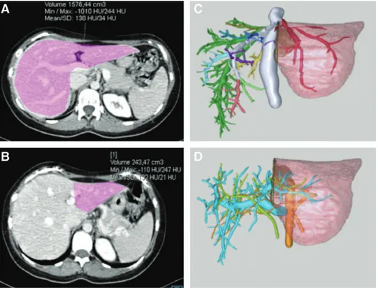
A 15-month-old boy (weight: 8 kg; height: 74 cm) with genetically confirmed PH1 and ESRD was referred for CLKT. At presentation, the patient was on peritoneal dialysis with secondary hyperparathyroidism, hyperphosphatemia, and anemia. Serum oxalates were 128 lmol/L and hemodi-alysis was started (in addition to nocturnal peritoneal dialy-sis) to reduce his level. The family was offered the option of a living-related SeqLKT, and the 32-year-old mother (weight: 67 kg; body mass index [BMI]: 23.5) expressed her willing-ness to donate. Her evaluation revealed no physical or psy-chological contraindications for living donation. Computed tomography (CT) and three-dimensional reconstruction were used to define the donor anatomy and the expected liver graft volume (Fig. 1). Liver vascular anatomy consisted in an ab-errant left hepatic artery (HA) arising from the left gastric artery and a right HA from the proper HA, while the left portal vein (PV), the hepatic veins (HVs), and biliary tree had standard anatomy. The expected left lateral segment (SII– SIII) volume was 243 gr (graft-to-recipient weight ratio
[GRWR]: 3%). At the CT, the left kidney had conventional anatomy (one renal artery, one renal vein, one ureter) and was chosen for SeqLKT.
First, in the donor full laparoscopic LLS was carried out as described by Soubrane et al.17The patient was placed in supine position, with her legs apart to apply the French position. The pneumoperitoneum was established at 12 mmHg and five tro-cars were inserted (Fig. 2A). Within the lesser omentum, the left HA was isolated. The round ligament was dissected and all the branches for segment IV were divided, exposing the left PV. After the sectioning of the Arantius ligament, the left HV was encircled extra-parenchymally. To obtain a modified ‘‘hanging maneuver,’’ an umbilical tape was placed between the left HV and the middle HV, and passed over the left PV and left HA. Dissection of the parenchyma was then performed to the right of the falciform ligament, using bipolar forceps, clips, sutures, and sealing devices, without Pringle maneuver. To preserve vascularization of the left bile duct, the hilar plate was sharply divided at the level of the Rex recessus, and the stump of the bile duct of the donor’s remnant liver was closed with sutures. When the parenchymal transection was completed, the left HA, the left PV, and the left HV were sequentially divided using mechanical staplers. A Pfannenstiel incision was used for graft extraction, and after harvesting the graft was flushed with preservation solution (Celsior) on the back table. During do-nor’s surgery, blood transfusion was not required. The donor had an uneventful postoperative course and was discharged after 5 days.
In the recipient, due to elevated preoperative oxalate levels a bilateral nephrectomy was performed. After hepatectomy with inferior vena cava (IVC) preservation, the left lateral segment was transplanted using the piggy-back technique.18
FIG. 1. Preoperative donor imaging of the liver. Preoperative donor’s CT scan showing the volumetry of (A) the whole liver and (B) the left lateral segment; three-dimensional reconstruction of (C) the hepatic vein outflow and (D) the portal vein, hepatic artery, and bile duct anatomy. CT, computed tomography.
After LT, liver function tests gradually normalized and he-modialysis was continued (six times/week). Recipient’s postoperative course was characterized by chronic throm-bocytopenia and anemia, and two spontaneous bone frac-tures, which required a delay of KT until complete recovery. After 8 months, the oxalate plasma level decreased to 50 lmol/L, and the living-related KT was carried out. In the donor, four trocars were inserted [three trocars using the same incision previously performed for LLS, one additional 12-mm trocar placed in the inferior left iliac fossa (Fig. 2A)]. At ab-dominal exploration, no adhesions were found, and a full laparoscopic left nephrectomy was performed as described by Ratner et al.19The pre-existing Pfannenstiel incision was used for kidney extraction. The mother had an uneventful postop-erative course and was discharged after 5 days. In the recipient, KT was performed without complications. Hemodialysis was continued for the first 10 days postsurgery to ensure complete removal of oxalate and stopped thereafter. After 12 months, the recipient had normal liver and renal functions (Fig. 3); the donor is currently in good condition with normal biochemical tests (creatinine: 0.8 mg/dL; aspartate aminotransferase [AST]: 23 IU/L; total bilirubin: 0.4 mg/dL).
Case 2
A 34-month-old boy (weight: 11 kg; height: 86 cm) af-fected by genetically confirmed PH1 and ESRD, on perito-neal dialysis since the age of 2 months, was referred for CLKT. During the pretransplant investigation, the CT showed a thrombosis of the right iliac vein, but a patent left iliac vein. Oxalate plasma level was 160 lmol/L, and he-modialysis was started to maximize its removal. The 40-year-old father (weight: 67 kg; BMI: 24) proposed himself as a
candidate donor and, after multidisciplinary evaluation, he was defined physically and psychologically suitable to do-nate. The father’s CT showed a conventional liver anatomy and an estimated left lateral segment of 328 gr (GRWR: 2.9%); three renal arteries and one renal vein were detected in the left kidney, while single renal artery and vein in the right kidney, which was chosen for SeqLKT.
The donor liver surgery consisted in laparoscopic LLS without Pringle maneuver and blood transfusion requirement as described in Case 1 (to identify the left HA arising from the proper HA, the left side of the hepatic pedicle was dissected). Trocar positions are detailed in Figure 2B. The donor had an uneventful postoperative course and was discharged 5 days after surgery. In the recipient, after bilateral nephrectomy the left lateral segment was transplanted as defined in Case 1. After LT, a rapid normalization of liver function tests was observed, while hemodialysis was continued six times/week. Two months from surgery the child developed ascites. The angio-CT and cavography showed normal suprahepatic outflow, but thrombosis of the retrohepatic IVC; the left iliac vein and subhepatic IVC outflows were passing through the lumbar veins into the azigos vein (Fig. 4). Anticoagulants and diuretics were started, with a progressive resolution of the ascites.
After 4 months from LT, serum oxalate level was 49 lmol/L and KT was planned. The father underwent a right-hand-assisted nephrectomy with standard technique,20 for which three trocar incisions previously performed for the LLS were used, and one 12-mm trocar incision was added in the inferior right iliac fossa (Fig. 2B). The pre-existing Pfannenstiel in-cision was used for the hand insertion and graft extraction. The donor postnephrectomy course was uneventful, and the patient was discharged after 4 days. In the recipient, KT was successfully performed in the left iliac fossa, and stable FIG. 2. Trocar position for sequential laparoscopic left lateral sectionectomy and nephrectomy in the same living donor. (A)Trocar position for laparoscopic left lateral sectionectomy (red) and left nephrectomy (green). (B) Trocar position for laparoscopic left lateral sectionectomy (red) and right-hand-assisted nephrectomy (green). For both sequential laparoscopic nephrectomies, two 10 mm-trocar and one 5-mm trocar incisions performed for the liver surgery were reused. The same Pfannenstiel incision was used for both liver and kidney extractions.
liver and renal functions were maintained throughout the entire follow-up (Fig. 3). After 12 months from surgery, the donor is in good clinical condition, with normal biochem-ical tests (creatinine: 1.1 mg/dL; AST: 35 U/L; total bili-rubin: 0.7 mg/dL).
Discussion
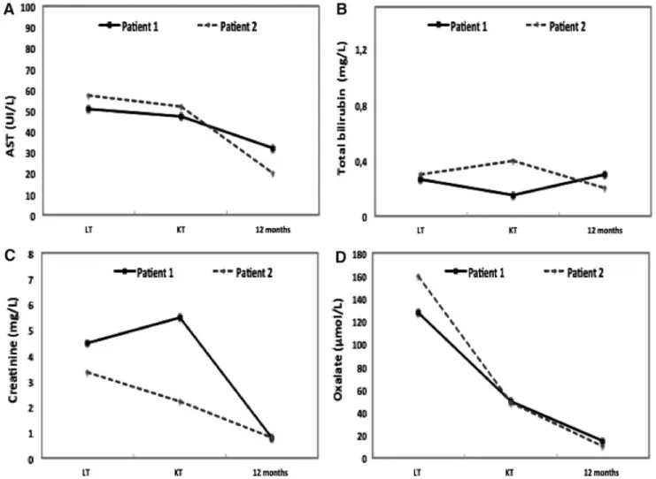
In children, PH1 represents the principal indication (37%– 61%) for CLKT, yet it is associated with inferior outcomes compared with other diseases requiring CLKT.21When severe FIG. 3. Recipients’ liver–kidney functions (A–C) and plasma oxalate levels (D). AST, aspartate aminotransferase; KT, kidney transplantation; LT, liver transplantation.
FIG. 4. Case 2 angio-CT scan and cavography. Case 2 recipient’s (A) angio-CT scan and (B) cavography, after 2 months from liver transplantation, showing the thrombosis of the inferior vena cava and right iliac vein but patent left ilac vein and good outflow of the lower extremities through the retroperitoneal vein system; (C) angio-CT scan after kidney trans-plantation in the left iliac fossa.
systemic oxalosis is present, SeqLKT (first LT, then dialysis until sufficient oxalate clearance from the body, followed by KT) seems to be preferable compared with simultaneous CLKT.1,22However, the timing for KT is influenced by the recipient’s oxalate pool as well as the deceased donor kidney availability.1
In the current era of organ shortage, the living-related SeqLKT in PH1 recipients combines the advantages of a sequential procedure—which optimizes the renal function by performing the KT when stable LT function and low plasma oxalate levels are achieved—and the benefits of a living-related transplantation, reducing the KT waiting-list time and the dialysis-related complications.
So far, SeqLKT from the same living donor in PH1 re-cipients has been reported only in small series (Table 1).5–12 Of 23 cases described in the literature, the median recipients’ age at SeqLKT was 6 (1–41) years and the majority of re-cipients (74%) received a left lateral segment graft. Mor et al. reported that SeqLKT from the same living donor could be safely performed also in young adults with PH1 using a right liver lobe graft.9The interval between LT and KT was *6 (1–10.6) months, during which period dialysis was per-formed according to center’s protocol (usually two to three times/week). Postoperative recipients’ complications in-cluded bleeding (n= 2), cytomegalovirus infection (n = 3) and bile leak (n= 1) and biliary sepsis (n = 1). After a follow-up of 2.3 (0.2–19.7) years, the overall recipient patient survival was 88.5%, while liver and kidney graft survivals were 88.5% and 90.5% (two KT lost for rejection), respectively, which ap-pears superior to those reported in pediatric CLKT from deceased donors (2-year survivals: 72.4% patient survival; 60.3% liver graft survival; 55.2% renal graft survival).21
On the donor side, no mortality and morbidity were re-ported after sequential liver–kidney procurements in the same living donor, except for one patient who developed an abdominal fluid collection after liver resection, resolved by percutaneous drainage. All donors had a rapid postoperative recovery, and showed normal liver and renal function tests in the long term (range: 2.7 months–19.7 years).5–12
Despite these promising results, the number of SeqLKT from the same living donor is limited, most likely because of the invasiveness of the two donor surgeries using the open technique and the lack of guidelines recommending this procedure.
In the last two decades, the laparoscopic hand-assisted or pure laparoscopic nephrectomies have been established as the gold standard, being preferred to the open procedure due to less postoperative pain, shorter hospital stay, and earlier re-turn to normal activities.15,16,23,24
Also for living liver donation, a laparoscopic approach to partial hepatectomy showed advantages over the open tech-nique in terms of intraoperative blood losses, postoperative recovery, and abdominal wall complications.13,14According to recent consensus guidelines, donor laparoscopic LLS for adult-to-child LT may be regarded as standard procedures, but it is still limited to few highly specialized centers.25,26 Recently, the feasibility of the combination of the two lapa-roscopic techniques in the same living donor was described by Gautier et al. for simultaneous CLKT.27
Of 227 pediatric LTs performed at our center between September 2008 and December 2018, 12 (5.3%) children underwent CLKT for PH1 (five simultaneous CLKT [one
from living donor], seven SeqLKT [four from living donor]). Following a 15-year learning curve in laparoscopic living donation, we adopted the laparoscopic approach for both li-ver and kidney procurements in the last two cases, which represent the first series of sequential laparoscopic LLS and nephrectomy in the same living donor.
In our experience, the sequential laparoscopic liver–kidney procurement in the same donor was safe and feasible, pro-viding several advantages: on the donor side, it combined the benefits of the two laparoscopic procedures (when performed separately) maintaining the surgical risks independent for each operation; on the recipient side, the sequential proce-dures permitted to optimize the KT timing according to re-cipients’ needs. Moreover, the donor LLS was associated with a rapid postoperative recovery without significant psy-chological/physical trauma (which would reduce the will to undergo a second operation) and permitted to reuse the same surgical incisions for the kidney procurement offering good esthetic results.
In the two PH1 recipients, liver and kidney functions rapidly recovered after transplantation, providing evidence that the laparoscopic donor procedures had no impact on graft out-comes. In the second case, the recipient developed IVC thrombosis after LT, which could be related to the pre-existing right iliac vein thrombosis and/or to the bilateral native ne-phrectomies performed after the hepatectomy. As proposed in other series,28,29in our recipients the bilateral nephrectomy at the time of LT was performed to remove the significant stores of oxalate contained in the native kidneys, which may cause damage to the newly transplanted renal graft.
In our experience, the living donor laparoscopic sequential liver–kidney procurement might be proposed in well-selected cases, requiring (i) a robust indication for SeqLKT in PH1 recipients (according to clinical status, age, and oxalate lev-els); (ii) meticulous living-related donor assessment (age p55 years, BMI <30, absence of active infections/malignancies/ systemic diseases, glomerular filtration rate q70–80 mL/min/ 1.73 m2, definition of liver/kidney anatomy and liver volume, psychological evaluation); and (iii) adequate donor–recipient matching (ABO compatibility, liver GRWR between 1.5 and 3). Yet, the laparoscopic living donor approach for SeqLKT has limitations related to the surgical technical complexity, which requires sufficient learning curve in mini-invasive liver–kidney procurement, and to the exposure of the same living donor to two major surgeries. So far, donor hepatectomy has imposed 0.1%–0.2% mortality and 25%–35% morbidity (mainly bili-ary leak) on healthy individuals,30while donor nephrectomy has shown<0.03% mortality and 10%–20% morbidity (major complication<3%).31Therefore, the potential living donor for SeqLKT has to be adequately informed of the risks for each surgical procedure.
Although additional experiences are needed, the two cases described herein suggest that sequential laparoscopic LLS and nephrectomy in the same living donor are safe and feasible, and could be considered as a preferential strategy to promote living-related SeqLKT in children affected by PH1.
Patient Consent
Informed consent was obtained from the patient for pub-lication of their information.
Table 1. Reported Experience of Sequential Liver and Kidney Transplantation from the Same Living Donor in Patients with Primary Hyperoxaluria Type 1 Authors No. of patients
Donor age (years) Donor liver surgery Donor kidney surgery
Donor postoperative complication Time between LT and KT (months) Recipient age (years) Recipient postoperative complication Time to last follow-up Recipient status Liver and kidney graft status Nakamura et al. 5 3 n.a. Open LLS Open LN None 1.7 1.3 CMV infection 2.8 Months Alive Function n.a. Open LLS Open LN None 3.6 2 Kidney acute mild rejection 5.4 Months Alive Function n.a. Open LLS Open LN None 2.2 3 None 2.7 Months Alive Function Sato et al. 6 3 n.a. Open LLS Open LN n.a. 1.7 15 CMV infection, L–K acute rejection 5 Years Alive Function (no immunosuppressive therapy) n.a. Open LL Open LN n.a. 10 41 Duodenal ulcer bleeding 1.5 Years Alive Function n.a. Open LLS Open LN n.a. 4.7 9 L–K acute rejection Bile leakage n.a. Alive Kidney explant for graft rupture after rejection Motoyoshil et al. 7 1 n.a. Open LLS n.a n.a 6 1 n.a 9 Months Alive Function Rosenblatt et al. 8 1 n.a. Open LLS Open LN None 8 1.1 None 1 Year Alive Function Mor et al. 9 2 4 7 Open RLH Open LN None 4.5 19 Relaparotomy for bleeding after LT 33 Months Alive Function 45 Open RLH Open LN Subfrenic fluid collection 0.7 17 None 16 Months Alive Function Khorsandi et al. 10 2 n.a Open LLS n.a n.a. 6 n.a n.a 5.6 Years Alive Function n.a Open LLS n.a n.a 6 n.a n.a 5.6 Years Alive Function Kitajima et al. 11 7 2 9 Open LLS n.a None 1.7 1 Kidney chronic rejection 19.7 Years Alive Kidney retransplant for chronic rejection (after 18.7 years) 43 Open LLH n.a None 10.6 41 None 16.7 Years Alive Function 46 Open LLS n.a None 4.7 9 None 2 Months Death for sepsis after 7 months from LT and 2 months from KT — 36 Open LLS n.a None 6.6 1 None 11.5 Years Alive Function 34 Open LLS n.a None 7.5 2 None 7.9 Years Alive Function 67 Open LLH n.a None 8.1 38 None 7 Years Alive Function 54 Open LLH n.a None 9.1 28 None 1.8 Years Alive Function Ozer et al. 12 4 28–42 Open LLS Laparoscopic LN (n = 2)/ RN (n = 1) None 4 1 0 (6–21) CMV infection, biliary sepsis 11 (2–15) Months Three alive, one died for pneumonia/ sepsis after 2 months from LT Function Current cases 2 3 2 Laparoscopic LLS Laparoscopic LN None 8 1.5 None 1 Year Alive Function 40 Laparoscopic LLS Laparoscopic hand- assisted RN None 4 2.8 Inferior vena cava partial thrombosis 1 Year Alive Function CMV, cytomegal ovirus; KT, kidney transplantation; LLH, left lobe hepatectomy; LLS, left lateral segmentectomy; LN, left nephr ectomy; LT, liv er tr ansplantation; n.a., not available; RLH, rig lobe hepatectomy; RN, right nephrectomy. 6
Disclosure Statement
No competing financial interests exist.
Funding Information
No funding was received for this article.
References
1. Cochat P, Rumsby G. Primary hyperoxaluria. N Engl J Med 2013;369:649–658.
2. Harambat J, Fargue S, Bacchetta J, Acquaviva C, Cochat P. Primary hyperoxaluria. Int J Nephrol 2011;2011:864580. 3. Bacchetta J, Mekahli D, Rivet C, Deme`de D, Leclerc AL. Pediatric combined liver–kidney transplantation: A 2015 update. Curr Opin Organ Transplant 2015;20:543–549. 4. Cochat P, Hulton SA, Acquaviva C, et al. Primary
hyper-oxaluria type 1: Indications for screening and guidance for diagnosis and treatment. Nephrol Dial Transplant 2012;27: 1729–1736.
5. Nakamura M, Fuchinoue S, Nakajima I, et al. Three cases of sequential liver–kidney transplantation from living-related donors. Nephrol Dial Transplant 2001;16:166–168. 6. Sato S, Fuchinoue S, Kimikawa M, et al. Sequential liver– kidney transplantation from a living-related donor in pri-mary hyperoxaluria type 1 (oxalosis). Transplant Proc 2003;35:373–374.
7. Motoyoshil Y, Hattori M, Chikamoto H, et al. [Sequential combined liver–kidney transplantation for a one-year-old boy with infantile primary hyperoxaluria type 1]. Nihon Jinzo Gakkai Shi 2006;48:22–28 (Article in Japanese). 8. Rosenblatt GS, Jenkins RD, Barry JM. Treatment of
pri-mary hyperoxaluria type 1 with sequential liver and kidney transplants from the same living donor. Urology 2006;68: 427.e7–427.e8.
9. Mor E, Nesher E, Ben-Ari Z, et al. Sequential liver and kidney transplantation from a single livingdonor in two young adults with primary hyperoxaluria type 1. Liver Transpl 2013;19:646–648.
10. Khorsandi SE, Samyn M, Hassan A, et al. An institutional experience of pre-emptive liver transplantation for pediatric primary hyperoxaluria type 1. Pediatr Transplant 2016;20: 523–529.
11. Kitajima K, Ogawa Y, Miki K, et al. Longterm renal al-lograft survival after sequential liver–kidney transplanta-tion from a single living donor transplantatransplanta-tion. Liver Transpl 2017;23:315–323.
12. Ozer A, Aktas H, Bulum B, Emiroglu R. The experience of combined and sequential liver and kidney transplantation from a single living donor in patients with primary hyper-oxaluria type 1. Pediatr Transplant 2019;23:e13406. 13. Park JI, Kim KH, Lee SG. Laparoscopic living donor
hepatectomy: A review of current status. J Hepatobiliary Pancreat Sci 2015;22:779–788.
14. Bekheit M, Khafagy PA, Bucur P, et al. Donor safety in live donor laparoscopic liver procurement: Systematic re-view and meta-analysis. Surg Endosc 2015;29:3047–3064. 15. Piros L, Langer RM. Laparoscopic donor nephrectomy techniques. Curr Opin Organ Transplant 2012;17:401–405. 16. O¨ zdemir-van Brunschot DM, Koning GG, van Laarhoven KC, et al. A comparison of technique modifications in laparoscopic donor nephrectomy: A systematic review and meta-analysis. PLoS One 2015;10:e0121131.
17. Soubrane O, Cherqui D, Scatton O, et al. Laparoscopic left lateral sectionectomy in living donors safety and
repro-ducibility of the technique in a single center. Ann Surg 2006;244:815–820.
18. Rogiers X, Malago´ M, Habib N, Broelsch CE. An easy technique for inferior vena cava control in pediatric liver transplantation. J Am Coll Surg 1996;182:555–556. 19. Ratner LE, Ciseck LJ, MooRE RG, Cigarroa FG, Kaufman
HS, Kavoussi LR. Laparoscopic live donor nephrectomy. Transplantation 1995;60:1047–1049.
20. Wolf JS, Jr., Tchetgen MB, Merion RM. Hand-assisted laparoscopic live donor nephrectomy. Urology 1998;52: 885–887.
21. Calinescu AM, Wildhaber BE, Poncet A, Toso C, McLin VA. Outcomes of combined liver–kidney transplantation in children: Analysis of the scientific registry of transplant recipients. Am J Transplant 2014;14:2861–2868.
22. Cochat P, Fargue S, Harambat J. Primary hyperoxaluria type 1: Strategy for organ transplantation. Curr Opin Organ Transplant 2010;15:590–593.
23. Fonouni H, Mehrabi A, Golriz M, et al. Comparison of the laparoscopic versus open live donor nephrectomy: An overview of surgical complications and outcome. Langen-becks Arch Surg 2014;399:543–551.
24. Burg JM, Scott DL, Roayaie K, Maynard E, Barry JM, Enestvedt CK. Impact of center volume and the adoption of laparoscopic donor nephrectomy on outcomes in pediatric kidney transplantation. Pediatr Transplant 2018;22:e13121. 25. Abu Hilal M, Aldrighetti L, Dagher I, et al. The South-ampton consensus guidelines for laparoscopic liver surgery: From indication to implementation. Ann Surg 2018;268: 11–18.
26. Han HS, Cho JY, Kaneko H, et al. Expert panel statement on laparoscopic living donor hepatectomy. Dig Surg 2018; 35:284–288.
27. Gautier S, Monakhov A, Miloserdov I, et al. Simultaneous laparoscopic left lateral sectionectomy and nephrectomy in the same living donor: The first case report. Am J Trans-plant 2019;19:1847–1851.
28. Villani V, Gupta N, Elias N, et al. Bilateral native ne-phrectomy reduces systemic oxalate level after combined liver–kidney transplant: A case report. Pediatr Transplant 2017;21:e12901.
29. Lee E, Ramos-Gonzalez G, Rodig N, Elisofon S, Vakili K, Kim HB. Bilateral native nephrectomy to reduce oxalate stores in children at the time of combined liver–kidney transplantation for primary hyperoxaluria type 1. Pediatr Nephrol 2018;33:881–887.
30. Au KP, Chok KSH. Minimally invasive donor hepatec-tomy, are we ready for prime time? World J Gastroenterol 2018;24:2698–2709.
31. Lentine KL, Lam NN, Segev DL. Risks of living kidney donation: current state of knowledge on outcomes impor-tant to donors. Clin J Am Soc Nephrol 2019;14:597–608.
Address correspondence to: Marco Spada, MD, PhD, FEBS Division of Abdominal Transplantation and Hepatobiliopancreatic Surgery Bambino Gesu` Children’s Hospital IRCCS Piazza Sant’Onofrio 4 Rome 00165 Italy
E-mail: [email protected]