NANOMATERIALS IN BIOLOGY AND MEDICINE
LIFE SCIENCES 229
MCM2019
14th MULTINATIONAL CONGRESS ON MICROSCOPY
S E P T E M B E R 1 5 – 2 0, 2 0 1 9 I N B E LG RAD E , S E RB I A
Hyaluronic acid-based nanocomplexes as an innovative therapeutic tool to treat
myotonic dystrophy
Mathieu Repellin1,2, Flavia Carton1,2, Federico Boschi3, Andrea Albano2, Giovanna Lollo2 and
Manuela Malatesta1
1Department of Neurosciences, Biomedicine and Movement Sciences, University of Verona, strada
Le Grazie 8, 37134 Verona, Italy
2University of Lyon, Université Claude Bernard Lyon 1, CNRS, LAGEPP UMR 5007, 43 bd 11
novembre 1918, 69100 Villeurbanne, France
3Department of Computer Science, University of Verona, strada Le Grazie 15, 37134 Verona, Italy
1. Introduction
Myotonic dystrophies (DMs) are the most common adult neuromuscular diseases of genetic origin that involve multiple organs and tissues, especially the skeletal muscles [1]. The molecular causes are linked to the pathological expansion of repeated nucleotide sequences in specific genes: these mutant sequences are transcribed into expanded RNAs that sequester splicing factors, with consequent alterations in the RNA and protein metabolism. Currently, no therapy is available for DMs, and treatments are only aimed at managing symptoms. Some molecules (e.g., pentamidine or morpholino oligonucleotides) proved to be efficient in binding expanded RNA thus mitigating the pathological signs of DMs in experimental models. Actually, the therapeutic applications of these compounds is impaired by their low bioavailability or high systemic toxicity. Nanoparticles (NPs) could be an innovative solution to face these problems thanks to their ability to protect drugs from enzimatic degradation and to target specific cells or tissues. The aim of the present investigation was to assess the biocompatibility of novel hyaluronic acid (HA)-based NPs, and prove their ability of delivering pentamidine isothionate (PTM) to skeletal muscle cells in vitro.
2. Materials and Methods
HA-NPs and HA-NPs loaded with PTM (Loaded-NPs) were manufactured by ionic gelation technique (Carton et al., Submitted). Fluorescent HA-NPs (Fluo-NPs) were obtained by using HA labelled with fluorescein isothionate. The size and polydispersity index (PdI) were assessed by Dynamic Light Scattering. Murine C2C12 myoblasts were grown in Dulbecco’s modified Eagle medium at 37°C in a 5% CO2 humidified atmosphere. For cytotoxicity assays, cells were seeded in
96-multiwell plates while for fluorescence and transmission electron microscopy (TEM) they were planted onto glass coverslips in 24-multiwell plates. After 24h, cells were treated with the NPs. Untreated cells were used as absolute control. MTT viability assay was performed following the administration of blank HA-NPs, free PTM or Loaded-NPs at different concentrations for 2, 24 and 48 h. For fluorescence microscopy, the cells were incubated with Fluo-NPs for 2 and 24 h, fixed with 4% paraformaldehyde, and stained with Phalloidin-Atto 594; nuclei were counterstained with Hoechst 33342. For TEM, cells were fixed with 2.5% glutaraldehyde/2% paraformaldehyde, post-fixed with OsO4 and potassium ferrocyanide, dehydrated and embedded in Epon resin.
3. Results and conclusions
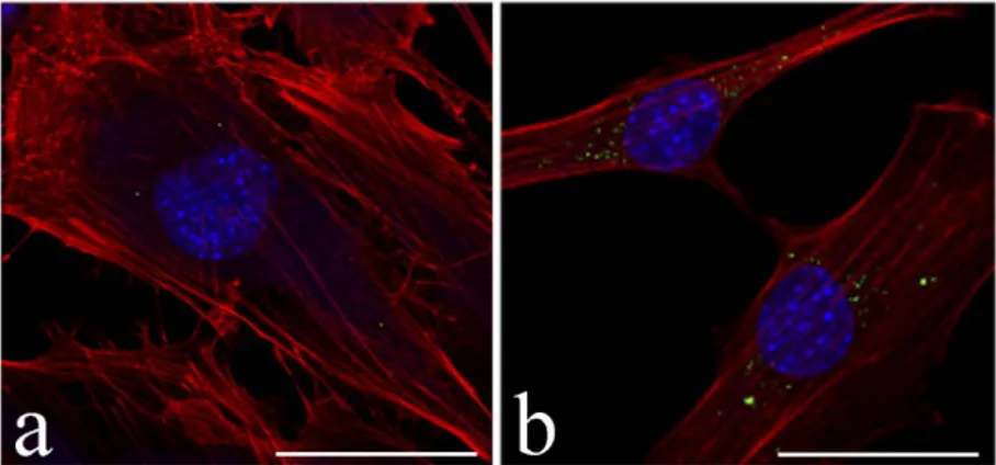
NPs ranged 250 to 300 nm in size, with PdI <0.1, and PTM encapsulation efficency of 80%. Viability assays showed that blank HA-NPs induce an increase of cell death only at the highest concentration tested for long incubation times (48 h), thus demonstrating their good biocompatibility; actually, no cytological alterations were ever observed at TEM following administration of blank HA-NPs. As expected, free PMT induced concentration-dependent cell death; similar evidence was obtained after PMT administration via HA-NP, when cell mortality was sometimes even higher. Fluorescence microscopy (Figure 1) showed that Fluo-NPs are rapidly (<30min) and efficiently internalized by the cells in a time-dependent manner. TEM (Figure 2)
Hyaluronic acid-based nanocomplexes as an innovative
therapeutic tool to treat myotonic dystrophy
MATHIEU REPELLIN1,2, FLAVIA CARTON1,2, FEDERICO BOSCHI3, ANDREA ALBANO2,
GIOVANNA LOLLO2 AND MANUELA MALATESTA1
1 Department of Neurosciences, Biomedicine and Movement Sciences, University of Verona, Italy; 2 University of Lyon, Université Claude Bernard Lyon 1, CNRS, LAGEPP UMR 5007, Villeurbanne, France;
NANOMATERIALS IN BIOLOGY AND MEDICINE
LIFE SCIENCES 230
MCM2019
14th MULTINATIONAL CONGRESS ON MICROSCOPY
S E P T E M B E R 1 5 – 2 0, 2 0 1 9 I N B E LG RAD E , S E RB I A
revealed that HA-NPs are internalized individually via endocytosis and localize throughout the cytoplasm, being never observed inside the cell nuclei. HA-NPs rapidly escape endosomes occuring free in the cytosol and, after 24 h, they may form large clusters (Figure 2b).
Figure 1. Confocal fluorescence micrographs of C2C12 cells treated with Fluo-NPs (green) for 30 min (a) and 24 h (b). Actin cytoskeleton in red (Phalloidin-Atto 594), nucleus in blue (Hoechst 33342). Bars: 50 µm.
Figure 2. TEM micrographs of C2C12 cells treated with HA-NPs (arrow) for 2 h (a) and 24 h (b). Note the
huge accumulation of NPs (asterisks) in the cytoplasm after 24 h. N: nucleus. Bars: 500 nm (a), 1 µm (b). We may conclude that, due to the good encapsulation efficiency and the excellent capability to enter and accumulate inside C2C12 cells, Loaded-NPs cause a rapid and massive intracellular release of PMT which can account for the high cell mortality observed. We may hypothesize that HA-NPs loaded with very low PMT concentrations would be able to induce therapeutic effects on DM muscle cells with reduced cytotoxicity. Studies are presently in progress to verify this hypothesis. 4. Acknowledgments
M.R. is an INVITE PhD student of the Nanoscience and Advanced Technologies program, University of Verona. INVITE project received funding from EU Horizon 2020 GA No 754345. References
[1] R. B. Siboni, M. J. Bodner, M. M. Khalifa, A. G. Docter, J. Y. Choi, M. Nakamori, M. M. Haley, J. Andrew Berglund, J Med Chem, 58 (2015) 5770-5780