“FEDERICO II” University of Naples
P
hD PROGRAM IN NEUROSCIENCE
XXXI CYCLE
PhD Thesis:
“Role of Autophagy in the regulation of
Angiogenesis in Stroke”
Coordinator
Chiar.mo prof. Maurizio Taglialatela
Tutor
Candidate
Dr. Antonio Vinciguerra
Dr. Chiara Vigliotti
- 1 - SUMMARY LIST OF ABBREVIATIONS………..………. 4 ABSTRACT………...………. 6 PREMISE………..………. 7 I. INTRODUCTION………..……….... 8
I.1. Vasculogenesis and Angiogenesis: an overview………..…...….……….. 8
I.1.1. Angiogenesis in the Central Nervous System……….…..…….... 10
I.1.2. Angiogenesis after ischemic stroke………. 11
I.1.3. Molecular mechanisms of new vessel sprouting…...………... 13
I.1.3.1. VEGF contribution to angiogenesis……….. 15
I.1.3.1.1. Role of VEGF on angiogenesis after stroke………..…….. 17
I.1.3.1.1.1. HIF involved in the activation of VEGF and autophagic process... 18
I.2. Autophagy, mechanism and molecular pathway…...……….. 19
I.2.1. The machinery of autophagy: role of Beclin1, LC3 and Atg5………….. 20
- 2 -
I.2.3. Relevance of autophagy in response to cerebral ischemia…….……… 25
I.2.4. The possible role of the autophagy in the angiogenesis after a stroke. 26 II. AIM OF THE STUDY………... 27
III. MATERIALS AND METHODS.……….. 29
III.1. Drugs and chemicals……….. 29
III.2. In vitro methods……….. 30
III.3. In vivo experiments………. 37
IV. RESULTS………..……… 41
IV.1. Tubule formation in bEND5 cells is not affected by Bevacizumab treatment………...……….. 41
IV.2. Metformin increases the capillary area during vessel formation in bEND5………... 45
IV.3. 3-Methyladenine decreases almost all parameters in the vessel Formation……….… 49
IV.4. LY294002 decreases all parameters on in vitro tubulogenesis at the high concentration.………. 53
- 3 -
IV.6. Bafilomycin blocks tubule formation in vitro……..………. 59
IV.7. Pantoprazole inhibits new tubule formation……… 62
IV.8. Beclin1 is increased in the early stages of in vitro tubulogenesis….….. 64
IV.9. The appearance of autophagic vacuoles during vessel formation in vitro indicates the link between autophagy and angiogenesis...……….. 65
IV.10. Atg5 is required for in vitro vessel formation………. 67
IV.11. CD31 and Beclin1 are both increased at 12 hours after PC and PC + tMCAO…...…………..……….……… 69
IV.12. Beclin1 and CD31 are not related to each other 24 hours from tMCAO and PC……….………. 71
V. DISCUSSION…………..……….. 72
VI. REFERENCES………..………... 76
- 4 -
LIST OF ABBREVIATIONS Ang: Angiopoietin
BBB: Blood Brain Barrier
bFGF: basic Fibroblast Growth Factor CCA: Artery Common Carotid
CNS: Central Nervous System DLL: Delta-like ligand
EC: Endothelial Cell
ECA: External Carotid Artery ECM: Extracellular Matrix
FGF-2: Fibroblast Growth Factor ICA: Internal Carotid Artery IPC: Preconditioning Ischemia 3-MA: 3-Methyladenine
MCA: Middle Cerebral Artery
mTOR: mammalian Target of Rapamycin NVU: Neurovascular Unit
- 5 -
PDGF: Platelet Derived Growth Factor PDGF-B: Platelet-Derived Growth Factor B PE: Phosphatidylethanolamine
PHD2: Prolyl Hydrolase Domain2 PlGF: Placental Growth Factor
pMBMECs: primary Mouse Brain Microvascular Endothelial Cells SHH: Sonic HedgeHog
Smc: Smooth muscular cells SVZ: SubVentricular Zone
tPa: tissue plasminogen activator TGF: Transforming Growth Factor TGF-β: Transforming Growth Factor-β
TIMPs: Tissue Inhibitor of Metalloproteinases tMCAO: transient Middle Cerebral Artery Occlusion ULK1: Unc-51 Like autophagy activating Kinase1 VE-cadherin: Vascular Endothelial cadherin VEGF: Vascular Endothelial Growth Factor VPF: Vascular Permeability Factor
- 6 -
ABSTRACT
Angiogenesis is the extension of new blood vessels from the preexisting vessels. This mechanism occurs in pathological conditions of the CNS such as stroke, brain tumors or neurotrauma. After ischemic stroke and brain injury, angiogenesis tries to restore tissue perfusion by developing new vessels. VEGF was traditionally regarded as the primary factor activated in the angiogenic reparation and it has been proposed to in the therapy of neurodegenerative diseases. Unfortunately, the VEGF is not a good drug candidate for its pharmacokinetic limitations and its capacity to permeabilize the cerebral endothelium causing brain leakage. Due to the increasing evidences from literature showing an involvement of autophagy in new vessels formation it has been verified whether autophagy could have a role in blood vessel formation in a pathological condition where the failure to supply blood to suffering brain is crucial, the ischemic stroke. There are different conditions in the brain that promote autophagy such as oxygen deprivation with activation of angiogenesis. To test our working hypothesis, in vitro experiments were performed in order to evaluate whether autophagy could be involved in vitro tubulogenesis by modulating the autophagic cascade. In these experiments, brain endothelial cells (bEND5) VEGF independents and forming a capillary-like network when plated on Matrigel, were used. Hereafter, it has been evaluated if the experimental hypothesis tested in vitro experiments could be confirmed also in vivo experiments in rodents thus allowing us to verify whether autophagy could be considered a mechanism involved in angiogenesis and therefore it could be proposed as a target for the therapy of stroke.
- 7 -
PREMISE
Still today ischemic stroke continues to represent the third leading cause of death in the western world. This cerebral insult is the leading cause of permanent disability with enormous costs in terms of suffering for patients and their caregivers. At present, the available therapies for the treatment of stroke do not appear satisfactory and there is an unmet medical need for effective therapies for that pathology. The Central Nervous System is a breakable organ and easily damage to the ischemic events. Stroke is caused by an interruption of blood flow to the brain that can occurs in two different mechanisms: the first, is due by a formation of a thrombus that block the cerebral artery; the second is a rupture of the cerebral vessel that causes hemorrhage and its fatal. Unfortunately, for logistical reasons it is often impossible to guarantee such a timely therapeutic intervention. In perspective, therefore, therapeutic approaches of regenerative medicine appear, which aim to intervene on the damage already realized to promote its repair. In the present experimental project, the objective is intended to acquire new information on the molecular mechanisms of such reparative processes and could lead to the identification of new approaches for their pharmacological activation. In this time, the only approved treatment is represented by vasal recanalization with the tissue plasminogen activator (tPa) or its derivatives which, however, are effective only if administered within a very narrow time window (between 3 and 6 hours) after the ischemic event. These drugs act by preventing neuronal death by early recovery of the circle while they are ineffective on the damage already established.
- 8 -
I. INTRODUCTION
I.1. Vasculogenesis and Angiogenesis: an overview
In the early stages of development, the human embryo receives nutrients and oxygen through the diffusion process. In the successive phases, the embryo undergoes to a rapid formation of a circulatory system constituted by a complex network of capillary and blood vessels that procure the transport of oxygen and nutrients to the various organs and tissues in the body. This initial phase of vessel formation in the embryo is called "Vasculogenesis" and starts from the hemangioblasts which are mesodermal cells derived from the extra-embryonic yolk sac of which proliferate and differentiate into angioblasts by Notch signals and for the follow “arterial-venous” cell fate transformation, it depends by on one of the two transmembrane proteins that they can have during the step of angioblast transformation, in which the first transmembrane protein is Ephrin-B2 for an arterial cell fate and from their aggregation generating the dorsal aorta, and the second is EphB4 for a venous cell fate that by their aggregation forming the cardinal vein. From the fusion of these two types of blood vessels there is the formation of the Primary capillary plexus which evolves in a complete circulatory system as shown in the Figure 1 (Chung and Ferrara 2011). The sprouting of new blood vessels from pre-existing vessels is stabilized by the recruitment of mural cell (Pericytes and Smooth-muscle cells) that proliferate and differentiate in response to Angiopoietins and to the TGF-β (Transforming Growth Factor-β) signaling and, in turn, are recruited to vessels by PDGF-B (Platelet-Derived Growth Factor B). This second stage of formation of new blood vessels from pre-existing vessels is termed “Angiogenesis” and this definition was introduced by John Hunter in 1794, to describe blood vessel growth in reindeer antlers.
- 9 -
Figure 1. A schematic representation of developmental vasculogenesis and angiogenesis during early stages of development in the human embryo: In the first step of vessel formation (A) the
hemangioblast precursors are marked by the Flk-1 that can generate the hematopoietic cells, or by Notch signaling become the angioblasts. They for a follow Notch signaling, can aggregate forming two types of vessels that depends from two signals that are: (B) Ephrin-B2 for the dorsal aorta (red) and EphB4 for the cardinal vein (blue). This aggregation mediated by signaling as VEGF, SHH and DLL (Delta-like ligand) conduces to the formation of primary capillary plexus (C). For a vascular remodeling, mural cells are recruited and initiate the process of vessels sprouting mediated by PDGF-B, TGF-β and ANG-1 to form the capillary bed (D) (Chung and Ferrara 2011).
In addition to these two terms of “vasculogenesis” and “angiogenesis”, the term neoangiogenesis is also used to describe the de novo formation of new blood vessels that is common in tumors where it contributes to nutrient supply both in primary tumors and in metastasis. This formation derives from either interstitial precursors or stem cells coming from peripheral circulation.
- 10 -
I.1.1. Angiogenesis in the Central Nervous System
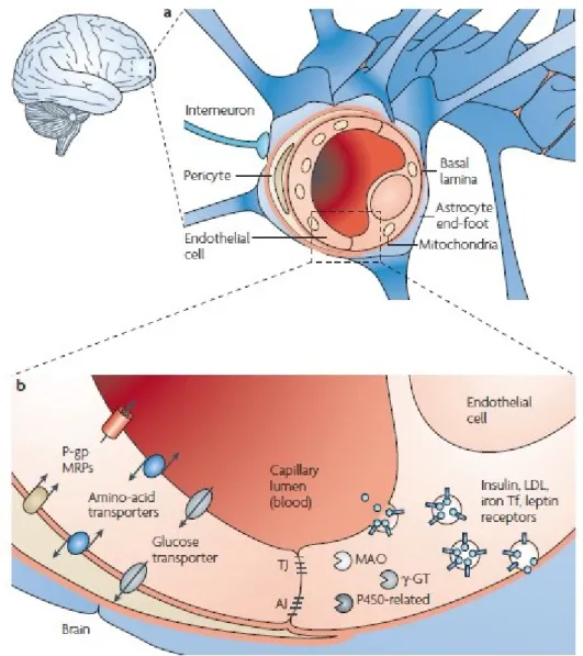
Central Nervous System (CNS) is a very complex organ that is supplied by a highly specialized vessels network configured to bring oxygen and nutrients through blood for the energetic requests of this metabolically highly active organ mainly constituted by the brain. The functions of cerebral circulation are controlled by cells involved in the neurovascular unit (NVU), that through their physical interaction form the Blood Brain Barrier (BBB) (Figure 2).
Figure 2. Focus on the BBB at the level of NVU: A) The structure of BBB. B) In addition to the
cell-cell adherent junctions, endothelial cell-cells form complex tight junctions through the interaction of several transmembrane crucial for the maintenance of brain integrity (Cecchelli, Berezowski et al. 2007).
- 11 -
In particular, the endothelial cells are kept in contact by the tight junctions (TJ) and the junctional adhesion molecules (JAMs) forming a continuous vessel structure. The junctions are transmembrane proteins and accessory cytoplasmic proteins that are located on the endothelium (Stamatovic, Keep et al. 2008). The essential function of BBB is the protection of the CNS integrity, by orchestrating different mechanisms such as ion regulation, balancing of neurotransmitters, by preventing the passage of both macromolecules and neurotoxins in the brain through the blood. (Cecchelli, Berezowski et al. 2007, Vallon, Chang et al. 2014). In pathological conditions of the CNS such as brain tumors, neurodegenerative diseases and arteriovenous malformations, angiogenesis is activated. For years, the growth of gliomas in CNS, has been identified with neoangiogenesis since this phenomenon was considered to be restricted only to the brain. More recently, it has become clear that angiogenesis also plays a role in non-neoplastic neurological diseases such as cerebral ischemia, neurotrauma and epilepsy. From the beginning of the 20th century, vascular anomalies and blood deficiency have been suspected of contributing to epileptogenesis (Bratz,1899, Spielmeyer,1927) and significant changes in blood flow have been described in the epileptic (Lennox and Gibbs 1932).
I.1.2. Angiogenesis after ischemic stroke
Among neurovascular multifactorial diseases that affect the brain a central role has the ischemic stroke, a pathological condition caused by an obstruction or rupture of a blood vessel causing a destabilization of the transport of nutrients and oxygen in the brain with neurologic deficit or death. Common causes of stroke are the follows: non-thrombotic occlusion of small, deep cortical arteries, embolism due to cardiac sources;
- 12 -
arterial thrombosis with hemodynamic disturbance leading to decrease of cerebral blood flow; artery to artery embolism. In response to this insult, reparative processes known as neurogenesis begin to activate in the brain which aim to reconstruct the tissue that was destroyed by replacing it with newly formed brain tissues (Beck and Plate 2009, Liman and Endres 2012). It is well established that the activation of this neurogenesis is elicited by the activation of the NVU that together to the angiogenesis is able to restore the blood flow It has been demonstrated that in the subventricular zone (SVZ) angiogenesis and neurogenesis are two closely related processes. The SVZ contain neural progenitor cells which are close to endothelial cells and during neurogenesis are differentiated in neuroblast and later in neurons through soluble factors that are secreted by endothelial cells. Other studies have reported an association between neurogenesis and vascular marker as CD31 after a stroke (Wang, Zhang et al. 2004, Jin, Wang et al. 2006, Ohab, Fleming et al. 2006). CD31 is an endothelial cell junctional protein also known as PECAM-1 important for the contribution to the maintenance of the endothelial cell permeability barrier. In addition to this function, CD31 is involved in the cell migration and angiogenesis (Lertkiatmongkol, Liao et al. 2016). For this functions, this is considered as a marker for endothelial cells and for a vessel structure in a immunofluorescence staining (DeLisser, Newman et al. 1993, DeLisser, Christofidou-Solomidou et al. 1997).
- 13 -
I.1.3. Molecular mechanisms of new vessel sprouting
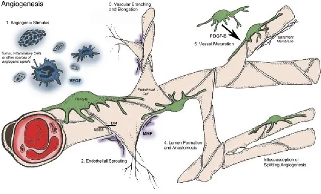
In normal and quiescent conditions, the physiology of vessel is ruled by a balance between pro- and anti- angiogenic factors. When a stimulus like hypoxia is triggered, this balance becomes impaired activating the vessels sprouting mechanisms. Anatomical structure of the blood vessel is constituted from a monolayer of endothelial cells colligated by junctional molecules (claudins and VE- cadherin) which are protected from the insults by the autocrine actions of signal such as VEGF, Notch, FGF’s and Ang-1 released from pericytes. Therefore, when the vessel detects an angiogenic signal (VEGF, FGF’s, chemokines), the pericytes separate from blood vessel and endothelial cells begin liberating from the junction causing a vasodilatation of blood vessel. Subsequently VEGF inducing an increase of permeability of the endothelial cells layer causing an emission of extracellular matrix that provide to constitute a scaffold for to build a new tube (Ferrara 2000, Ferrara, Gerber et al. 2003, Carmeliet and Jain 2011). Summarizing angiogenic process occurs in several steps: 1) Angiogenic stimulus; 2) Endothelial Sprouting; 3) Vascular Branching and Elongation; 4) Lumen formation and anastomosis; 5) Vessel maturation and Intussusception or splitting angiogenesis (Figure 3).
- 14 -
Figure 3. Schematic angiogenetic process observed in a blood vessel during physiological conditions: After an angiogenic stimulus (1) due by Hypoxia, Inflammatory cells or VEGF, the Pericytes
separate from the vessel and the permeability of the vessel increases. The MMP (Matrix -Metalloproteinases) degrade the collagen and other components of the extracellular matrix that evenlope the vessel with a disruption of the barrier allowing the Endothelial Cells (EC) migration for an endothelial sprouting (2) that continues in the vessel elongation (3) by different mechanism as sprouting or intussusception (4) and by the PDGF-B the mural cells are recruited for the vessel maturation with a complete formation of the vessel (Logsdon, Finley et al. 2014).
The new vessel sprouting can occur in different modes as follows: sprouting angiogenesis, vasculogenesis, intussusception, vessel co-option, vascular mimicry, tumor cell-EC differentiation (Figure 4), and the causes that triggers these different mechanisms of new vessel formation is not well understood.
- 15 -
.
Figure 4. Different types of vessel sprouting : (A) It is represented the traditional vessel expansion
known as angiogenesis while the figure (B) is the vasculogenesis;(C) the intussusception is a mode of capillary formation in which there is a creation of a central perforation and fusion of the cellular bilayer, the endothelial cells form a wall separating the two new vessels formed (Kurz, Burri et al. 2003). (D) this model of sprouting happens when tumor cells take possession of the existing vasculature; (E) for instance aggressive cancer cells are able to form an alternative vascular network involving endothelial cells with a phenomenon called mimicry (Carmeliet and Jain 2011).
I.1.3.1. VEGF contribution to angiogenesis
In 1989, Ferrara and Henzel and Ploüet independently reported the genomic sequencing of the first regulator of angiogenesis by the isolation of an endothelial cell specific mitogen, they called VEGF, from medium conditioned from bovine pituitary follicular cells (Ferrara and Henzel 1989, Plouet, Moro et al. 1997). The VEGF mitogen family includes the VEGF-A (or VEGF, the originally identified), VEGF-B, VEGF-C, VEGF-D, VEGF-E and PlGF. In addition, alternative VEGF isoform resulted by the differential splicing of the VEGF gene: VEGF121, VEGF145, VEGF165, VEGF1831
- 16 -
which is the binding determinant for heparin, encoded for by exons 6 and 7. The different members of the VEGF family exert their biological activities through the family of specific of binding to a receptors family: VEGFR1-(Flt1); VEGFR2 (Flt1/KDR) that is mainly expressed in activated endothelial cells and their embryonic precursors and is required for angioblastic differentiation (during vasculogenesis); and VEGFR-3 is predominantly expressed in lymphatic endothelium (Figure 5). VEGFR-2 is the most responsible for the majority of angiogenic effects activated by VEGF. VEGF is involved in the development and spread of many diseases as in neoangiogenesis as it plays an essential for tissue repairing and is required for the growth and metastasis of all solid tumors.
- 17 -
As shown in the Figure 5, VEGF-A, VEGF-B and PlGF bind to VEGFR1, VEGF-A and VEGF-E bind VEGFR2, and VEGF-C and VEGF-D bind VEGFR3. VEGF and its receptor are expressed in pre- and post-natal neurons that after an insult, for example a stroke, their expression increases. Of these, VEGF-A is the only one with the most attention for its biological activity and for its only hypoxia-dependent angiogenic mediator (Ferrara, Gerber et al. 2003).
I.1.3.1.1. Role of VEGF on angiogenesis after stroke
VEGF is involved in many important processes of the central nervous system and for this it was traditionally regarded as the primary factor activated in the angiogenetic reparation and it has been proposed to use in the therapy of neurodegenerative diseases (Greenberg and Jin 2013). Many studies on rodents, demonstrated that after stroke, there is an increased expression of VEGF in ischemic brain that contributes to the repair of brain ischemic regions by activating the neurovascular niche. It has been suggested that VEGF could represent a useful therapeutic strategy for the treatment of stroke. Since encouraging results have been obtained in experimental model of stroke in rodents showing that post ischemic administration of VEGF promotes angiogenesis. Nevertheless, high doses of VEGF administrated led to significantly increased BBB permeability that can worsen the ischemic lesions (Zhang, Zhang et al. 2000, Singh, Sharma et al. 2012). These unsuccessful events due from the VEGF administration raises an interest in the research to identify new mechanisms that can repair the damages caused by ischemic stroke without causing the same events of VEGF.
- 18 -
I.1.3.1.1.1. HIF involved in the activation of VEGF and autophagic process The factor mainly responsible for the activation of post ischemic endothelial proliferation is the VEGF whose synthesis is regulated by HIF-1 which is activated in response to a cerebral ischemic insult (Marti, Bernaudin et al. 2000, Beck and Plate 2009). HIF-1 is structured as an α-β heterodimer and the α subunit is the determining factor for the principal activity of HIF-1 that depends from the oxygen. During a stroke causing oxygen deprivation and damage to the tissues, HIF-1 activates different genes involved in angiogenesis for restore the flux of oxygen in particularly VEGF (Singh, Sharma et al. 2012). As previously suggested that VEGF's administration in the treatment of post ischemia has not been successful for its pharmacokinetic properties and for its toxicity (induces a permeabilization of the blood brain barrier) that discourage the development and the use of VEGF as a drug and for these limitations there is interest to identify the mechanism for the regeneration of ischemic tissue activating the neuro-vascular niche through a pharmacological approach that does not involve the administration of VEGF. In this perspective it appears interesting that in parallel to the activation of HIF-1 responsible for the induction of the synthesis of VEGF, in response to cerebral ischemia there is also a significant activation of autophagy (Wen, Sheng et al. 2008).
- 19 -
I.2. Autophagy, mechanism and molecular pathway
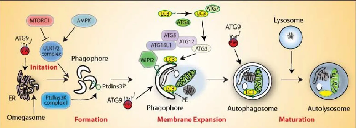
Autophagy as a new term was introduced in the 1963 by Christian de Duve, a Belgian biochemist who previously had discovered lysosomes and peroxisomes. Autophagy is an orchestrated process useful to eliminate unwanted proteins and damaged organelles and intracellular microbial pathogens. This mechanism is involved in biological and physiological cellular processes such as cellular differentiation and development. It has a double possible significance because if it can contribute to cell death, on the other it can also represent an important protective factor because it provides energy for cell survival in conditions of energy deprivation. At least three different forms of autophagy have been identified: the “Microautophagy”, the “Chaperone-mediated autophagy” and the “Macroautophagy” as shown with the term Autophagy. These forms differing for their function and the mode of delivery of the cargo to the lysosomes. Autophagy is a highly controlled process that progresses through consecutive steps: Initiation, Nucleation, Elongation, Maturation and Degradations. This process is initiated by the activation of ULK1 complex from AMPK and inhibited by the TORC1 complex. ULK1 mediates the activation of the PAS (Phagophore Assemble Site) which is a specialized intracellular membrane region that through a double-membrane known as phagophore is assembled. It’s the precursor of autophagosome that through the elongation of this phagophore, forms an omegasome and finally closes forming the autophagosome. An ULK1 interacting protein, Atg9, acts as a lipid cargo system between the ER (Endoplasmic Reticulum) and PAS and provide for the lipids during phagophore maturation. In the elongation and maturation of the phagophore in the autophagosome, it’s required ULK1 mediated activation of the PI3KIII complex and directs the assembly and activation of two ubiquitin like system:
- 20 -
the Atg5/atg12 system and LC3 system. After activation, LC3 is first cleaved in two fragments: LC3-I and LC3-II and the latter is involved in a reaction requiring Atg12-Atg5-Atg16L complex that concludes for the omegasome closure with the formation of a mature autophagosome containing cell components committed to lysosomal degradation (Xie and Klionsky 2007). Finally, it fuses with lysosomes to form autolysosome where degradation begins (Figure 6) (Klionsky, Abdelmohsen et al. 2016, Yin, Pascual et al. 2016).
Figure 6. Schematic representation of the pathway of autophagy (Yu, Chen et al. 2018).
I.2.1. The machinery of autophagy: role of Beclin1, LC3 and Atg5
As described in the previous paragraph, the autophagy evolves in distinct step in which different proteins are involved assuming an importance as a marker for study this biological process. Among them, Beclin1 is involved in the nucleation step, it’s the
- 21 -
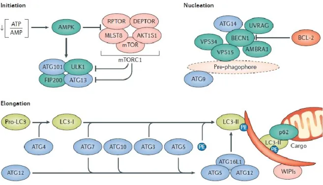
orthologue of yeast Atg6 and has a major role in the identification of the localization of the PAS. Beclin1 together with AMBRA1 (Activating molecule in beclin1 regulated autophagy) forms a complex with VPS15 and VPS34 called PI3KC3 (class III phosphatidylinositol 3-kinase) (Figure 7) that by a phosphorylation produces PI3P (Phosphatidylinositol-3-phosphate) essential for the autophagosome formation along with the other proteins involved in this process as the complex Atg12-Atg5 and Atg16-L1 (Kang, Zeh et al. 2011). In particular Beclin-1 has been identified as an interactor of the anti-apoptotic protein Bcl2 that negatively regulates autophagy (Levine, Sinha et al. 2008). In the step of elongation, as shown in figure 7, LC3 has an important role in the autophagosome formation. This protein exists in two forms that are known as LC3-I and LC3-II. The first is the complete protein, while the second is incomplete of an amino acid sequence and is a carrier of the PE (Phosphatidylethanolamine). Through the PE, LC3 is associated with the vesicle that being formed. When the synthesis process is completed, LC3 is removed from the outer membrane and recycled while that is located in the inner membrane will be degraded together it. This protein is the most used as an autophagosome marker (Kabeya, Mizushima et al. 2000, Kimura, Noda et al. 2007, Kimura, Fujita et al. 2009). Furthermore, Atg5 is essential for the extension of the autophagosomal membrane and it is coordinated with the conjugation system as LC3-PE complex (Figure 7). The Atg5-Atg16L1-Atg12 complex is the first direct to the expansion and curvature of the autophagosome membrane during formation. LC3 is initially cut off from the Atg4 protease. After the LC3 process, the Atg5-Atg16L1-Atg12 complex dissociates from the membrane just before completion of autophagosome formation and LC3 II continues it process (Mizushima, Sugita et al. 1998, Kabeya, Mizushima et al. 2000).
- 22 -
Figure 7. The molecular pathway of autophagy: It is showed how the regulation of autophagy is
determined by specific proteins in each step: Initiation, Nucleation, Elongation (Galluzzi, Bravo-San Pedro et al. 2017).
- 23 -
I.2.2. Interest in the pharmacological modulation of the autophagy
It becomes interesting to consider that the biochemical reactions involving all the steps of the autophagic cascade recruit at least 100 different proteins (Noda and Inagaki 2015). Therefore,it’s of particular interest to study the autophagy process through the specific drugs directed on proteins involved in each step of the autophagic cascade (Figure 8) (Galluzzi, Bravo-San Pedro et al. 2017).
Figure 8. A synthetic representation of the drugs that act selectively in each step of autophagy cascade: The drugs are grouped as follows: in the blue panels are represented the inhibitor drugs for
autophagy whereas in orange the drugs activators of autophagy. And in each panel, there are represented the protein target for the drugs listed in the panel (Galluzzi, Bravo-San Pedro et al. 2017).
- 24 -
As shown in Figure 8, during the first step of Initiation, one of the drugs used for the activation of autophagy is the Metformin that is a well known pharmacological compound largely used to treat type 2 diabetes mellitus (DM2) and this drug is of particular interest for its ability to activate the AMPK cascade (Zhou, Myers et al. 2001, Viollet, Guigas et al. 2012, Wrobel, Marek et al. 2017). In fact, the autophagy activation by AMPK can be proceeded through two mechanisms: inhibition of mTOR or phosphorylation of ULK-1 (Steinberg and Kemp 2009, Villanueva-Paz, Cotan et al. 2016). In the phase following initiation, that is named nucleation, several researchers have shown that it can be inhibited by drugs as LY290042 or 3MA through an inhibition mechanism of the class III PI3K complex that is required for the formation of PAS (Caro, Plomp et al. 1988, Blommaart, Krause et al. 1997, Stroikin, Dalen et al. 2004, Wu, Tan et al. 2010, Zou, Zhang et al. 2014). More, in the step before the degradation, the lysosome is fused with the autophagosome exposing the organelles and compounds to the degradation caused by the action of the lysosomal hydrolases. In this phase, the Chloroquine acts with a mechanism of prevention of the endosomal acidification. It accumulates in the lysosomes causing an increase in lysosomal pH, which inhibits lysosome function and may block fusion of the autophagosome with the lysosome (Beil, Staar et al. 1992, Beil, Sewing et al. 1999). Or the Bafilomycin (Hanada, Moriyama et al. 1990, Oda, Nishimura et al. 1991) and Pantoprazole (Moreira Dias 2009, Vegger, Bruel et al. 2017) that are two inhibitors of the Vacuolar H+ATPase, which controls the pH in the lysosome and causing an elevated lysosomal
pH preventing the acidification of endosomes and lysosomes inhibiting the autophagic flux. Furthermore, there are still experimental obstacles that do not allow pharmacological modulation in the clinic. In the light of these evidences, it is of
- 25 -
considerable interest to identify and creating selective autophagy modulators and specific biomarkers for the autophagic flux (Galluzzi, Bravo-San Pedro et al. 2017).
I.2.3. Relevance of autophagy in response to cerebral ischemia
Autophagy is activated in conditions of nutrients and oxygen deprivation and help cells to survive by recycling part of their components. More recently, relevant implications of autophagy in diseases progression have been described. In particular, autophagy is involved in neurodegenerative diseases, in microbial infections, in aging and in obesity (Kimura, Isaka et al. 2017, Qian, Fang et al. 2017). Recent evidence suggests that autophagy could also have a prominent role in vascular biology (Nussenzweig, Verma et al. 2015) where it has been shown that autophagy has a protective role against atherogenesis, that it’s essential for the increase in nitric oxide (NO) production induced by shear stress and consequently, an impairment of autophagy leads to endothelial dysfunction. So far, contrasting data have been accumulated on the role of autophagy in angiogenesis because some studies suggested that it inhibits and other that it stimulates this process. Antiangiogenic factors seem to exert their effects by inducing autophagy. An increase in autophagic flux has been observed upon exposure to dermatan proteoglycan decorin that inhibits KDR/VEGFR2 and increases the expression of PEG3/PW1 a transcription factor that controls the transcription of Beclin1 and LC3 (Buraschi, Neill et al. 2013, Neill, Torres et al. 2013) and this antiangiogenic factor also activates AMPK. Some evidence suggests that autophagy is required for angiogenesis during development.
- 26 -
I.2.4. The possible role of the autophagy in the angiogenesis after a stroke After a stroke, different signals are triggered in response to the damage in order to try to restore the physiological conditions of nutrients and oxygen in the area of injury. When a reduction of blood flow occurs, the angiogenic signals are stimulated for to activate the angiogenic sprouting and new vessels will form. As the hypoxia activates the VEGF pathway, in turn it activates the autophagy process able to sustain cell survival. Although many studies on the angiogenesis were performed on the low nutrient conditions, there are still several studies that investigate the angiogenesis under nutrient deprivation. It’s well known that autophagy is a biological process of degradation that is fundamental for the cell survival in low nutrient-deprived conditions (Du, Teng et al. 2012). Recent studies reported evidences that autophagy is involved in angiogenic activation in conditions of nutrient deprivations, and that if the angiogenesis is inhibited there is an increase in autophagy signals (Nguyen, Subramanian et al. 2007, Ramakrishnan, Nguyen et al. 2007). Nowadays it is unknown whether autophagy has a role on the endothelial cells in angiogenesis. It is become interesting the study of the expression of proteins involved in the autophagic pathway after an ischemic insult (Rami, Langhagen et al. 2008, Dai, Xu et al. 2017, Wang, Shao et al. 2018). As Beclin1 is the protein involved in the initial process of autophagy, it can be a marker to evaluate the initial involvement of autophagy in the stroke by the reparative processes carried out by angiogenesis. The complicated pathway of autophagy not help to investigate the specific phases of angiogenesis on in vitro and
- 27 -
II. AIM OF THE STUDY
After ischemic stroke and brain injury, angiogenesis occurs to restore tissue perfusion by developing new vessels (Beck and Plate, 2009). VEGF was traditionally regarded as the primary factor activated in the angiogenic reparation and it has been proposed as a therapeutic tool for neurodegenerative diseases (Zhang, Zhang et al. 2000). Contrary to the expectations, VEGF is discouraged in the use as a therapeutic drug for its pharmacokinetic limitations and its potential to induce toxicity. From these reasons, researchers are looking for an alternative mechanism able to modulate angiogenesis in the central nervous system independent from the pathway of VEGF. Since the hypoxia has been widely characterized as a powerful stimulus for the activation of the angiogenic process and as an inducer of the autophagic cascade, the aim of the present thesis has been to determine whether autophagy represents a control of the angiogenic mechanism in alternative to VEGF. Preliminary studies are focusing on the role played by endothelial cells bEND5 in angiogenesis. These cells have the peculiarity to form a capillary like network in the absence of exogenous VEGF. In details, the aims of the present thesis work have been:
• To investigate the effects of drugs acting on the autophagic cascade on capillary tubule formation in vitro.
• To evaluate the expression of autophagic proteins: Beclin-1 and LC3, in cell cultures after specific pharmacological modulation.
• To investigate the time course of Beclin-1 expression after endothelial cell seeding on Matrigel with confocal.
- 28 -
• To investigate whether also autophagosome is activated during tubule formation. For that reason, we treated the bEND5 cells with Monodansylcadaverine a fluorescent compound selectively incorporated in activated autophagosomes by both an ion trapping mechanism and by the interaction with membrane lipids.
• To evaluate the effects on the silencing of the key gene involved in the autophagy pathway by silencing Atg5 in bEND5 cells.
• To perform immunocytochemical analysis of Beclin1 and CD31 expression in the cerebral cortex at different times point after tMCAO and ischemic preconditioning.
- 29 -
III. MATERIALS AND METHODS
III.1. Drugs and chemicals
To validate bEND5 as a model of VEGF independent cell line for the study of angiogenesis, the drug Bevacizumab was used (purchased from Roche Pharmaceutical). For experiments on the pharmacological modulation of autophagic cascade during tubulogenesis in vitro, the following drugs were used: the antidiabetic drug Metformin hydrochloride (Cod.PHR1084) (purchased from Sigma Aldrich); an inhibitor of the PI3K complex in the autophagy, namely 3-Methyladenine (3-MA) (Cod.M9281) (purchased from Sigma Aldrich); another inhibitor of autophagy with the same mechanism as observed for 3-MA, LY294002 (Cod.L9908) (purchased from Sigma Aldrich); Chloroquine diphosphate salt (Cod.C6628), Bafilomycin A1 from Streptomyces (Cod. B1793) and Pantoprazole sodium hydrate (Cod.P0021) (all purchased from Sigma Aldrich). For the assay of tubulogenesis in vitro, Geltrex™ LDEV-Free Reduced Growth Factor Basement Membrane Matrix (purchased from InvitrogenTM /Thermo Fisher Scientific) were used. The siRNA used for the Atg5
silencing were Atg5MSS247019 (RNA) -(5’GCGAGCAUCUGAGCUACCCAGAUAA3’)
and Atg5MSS247019 (RNA)-(5’UUAUCUGGGUAGCUCAGAUGCUCGC3’)
(purchased from InvitrogenTM /Thermo Fisher Scientific). The kit for
Autophagy/Cytotoxicity dual staining detection (purchased from Cayman Chemical Company) that contains Monodansylcadaverine, a fluorescent compound that was incorporated into multilamellar bodies by both an ion trapping mechanism and the interaction with membrane lipids, as a probe for detection of autophagic vacuoles in cultured cells (Biederbick, Kern et al. 1995, Munafo and Colombo 2001).
- 30 -
III.2. In vitro methods
III.2.1. Cell culture
The bEND5 cells were purchased from ECACC (European Collection of Authenticated Cell Cultures-cod 96091930). These cells line has been established from brain endothelial cells of BALB/C mice by immortalization through the infection of primary cells with retrovirus coding for the Polyoma virus middle T-antigen. The bEND5 cells possess an endothelial-like morphology, and are testes by fluorescent activated cell sorting (FACS), are positive for endothelial specific proteins (PECAM-1, Endoglin, MECA-32, FIK-1) (Yang, Roder et al. 2007, Steiner, Coisne et al. 2011). bEnd5 cells are well distinguished for its expression of EC specific proteins, such as the vascular endothelial-cadherin (VE-cadherin), von Willebrand factor, PECAM-1 (platelet endothelial cell adhesion molecule-1), endoglin, ICAM-2, and claudin-5. Nonetheless, despite previous studies indicating the expression of occludin in brain ECs, it’s still in question since a study by Steiner et al. (2011) found a significant decrease in occludin mRNA expression in bEnd5 cells when compared to primary mouse brain microvascular endothelial cells (pMBMECs). Cytokines induce the expression of protein such as (CAM-1, V-CAM-1 and E-Selectin) and have endothelial-like morphology. Injection of this cell line into mice causes development of haemangiomas. These cells express BBB markers like GLUTs, P-glycoprotein, Na+/K+/Cl cotransporter,
occludin and the tight junction protein 1, ZO-1 and form BBB-like polarized monolayers in vitro. The important feature of these cells is that form blood vessels in vitro.
- 31 -
The bEND5 grows in the Dulbecco’s Modified Eagle’s Medium (DMEM) with 2mM L-Glutamine, 1mM Sodium Pyruvate (NaP), 1% Minimal Essential Medium MEM, 10% Fetal Bovine Serum (FBS), 50 Units/mL penicillin, and 50 μg/mL streptomycin. At confluence, cells were trypsinated using 0.25% EDTA-trypsin (Whitehead Scientific, BE02-007E). bEnd5 culture conditions for cell growth was 37°C with 95% humidity and 5% CO2. When bEND5 cells are plated on plastic culture dishes they are grow as a monostrate culture; they are showing a cobbled appearance similar to that of well differentiated endothelial cells. At confluence the culture virtually covered the entire surface of the dish. Most of the cells were elongated or fusiform. An aspect can be noticed when we plated on multiwell coated with extracellular matrix Geltrex™ LDEV-Free Reduced Growth Factor Basement Membrane Matrix, bEND5 cells formed a well differentiated capillary-like network. Since the first two/three hours from plating, the cells started to arrange in strings around central holes though they were still rounded. Well defined capillary structures were evident at 22 hours from plating (DeCicco-Skinner, Henry et al. 2014).
III.2.2. In vitro tubulogenesis
The angiogenesis can be studied on in vitro methods that consist of mimicking the growth of endothelial cells in a matrix allowing the development of tubules (Folkman and Haudenschild 1980, Staton, Stribbling et al. 2004, DeCicco-Skinner, Henry et al. 2014, Nowak-Sliwinska, Alitalo et al. 2018). The experiments performed on in vitro tubulogenesis were performed to evaluate the vessel formation treated with drugs that act on different steps of autophagic cascade. Before the experiment, the 96 multiwell plate and plastic tips were kept overnight at -20° and Geltrex™ Matrigel was thawed
- 32 -
by keeping it in ice in the cold room overnight. In the day of experiment. The Geltrex™ LDEV-Free Reduced Growth Factor Basement Membrane Matrix purchase from InvitrogenTM /Thermo Fisher Scientific); 40 µl of Geltrex were gently and
homogeneously dispensed on the bottom of each well avoiding bubbles. The matrix was left to gel by leaving the multiwell at 37°C for 30 minutes in a 5% CO2 incubator.
In the meanwhile, bEND5 cells were plated in each well, approximately 85.000 cells in the presence of vehicle for the control and with the appropriate concentration of the test drug that were: Metformin hydrochloride that the stock solution used is prepared at the concentration of 10 mM and through dilutions with the bEND5 medium were obtained the concentrations of 3 and 1 mM; 3-Methyladenine an inhibitor of the PI3K complex in the autophagy, the stock solution is prepared at 10 mM and through dilutions with the bEND5 medium were obtained the concentrations of 3 and 1 mM; LY294002 is another inhibitor of autophagy with the same mechanism of the 3-Methyladenine, this drug is prepared by a stock solution of 25mM dissolved in a solution of DMSO and from this were prepared the solutions of 100, 30 and 10 µM through a dilution with bEND5 medium. Chloroquine with a stock solution of 30 µM and through dilution with bEND5 medium were prepared concentrations of 3 and 1mM. Bafilomycin was prepared by a stock of 100µM dissolved in DMSO and from this there were prepared the concentrations of 100, 10 and 1 nM with dilution by bEND5 medium. The Pantoprazole stock solution was prepared at the concentration of 1mM, and by dilution there were obtained the concentrations of 0,3 and 0,1 mM. Experiments were performed on individual multiwell for each drug used. Unless otherwise specified, microphotographs for image analysis were taken at 4X magnification 22 h after plating.
- 33 -
III. 2.2.1 Statistical analysis of in vitro vessel formation
The results related to the effect of the different pharmacological treatments on capillary network formation was analyzed using the Image J software which can be downloaded for free from the website http://rsbweb.nih.gov/ij/. This software program can analyze and measure specific parameters defined with the name of shape descriptors that include: tubule number/field, cumulative area of the lumina/field, mean tubule circularity, mean tubule solidity. The tubule number/field indicates the number of vessels that are formed with the pharmacological treatments; the cumulative area gives a better idea on how diffuse is tubule formation in the microscope field; the circularity is represented by this formula: 4π * [𝐴𝑟𝑒𝑎]
[𝑃𝑒𝑟𝑖𝑚𝑒𝑡𝑒𝑟]2. A circularity value of 1 indicates a perfect circle. The closest is the value to 0 the more elliptical is the shape; the solidity is represented by this formula: [𝐴𝑟𝑒𝑎]
[𝐶𝑜𝑛𝑣𝑒𝑥 𝑎𝑟𝑒𝑎]. The convex hull or convex
envelope of a set X of points in the Euclidean plane is the smallest convex set that contains X; solidity is a measure of asymmetry. For a complete guide see the website:
http://rsbweb.nih.gov/ij/.
III.2.3. Western Blot on cells
After drug treatment of bEND5, the cells were washed in phosphate-buffered saline (PBS) and collected by gentle scraping in ice-cold lysis buffer containing (in mM): 20 Hepes pH 7.4, 1 sodium azide, 0.2 sodium orthovanadate, 0.05 sodium fluoride, 1% TRITON X-100, and a protease inhibitor cocktail (0.1% aprotinin, 0.7 mg/ml pepstatin, 1 μg/ml leupeptin) (Roche Diagnostic, Monza, Italy). Samples were cleared by centrifugation and supernatants were used for Western Blot. Protein concentration in supernatants was determined by Bradford method (Bradford, 1976). Protein samples
- 34 -
were loaded and separated on 10% (for Beclin-1) or 15% (for LC3) SDS-polyacrylamide gels. Proteins were transferred onto Hybond-ECL nitrocellulose membrane (GE Healthcare, Little Chalfont, UK). The filters were blocked for 1h at room temperature with a blocking solution composed of 5% non-fat dry milk (Bio-Rad Laboratoires) in TBS-T (2mM Tris-HCl, 50 mM NaCl, pH 7.5 plus 0.1% Tween20) and were then incubated with the appropriate primary antibody dissolved in 5% non-fat dry milk. Primary antibodies were used at the following final diluitions: 1:1000 for the polyclonal rabbit anti Beclin1 antibody, 1:1000 for the polyclonal rabbit anti LC3 antibody were purchased from the Sigma Aldrich. After overnight incubation with the primary antibody, the membranes were washed with 0.1 Tween20 and incubated with the appropriate secondary antibodies dissolved in 5% non-fat dry milk (1:1000) for 1h. Immunoreactive bands were detected with the ECL (Amersham). The optical density of the bands was measured was determined using a Chemi Doc Imaging System (Biorad). Optical density data were normalized to the density of α-Tubulin in the case of the LC3, or β-actin in the case of Beclin1 using the ImageJ software for the density analysis.
III.2.4. MTT assay
Assessment of cell proliferation ability was performed according to the MTT [3-(4,5-dimethylthiazol-2-yl)-2,5-diphenyl-2H-tetrazolium bromide] assay. The cells were seeded in a 24-well plate at a density of 130.000 cells/well, three parallel wells for group. The cells were divided into control and drug groups according to the different concentrations tested. The cells were pretreated with the drugs as indicated here before: Metformin hydrochloride was used from a 10mM stock solution through
- 35 -
dilutions with the bEND5 medium to reach the final concentrations of 3 and 1 mM; 3-Methyladenine, an inhibitor of the PI3K complex in the autophagy, whose stock solution was 10 mM and through dilutions with the bEND5 medium the concentrations of 3 and 1 mM were obtained; LY294002 is another inhibitor of autophagy with the same mechanism of the 3-Methyladenine, whose stock solution was prepared by a concentration of 25mM dissolved in DMSO and from this stock, the diluted solutions of 100, 30 and 10 µM using bEND5 medium were prepared. Chloroquine was used as a stock solution of 30 µM diluted with bEND5 medium to 3 and 1 mM. Bafilomycin was prepared by a stock of 100µM dissolved in DMSO and from this stock, the dilutions of 100, 10 were obtained. The Pantoprazole stock solution was prepared at the concentration of 1mM, and then diluted to 0,3 and 0,1 mM. At 22 hours after incubation, the medium was aspirated and 0,5 mg/ml MTT (Sigma) was added to the wells, and the cells continue to be incubated for 2 h. After this, formazan salt in the well was dissolved with 500 μl dimethylsulfoxide (Sigma). Absorbance values were determined at 595 nm with BioPhotometers and the cells viability was evaluated.
III.2.5. Confocal microscopy analysis
bEND5 cells, after 22 hours from tubulogenesis in vitro using the protocol described previously for the vessel formations, were fixed in 4% paraformaldehyde for 20 min at room temperature and blocked in 1x PBS, 0.25% Triton X-100 (v/v), and 3% bovine serum albumin (BSA). Cells were subsequently incubated with anti-Beclin-1 antibody overnight at 4°C. Secondary antibody goat anti-rabbit was used. Nuclear counter stain was visualized with DAPI (4,6-diamidino-2-phenylindole, 0.5 μg/ml). The Beclin-1 immunofluorescence was visualized using an Olympus multiphoton FV-1000 MER
- 36 -
microscope with Argon laser 488 nm line and diode laser 405 nm line. The images were taken with a UPlanSApo 10-60X/1.35W objective lens.
III.2.6. Autophagy/Cytotoxicity dual staining test
bEND5 cells after seeding on matrigel (Geltrex) as described in a previous paragraph, are treated following the protocol of the kit (purchased from Cayman) and were immediately analyzed by fluorescence microscopy. Dead cells can be detected with a fluorescent filter usually designed to detect Rhodamine (excitation/emission= 540/570 nm). Autophagic vacuoles stained by MDC can be detected with a UV filter usually designed to detect DAPI. The images were taken with a DMI4000 B Inverted Microscope from Leica 20X objective lens.
III.2.7. Silencing of Atg5
Silencing of ATG5 in bEND5 cells was performed by using the standard protocol of transfection, by using two different siRNA for Atg5, Atg5MSS247019 (RNA) -(5’GCGAGCAUCUGAGCUACCCAGAUAA3’) and Atg5MSS247019 (RNA) - (5’UUAUCUGGGUAGCUCAGAUGCUCGC3’) purchased from (InvitrogenTM /Thermo
Fisher Scientific). Cells were incubated with DMEM (Sigma) supplemented with the Dharmafect Transfection (Invitrogen) and 5 nM of each siRNA for 72h and 96h following the protocol. After the time of incubation, the cells were trypsinated and re-plated on matrigel for tubulogenesis procedure as previously described. Unless otherwise specified, microphotographs for image analysis were taken at 4X and 10X magnification 72 and 96h after plating. For an optimal confirm there were performed
- 37 -
experiments of western blot for evaluate the expression of Atg5 in the cells after transfection.
III.3. In vivo experiments
To compare the expression of the proteins Beclin1 and CD31 in different experimental conditions like ischemia (tMCAO), preconditioning (PC) and ischemic preconditioning (PC + tMCAO) at different time points, we have been planned the following experimental groups:
o Group1: 8 rats sham subjected to sham intervention and sacrificed. Brain samples of ischemic cortex was collected.
o Group 2: 8 rats subjected to tMCAO and sacrificed 12 hours after the cerebral reperfusion. Brain samples of ischemic cortex was collected.
o Group 3: 8 rats subjected to tMCAO and sacrificed 24 hours after the cerebral reperfusion. Brain samples of ischemic cortex was collected.
o Group 4: 8 rats subjected to ischemic preconditioning and sacrificed 12 hours after the cerebral reperfusion. Brain samples of ischemic cortex was collected.
o Group 5: 8 rats subjected to ischemic preconditioning and sacrificed 24 hours after the cerebral reperfusion. Brain samples of ischemic cortex was collected.
o Group 6: 8 rats subjected to ischemic preconditioning and tMCAO and sacrificed 12 hours after the cerebral reperfusion. Brain samples of ischemic cortex was collected.
o Group 7: 8 rats subjected to ischemic preconditioning and tMCAO and sacrificed 24 hours after the cerebral reperfusion. Brain samples of ischemic cortex was collected.
- 38 -
III.3.1. Transient Middle Cerebral Artery Occlusion model (tMCAO)
Rats were subjected to surgical procedure as previously described (Longa, Weinstein et al. 1989). Before the surgical intervention, the animals were anesthetized with a gaseous mixture containing O2 98% and sevoflurane 2% through a mask connected at
the vaporized by means a polypropylene tube. The rats under anesthesia were placed, in dorsal decubitus, on a heating pad connected with a rectal thermometer in order to maintain stable body temperature during the surgical procedure. Then a medial incision of about 3 cm on the ventral part of the neck was practiced and, under the guidance of a stereomicroscope and with the aid of microsurgical tweezers, the muscular plane and the underlying bands were detached until reaching the artery common carotid (CCA). Then, through a blunt dissection it was isolated the CCA from the vagus nerve and, similarly, from the external carotid artery (ECA) and the right internal carotid artery (ICA). Once the vessels were isolated a nylon filament (5-0, Ethylon, 19mm long) was inserted into the right internal carotid artery (ICA), at the level of the branch from the artery common carotid. The filament was pushed in the cranial direction until it reached the branch of the middle cerebral artery (MCA), thus blocking the blood flow. After 100 minutes the animal was anesthetized with the precedent gaseous mixture in order to allow the reperfusion to occur by removing the filament.
- 39 -
III.3.2. Ischemic Preconditioning (IPC)
Ischemic preconditioning was obtained by 30-minute occlusion of the middle cerebral artery using the same procedure above described.
III.3.3. PC + tMCAO model
Animals were subjected to the preconditioning by 30 minutes MCA occlusion and, 72 hours after reperfusion the filament was reinserted and the middle cerebral artery thereby reoccluding it for 100 minutes and finally animals were sacrificed at the time established for the experimental group.
III.3.4. Tissue processing and confocal immunofluorescence
Animals were anesthetized and transcardially perfused with saline solution containing 0.01 ml heparin and followed by 4% Paraformaldehyde in 0.1 mol/l of PBS saline solution. Brains were rapidly removed from the cranium and fixed in the Paraformaldehyde overnight at +4°C. The day after the brains are posted in 30% sucrose in 0.1 M phosphate buffer (PB) with sodium azide 0.02% for 24 h at 4°C for the cryoprotection. When the brains are on the bottom of the container, they were sectioned frozen on a sliding cryostat at 40 μm thickness, in rostrum-caudal direction. Afterwards, free floating serial sections were incubated with PB Triton X 0.3% and blocking solution (0.5% milk, 10% FBS, 1% BSA) for 90 min. The sections were incubated overnight at +4 °C with the following primary antibodies: anti-Beclin-1 1:1000; (Rabbit polyclonal antibody purchased from Abcam, Cambridge, UK), anti-CD31 1:1000 (mouse monoclonal antibody; purchased from Abcam, Cambridge, UK). The sections were then incubated with the corresponding florescent-labeled secondary
- 40 -
antibodies, Alexa 488/Alexa 594 conjugated antimouse/antirabbit IgGs (Molecular Probes, Invitrogen S.R.L., Milan, Italy). Nuclei were counterstained with Hoechst (Sigma-Aldrich, Milan, Italy). Images were observed using a Zeiss LSM700 META/laser scanning confocal microscope (Zeiss, Oberkochen, Germany). Single images were taken with an optical thickness of 0.7 m and a resolution of 1024 × 1024. Quantification of Beclin1, CD31 fluorescence intensity on tissue sections at the level of the cortex, was quantified in terms of pixel intensity value by using the NIH image software. After, digital images were taken with ×40 or ×10 objective and identical laser power settings and exposure times were applied to all the photographs from each experimental set.
III.3.5. Statistical analysis
The data obtained in bEND5 cells experiments were expressed as mean ± standard error and statistical significance of differences between groups were performed by Two-Way ANOVA analysis followed by Newman Keuls post-hoc test. P-value was set to p<0.05.
- 41 -
IV. RESULTS
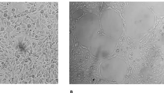
IV.1. Tubule formation in bEND5 cells is not affected by Bevacizumab-treatment bEND5 cells form a well differentiated capillary-like network when plated on multiwell coated with extracellular matrix Geltrex™ (Figure 9).
Figure 9. bEND5 cells form capillary-like networks in vitro: A) bEND5 plated on plastic dish, at
confluence have a cobbled appearance. B) bEND5 plated on plastic dish coated with extracellular matrix form a well differentiated capillary-like network. The data are reported as mean ± SEM of n=3 experiments and the capillary like network formation was observed after 22 hours after seeding on Matrigel.
The time for a complete in vitro tubulogenesis is between 22-24 hours from seeding on Matrigel. The results show the independence of these cells from the VEGF during the vessels formation by the treatment with the anti-VEGF drug, the Bevacizumab (Avastin), a humanized monoclonal antibody that bind the VEGF an angiogenic stimulator. As widely demonstrated this drug, neutralizes VEGF and blocked its signal transduction through VEGF receptors (Wang, Fei et al. 2004, Ferrara, Hillan et al. 2005). For this experiment, the drug has been tested on bEND5 during in vitro
- 42 -
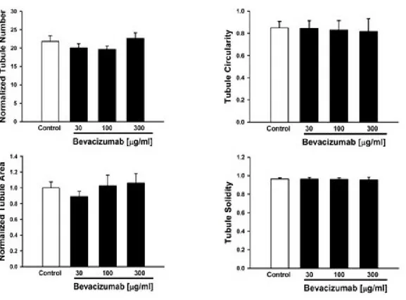
tubulogenesis, and the microscope analysis was performed at 22 hours that represents the time for a complete in vitro tubulogenesis of bEND5, As shown in Figure 10, tubule formation in our bEND5 model has been not inhibited in presence of increasing concentrations of Bevacizumab. In particular, three different drug concentrations have been used 30µg/ml, 100 µg/ml and 300 µg/ml. In addition, to test the hypothesis that Bevacizumab treatment could affect the morphology of tubule formation in bEND5 model it has been performed a set of further experiments (Figure 11).
Figure 10. Effects of Bevacizumab on tubulogenesis in vitro: bEND5 cells plated on multiwell
coated with extracellular matrigel have been observed upon treatment at different concentration of Bevacizumab (30-100-300 µg/ml). Results have been compared to the control were bEND5 cells were plated in normal medium.
- 43 -
Figure 11. Statistical analysis of tubule formation: As shown in the panels, the concentrations of
Bevacizumab used in these experiments has not affected in vitro capillary tubule formation at different concentrations as shown in the Figure 10. Bevacizumab, an antiVEGF drug not inhibite tubule formation
in vitro at the concentrations performed for the experiment. The data of these results are reported as
mean ± SEM of n=4 experiments performed on bEND5.
In order to confirm the validity of our in vitro model for the study of angiogenesis, the level of expression of the receptor-1 and 2 for VEGF (VEGFR1 and VEGFR2) were evaluated. In bEND5 cells treated with Bevacizumab, the VEGFR1 and VEGFR2 expression has been compared to the physiological expression from lysates of a cortex tissue collected from a brain obtained through extraction procedure from a mouse of our work group that was exposed to the Bevacizumab treatment.
- 44 -
Figure12. Western Blot on VEGFR expression: As known that VEGF is essential for tubule formation,
in order to confirm that bEND5 cells express the VEGF receptor, WB analysis has been performed in order to evaluate the presence of VEGF in these cells after Bevacizumab treatment in comparison with a lysate of a cortex tissue collected from a brain of a mouse exposed to Bevacizumab treatment. In that way, the confirmation of the presence of VEGFR-1 and VEGFR-2 in bEND5 cells indicates that, tubule formation in these cells are VEGF independent. The data of these results are reported as mean ± SEM of n=4 experiments performed on bEND5.
From these results it can be conclude that bEND5 cells are VEGF independent, and this represent a valitate experiment model to study angiogenesis in vitro.
- 45 -
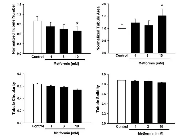
IV.2. Metformin increases the capillary area during vessel formation in bEND5 Metformin, an inducer of autophagy acting on AMPK (Zhou, Myers et al. 2001), administered at increasing concentrations (1, 3, 10 mM) in cultured bEND5 cells on Matrigel, is able to increase the capillary area of new vessels (Figure 13A). The efficacy of Metformin treatment has been assessed at by a microscope analysis after 22 hours from the exposure to the drug, as previously show. In particular Metformin treatment has not affected the other parameters of new tubules formation, as tubule number or circularity but only presented an effect on tubule area. This result clearly indicates an involvement of Metformin in new tubule formation (Figure 13A).
- 46 -
Figure 13. Metformin affects new tubule formation in Bend5 cells: (A) After 22 hours Metformin
increases tubule area during tubulogenesis as shown in the panel at a concentration of 10mM, this evidence indicates that the drug helps the tubule formation in vitro. (B) There is a reduction of tubule number at the highest concentration of Metformin, tested (10mM) conversely tubule area results increased the same in vitro concentration. The values are expressed as mean ± SEM of 3 experiments *p<0.05 versus their respective controls.
To verify which phase of the autophagic cascade is involved in angiogenesis observed in our in vitro model of bEND5 cells mimicking BBB, Beclin1 expression levels has been observed upon treatment with Metformin. As shown in the Figure 14, Metformin at 3mM concentration is strongly expressed in bEND5 cells compared to control cells. This evidence clearly indicated that the initiation of autophagic pathway in bEND5 has been activated by the drug.
- 47 -
Figure 14. Beclin1 expression after Metformin treatment at different concentrations: Beclin1
expression evaluated by WB after Metformin treatment (A); Beclin1 is increased in bEND5 treated with the drug starting from the concentration of 3mM (B). Beclin1 levels are expressed normalized with β-actin. The values are expressed as mean ± SEM of 3 experiments *p<0.05 versus their respective controls.
Further, to dissect the possible involvement of the later stage of autophagy in the pharmacological effect of Metformin, able to stimulate an increase in capillary density area, LC3 expression has been analyzed. What we found is that the increase in LC3 levels of expression is similar to that observed for Beclin1 (Figure 15).
Figure 15. LC3 expression after Metformin treatment at different concentrations: (A). LC3 is
increased in bEND5 treated with the drug starting from the concentration of 3mM (B). LC3 levels are expressed normalized with α-tubulin. The values are expressed as mean ± SEM of 3 experiments. *p<0.05 versus their respective controls.
A B
- 48 -
In addition, the highest concentration of Metformin (10mM) significantly reduces bEND5 cells number, but not causes an increase in cell mortality as shown in the Figure 16.
Figure 16. Effects of Metformin on cell viability: After 22 hours from the Metformin starting treatment.
- 49 -
IV.3. 3-Methyladenine decreases almost all parameters in the vessel formation To analyze the capability of a compound, able to block the nucleation step of autophagy (Wu, Tan et al. 2010), to reduce new vessel formation during angiogenesis, the 3-metil-adenine (3-MA) has been administered at different concentrations (1, 3, 10 mM) in plated bEND5 cells on Matrigel monitored by microscope analysis after 22 hours from the beginning of the treatment. As observed in Figure 17, 3-MA treatment causes a decrease of bot tubule area, circularity and solidity that are all parameters that give a better idea on how is diffused the new vessel formation during angiogenesis.
- 50 -
Figure 17. Effects of 3MA on tubule formation: (A) 3MA at 10mM decreases new tubule formation.
(B) 3-MA at 5- and 10-mM concentration reduced tubule area and tubule solidity during tubulogenesis. These data are reported as mean n=3 experiments. The values are expressed as mean ± SEM of 3 experimental series. *p<0.05 versus their respective controls.
Trying to understand whether LC3 expression, involved in later phases of autophagic cascade, could be affected by pharmacological treatment of 3-MA at different concentrations, its expression has been detected by Western Blotting experiments (Figure 18).
- 51 -
Figure 18. LC3 expression at different concentrations of 3MA: (A) LC3 expression evaluated in
bEND5 22 hours after the treatment with 5 and 10 mM of 3-MA is reduced because autophagy is inhibited by 3MA. (B) The given quantization of LC3 is expressed normalized with α-tubulin. These data are reported as mean of n=3 experiments. The values are expressed as mean ± SEM. *p<0.05 versus their respective controls.
From the WB results, LC3 expression shows a decrease when bEND5 cells are treated with 5 and 10 mM of 3-MA, thus indicating that autophagy is inhibited by 3MA also in the later stages of in vitro tubulogenesis. At the same time, both 3mM and 10mM do not influence significantly the mortality rate of the cells as evidenced by the MTT assay performed at 24 hours after the treatment (Figure 19).
- 52 -
Figure 19. Effects of 3MA on cell viability: 3-MA does not reduce bEND5 cells number compared to
the control one. These data are reported as mean of n=3 experiments. The values are expressed as mean ± SEM.
- 53 -
IV.4. LY294002 decreases all parameters on in vitro tubulogenesis at the high concentration
To confirm the capacity of specific step autophagic inhibitor (es. 3-MA) to block the autophagic cascade, and, consequently, new vessels formation, another autophagy selective for the nucleation step of autophagy, namely the LY294002 (Blommaart, Krause et al. 1997), has been tested in our model of in vitro tubulogenesis. As it is shown thereafter, when administered in bEND5 cultures at high concentrations, LY294002 induces a decrease of all tubule parameters (Figure 20A and B).
- 54 -
Figure 20. Effects of LY294002 on tubule formation: (A) The treatment with 100 µM of LY294002 is
able to reduce new tubule formation. (B) All parameters related to new tubule formation are modified by this concentration of LY294002. The data are reported as mean of n=3 experiments. The values are expressed as mean ± SEM of 3 experimental series. *p<0.05 vs controls.
As shown by testing increasing concentration of the compound in order to find the effective concentration able to abrupt stop new vessels sprouting from the plated bEND5, at 100 µM it is evident a regression on angiogenesis thus indicating that autophagy in closely correlated to tubule formation.
The direct interaction between LC3 and LY294002 has been confirmed by WB experiments showing that the drug induced a concentration dependent not significantly decrease in LC3 compared to control. This last result supports the hypothesis that autophagy is inhibited by LY294002.