Actin Filaments Are Involved in the Coupling of V
0
-V
1
Domains of Vacuolar H
?
-ATPase at the Golgi Complex
*
□SReceived for publication, June 26, 2015, and in revised form, February 11, 2016 Published, JBC Papers in Press, February 12, 2016, DOI 10.1074/jbc.M115.675272 Carla Serra-Peinado‡1, Adria` Sicart‡2, Juan Llopis§, and Gustavo Egea‡¶?3
From the‡Department de Biologia Cellular, Immunologia i Neurocie`ncies, Facultat de Medicina, Universitat de Barcelona, E-08036 Barcelona, the¶Institut d’Investigacio´ Biome`dica August Pi i Sunyer, E-08036 Barcelona, the?Institut de Nanocie`ncia i
Nanotecnologia (IN2UB), E-08036 Barcelona, and the§Facultad de Medicina de Albacete and Centro Regional de Investigaciones Biome´dicas, Universidad de Castilla-La Mancha, E-0200 Albacete, Spain
We previously reported that actin-depolymerizing agents promote the alkalization of the Golgi stack and the trans-Golgi network. The main determinant of acidic pH at the Golgi is the vacuolar-type H?-translocating ATPase (V-ATPase), whose V1 domain subunits B and C bind actin. We have generated a GFP-tagged subunit B2 construct (GFP-B2) that is incorporated into the V1domain, which in turn is coupled to the V0sector. GFP-B2 subunit is enriched at distal Golgi compartments in HeLa cells. Subcellular fractionation, immunoprecipitation, and inversal FRAP experiments show that the actin depolymerization pro-motes the dissociation of V1-V0domains, which entails subunit B2 translocation from Golgi membranes to the cytosol. More-over, molecular interaction between subunits B2 and C1 and actin were detected. In addition, Golgi membrane lipid order disruption byD-ceramide-C6 causes Golgi pH alkalization. We conclude that actin regulates the Golgi pH homeostasis main-taining the coupling of V1-V0domains of V-ATPase through the binding of microfilaments to subunits B and C and preserving the integrity of detergent-resistant membrane organization. These results establish the Golgi-associated V-ATPase activity as the molecular link between actin and the Golgi pH.
The secretory pathway is characterized by progressive lumen acidification of its organelles, from almost neutral in the endo-plasmic reticulum (ER)4(pH ?7.1–7.2), along the cis-to-trans Golgi stack (pH ?6.7– 6.0), to more acidic in the trans-Golgi
network (TGN) and secretory vesicles/granules (pH ?5.0) (1–3). This pH gradient is crucial for post-translational modi-fications and membrane trafficking events (4, 5). The main molecular determinant of the progressive fall in pH along the secretory pathway is the vacuolar [H?]ATPase (V-ATPase) (6 – 8). V-ATPase is a multisubunit complex composed of two large domains, V0and V1. The V0domain is a 260-kDa integral membrane complex made up of five different subunits (a, b, c, c?, c?, d, and e), which mediates proton translocation; the V1 domain is a 600 – 650-kDa peripheral complex composed of eight different subunits (A, B, C, D, E, F, G, and H), which is responsible for the ATP hydrolysis that provides the mechani-cal force necessary for proton (H?) translocation (7, 9 –11). Whereas they are the primary source of proton delivery to endomembranes consuming ATP, the final steady-state pH in the secretory pathway is the result of the balance between active H? pumping by the V-ATPase, passive H? efflux through organelle endogenous H? permeability, and differences in counter-ion conductance (3, 12).
How differences in the pH of individual secretory compart-ments are generated is not well understood. Differential V-ATPase density and/or local regulatory mechanisms in secre-tory organelles and subcompartments are possible (13). In this respect, V-ATPase-dependent proton translocation could be regulated by several mechanisms, which include the following: (a) differential V-ATPase subunit expression; (b) intracellular targeting and recycling of V-ATPase-containing transport car-riers to and from the plasma membrane and endomembranes (mainly late endosomes and lysosomes); (c) modulation of the coupling ratio between ATP hydrolysis and proton pumping; and (d) the reversible association of the V0and V1domains (14). This latter phenomenon is characterized by the release of the V1domain into the cytosol, leading to the inhibition of ATPase and proton transport activities (15). This mechanism was first identified in tobacco hornworm and yeast after glucose depri-vation (16), which was subsequently reported in mammalian cells (17, 18). When cells are cultured in the absence of glucose, a rapid dissociation of V1and V0domains occurs, which is completely reassembled after glucose re-addition, which obvi-ates the need for de novo protein synthesis (19 –21). Impor-tantly, the cytoskeleton also seems to play a role in V-ATPase assembly and activity. In particular, microtubule integrity is necessary for the reversible dissociation of the two domains, because their disruption with nocodazole blocked the V0-V1 dissociation in response to glucose depletion in yeast (22). In *This work was supported in part by Grants BFU2009-07186 and 2012-33932
from Ministerio de Economía y Competitividad del Gobierno de Espan˜ a (to G. E.). The authors declare that they have no conflicts of interest with the contents of this article.
□S
This article containssupplemental Movies S1–S5. 1
Recipient of a predoctoral fellowship from the Spanish Ministerio de Econo-mia y Competitividad.
2
Present address: Vlaams Instituut voor Biotechnologie (VIB) Centre for the Biology of Disease and KU Leuven, Dept. of Human Genetics, Campus Gasthuisberg, 3000 Leuven, Belgium.
3
To whom correspondence should be addressed: Dept. de Biologia Cellular, Immunologia i Neurocie` ncies, Facultat de Medicina, Universitat de Barce-lona, C/Casanova 143, E-08036 BarceBarce-lona, Spain. Tel.: 34-93-4021909; E-mail: [email protected].
4
The abbreviations used are: ER, endoplasmic reticulum; TGN, trans-Golgi network; V-ATPase, vacuolar H?-ATPase; DRM, detergent-resistant mem-brane; F-actin, filamentous actin; G-actin, globular actin; TfR, transferrin receptor; TRITC, tetramethylrhodamine isothiocyanate; FRAP, fluores-cence recovery after photobleaching; iFRAP, inversal FRAP; LtB, latrunculin B; CytD, cytochalasin D; JpK, jasplakinolide; Endo H, endoglycosidase H; GDI, guanidine dissociation inhibitor; NT siRNA, non-targeting siRNA.
THE JOURNAL OF BIOLOGICAL CHEMISTRY VOL. 291, NO. 14, pp. 7286 –7299, April 1, 2016 © 2016 by The American Society for Biochemistry and Molecular Biology, Inc. Published in the U.S.A.
contrast, actin interacts with the B and C subunits of the V1 domain, both of which contain at least one actin-binding domain (23–25). Filamentous actin (F-actin) binds to the amino terminus of the kidney and brain B1 and B2 subunit isoforms, respectively (26). This actin interaction is necessary for the transport of the V-ATPase to the plasma membrane during osteoclast inactivation (27). Recombinant subunit C binds both filamentous (F) actin and globular (G) actin (25, 28). Notice that these studies were focused on the V-ATPase located in the plasma membrane or assayed in vitro utilizing purified proteins. Little is known about the functional relevance of such interaction and even less about whether it also takes place in endomembrane systems.
The presence of an H?-translocating ATPase in Golgi mem-branes of animal and plant cells has long been known (29 –34), and it is particularly enriched in lipid rafts (35). Besides V-ATPase, other ion channels have been reported in the Golgi, such as the Golgi chloride channel pH regulator (36), Golgi anion channels GOLAC-1 and GOLAC-2 (37), and Na?/H? exchanger isoforms NHE7 and NHE8 (38). The presence of all these ion channels, including V-ATPase, has contributed to our understanding of the importance of Golgi pH homeostasis in health and disease. In particular, both glycosylation and pro-tein-sorting events are known to be highly sensitive to changes in intra-Golgi pH (39 – 46). In contrast, much less is known about the subcellular localization and regulatory mechanisms of V-ATPase in the Golgi.
Our group previously reported significant similarities between events occurring after microfilament disruption (with actin-depolymerizing agents) (47– 49) and those seen after the pharmacological inhibition of V-ATPase (with bafilomycin A1 and concanamycin A) (43, 44, 49). These similarities include the following: (a) membrane trafficking alterations in the Gol-gi-to-ER and post-Golgi protein transports; (b) alkalization of the Golgi complex, and (c) strong dilatation of cisternae, observed under the electron microscope. Knowing that B and C subunits of the V1domain bind to F- and/or G-actin (25, 26, 28, 50), we hypothesized that actin could participate in Golgi pH homeostasis through the regulation of V-ATPase activity. In particular, we hypothesized that microfilaments are crucial to the maintenance of V0and V1domain association (49, 51). In this study, we provide experimental evidence that microfila-ments do indeed maintain V0and V1domain association via two mechanisms as follows: one through the interaction between actin and subunits B2 and C1, and other based on the actin-dependent integrity of detergent-resistant membranes (DRMs) where V-ATPase localizes.
Experimental Procedures
Antibodies and Reagents—Mouse monoclonal antibodies to GM130 and Golgin97 were from Transduction Laboratories (Lexington, KY) and Molecular Probes, Life Technologies, Inc. (Paisley, UK), respectively. Sheep polyclonal anti-TGN46 was from Abcam (Cambridge, UK). Mouse monoclonal anti-trans-ferrin receptor (TfR), rabbit polyclonal anti-Rho-GDI, rabbit polyclonal anti-actin, and mouse monoclonal anti-caveolin1 were from Sigma, Santa Cruz Biotechnology (Santa Cruz, CA), Sigma, and BD Transduction Laboratories, respectively. Mouse
monoclonal anti-GFP was from Molecular Probes. Mouse monoclonal and polyclonal antibodies against subunits A or B2 from V-ATPase V1domain were from Abnova (Taipei, Taiwan) and Abcam, respectively. Rabbit polyclonal against subunit a1 from the V0domain was from Santa Cruz Biotechnology. Rab-bit polyclonal against subunit C1 was from Abcam. Mouse monoclonal antibody against subunit B1 was from OriGene (Rockville, MD). Mouse monoclonal antibody against actin used for immunoprecipitation experiments was agarose-conju-gated (Santa Cruz Biotechnology). Cy3-conjuagarose-conju-gated rabbit sec-ondary antibodies were from Jackson ImmunoResearch (West Grove, PA), and Alexa Fluor 488-conjugated or Alexa Fluor 546-conjugated mouse, rabbit, and sheep anti-bodies were from Molecular Probes, Life Technologies, Inc. Peroxidase-conjugated secondary antibodies were from Pro-mega (Eugene, OR). Cholera toxin B subunit, from Vibrio chol-erae-HRP, was from Sigma. Protein A-agarose beads were from Santa Cruz Biotechnology. Cycloheximide, cytochalasin D, nigericin, monensin, and FITC-phalloidin were from Sigma. DAPI Fluoromount G was from SouthernBiotech. Latrunculin B and Mowiol were from Calbiochem (Darmstadt, Germany). Jasplakinolide was from Invitrogen, Life Technologies, Inc., and dithiobis[succinimidylpropionate] was from Thermo Scien-tific, Pierce.35S-Labeled protein labeling mix (35S-EXPRESS) was from PerkinElmer Life Sciences. N-Hexanoyl-D -erythro-sphingosine (D-ceramide-C6) and N-hexanoyl-L -erythrosphin-gosine (L-ceramide-C6) were from Matreya, and dissolved in ethanol as a stock solution.
Transfection of Plasmids and siRNA—Plasmids were trans-fected with FuGENE? HD transfection reagent (Promega, Eugene, OR) following the manufacturer’s recommendations. siRNA pool for subunit B2 was purchased from Dharmacon (ON-TARGETplus SMARTpool, reference number L-011589-01-0005). siRNAs (20 nM) were transfected in cells in suspen-sion using Lipofectamine? 2000 reagent (Invitrogen) following the manufacturer’s instructions. Experiments were performed 72 h after treatment. All siRNA-mediated knockdown experi-ments were validated with a pool of four non-targeting siRNAs (Dharmacon, reference number D-001810-10?05).
Plasmids—Plasmids encoding pHluorin and pHluorin-TGN constructs (52) were kindly provided by Juan L. Llopis (Univer-sidad de Castilla-La Mancha, Albacete, Spain). Plasmid encod-ing Cherry-ts045VSV-G was kindly provided by K. Simons (Max Planck Institute, Dresden, Germany). The GFP-tagged subunit B2 in amino-terminal plasmid (GFP-B2) was amplified from untagged subunit B2 plasmid (Origene Technologies, SC119083, Rockville, MD) with the forward and reverse prim-ers as follows: 5?-ACGTAAGCTTATGGCGCTGCGG-GCG-3? and 5?-ACGTGGATCCCTAATGCTTTGCAGA-3?. The PCR product was then subcloned into pEGFP-C3 vector after cutting with HindIII and BamHI restriction enzymes.
Immunofluorescence Microscopy—HeLa cells were fixed and processed as described previously (53). Working dilutions of antibodies were as follows: anti-GM130 (1:1000); anti-Gol-gin97 (1:300); anti-TGN46 (1:500). GFP-B2 was visualized directly by the fluorescent emission of GFP. The images were processed using ImageJ software. Colocalization quantitative analysis was done by calculating the number of green pixels that
colocalize with red pixels divided by the total number of green pixels of each cell. Background subtraction was performed before the analysis.
Inversal FRAP—iFRAP experiments were carried out using a Leica TCS SL with argon and HeNe lasers attached to a Leica DMIRE2 inverted microscope equipped with an incubation system with temperature and CO2control as reported previ-ously (48). All experiments were performed at 37 °C and 5% CO2. Cells were grown on 35-mm dishes, transfected with GFP-subunit B2, and incubated for 24 h at 37 °C. For visualization of GFP, images were acquired using a ?63 oil immersion objective lens (NA 1.32), 488-nm laser line (20% powered), excitation beam splitter RSP 500, and emission range detection as follows: 500 – 600 nm with the confocal pinhole set at 4.94 Airy units to minimize changes in fluorescence due to protein-GFP moving away from the plane of focus. The whole cytoplasm staining (with the exception of the Golgi complex) of the GFP-subunit B2-transfected cell was photobleached using 50 – 80 scans with the 488-nm laser line at full power. Post-bleach images were monitored at 5-s intervals for 15 min. The excitation intensity was attenuated to ?5% of the half-laser power to avoid signifi-cant photobleaching. To evaluate the results, the observed fluo-rescence equilibration in the unbleached region (the Golgi complex) was quantified using ImageJ software. For each time point, the loss of total fluorescent intensity in the unbleached region of interest was calculated as shown in Equation 1,
Irel??It?/?I0? (Eq. 1)
where Itis the average intensity of unbleached region of interest at point t, and I0is the average pre-bleach intensity of the region of interest. Fitting of iFRAP curves was performed with Graph-pad Prism software version 5.0 (GraphGraph-pad Software, San Diego) and modeled assuming two-phase exponential decay iFRAP, whereas they were equally well modeled with the one-phase exponential decay Equation 2,
Y ? fluorescence decay? ? span ? exp??K ? X? ? plateau (Eq. 2)
where Y started at span ? plateau and decayed to plateau with the rate constant K. Half-time was calculated as 0.69/K. After-ward, data were plotted as fluorescence intensity that remained in the Golgi versus time. Mobile fraction (MF) was calculated as shown in Equation 3,
M F1 / 4?Fpre?Fend (Eq. 3)
where Fprewas the initial fluorescence intensity and Fend the final recovered fluorescence intensity. Statistical analysis was performed by one-tailed Student’s t test.
Golgi and TGN pH Measurements—To measure the pH of the Golgi stack and the TGN, cells were grown on 35-mm dishes and transfected with GalT-pHluorin (for the Golgi stack) or TGN38-pHluorin (for the TGN) and incubated overnight at 37 °C. Golgi-associated Golgi-pHluorin or TGN-associated TGN-pHluorin ratiometric fluorescence intensities were mea-sured. Thereafter, cells were incubated with seven different pH calibration buffers containing 70 mMNaCl, 70 mMKCl, 1.5 mM
K2HPO4, 1 mM MgSO4, 10 mMHEPES, 10 mM MES, 2 mM CaCl2, 10 mMglucose, 0.01 mMnigericin, and 0.01 mM
monen-sin. These seven ratio measurements established a linear regression from which initial in situ Golgi or TGN pH measure-ments were extrapolated. Measuremeasure-ments were obtained with a Leica TCS-SP5 confocal microscope (Leica Microsystems Heidelberg, Manheim, Germany). The excitation was per-formed with 470- and 405-nm filters, and emission was detected at 508 nm with the confocal pinhole set at 1 Airy unit. LifeAct Assay—Cells were grown on 35-mm dishes and cotransfected with LifeAct-RFP and GFP subunit B2 plasmids for 24 h at 37 °C. A single cell was recorded with a Leica TCS-SP5 confocal microscope. Images were taken at 0.5-s intervals for 2.5 min with ?63 glycerol immersion objective lens at 1 Airy unit with 512 ? 512 pixel frame size. The microscope was equipped with temperature and CO2control.
Protein Transport Assays—To examine the VSV-G protein transport, HeLa cells were grown on 35-mm dishes, transfected with the respective pools of siRNAs, and incubated at 37 °C for 72 h. The cells were then transfected with Cherry-ts045VSV-G, seeded on coverslips, and incubated overnight at 40 °C. For post-Golgi transport assay, cells were incubated for 2 h at 20 °C and then shifted to 32 °C to synchronize VSV-G transport. For the ER to the Golgi pathway, cells were shifted directly to 32 °C. Cycloheximide (100 ?g/ml) was added 30 min before the tem-perature shift. At the indicated times, cells were fixed and pro-cessed for immunofluorescence analysis.
To examine the VSV-G protein transport biochemically, sta-ble HeLa cells that constitutively express VSV-G-GFP were grown on 35-mm dishes, transfected with the respective pools of siRNAs, and incubated at 37 °C for 72 h. Cells were then incubated at 40 °C for 24 h. Cycloheximide was added to a final concentration of 100 ?g/ml (for the last 30 min), and cells were then shifted to 32 °C to allow protein transport. At the indicated times, cells were solubilized with 0.5% SDS and 1% 2-mercap-toethanol (0.1 ml/dish) and heated to 100 °C for 10 min. A portion of lysate was digested with Endo H following the man-ufacturer’s protocol and then subjected to SDS-PAGE on 8% gels. VSV-G-GFP was visualized by immunoblotting with a polyclonal anti-GFP.
For the soluble protein secretion experiments, HeLa cells were starved for 30 min in Met/Cys-free medium and then pulse-labeled with 20 ?Ci of [35S]Met/Cys mix per well in a six-well plate for 10 min. Cells were then rinsed in cold com-plete medium and shifted to 19 °C for 3 h to allow accumulation at the Golgi of35S-labeled protein synthesized de novo. There-after, cells were washed twice with 5% BSA in PBS and then transferred to 37 °C. To determine the kinetics of secretion, culture supernatants were collected at the indicated times.35 S-Labeled secreted proteins were precipitated with 20% trichlo-roacetic acid (TCA), washed in cold acetone, and quantified by scintillation counting. To determine total incorporation of [35S]Met/Cys into cellular proteins, cells were lysed with 0.1N
sodium hydroxide in 0.1% sodium deoxycholate, treated with TCA, and processed as above. As a positive control, cells were treated with 5 ?g/ml of brefeldin A.
Isolation of Golgi Membranes—Golgi-enriched fractions from rat liver were obtained as reported previously (54) Adult female Wistar rats were starved for 24 h. Rat livers were extracted after surgery and placed into 200 ml of cold 0.5M
phosphate buffer, pH 6.7, with 0.5 M sucrose. Livers were swirled, squeezed, and cut into several pieces; excess buffer was removed, and liver pieces were homogenized by gently pressing them through a 150-?m mesh stainless sieve with the bottom of a conical flask. 13 ml of this homogenate was added to a discon-tinuous gradient and centrifuged in an SW-28 rotor at 28,000 rpm for 1 h at 4 °C. 2–3 ml of the Golgi fractions were collected from between the 0.5 and 0.86Msucrose fractions using a Pas-teur pipette. Golgi fractions were diluted to 0.25Msucrose and centrifuged at 7,000 rpm for 30 min at 4 °C in an SW-28 rotor. Pellets were resuspended in 2 ml of phosphate buffer contain-ing 0.25Msucrose and centrifuged again at 7,000 rpm for 30 min at 4 °C. The final pellet was resuspended in 4.5 ml of phos-phate buffer with 0.25Msucrose, aliquoted, and frozen in liquid nitrogen. The purity of the Golgi-enriched fractions was checked by Western blot by enrichment of Golgi marker GM130.
Subcellular (Membrane and Cytosol) Fractionation—Cells were grown on 100 mm dishes. After treatments, cells were washed twice in PBS and scraped into XB buffer (20 mMHEPES, 150 mMKCl, pH 7.7) supplemented with protease inhibitors. Extracts were mechanically cracked with an insulin syringe and centrifuged at 1,000 ? g for 10 min to remove cell debris and nuclei. The supernatant was subsequently subjected to 60 min of ultracentrifugation at 60,000 rpm using an MLA-130 rotor (Beckman Coulter Inc., Brea, CA) at 4 °C. The resulting super-natant was the cytosolic fraction. The pellet was resuspended in the same volume as the cytosolic fraction in XB buffer and again subjected to 60,000 rpm centrifugation for 60 min at 4 °C. The resulting pellet (membrane fraction) was solubilized in radio-immunoprecipitation assay buffer (50 mMTris-HCl, pH 7.4, 150 mMNaCl, 1% Triton X-100, 1% sodium deoxycholate, 0.1% SDS, 1 mMEDTA plus protease inhibitors). All subcellular frac-tions were subjected to 10% (v/v) SDS-PAGE.
Immunoprecipitation and Western Blotting Experiments— Whole cell extracts (total lysates) were obtained by adding 200 ?l of lysis buffer (20 mMTris-HCl, pH 7.4, 0.6% CHAPS, 1 mM
EDTA, 1 mMEGTA, 1.5% octyl ?-D-glucopyranoside, and 10% glycerol) containing protein and phosphatase inhibitors (apro-tinin, leupeptin, and pepstatin A and sodium orthovanadate and phenylmethylsulfonyl fluoride, respectively). Samples were passed 10 times through a 25-gauge needle, incubated for 30 min on ice, and centrifuged (14,000 ? g for 10 min at 4 °C).
For immunoprecipitation experiments, equal amounts of total lysates (100 ?g) were incubated overnight at 4 °C with 30 ?l of protein A-Sepharose beads (pre-cleaned lysates). In par-allel, 30 ?l of protein A-Sepharose beads was incubated for 2 h at 4 °C with the antibody of interest (10 ?g) to generate the immunobeads, which were subsequently mixed with pre-cleaned lysates and incubated overnight at 4 °C. The next day, beads were rinsed three times in TA buffer (20 mMTris-HCl, pH 7.5, 5 mMsodium azide, 1 mMPMSF, 1 mMEGTA). Proteins were eluted from Sepharose beads by adding 20 ?l of loading buffer 5? (containing 10% ?-mercaptoethanol). Subsequently, samples were processed for Western blotting (18).
For Western blotting experiments, 20 ?g of protein were loaded per well. Dilutions of the primary antibodies used were as follows: for anti-subunit A, 1:1,000; anti-subunit B2, 1:4,000;
subunit C1, 1:1,1000; subunit a1, 1:4,000; RhoGDI, 1:1,000; TfR, 1:5,000; GFP, 1:25,000; anti-actin, 1:25,000; and anti-caveolin, 1:1,000. Band intensities were measured by densitometry scanning of the film using ImageJ software.
Filamentous/Globular-Actin Isolation—After treatment, cells (grown on 100-mm dishes) were washed twice in PBS and incu-bated for 30 min in PBS containing 1 mM dithiobis[succinimi-dylpropionate] containing 250 nMTRITC-phalloidin, rinsed in PBS, and blocked for 15 min in 20 mMTris-HCl, pH 7.5, diluted in PBS containing 250 nM TRITC-phalloidin. Subsequently, cells were homogenized in lysis buffer (50 mMPIPES, pH 6.9, 50 mMKCl, 5 mMMgCl2, 5% (v/v) glycerol, 0,1% Nonidet P-40, 0.1% Triton X-100, 0.1% Tween 200, 1% 2-mercaptoethanol, and 0.001% antifoam C) containing 250 nMTRITC-phalloidin and protease inhibitors. Lysates were centrifuged at 1,000 ? g for 10 min at room temperature to remove cell debris and nuclei. Supernatants were collected and ultracentrifuged at 45,000 rpm for 1 h at room temperature using an MLA-130 rotor (Beckman Coulter Inc., Brea, CA). The pellet contained the F-actin fraction and the supernatant the G-actin fraction. Samples were subsequently analyzed by immunoblotting.
DRM Isolation—Cells were grown on five 100-mm plates per condition. After treatments, cells were washed three times in PBS, collected in 1 ml of HES buffer (20 mM HEPES, 1 mM
EDTA, 250 mMsucrose, pH 7.4) containing protease and phos-phatase inhibitors, and homogenized by passing 10 times through a 22-gauge needle. Samples were ultracentrifuged at 67,000 rpm for 90 min at 4 °C using an MLA-130 rotor (Beck-man Coulter Inc., Brea, CA). The pellets were resuspended with 1 ml of MBS buffer (25 mM MES, 150 mM NaCl, 1% Triton X-100, pH 6.5) containing protease inhibitors, incubated for 20 min at 4 °C, and passed 10 times through a 22-gauge needle. A discontinuous sucrose gradient was then prepared as follows. At the bottom, 1 ml of resuspended pellet was mixed with 1 ml of MBS buffer, 80% sucrose. Subsequently, 2 ml of MBS buffer, 30% sucrose was added. On the top, 1 ml of MBS buffer, 5% sucrose was added. The gradients were ultracentrifuged at 35,000 rpm for 17 h at 4 °C using an SW-55 Ti rotor (Beckman Coulter Inc.). The next day, fractions (500 ?l each) were col-lected from the top to the bottom. Protein was precipitated from each fraction by adding the same volume of acetone and incubated overnight at ?20 °C. Thereafter, precipitated pro-tein was resuspended with 100 ?l of loading buffer 1? (contain-ing 10% ?-mercaptoethanol) and processed by Western blot.
Cell Viability Assay—HeLa cells were transfected with the indicated siRNA (siRNA non-targeting or siRNA against sub-unit B2). After transfection (72 h), the same number of cells were cultured and fixed. Cell viability was assessed by nuclear DNA staining with Hoechst 33258 as described previously (55). Twenty fields were counted by condition and experiment, com-prising a total of at least 200 cells.
Results
V-ATPase Revealed by GFP-tagged Subunit B2 of the V1 Domain Is Enriched in Distal Golgi Compartments and Supports Post-Golgi Protein Transport—We first examined whether V-ATPases indeed localized in the Golgi complex as
reported previously. We first tested for the presence of V-ATPase in the Golgi complex of HeLa cells, as reported previ-ously (31), using a variety of commercially available antibodies against the subunits of V0and V1domains. Anti-V1subunits A and B2 and anti-V0subunit a1 antibodies recognized bands of the appropriate molecular weight in Golgi fractions isolated from rat liver (Fig. 1A), but unfortunately these antibodies were not suitable for immunocytochemistry. To overcome this drawback, we decided to clone subunits B2 and C1 tagged to GFP, because they both contain actin-binding domains (25, 27). We only succeeded for subunit B2, which was tagged to GFP at the NH2or COOH terminus (B2-GFP and GFP-B2, respec-tively). As shown in Fig. 1B (upper panel), subunit B2 anti-body recognizes both the endogenous and overexpressed (GFP-B2) proteins at their expected molecular weights. We next examined whether GFP-tagged subunit B2 protein was inte-grated into V-ATPase holoenzyme. To this end, GFP-B2 or B2-GFP plasmids were expressed in HeLa cells, and cell extracts were immunoprecipitated with anti-GFP antibodies and revealed for subunit A by Western blotting. Endogenous
sub-unit A coimmunoprecipitated with the expressed GFP-B2 form (NH2-tagged) (Fig. 1B, middle panel), but not with the B2-GFP form (COOH-tagged) (data not shown). Consequently, there-after we used only the GFP-B form. Next, we investigated whether the V1domain containing GFP-B2 interacts with the V0domain to form the V-ATPase complex. To this aim, lysates of cells expressing GFP-B2 were immunoprecipitated with antibodies to the V0subunit a1 and subsequently subjected to Western blotting to GFP. We observed that subunit a1 coim-munoprecipitated with expressed GFP-B2 (Fig. 1B, lower panel), which indicates that the incorporation of GFP-B2 into the V1domain does not interfere with its interaction with the V0domain.
We next examined the subcellular localization of expressed GFP-B2 in HeLa cells. Cells were transfected with GFP-B2 and fixed after several expression times (from 6 to 24 h). GFP-B2 was localized both to cytoplasmic vesicular structures that may correspond to endosomes and lysosomes (identified by their annular fluorescence staining) and to a juxtanuclear and reticular structure identified as the Golgi complex by its FIGURE 1. V-ATPase is present in Golgi membranes and enriched in distal Golgi compartments. A, Western blotting analysis for the presence of subunits A and B2 of V1domain and subunit a1 of V0domain in HeLa cells lysates (L) and in Golgi-enriched fractions (GM) from rat liver. B, cells expressing for 24 h the subunit B2 tagged to GFP in carboxyl terminus (GFP-B2) and non-transfected cells (Mock) were lysed, subjected to Western blotting, and stained with specific antibodies to subunit B2 to see the endogenous and overexpressed subunits (upper panel). GFP-B2-transfected cells were immu-noprecipitated (IP) with antibodies to GFP and subsequently subjected to Western blotting for the presence of the V1subunit A domain (middle panel). Cells expressing GFP-B2 were equally lysed but immunoprecipitated with antibodies to subunit a1 and subsequently subjected to Western blotting for the presence of the GFP tag (lower panel). C, HeLa cells transfected with GFP-subunit B2 construct (expressed for 24 h) were fixed and stained with antibodies against cis- (GM130), trans-Golgi (Golgin97), or TGN (TGN46) markers. Bar, 10?m. D, quantitative analysis of results shown in B from at least 45 cells/marker of three independent experiments.
colocalization with a variety of Golgi markers. Quantitative confocal image analysis showed that GFP-B2 mainly local-ized in distal Golgi cisternae, as indicated by its strong colo-calization with trans (Golgi97) and trans-Golgi network (TGN46) protein markers, in comparison with other more proximal Golgi markers (GM130) (Fig. 1, C and D). This subcellular Golgi distribution of the V-ATPase correlates well with the diminishing pH gradient recorded in the Golgi of HeLa cells (Table 1).
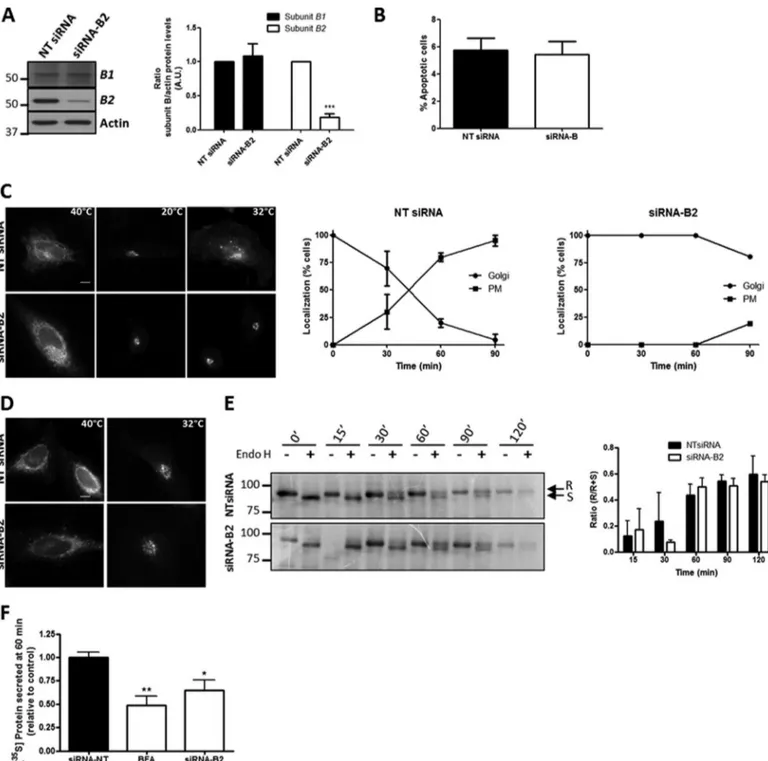
The pharmacological inhibition of V0 domain blocks V-ATPase activity and in turn alters the pH (49) as well as the Golgi-associated protein transport (44, 46, 56), but little is known about the specific contribution of each V1subunit in V-ATPase activity. We then knocked down the expression of subunit B2 using siRNA technology to see whether the subunit is essential for V-ATPase activity and its role in Golgi-associ-ated protein transport. The silencing of subunit B2 (?80% without alteration in the subunit B1 expression; Fig. 2A) did not perturb cell viability (Fig. 2B). HeLa cells transiently transfected with the transmembrane Cherry-VSV-G protein and silenced for subunit B2 showed a large accumulation of Cherry-VSV-G at the Golgi 90 min after the temperature shift. In comparison, in non-targeting siRNA (NT siRNA) transfected cells, the viral glycoprotein was observed in transit to the plasma membrane at 90 min (Fig. 2C). In contrast, ER-to-Golgi protein transport of VSV-G was not altered in subunit B2-depleted cells (Fig. 2, D and E), which was in accordance with previous results obtained using V-ATPase pharmacological inhibitors (44). In addition, the secretion of radioactive-labeled soluble proteins trans-ported from the Golgi to the extracellular medium was also significantly blocked in knockdown cells (Fig. 2F). These impairments in protein transport caused by the depletion of subunit B2 could be attributable to the disruption of Golgi pH, because V-ATPase is the main supplier of protons in endo-membranes, including the Golgi complex (6, 7). To explore this, we measured the pH both in the Golgi stack (Golgi pH) and in the TGN (TGN pH) using pHluorin-tagged sensors (52). As expected, control cells showed that the pH in the TGN was lower than in the Golgi (Table 1), and knockdown cells dis-played severe alkalization of both Golgi compartments. Curi-ously, the alkalization effect was stronger in the Golgi stack than in the TGN (Table 1).
Overall, these results show that V-ATPase revealed by GFP-tagged subunit B2 localized in the Golgi, particularly in distal compartments, where it is necessary for post-Golgi trafficking. GFP-B2 subunit is a tool that facilitates the study of the
dynam-ics and molecular interactions of V-ATPase at the Golgi complex.
Actin-depolymerizing Agents Induce the V1-V0Disassociation of the V-ATPase at the Golgi Complex—On the one hand, we previously reported that actin depolymerization raises the pH in the Golgi and alters Golgi-to-ER and post-Golgi trafficking (47– 49). These results are similar to those recorded after the pharmacological inhibition of V-ATPase using bafilomycin and concanamycin (44). On the other hand, it has been reported that subunits B and C of V-ATPase V1domain interact with F-actin and/or G-actin (26, 28). Taking all these observations into account, we hypothesized that V-ATPase activity at the Golgi could be regulated by actin dynamics (49, 51, 57). To test this hypothesis, we first examined whether the V1-V0 associa-tion is perturbed in the presence of actin toxins that either depolymerize (latrunculin B (LtB); cytochalasin D (CytD)) or stabilize (jasplakinolide (JpK)) actin. As a positive control, we cultured HeLa cells in the absence of glucose and fetal bovine serum (?Glu/FBS), because this treatment reversibly uncou-ples V-ATPase domains both in yeast (16) and in mammalian cells (17). Cells were incubated with LtB (500 nM/90 min), CytD (1 ?M/90 min), JpK (500 nM/90 min), or ?Glu/FBS (4 h), lysed, and immunoprecipitated with anti-subunit A antibodies and subjected to Western blotting for subunit a1 (which forms part of V0complex). The ratio between a1/A (Fig. 3A) subunits was significantly lower in LtB- and in ?Glu/FBS-treated cells (but not in JpK-treated cells) when compared with untreated cells (control) (Fig. 3A). Similar results were obtained with CytD (data not shown). Therefore, taking these results into account, we postulated that actin depolymerization would lead to an enrichment of V1subunits in the cytosol. We obtained highly pure membrane and cytosol fractions (respectively identified by the presence of transferrin receptor/TfR and RhoGDI mark-ers) from LtB- or JpK-treated cells, and we evaluated the pres-ence of subunits A and B2 in both fractions by Western blotting. Unlike JpK, LtB significantly enriched both subunits in the cytosol (Fig. 3B).
Because we observed that the GFP-B2 subunit localizes in the Golgi complex (Fig. 1B), we next used iFRAP microscopy to visualize whether actin depolymerization induces the release of the GFP-B2 subunit from the Golgi, as a demonstration of the dissociation of V1-V0 domains in vivo. To this end, in cells expressing GFP-B2, we bleached the cytoplasm, except the Golgi, and we measured the dissociation of the V1domain by the loss of fluorescence at the Golgi (Fig. 4A). Sequential pic-tures obtained fromsupplemental movies 1–3indeed showed the loss of the Golgi-associated GFP-B2 fluorescence after incu-bation of cells in the absence of glucose and FBS (?Glu/FBS) and with LtB (Fig. 4B; also compare supplemental movie 2/?Glu/FBS, andsupplemental movie 3/LtB with supplemen-tal movie 1/control). Quantitative image analysis showed that LtB and ?Glu/FBS caused a similar loss of the total GFP-B2 fluorescence at the Golgi at 15 min (Fig. 4C and mobile frac-tion/MF values shown in D). However, their respective kinetics was different, the loss being much faster for LtB than for ?Glu/ FBS, as shown by their respective t1⁄2(Fig. 4D). Therefore, actin depolymerization promotes the V0-V1dissociation of Golgi-as-TABLE 1
Depletion of subunit B2 raises Golgi and TGN pH
Ratiometric measurements of pH in the Golgi stack and in the TGN in HeLa cells expressing (72 h) non-targeting siRNA (NT siRNA) or siRNA to subunit B2 (siRNA-B2). Thereafter, cells were transfected with Golgi-pHluorin or TGN-pHluorin constructs for 18 h. Data represent means ? S.D. of at least three inde-pendent experiments. Significant differences with respect to NT siRNA (***, p ? 0.001) and to intra-Golgi pH (¶, p ? 0.001) using Student’s t test are shown. n is number of measured cells.
Golgi pH TGN pH
NT siRNA 6.88 ? 0.07 (n ? 85) 6.16 ? 0.06 (n ? 32)¶
FIGURE 2. V-ATPase subunit B2 depletion blocks post-Golgi trafficking of transmembrane and soluble secretory proteins. A, total cell lysates from HeLa cells previously transfected (72 h) with non-targeting siRNA pool (NT siRNA) or specific siRNA pool to subunit B2 were subjected to immunoblot analysis using monospecific antibodies to subunit B2 and subunit B1. On the right, quantitative analysis of immunoblots is also shown. Values are the mean ? S.D. from four independent experiments. Statistical analysis versus control (NT siRNA) using Student’s t test, ***, p ? 0.001, is shown. A.U., arbitrary units. B, after transfection with NT siRNA or siRNA-B2, HeLa cells were stained with DAPI to study cell viability. Apoptotic cells were counted. Graph represents the mean of percentage of apoptotic cells ? S.D. of four independent experiments. C, NT siRNA- and siRNA-B2-expressing HeLa cells were transfected with Cherry-ts045VSV-G and incubated overnight at 40 °C and then at 20 °C for 2 h, showing the viral protein accumulated in the ER and in the Golgi, respectively. When cells were shifted from 20 to 32 °C for 90 min, VSV-G is seen in transport carriers and at the plasma membrane in the case of cells transfected with NT siRNA but not to those cells with siRNA-B2. Bar, 10?m. Graphs on the right correspond to quantitative analysis of results shown in B. D, representative images of HeLa cells transfected with Cherry-ts045VSV-G and non-targeting (NT siRNA) or subunit B2 (siRNA-B2) siRNAs. After overnight incubation of cells at 40 °C, temperature was shifted to 32 °C for 30 min and VSV-G protein was transported to the Golgi. Bar, 10?m. E, biochemical transport assay for VSV-G-GFP using Endo H assay. HeLa cells constitutively expressing VSV-G-GFP were transfected for 72 h with NT siRNA or siRNA-B2 and incubated at 40 °C for the last 24 h. The cells were then shifted at 32 °C to induce the transport of VSV-G from the ER, lysed at indicated times, and subjected to Endo H treatment. R and S indicate Endo H-resistant and Endo H-sensitive forms, respectively. The ratio of the amount of Endo H-resistant form to that of total (R ? S) amount is plotted. Values are represented as the mean ? S.D. of three independent experiments, and no significant differences were found. F, HeLa cells transfected with NT siRNA or siRNA-B2 for 72 h were pulse-labeled with [35S]Met/Cys, incubated at 19 °C for 3 h, and shifted to 37 °C for 60 min. Secreted proteins of the culture medium and cell lysates were precipitated and quantified by scintillation counting. As positive control, the secretion assay was performed in cells treated with brefeldin A (BFA, 5?g/ml). Results are the mean ? S.D. from three independent experiments. Significant differences with respect to the control (NT siRNA) using Student’s t test; *, p ? 0.05, and **, p ? 0.01.
sociated V-ATPase and therefore alterations in its activity and the resulting pH.
V1Domain Subunits B2 and C1 Interact with Actin in Vivo— We next approached how microfilaments regulate V-ATPase activity at the Golgi. It is important to note that in HeLa cells V-ATPase is not present at the plasma membrane, which so far is the only molecular interaction where actin and V-ATPase have been described (25, 27). Therefore, to examine whether microfilaments interact with subunits B2 and C1 at endomem-branes in HeLa cells, we obtained F- and G-actin-enriched frac-tions from untreated and LtB-treated cells, and we subse-quently used Western blotting to examine the presence of both subunits. In untreated cells, B2 and C1 were indeed present in F- and G-actin fractions, with the former almost equally local-ized in both fractions and the latter more enriched in the G-ac-tin fraction. After LtB treatment, unlike subunit B2, subunit C1 showed a significant reduction of the F-/G-actin ratio and was
thus more enriched in the G-actin fraction (Fig. 5A). These results suggest that subunit C1 shows more affinity for G-actin, unlike subunit B2 that does not show any preference for F- or G-actin. Immunoprecipitation experiments with anti-actin antibodies and subsequent Western blotting for subunits B2 or C1 showed that indeed both V1subunits interacted with actin, with apparently stronger affinity for C1 (Fig. 5B). Overall, our results suggest that in HeLa cells the molecular interaction between actin and V-ATPase in endomembranes indeed occurs through subunits B2 and C1.
We next analyzed whether F-actin colocalized with V-ATPase at the Golgi complex. The difficulty to visualize F-actin at the Golgi is well known, although there is both functional and morphological evidence of its presence (57–59). To this end, we used the LifeAct-RFP probe (Fig. 5C) (60). HeLa cells co-ex-pressing LifeAct and GFP-B2 showed at the Golgi a small frac-tion of discrete actin-positive punctae adjacent to GFP-B2-pos-FIGURE 3. Actin depolymerization induces the disassociation of V
0and V1domains. A, HeLa cells treated with LtB (500 nMfor 90 min), JpK (500 nMfor 90
min), or cells starved of glucose and FBS (?Glu/FBS; for 4 h) were lysed and subjected to Western blotting (WB) (input) or immunoprecipitation (IP) with anti-subunit A antibodies and Western blotting to subunits a1 or A. Supernatant (SN) from immunoprecipitation. A representative experiment is shown. Quantitative analysis showing the ratio between V
0and V1subunits (ratio a1/A) from four independent experiments. Results are represented as means ? S.D. Statistical analysis using Student’s t test versus control; **, p ? 0.01. C, control; Lt, total lysate; P, pellet. B, post-nuclear supernatants of HeLa cells untreated (C) or treated with LtB (1?Mfor 90 min) or Jpk (1?Mfor 90 min) were ultracentrifuged, and the pellet (membrane fraction) and the supernatant (cytosol fraction) were subjected to SDS-PAGE. Western blotting was revealed with antibodies to TfR (membrane protein marker), Rho guanidine dissociation inhibitor (RhoGDI; a cytosolic protein marker), and to subunits A and B2. Quantitative results are represented as means ? S.D. from five independent experiments. Amounts of subunits A and B2 in cytosol and membrane fractions were normalized to RhoGDI and TfR, respectively, before the cytosol/membrane ratio was determined. Statistical analysis using Student’s t test versus control; *, p ? 0.05, is shown.
itive zones (Fig. 5C, arrows; supplemental movies 4 and 5), which suggests that F-actin could transiently associate with V-ATPase in Golgi membranes.
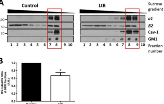
Actin Depolymerization-induced V1-V0Domain Disassocia-tion also Occurs by DisrupDisassocia-tion of DRMs—It has been reported that several V-ATPase subunits are major components in Golgi membrane-derived detergent-insoluble complexes, which are the Golgi equivalent to sphingomyelin- and cholesterol-en-riched domains isolated as detergent-resistant membranes from total cell extracts (DRMs) (35). In contrast, actin filaments are reported to maintain the integrity of DRMs (61). Then, we reasoned that besides its interaction with subunits B and C, actin could also indirectly regulate association of V1-V0 domains by maintaining the integrity of DRMs. To test this possibility, we isolated total DRMs from control (untreated) and LtB-treated HeLa cells and examined the presence of V0
and V1subunits a1 and B2, respectively, in DRMs and deter-gent-sensitive membrane fractions (Fig. 6A). Unlike polarized epithelial cells (14), HeLa cells do not express V-ATPase at the plasma membrane, and therefore, we assumed that the putative source of V-ATPase in DRMs would be endomembranes, mainly from the Golgi (35) and late endosomes and lysosomes (62). DRMs were identified by the presence of caveolin-1 and the GM1 ganglioside. Subunit B2 distributed both in detergent-sensitive membranes and in DRMs (red square) (Fig. 6A), but subunit a1 was exclusively located in DRMs. This difference in distribution is explained because subunit B2 is synthesized in the cytosol and is not always coupled to the V0domain. When HeLa cells were treated with LtB, DRMs were partially dis-rupted, as revealed by the presence of DRMs markers in heavier sucrose fractions. The ratio of B2/a1 (V1–V0) subunits in DRMs was significantly reduced (Fig. 6B). To confirm that the FIGURE 4. Actin depolymerization promotes the release of GFP-B2 subunit from the Golgi to the cytosol. A, schematic representation of the iFRAP experiment, whose results are shown in B. The cytoplasm of HeLa cells expressing GFP-B2 was bleached except for the Golgi area, and subsequently the fluorescence of the Golgi was recorded. B, representative sequential images of time-lapse microscopy of GFP-B2 expressing HeLa cells treated with LtB (500 nM) or cells that were grown in normal culture medium (control) or in medium without glucose and FBS (?Glu/FBS; 4 h). Bar, 10?m. C, inversal FRAP decay curves of the Golgi fluorescence from GFP-B2 shown in B. Golgi fluorescence values were obtained every 5 s and for a total period of 15 min. Inset, corresponds to a graphic representation of parameters measured in the table. A.U., arbitrary units. D, mobile fraction (MF) and t1
⁄2(min) for GFP-B2 fluorescence at the Golgi.
presence of cholesterol- and sphingomyelin-enriched domains in Golgi membranes are necessary for V-ATPase function, we manipulated lipid homeostasis in this organelle. Thus, we exog-enously added short chain ceramide-C6, the non-metaboliza-ble enantiomer L-ceramide-C6 (N-hexanoyl-L -erythrosphin-gosine) as a control, and the enantiomer D-ceramide-C6 (N-hexanoyl-D-erythrosphingosine), which is incorporated into Golgi membranes and locally increases levels of short-chain sphingomyelin, short-short-chain glucosylceramide, and dia-cylglycerol. As a consequence, there is a reduction in the for-mation of cholesterol/sphingomyelin-enriched domains into the Golgi (63). Thereafter, we measured Golgi and TGN pH in cells transfected with Golgi- or TGN-pHluorin constructs, and we subsequently cells treated withD- orL-ceramide-C6 (20 ?M
for 30 min each).D-Ceramide-C6, but notL-ceramide-C6, sig-nificantly increased both Golgi and TGN pH (Table 2). Know-ing that V-ATPase is the main source of H?
in the Golgi lumen, our results strongly suggest that the integrity of DRMs in Golgi membranes is essential for V-ATPase activity. Altogether, these results suggest that actin could also indirectly regulate
V1-V0association of V-ATPase in Golgi membranes by main-taining the integrity of DRMs.
Discussion
We report here that actin regulates the activity of the V-ATPase in the secretory pathway by controlling the association of V1-V0domains. We suggest that actin carries out such reg-ulation through two non-mutually exclusive mechanisms as follows: (a) its direct interaction with subunits B2 and C1; and (b) indirectly maintaining the organization of lipid rafts. Although V-ATPase localization at the Golgi complex is well known (29 –34), we provide here a more detailed picture of V-ATPase distribution along the Golgi stack. Because commer-cially available antibodies were unsuitable for immunocyto-chemistry, we cloned the V1domain subunit B2, which, like subunit C, contains actin-binding sites (27). Importantly, expressed GFP-B2 (GFP tagged to its amino end) was effec-tively incorporated into the V1domain without interfering with its interaction with the V0domain, which indicates that GFP-B2 is indeed a representative tool for studying V-FIGURE 5. Actin interacts with V-ATPase V1domain subunits B2 and C1. A, F-/G-actin fractionation of HeLa cells untreated (control, C) or treated with LtB (1
?M; 90 min). Samples were subjected to Western blotting analysis for actin and subunits B2 and C1. On the right, quantitative analysis of results shown on the
left. Statistical analysis using Student’s t test of results versus respective control from three independent experiments; ***, p ? 0.001. B, coimmunoprecipitation
experiments with anti-actin (IP: actin) antibody beads conjugated from HeLa cell lysates and subsequent Western blotting analysis for the presence of subunits
B2 and C1. C, high magnification of Golgi region snapshots of a 10-s interval time-lapse confocalsupplemental movie 4(upper panel) andsupplemental movie 5(bottom panel) from HeLa cells co-expressing GFP-B2 (green) and the F-actin probe Lifeact-RFP (red). Discrete accumulations of F-actin colocalizing with GFP-B2 at the Golgi are indicated by arrows. Scale bar, 1?m.
ATPase localization and dynamics in living cells. Expressed GFP-B2 localized V-ATPase in the Golgi complex, where it was visualized along all the compartments, although it was more enriched in distal ones. This Golgi distribution corre-lates well with the pH gradient in the Golgi (1). Because the mechanisms that generate this pH gradient are unclear (3), our results suggest that a progressive increase in V-ATPase along the cis-to-trans-Golgi axis is the main factor responsi-ble for the progressive fall in pH.
Our group previously reported that the interference in actin dynamics induced by a variety of actin toxins blocks Golgi-to-ER and post-Golgi trafficking alkalinizes the Golgi and induces cisternae swelling. All these were similar to the effects caused by the interference of V-ATPase activity with specific pharmacological agents targeted to V0domain, such as bafilo-mycin and concanabafilo-mycin A (5, 47, 48). Therefore, we postu-lated that one of the potential mechanisms by which actin par-ticipates in Golgi protein transport and flat cisternae morphology, as well as in the maintenance of pH, might be through the regulation of V-ATPase activity, taking into account that both V1domain subunits B and C contain
actin-binding domains (25, 27). Studies on actin interaction with sub-unit B have focused on V-ATPase of the plasma membrane in osteoclasts and on the role of actin in the recycling of the proton pump to and from intracellular membranes (50, 64). In con-trast, the interaction of subunit C with actin cytoskeleton was studied only in vitro using recombinant proteins (25, 28). How-ever, nothing is known about the potential interaction of B and C subunits with actin filaments in the secretory pathway endo-membranes and in the Golgi in particular. Our results show an interaction between actin and V1subunits B2 and C1, because both coimmunoprecipitated with actin. Besides this biochemi-cal evidence for subunit B2-actin interaction, we furthermore visualized this interaction at the Golgi in living cells expressing LifeAct-RFP and GFP-B2. Some spots showed colocalization with both fluorescent probes, although it is true that they were not the majority. It is reasonable to postulate that the interac-tion between F-actin (revealed by LifeAct) and V-ATPase (revealed by GFP-B2) could be highly dynamic and/or tran-sient. In respect of actin-subunit C1, immunoprecipitation results support the previously suggested role of subunit C directly connecting and modulating the interaction between V1 and V0domains of the V-ATPase (24), and actin could regulate such a function. We show here that the interaction of the two domains is reduced by LtB, and the V1domain is concomitantly enriched in the cytosol, which clearly indicates that F-actin maintains the association of the two domains. These biochem-ical observations were morphologbiochem-ically confirmed by iFRAP experiments, where the LtB treatment GFP-B2 is shifted from the Golgi to the cytoplasm. Taken together, our results strongly suggest that actin could stabilize and/or reinforce functional V-ATPase V1-V0domain association in vivo through its inter-action with both subunits.
FIGURE 6. Disassociation V1-V0domains by actin depolymerization also occurs by the disruption of DRMs. A, DRMs were obtained from HeLa cells
untreated or treated with LtB (1?M; 90 min) submitted to solubilization in 1% Triton X-100 at 4 °C. The Triton X-100 fractions were subsequently loaded at the bottom of a sucrose gradient as indicated under “Experimental Procedures.” After centrifugation, fractions were collected from the top (from 10/top to 1/bottom) and analyzed by SDS-PAGE followed by Western blotting to detect caveolin 1 (Cav-1) and subunits B2 and a1. For GM1, dot blot was carried out from each fraction collected and before the SDS-PAGE using cholera toxin-HRP. B, quantitative analysis examining the subunits B2/a ratio present in DRMs fractions (indicated with a red box in A). Statistical analysis using Student’s t test of results versus control from three independent experiments; *, p ? 0.01.
TABLE 2
Reduction of Golgi membrane lipid order increases Golgi and TGN pH
Ratiometric measurements of Golgi and TGN pH in HeLa cells treated with 20 ?M
of eitherL-ceramide-C6 orD-ceramide-C6 during 30 min. Thereafter, cells were transfected with Golgi-pHluorin or TGN-pHluorin constructs for 18 h. Data rep-resent means ? S.D. of three independent experiments. Significant differences with respectL-ceramide-C6 (***, p ? 0.001) using Student’s t test is shown. n, number of measured cells.
Golgi pH TGN pH
L-Ceramide-C6 6.12 ? 0.04 (n ? 48) 6.56 ? 0.08 (n ? 35)
Although the B2 and C1 subunits do interact with actin, F/G-actin fractionation and immunoprecipitation experiments sug-gest more affinity and sensitivity to actin for subunit C1 than for subunit B2. The reason for this is unknown, but it could be related to the functional relevance that both could have in vivo. F-actin interaction to B2 could interfere with its ATP-binding role and consequently with the ATPase activity of subunit A. We cannot discard that such differences might be attributable to the different affinity of antibodies used or even to the pres-ence of an intermediate protein that connects actin with sub-unit B2. In contrast, F-actin interaction with C1 could promote or stabilize the complex formed by the V0-V1domains, which is essential for the function of V-ATPase. The question then arises as to the functional relevance of actin-binding sites of both Golgi-associated V1subunits. As expected, subunit B2 is crucial for the activity of V-ATPase at the Golgi. This conclu-sion is based on the finding that silencing of subunit B2 raised Golgi and TGN pH values and significantly impaired post-Golgi transport (but not that between the ER to the post-Golgi) of membrane VSV-G glycoprotein and luminal secretory cargo. The physiological significance of this blockade can be explained by the reduction of subunit B-associated ATP consumption of the V1 domain (9, 65). Similar results were obtained after knockdown of subunit a of the V0domain (45).
Furthermore, it has been described that V-ATPase also local-izes to the lipid rafts in Golgi membranes (35). Our results dem-onstrate that the LtB treatment induced a partial disorganiza-tion of cholesterol and sphingomyelin enriched domains. Moreover, actin depolymerization induces V1-V0domain dis-sociation in these domains, which could result from the lipid raft disorganization primarily caused by the disassembly of microfilaments. The role of the actin cytoskeleton in the
orga-nization of lipid rafts has been described only in the plasma membrane (61). Taking into account that V-ATPase (revealed by GFP-B2) in HeLa cells is in the Golgi and not in the plasma membrane, and that the disruption of lipid rafts also affects the V0domain subunit a1, we suggest that actin filaments also par-ticipate in the functional association of V1-V0domain thanks to their role in maintaining lipid raft organization in Golgi mem-branes. This postulate is supported by results obtained with
D-ceramide-C6. This lipid derivative causes the disruption of cholesterol- and sphingomyelin-enriched domains at the Golgi (63), which is accompanied by a swelling of cisternae and alter-ations in N-glycosylation (63, 66), both strikingly coincidental with impairments at the Golgi occurring after actin depolymer-ization and pharmacological V-ATPase inhibition (49, 57, 58). Importantly,D-ceramide-C6 treatment also caused Golgi and TGN pH alkalization, which also occurs after actin-depolymer-izing toxins (49). Therefore, we establish a functional link at the Golgi between actin dynamics and lipid raft integrity, and con-sequently V-ATPase activity, which help to maintain pH home-ostasis and secretory transport activity (Fig. 7). This new role of actin in the activity of the V-ATPase holoenzyme is compatible with other direct roles of actin in Golgi membranes, such as membrane fission (59) and calcium import (67). It is clear that actin participates in the Golgi architecture and transport func-tions at different levels (57).
Author Contributions—C. S. P. and G. E. designed and coordinated the study and wrote the paper. C. S. P. performed and analyzed all the experiments. A. S. designed, performed, and analyzed experiments shown in Fig. 2D. J. L. provided technical assistance and DNA con-structs for experiment shown in Table 1. All the authors reviewed the results and approved the final version of the manuscript.
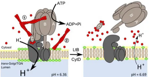
FIGURE 7. Model of the mechanisms of action by which actin depolymerization promotes dissociation of V
1-V0domains of V-ATPase in Golgi
membranes. We suggest two mechanisms, not mutually exclusive, for the regulation of V
0-V1coupling in distal compartments of the Golgi complex. One of them was through the direct interaction between F-actin and V1subunits B2 and C1; the other one was through the actin-dependent mainte-nance of the lipid raft domain integrity, which is essential to support V-ATPase activity. When Golgi-associated F-actin is depolymerized through the physiological actin dynamics cycle or widely by the action of actin toxins (LtB or CytD as indicated in the figure), the F-actin-subunit C interaction is not maintained anymore and V
1uncouples from V0, which leads to the rise of the Golgi pH (values are indicated; see Ref. 49). Under physiological conditions, this uncoupling would be very transient thanks to the fast Golgi-associated actin dynamics. However, after massive actin depolymerization caused by actin toxins, a large G-actin pool is generated, and the higher affinity of C subunit for G-actin prevents that the subunits could be reutilized for recoupling both V domains, leading to the permanent dysfunction of V-ATPase at the Golgi and its alkalization. This mechanistic dysfunction could be additionally enhanced by the concomitant disruption of lipid rafts (represented in green) of Golgi membranes where V-ATPase resides. These mecha-nisms could well occur in other endomembrane systems such endo/lysosomal compartments where V-ATPase is present. The different size of letter H? indicates different proton concentration consistently to its size. C subunit binds F- or G-actin at both extremes (26). Leakage of luminal Golgi protons to the cytosol is indicated by dashed lines.
Acknowledgments—We thank Francisco La´ zaro-Die´guez for critical reading of the manuscript; Miriam Esgles-Izquierdo and Darya Gor-benko del Blanco (University of Barcelona School of Medicine, Barce-lona, Spain) and Agustín Herna´ ndez (Universidad Pablo Olavide, Sevilla, Spain) and Yuri Y. Sautin (University of Florida College of Medicine) for technical advice in cloning and immunoprecipitation experiments, respectively; Stephanie Miserey-Lenkei (Institut Curie, Paris) for LifeAct-RFP; Fe´lix Campelo and Juan M. Dura´ n (Centre of Genomic Regulation, Barcelona) for advice withD-/L-ceramide C6; Maite Mun˜ oz for general technical support; and the personnel in charge of Plataformes Tecnolo`giques (Campus Casanova) for techni-cal support with confotechni-cal microscopy and pH measurements. We also thank Robin Rycroft for editorial assistance.
References
1. Wu, M. M., Grabe, M., Adams, S., Tsien, R. Y., Moore, H. P., and Machen, T. E. (2001) Mechanisms of pH regulation in the regulated secretory path-way. J. Biol. Chem.276, 33027–33035
2. Paroutis, P., Touret, N., and Grinstein, S. (2004) The pH of the secretory pathway: measurement, determinants, and regulation. Physiology 19,
207–215
3. Casey, J. R., Grinstein, S., and Orlowski, J. (2010) Sensors and regulators of intracellular pH. Nat. Rev. Mol. Cell Biol.11, 50 – 61
4. Weisz, O. A. (2003) Organelle acidification and disease. Traffic4, 57– 64
5. Maeda, Y., and Kinoshita, T. (2010) The acidic environment of the Golgi is critical for glycosylation and transport. Methods Enzymol.480, 495–510
6. Forgac, M. (2007) Vacuolar ATPases: rotary proton pumps in physiology and pathophysiology. Nat. Rev. Mol. Cell Biol.8, 917–929
7. Jefferies, K. C., Cipriano, D. J., and Forgac, M. (2008) Function, structure and regulation of the vacuolar (H?)-ATPases. Arch. Biochem. Biophys.
476, 33– 42
8. Kane, P. M. (2006) The where, when, and how of organelle acidification by the yeast vacuolar H-ATPase. Microbiol. Mol. Biol. Rev.70, 177–191
9. MacLeod, K. J., Vasilyeva, E., Baleja, J. D., and Forgac, M. (1998) Muta-tional analysis of the nucleotide binding sites of the yeast vacuolar proton-translocating ATPase. J. Biol. Chem.273, 150 –156
10. Drory, O., and Nelson, N. (2006) The emerging structure of vacuolar ATPases. Physiology21, 317–325
11. Inoue, T., Wang, Y., Jefferies, K., Qi, J., Hinton, A., and Forgac, M. (2005) Structure and regulation of the V-ATPases. J. Bioenerg. Biomembr.37,
393–398
12. Demaurex, N. (2002) pH homeostasis of cellular organelles. News Physiol.
Sci.17, 1–5
13. Marshansky, V., and Futai, M. (2008) The V-type H?-ATPase in vesicular trafficking: targeting, regulation and function. Curr. Opin. Cell Biol.20,
415– 426
14. Breton, S., and Brown, D. (2007) New insights into the regulation of V-ATPase-dependent proton secretion. Am. J. Physiol. Renal. Physiol.292,
F1–F10
15. Kane, P. M. (2012) Targeting reversible disassembly as a mechanism of controlling V-ATPase activity. Curr. Protein Pept. Sci.13, 117–123
16. Kane, P. M. (1995) Disassembly and reassembly of the yeast vacuolar H? -ATPase in vivo. J. Biol. Chem.270, 17025–17032
17. Nakamura, S. (2004) Glucose activates H?-ATPase in kidney epithelial cells. Am. J. Physiol. Cell Physiol.287, C97–C105
18. Sautin, Y. Y., Lu, M., Gaugler, A., Zhang, L., and Gluck, S. L. (2005) Phos-phatidylinositol 3-kinase-mediated effects of glucose on vacuolar H? -ATPase assembly, translocation, and acidification of intracellular com-partments in renal epithelial cells. Mol. Cell. Biol.25, 575–589
19. Parra, K. J., and Kane, P. M. (1998) Reversible association between the V1 and V0 domains of yeast vacuolar H?-ATPase is an unconventional glu-cose-induced effect. Mol. Cell. Biol.18, 7064 –7074
20. Rubenstein, E. M., and Schmidt, M. C. (2010) The glucose signal and metabolic p[H?]lux. EMBO J.29, 2473–2474
21. Lu, M., Sautin, Y. Y., Holliday, L. S., and Gluck, S. L. (2004) The glycolytic
enzyme aldolase mediates assembly, expression, and activity of vacuolar H?
-ATPase. J. Biol. Chem.279, 8732– 8739
22. Xu, T., and Forgac, M. (2001) Microtubules are involved in glucose-de-pendent dissociation of the yeast vacuolar [H?
]ATPase in vivo. J. Biol. Chem.276, 24855–24861
23. Zuo, J., Vergara, S., Kohno, S., and Holliday, L. S. (2008) Biochemical and functional characterization of the actin-binding activity of the B subunit of yeast vacuolar H?
-ATPase. J. Exp. Biol.211, 1102–1108
24. Pe´rez-Saya´ns, M., Sua´rez-Pen˜ aranda, J. M., Barros-Angueira, F., Diz, P. G., Ga´ndara-Rey, J. M., and García-García, A. (2012) An update in the struc-ture, function, and regulation of V-ATPases: the role of the C subunit. Braz. J. Biol.72, 189 –198
25. Vitavska, O., Wieczorek, H., and Merzendorfer, H. (2003) A novel role for subunit C in mediating binding of the H?-V-ATPase to the actin cytoskel-eton. J. Biol. Chem.278, 18499 –18505
26. Holliday, L. S., Lu, M., Lee, B. S., Nelson, R. D., Solivan, S., Zhang, L., and Gluck, S. L. (2000) The amino-terminal domain of the B subunit of vacu-olar H?-ATPase contains a filamentous actin binding site. J. Biol. Chem.
275, 32331–32337
27. Lee, B. S., Gluck, S. L., and Holliday, L. S. (1999) Interaction between vacuolar H?-ATPase and microfilaments during osteoclast activation. J. Biol. Chem.274, 29164 –29171
28. Vitavska, O., Merzendorfer, H., and Wieczorek, H. (2005) The V-ATPase subunit C binds to polymeric F-actin as well as to monomeric G-actin and induces cross-linking of actin filaments. J. Biol. Chem.280, 1070 –1076
29. Zhang, F., and Schneider, D. L. (1983) The bioenergetics of Golgi appara-tus function: evidence for an ATP-dependent proton pump. Biochem. Biophys. Res. Commun.114, 620 – 625
30. Chanson, A., and Taiz, L. (1985) Evidence for an ATP-dependent proton pump on the Golgi of corn coleoptiles. Plant Physiol.78, 232–240
31. Moriyama, Y., and Nelson, N. (1989) H?
-translocating ATPase in golgi apparatus. Characterization as vacuolar H?
-ATPase and subunit struc-tures. J. Biol. Chem.264, 18445–18450
32. Hurley, D., and Taiz, L. (1989) Immunocytochemical localization of the vacuolar H?
-ATPase in maize root tip cells. Plant Physiol.89, 391–395
33. Young, G. P., Qiao, J. Z., and Al-Awqati, Q. (1988) Purification and recon-stitution of the proton-translocating ATPase of Golgi-enriched mem-branes. Proc. Natl. Acad. Sci. U.S.A.85, 9590 –9594
34. Glickman, J., Croen, K., Kelly, S., and Al-Awqati, Q. (1983) Golgi mem-branes contain an electrogenic H?
pump in parallel to a chloride conduct-ance. J. Cell Biol.97, 1303–1308
35. Gkantiragas, I., Bru¨ gger, B., Stu¨ ven, E., Kaloyanova, D., Li, X. Y., Lo¨hr, K., Lottspeich, F., Wieland, F. T., and Helms, J. B. (2001) Sphingomyelin-enriched microdomains at the Golgi complex. Mol. Biol. Cell. 12,
1819 –1833
36. Maeda, Y., Ide, T., Koike, M., Uchiyama, Y., and Kinoshita, T. (2008) GPHR is a novel anion channel critical for acidification and functions of the Golgi apparatus. Nat. Cell Biol.10, 1135–1145
37. Thompson, R. J., Nordeen, M. H., Howell, K. E., and Caldwell, J. H. (2002) A large-conductance anion channel of the Golgi complex. Biophys. J.83,
278 –289
38. Nakamura, N., Tanaka, S., Teko, Y., Mitsui, K., and Kanazawa, H. (2005) Four Na?
/H?
exchanger isoforms are distributed to Golgi and post-Golgi compartments and are involved in organelle pH regulation. J. Biol. Chem.
280, 1561–1572
39. Ono, M., and Hakomori, S. (2004) Glycosylation defining cancer cell mo-tility and invasiveness. Glycoconj. J.20, 71–78
40. Zhao, Y.-Y., Takahashi, M., Gu, J.-G., Miyoshi, E., Matsumoto, A., Kita-zume, S., and Taniguchi, N. (2008) Functional roles of N-glycans in cell signaling and cell adhesion in cancer. Cancer Sci.99, 1304 –1310
41. Kornak, U., Reynders, E., Dimopoulou, A., van Reeuwijk, J., Fischer, B., Rajab, A., Budde, B., Nu¨ rnberg, P., Foulquier, F., ARCL Debre´-type Study Group, Lefeber, D., Urban, Z., Gruenewald, S., Annaert, W., Brunner, H. G., et al. (2008) Impaired glycosylation and cutis laxa caused by muta-tions in the vesicular H?-ATPase subunit ATP6V0A2. Nat. Genet.40,
32–34
42. Rivinoja, A., Pujol, F. M., Hassinen, A., and Kellokumpu, S. (2012) Golgi pH, its regulation and roles in human disease. Ann. Med.44, 542–554
43. Muroi, M., Shiragami, N., Nagao, K., Yamasaki, M., and Takatsuki, A. (1993) Folimycin (concanamycin A), a specific inhibitor of V-ATPase, blocks intra-cellular translocation of the glycoprotein of vesicular stoma-titis virus before arrival to the Golgi apparatus. Cell Struct. Funct.18,
139 –149
44. Palokangas, H., Metsikko¨, K., and Va¨a¨na¨nen, K. (1994) Active vacuolar H?
ATPase is required for both endocytic and exocytic processes during viral infection of BHK-21 cells. J. Biol. Chem.269, 17577–17585
45. Sobota, J. A., Ba¨ck, N., Eipper, B. A., and Mains, R. E. (2009) Inhibitors of the V0 subunit of the vacuolar H?
-ATPase prevent segregation of lyso-somal- and secretory-pathway proteins. J. Cell Sci.122, 3542–3553
46. Yilla, M., Tan, A., Ito, K., Miwa, K., and Ploegh, H. L. (1993) Involvement of the vacuolar H(?)-ATPases in the secretory pathway of HepG2 cells. J. Biol. Chem.268, 19092–19100
47. Valderrama, F., Luna, A., Babía, T., Martinez-Mena´rguez, J. A., Ballesta, J., Barth, H., Chaponnier, C., Renau-Piqueras, J., and Egea, G. (2000) The Golgi-associated COPI-coated buds and vesicles contain?/?-actin. Proc. Natl. Acad. Sci. U.S.A.97, 1560 –1565
48. La´zaro-Die´guez, F., Colonna, C., Cortegano, M., Calvo, M., Martínez, S. E., and Egea, G. (2007) Variable actin dynamics requirement for the exit of different cargo from the trans-Golgi network. FEBS Lett.581, 3875–3881
49. La´zaro-Die´guez, F., Jime´nez, N., Barth, H., Koster, A. J., Renau-Piqueras, J., Llopis, J. L., Burger, K. N., and Egea, G. (2006) Actin filaments are involved in the maintenance of Golgi cisternae morphology and intra-Golgi pH. Cell Motil. Cytoskeleton63, 778 –791
50. Chen, S.-H., Bubb, M. R., Yarmola, E. G., Zuo, J., Jiang, J., Lee, B. S., Lu, M., Gluck, S. L., Hurst, I. R., and Holliday, L. S. (2004) Vacuolar H?
-ATPase binding to microfilaments: regulation in response to phosphatidylinositol 3-kinase activity and detailed characterization of the actin-binding site in subunit B. J. Biol. Chem.279, 7988 –7998
51. Egea, G., La´zaro-Die´guez, F., and Vilella, M. (2006) Actin dynamics at the Golgi complex in mammalian cells. Curr. Opin. Cell Biol.18, 168 –178
52. Miesenbo¨ck, G., De Angelis, D. A., and Rothman, J. E. (1998) Visualizing secretion and synaptic transmission with pH-sensitive green fluorescent proteins. Nature394, 192–195
53. Gutie´rrez-Martínez, E., Ferna´ndez-Ulibarri, I., La´zaro-Die´guez, F., Jo-hannes, L., Pyne, S., Sarri, E., and Egea, G. (2013) Lipid phosphate phos-phatase 3 participates in transport carrier formation and protein traffick-ing in the early secretory pathway. J. Cell Sci.126, 2641–2655
54. Balch, W. E., Dunphy, W. G., Braell, W. A., and Rothman, J. E. (1984) Reconstitution of the transport of protein between successive compart-ments of the Golgi measured by the coupled incorporation of N-acetylg-lucosamine. Cell39, 405– 416
55. Xifro´, X., García-Martínez, J. M., Del Toro, D., Alberch, J., and Pe
´rez-Navarro, E. (2008) Calcineurin is involved in the early activation of NMDA-mediated cell death in mutant huntingtin knock-in striatal cells. J. Neurochem.105, 1596 –1612
56. Huang, C., and Chang, A. (2011) pH-dependent cargo sorting from the Golgi. J. Biol. Chem.286, 10058 –10065
57. Egea, G., Serra-Peinado, C., Salcedo-Sicilia, L., and Gutie´rrez-Martínez, E. (2013) Actin acting at the Golgi. Histochem. Cell Biol.140, 347–360
58. Valderrama, F., Dura´n, J. M., Babia`, T., Barth, H., Renau-Piqueras, J., and Egea, G. (2001) Actin microfilaments facilitate the retrograde transport from the Golgi complex to the endoplasmic reticulum in mammalian cells. Traffic2, 717–726
59. Miserey-Lenkei, S., Chalancon, G., Bardin, S., Formstecher, E., Goud, B., and Echard, A. (2010) Rab and actomyosin-dependent fission of transport vesicles at the Golgi complex. Nat. Cell Biol.12, 645– 654
60. Riedl, J., Crevenna, A. H., Kessenbrock, K., Yu, J. H., Neukirchen, D., Bista, M., Bradke, F., Jenne, D., Holak, T. A., Werb, Z., Sixt, M., and Wedlich-Soldner, R. (2008) Lifeact: a versatile marker to visualize F-actin. Nat. Methods5, 605– 607
61. Chichili, G. R., and Rodgers, W. (2009) Cytoskeleton-membrane interac-tions in membrane raft structure. Cell. Mol. Life Sci.66, 2319 –2328
62. Lafourcade, C., Sobo, K., Kieffer-Jaquinod, S., Garin, J., and van der Goot, F. G. (2008) Regulation of the V-ATPase along the endocytic pathway occurs through reversible subunit association and membrane localization. PLoS ONE3, 2758
63. Duran, J. M., Campelo, F., van Galen, J., Sachsenheimer, T., Sot, J., Egorov, M. V., Rentero, C., Enrich, C., Polishchuk, R. S., Gon˜ i, F. M., Bru¨ gger, B., Wieland, F., and Malhotra, V. (2012) Sphingomyelin organization is re-quired for vesicle biogenesis at the Golgi complex. EMBO J. 31,
4535– 4546
64. Holliday, L. S., Bubb, M. R., Jiang, J., Hurst, I. R., and Zuo, J. (2005) Inter-actions between vacuolar H?-ATPases and microfilaments in osteoclasts. J. Bioenerg. Biomembr.37, 419 – 423
65. Vasilyeva, E., Liu, Q., MacLeod, K. J., Baleja, J. D., and Forgac, M. (2000) Cysteine scanning mutagenesis of the noncatalytic nucleotide binding site of the yeast V-ATPase. J. Biol. Chem.275, 255–260
66. van Galen, J., Campelo, F., Alonso, E., Scarpa, M., Martínez-Mena´rguez, J. A´ ., and Malhotra, V. (2014) Sphingomyelin homeostasis is required to form functional enzymatic domains at the trans-Golgi net-work. J. Cell Biol.206, 609 – 618
67. von Blume, J., Alleaume, A.-M., Cantero-Recasens, G., Curwin, A., Carre-ras-Sureda, A., Zimmermann, T., van Galen, J., Wakana, Y., Valverde, M. A., and Malhotra, V. (2011) ADF/cofilin regulates secretory cargo sort-ing at the TGN via the Ca2?ATPase SPCA1. Dev. Cell.20, 652– 662