UNIVERSITA' POLITECNICA DELLE MARCHE
FACOLTA' DI MEDICINA E CHIRURGIA
Dottorato di Ricerca i Scie ze Bio ediche XXIX ciclo
Circannual pattern and TEMPerature-related
incidence of Electrical STorm: the TEMPEST study
Tesi di Dottorato di:
Relatore:
Dott.ssa Laura Cipolletta Dr. Federico Guerra
Table of contents: 1. Abstract 2. Introduction
2.1 Definition
2.2 Incidence and Prognostic Implications 2.3 Initial Evaluation of Electrical Storm 2.4 Clinical Syndromes of Electrical Storm
2.4.1 Monomorphic Ventricular Tachycardia 2.4.2 Polymorphic Ventricular Tachycardia 2.4.5 Ventricular Fibrillation
2.5 Pharmacologic Therapy for Electrical Storm 2.5.1 Adrenergic Blockade
2.5.2 Amiodarone
2.5.3 Class I Antiarrhythmic (Sodium Channel-Blocking) Agents 2.5.4 Anesthetic Agents
2.5.5 Nonpharmacological Therapy 2.5.6 Electrical Storm in ICD paitients 2.5.7 Study Hypothesis
3. Methods
3.1 Study selection 3.2 Data collection
3.3 Temperature data collection 3.4 Statistical analysis
4. Results
4.1 Population characteristics
4.2 Electrical storm incidence over time 4.3 Temperature and electrical storm
5. Discussion
5.1 Temperature related variations 5.2 Weekly and daily variations 5.3 Limitations
6. Conclusions 7. References
1. Abstract
Background: The occurrence of ventricular tachyarrhythmias appears to follow
circadian, daily, and seasonal distributions. We aim to identify potential temporal patterns of electrical storm (ES), in which a cluster of ventricular tachycardias (VT) or ventricular fibrillation (VF), negatively affects short- and long-term survival.
Methods and results: The Circannual pattern and TEMPerature-related incidence
of Electrical Storm (TEMPEST) study is a patient-level, pooled analysis of previously published datasets. Study selection criteria included: diagnosis of ES; absence of acute coronary syndrome as the arrhythmic trigger; ≥10 patients included. At the end of the selection and collection processes, five centres had the dataset from their papers pooled into the present registry. Temperature data and sunrise and sunset hours were retrieved from Weather Underground, the largest weather database available online.
Results: 246 patients admitted for ES (221 males, age 66±9 years) were enrolled.
Each ES episode included a median of seven VT/VF episodes. Fifty-nine percent of patients experienced ES during daytime hours (p<0.001). The incidence of ES was significantly more prevalent during work-days, with Saturdays and Sundays
registering the lowest prevalence of ES (10.4 and 7.2% respectively, vs 16.5% daily mean from Monday to Friday; p<0.001). ES incidence was significantly associated with increased monthly temperature range when compared to the month before (p=0.003).
Conclusions: ES incidence is not homogenous over time but seems to recognize a
clustered or "hot-spot" pattern, with a higher incidence during daytime hours and working days. ES is associated with an increase in monthly temperature variation.
2. Introduction
2.1 Definition
Electrical storm (ES) is usually defined as the occurrence of three or more episodes of sustained ventricular tachycardia (VT) or ventricular fibrillation (VF) within 24 hours requiring either anti-tachycardia pacing (ATP) or cardioversion/defibrillation (1). Sustained VT are defined as lasting 30 seconds, involving hemodynamic compromise, or requiring intervention to terminate the episode. Treatment and management of electrical storm requires a tailored approach according to the underlying cause. ES could verify during the acute phase of myocardial infarction (MI) or in patients with structural heart disease, with ICD, or an inherited arrhythmic syndrome. The commonest symptoms are palpitations, dizziness, and syncope. The dramatic clinical presentation can provoke cardiac arrest or multiple episodes of life threatening arrhythmias. Patients with ICD can suffer from recurrent shocks. A deep knowledge of arrhythmia mechanisms is the key for the effective management of electrical storm involving different therapeutic options, ICD programming, and invasive techniques for the treatment of refractory cases.
2.2 Incidence and Prognostic Implications
ES represents a true medical emergency that requires a multi-disciplinary approach, and its prevalence is steadily rising along with the number of patients bearing an implantable cardioverter-defibrillator (ICD) in developed countries (2).
The incidence of electrical storm varies depending on the clinical status. ES can occur in 10% to 20% of ICD recipients. Patients suffering from an acute MI or with previous MI or having an inherited arrhythmic syndrome are at risk. The rising prevalence of congestive heart failure causes an increase of ICD implantations (3). The incidence of
ES is higher in secondary compared to primary prevention. In a MADIT-II sub-study, involving 719 patients, (4) electrical storm occurred in 4% over an average follow-up of 20.6 months. Baseline characteristics were comparable between patients with ES and those with isolated episodes of VT/VF. In another trial, ES occurred in 20% of secondary prevention ICD recipients during a 31-month period (5). Data derived from recorded intracardiac electrograms of those episodes showed that VT was the most frequent arrhythmia and it occurred an average of 9.2 months after ICD implantation.
Data on the prognostic significance of electrical storm strongly suggest that ES is a risk factor of a poor outcome and also it might be an independent risk factor for cardiac death. In the AVID trial (5), nonsudden cardiac death was higher in patients with ES (risk ratio, 2.4). In the Madit-II substudy, a 7.4-fold higher risk of death was documented in patients with ES compared with patients without it (4). Also both studies showed that the highest risk of death was recorded within the first 3 months after a storm. Moreover, the prognosis remained poor for patients who survived the initial period of electrical instability, because of many sustained recurrent electrical storms and refractory heart failure. It is still unclear if electrical storm is simply an epiphenomenon of advanced structural heart disease or if it strongly contributes to a poor outcome. In fact, recurrent VT/VF and consequent ICD shocks worsen left ventricular (LV) systolic dysfunction and cause myocardial injury, which can lead to adrenergic neurohormonal activation and exacerbate HF (6).
2.3 Initial Evaluation of Electrical Storm
The management of patients with ES starts with the accurate diagnosis of the clinical arrhythmia. In patients with wide QRS tachycardia, bundle branch block, ventricular preexcitation (Wolff-Parkinson-White syndrome), or a rate related aberrancy,
supraventricular tachycardia (SVT) need a differential diagnosis with VT. Wellens, Kindwall, and Brugada (7-9) accurately described the differentiation of VT from SVT with aberrant conduction. Hemodynamic stability of the patient is not helpful in the differential diagnosis, because VTs may provoke minimal symptoms and lead to an erroneous diagnosis of SVT with aberrant conduction. Due to this reason, an unclear wide-complex tachycardia should be interpreted as a VT, in particular in patients with structural heart disease. Following this rule, the diagnosis of electrical storm will be accurate in 80% of all patients with tachycardia and in 95% of patients with previous MI (10). Moreover, while SVT might stop with a specific VT treatment, calcium-channel blockers or adenosine can precipitate cardiac arrest if a VT is thought to be an SVT. If VT-induced ES causes a cardiac arrest, it is of paramount importance to incorporate all aspects of critical care in this emergency setting, such as prompt management of a compromised airway, post-shock bradycardia, hypotension, ischemia, and defibrillation of hemodynamically unstable patients. Simultaneous therapies are usually necessary. Sometimes multiple electrical cardioversions or defibrillations are required, especially in patients with poor systolic function or rapid VT. If VT is hemodynamically stable, antiarrhythmic medication can be tried. Thus, if medical therapy is unsuccessful, cardioversion under sedation is indicated. Patients who have underlying structural heart disease and chronic renal failure often have with refractory VT or VF. Advanced age, male sex, a low LV ejection fraction (LVEF), and New York Heart Association functional class III or IV are other risk factors for ES. Also antiarrhythmic agents can provoke ES (11). Of note, a lower incidence of electrical storm is reported in ICD recipients with diabetes mellitus and patients who are taking lipid-lowering drugs. Identifying and reversing the causative factors is an important step in the management of ES. Acute ischemia, worsening heart failure, hypokalemia,
hypomagnesemia, arrhythmogenic drug therapy, hyperthyroidism, infection and fever are specific precipitants. Acute ischemia, decompensated HF, and electrolyte imbalances should be treated aggressively. Increased catecholamine levels related to a worsening anxiety and can amplify the severity and perpetuate ES (12). The complex interactions between multiple predisposing factors which culminate in an electrical storm are poorly understood.
2.4 Clinical Syndromes of Electrical Storm
When a vulnerable anatomic substrate (e.g. a structural heart disease or a scar after a MI), is affected by a triggering event such as premature ventricular contractions (PVCs) or an electrolyte imbalance can cause an ES. Treatment should target the underlying mechanism, so understanding the cause of ES is fundamental. 3 electrocardiographic (ECG) surface morphologies permit to classify ES: monomorphic VT, polymorphic VT, or VF.
2.4.1 Monomorphic Ventricular Tachycardia
Often, ES is caused by a sustained monomorphic VT associated with structural heart disease. Monomorphic VT is defined as an identical ventricular activation sequence without any variation in the QRS complexes. An electrical wavefront reentry around a fixed anatomic barrier, most commonly due to scar tissue after MI is the substrate of most monomorphic VT which does not require active ischemia as a trigger, and it is uncommon in patients who are having an acute MI. Heterogeneous areas of scarred myocardium are the vulnerable substrate for reentry in ischemic or nonischemic cardiomyopathy. Structural changes occur after an acute MI, or in advanced nonischemic cardiomyopathy. Scar formation due to fibrosis creates areas of conduction block. However, around the border of a scar, bundles of myofibrils can
survive and provide a pathway for electrically stable reentry if the conduction through these regions is slow. So, premature ventricular depolarizations which are usually a harmless trigger, in this case, can initiate monomorphic VT. The location of the scar and the exit site into the ventricle determine the surface ECG morphology during monomorphic VT. The VT may occur early or late after MI. When inadequate reperfusion or large areas of infarction are present, the burden of ventricular arrhythmias is higher. Monomorphic VT can be asymptomatic or can present as cardiac arrest. The ventricular rate, LV function, the presence of heart failure, any loss of atrioventricular synchrony, and the pattern of ventricular activation determines the degree of hemodynamic compromise (13). Amiodarone and β-blockers are the preferred pharmacological treatment.
2.4.2 Polymorphic Ventricular Tachycardia
Polymorphic VT is defined as beat-to-beat variations in the QRS complexes on surface telemetry or ECG. It has a different mechanism compared to monomorphic VT. In polymorphic VTs multiple wavefronts propagate throughout the heart or appear simultaneously in several parts of the heart and generate the peculiar surface ECG. It can be associated with a normal or a prolonged QT interval in sinus rhythm. Although seldom it is documented in the absence of organic heart disease, polymorphic VT is most often associated with acute ischemic syndromes or acute myocarditis or hypertrophic cardiomyopathy. Therapy for polymorphic VT and VF varies, depending on the underlying QT interval in sinus rhythm and the mode of initiation. The initial manifestation of ischemia could be ES. In contrast, unless the patient has a remote myocardial infarction, that serves as a substrate for reentry, monomorphic VT is infrequent during the first 72 hours of infarction. Acute myocardial ischemia causes almost always polymorphic VT. In these cases, the baseline QT interval may be
normal. In acute MI, ischemia, altered membrane potential, triggered activity, necrosis, or scar formation are responsible of polymorphic VT induction. Ischemia contribute to the creation of a dispersion of electrical refractory periods between the endocardium and epicardium, which is a requirement for multiple waves of reentry. Also, Purkinje cell automaticity is increased by ischemia increases, and polymorphic VT or VF are triggered by the spontaneous firing of these fibers. During episodes of ischemia intense electrical storms of polymorphic VT are not uncommon. Reversing the ischemia with emergency coronary revascularization or with anti-ischemic, antiplatelet, or thrombolytic agents is the most effective treatment. The most effective antiarrhythmic drugs are Amiodarone and β-blockers. Nowadays, randomized trials have not confirmed that lidocaine is superior to other antiarrhythmic medications in the presence of ischemia, although initially it was thought to be the optimal therapy for VT. In polymorphic VT associated with normal QT intervals, magnesium therapy is scarcely effective. Indeed, baseline (sinus-rhythm) ECG should be carefully evaluated for a prolonged QT interval in patients with recurrent polymorphic VT, to guarantee the best treatment. For example, pause-dependent polymorphic VT associated with bradycardia and long QT interval, are defined torsade de pointes. Careful attention to QT interval in sinus rhythm may underline marked QT prolongation. Distinguishing the common U waves from abnormal T waves might be difficult (14). Female sex, bradycardia, heart block, QT-prolonging drugs, hypokalemia, and inherited long QT syndrome are risk factors for torsades de pointes. Consideration of inherited and acquired causes represents the initial evaluation of polymorphic VT with a long QT interval. Inherited long QT syndromes increase the risk of sudden cardiac death, but they rarely can appear as an ES. The use of catecholamines, also isoproterenol, should be avoided in these patients. In case of a polymorphic VT with a long QT
interval a search for acquired causes, including electrolyte imbalances (hypokalemia, hypocalcemia, or hypomagnesemia), hypothyroidism, and the use of medications that are known to prolong the QT interval, including sotalol, haloperidol, methadone, and erythromycin should be prompted. Only in case of bradycardia or heart block related torsades de pointes, they should be treated with isoproterenol therapy or temporary pacing, followed by permanent pacemaker implantation in refractory cases. In patients with polymorphic VT and a long QT interval, intravenous magnesium administration could be used. In all cases, a serum potassium level above 4.5 mmol/L is recommended (10).
2.4.3 Ventricular Fibrillation
Ventricular fibrillation is a fatal arrhythmia if it is not treated promptly. VF may recur repeatedly, even with defibrillation, determining an ES. Mortality rates related to VF storm are between 85% and 97% (15). The primary mechanism of VF storm is ischemia, which should be the target of treatment. Also in patients with a normal heart, closely coupled monomorphic PVCs may trigger VF storm. The characteristic of this syndrome is the presence of identical PVCs (in terms of morphology and coupling intervals relative to the preceding QRS complexes) that can be documented during sinus rhythm and can lead to VF. A similar presentation has been described late after MI. The VF trigger is the PVC which often originates in the distal Purkinje system. Radiofrequency (RF) catheter ablation at these sites can eliminate further VF episodes (16). Brugada syndrome, an inherited arrhythmic disease provoked usually by a defective cardiac sodium channel gene, is characterized by a typical ECG pattern of right bundle branch block with ST-segment elevation in leads V1 through V3 and recurrent VF can happen in adulthood also organized in ES. The prevalence of malignant arrhythmias ranges from 5% in patients without previous arrhythmias to
40% in those with a history of cardiac arrest. Predisposing factors for ES are hypokalemia, a high vagal tone, bradycardia, and fever. However, in a study which evaluated patients with Brugada syndrome and a history of ES, no predictive clinical, laboratory, ECG, or electrophysiologic characteristics could be identified by Ohgo and colleagues. Also it was found that, continuous isoproterenol infusion completely normalized ST-segment elevation and suppressed ES. Recurrent VF can occur if attempts to wean patients from isoproterenol are made without oral antiarrhythmic therapy. Class I antiarrhythmic agents are contraindicated in patients with Brugada syndrome, because are potent sodium-channel blockers. However, quinidine, that blocks the transient outward potassium channel that is responsible for phase 1 of the action potential, has prevented ventricular arrhythmias in these patients. So, Quinidine is recommended therapy for recurrent cases of ES in Brugada syndrome (17).
2.5 Pharmacologic Therapy for Electrical Storm 2.5.1 Adrenergic Blockade
The sympathetic nervous system is activated during an ES. Although during cardiac arrest levels of endogenous catecholamines are extremely high, the current guidelines for advanced cardiac life support declare that epinephrine or vasopressin should be administered in cases of pulseless VT or VF. Epinephrine stimulates the -adrenergic receptor causing intense vasoconstriction and redirects blood flow to the central circulation, thereby increasing coronary perfusion. Increased rates of spontaneous circulation, coronary blood flow, and short-term survival are related to the administration of epinephrine. However, ventricular arrhythmias can be worsened by catecholamines which are proarrhythmic. Epinephrine stimulates the β-adrenergic receptor making the patient more susceptible to VF, contributing to myocardial
dysfunction, and increasing myocardial oxygen demand. the detrimental effects of the β-adrenergic receptor on VF susceptibility and the increased demand for myocardial oxygen may outweigh the beneficial -adrenergic effects of catecholamines on coronary perfusion pressure. So, β-Blockers play a key role in the treatment of ES. in the 1970s their effects were discovered, using them as therapy for acute MI. Propranolol significantly reduces the incidences of fatal VF during acute MI and sudden cardiac death after MI. Although several β-blockers decrease susceptibility to VF, most of the studies have focused on propranolol. In a canine study, in ischemic and nonischemic models, β-blockers increased the fibrillation threshold 6-fold. Potent β-blockers that antagonized both the β1 and β2 receptors were responsible for the best improvement. In HF patients, propranolol reduces sympathetic outflow more than metoprolol, maybe because β2 receptors number are higher in failing hearts. Propranolol is highly lipophilic and consequently able to actively penetrate in central nervous system and block central and prejunctional receptors in addition to peripheral β receptors. Even when metoprolol has failed, Propranolol may effectively suppress an ES (18). Therefore, propranolol is the first choice β-blocker, pending further clinical studies. Nademanee and colleagues studied the efficacy of sympathetic blockade in ES by comparing propranolol, esmolol, and left stellate ganglionic blockade to combined lidocaine, procainamide, and bretylium therapy. The study-population had experienced a recent MI and more than 20 episodes of VT within 24 hours or more than 4 episodes per hour. Although the trial was nonrandomized, a marked survival advantage was provided by sympathetic blockade (78% vs 18% at 1 wk, and 67% vs 5% at 1 yr) and HF was not exacerbated, despite the high doses of propranolol. Several authors have recommended that the combination of amiodarone and propranolol improves survival rates and should be the first-choice therapy in treatment
of ES (19). In patients with poor systolic function propranolol use should be carefully monitored because of the risk of worsening HF.
2.5.2 Amiodarone
Amiodarone is one of the main stones in treatment of electrical storm. Rapid intravenous administration blocks fast sodium channels in a use-dependent fashion (producing more channel blockade at faster heart rates), inhibits norepinephrine release, and blocks L-type calcium channels but does not prolong ventricular refractoriness, if administered acutely. Conversely, oral amiodarone therapy, prolongs ventricular refractory periods in days or weeks. Amiodarone is safe in patients with depressed systolic function having few negative inotropic effects. Moreover, despite the potential for significant prolongation of the QT interval, the incidence of torsades de pointes is low. Amiodarone resolved electrical storm in approximately 60% of cases. In the Arrest trial, in which was compared with placebo, amiodarone improved survival-to-hospital admission rates in patients who had an ES due to VF or pulseless VT. The statistical power of the trial was too low to detect differences in rates of survival to hospital discharge (20). Amiodarone can be effective even when other agents have been ineffective. 273 hospitalized patients with ES refractory to lidocaine, procainamide, and bretylium therapy were examined by Levine and colleagues. When amiodarone was given, 46% of the patients survived for 24 hours without another episode of VT, and another 12% responded after taking amiodarone plus another agent (21). short-term side effects were rare. Amiodarone is effective as an adjunctive therapy to prevent ICD shock recurrences. Long-term amiodarone therapy is highly effective, but side effects include pulmonary fibrosis, hypothyroidism, liver toxicity, and corneal deposits. Furthermore, amiodarone may increase defibrillation threshold.
Patients with ES recurrences despite amiodarone therapy should be candidates for RF ablation.
2.5.3 Class I Antiarrhythmic (Sodium Channel-Blocking) Agents
Fast sodium channels are bound by Lidocaine in a use-dependent fashion and also under cellular conditions common in ischemic VT, such as a reduced pH, a faster stimulation rate, and a reduced membrane potential. However, lidocaine has relatively weak antiarrhythmic properties, outside the setting of ischemia: conversion rates from VT to sinus rhythm range from 8% to 30%. In 1 study, 347 patients suffering from out-of-hospital, shock-resistant VT or VF, were randomized to receive lidocaine versus amiodarone; 12% of patients treated with lidocaine survived to hospital admission, compared to 23% of patients taking amiodarone. Thus, after this and others trial, amiodarone has replaced lidocaine as 1st-line therapy for refractory VT and VF (22). According to guidelines for ventricular arrhythmias treatment, intravenous lidocaine has a IIb recommendation only in the treatment of polymorphic VT related to ischemia (10). Lidocaine should be administered as an intravenous bolus of 0.5 to 0.75 mg/kg and repeated every 5 to 10 min as needed. The maintenance infusion is 1 to 4 mg/min. The maximum total dose is 3 mg/kg over 1 hr.
Procainamide blocks fast sodium channels in a usedependent fashion. However, the active metabolite of procainamide, N-acetylprocainamide, blocks potassium channels and the majority the antiarrhythmic effect is in vivo. Procainamide prolongs the QT interval and increases the risk of torsades de pointes. Therefore, patients with impaired renal function should not be treated with it, because N-acetylprocainamide is excreted by the kidneys. Given as a loading dose of 100 mg over 5 min, procainamide is a good choice for terminating monomorphic VT. In patients with systolic disfunction,
procainamide can provoke hypotension or widen QRS complex by more than 50%, which would necessitate discontinuation of the drug.
2.5.4 Anesthetic Agents
Arrhythmias are often perpetuated by the physical and emotional stress experienced with ES and multiple electrical cardioversions. All patients with ES should be sedated. Conversion and suppression of VT has been related to the use of short-acting anesthetics such as propofol, benzodiazepines, and some agents of general anesthesia. It has been reported that left stellate ganglion blockade and thoracic epidural anesthesia suppressed electrical storms refractory to multiple antiarrhythmic agents and β blockade, because they directly target nerve fibers for the myocardium, and consequently reduce adrenergic tone (23).
2.5.5 Nonpharmacological Therapy
Intra-aortic balloon pump or percutaneous LV assist device placement are effective for the suppression of malignant arrhythmias. The increasing in coronary perfusion pressure and the relief of the ischemic substrate are the main reason for their efficacy. The mechanical effects of balloon counterpulsation might be antiarrhythmic, because is has been demonstrated that this therapy has been effective in treating electrical storm outside ischemia (10). The reasons involved are mainly the reduction in afterload, LV size, and wall tension. refractory ventricular arrhythmias have been terminated by the use of extracorporeal life support. The deployment of life support early during electrical storm is important for achieving successful outcomes, preventing secondary organ damage, maintaining sufficient cardiac unloading, and avoiding complications.
Furthermore, intracardiac mapping and RF ablation can alter the myocardial substrate for reentry. Electroanatomic mapping is frequently used as a gold-standard method to ablate multiple and unstable VTs. Percutaneous LV assist devices provide hemodynamic support and help the mapping and ablation of unstable VT. In the past, RF ablation to resolve ES or to avoid frequent ICD shocks was considered only after failure of multiple antiarrhythmic drugs. However, nowadays, it has been demonstrated that RF ablation of VT effectively reduced appropriate ICD shocks in patients with multiple VTs. stored intracardiac electrograms can indicate the cycle length of the clinical VT, when frequent ICD therapy is the indication for RF ablation. Prophylactic RF ablation right after ICD implantation can be beneficial. In a study of patients with unstable VT, cardiac arrest, or syncope with inducible VT, prophylactic VT ablation plus ICD implantation was effective to reduced ICD shocks during follow-up compared with patients undergone ICD implantation only (10). In a multicenter trial, patients with stable VT, a history of MI, and low LVEF undergone prophylactic RF ablation plus ICD implantation had less recurrence of VT than did patients who received an ICD without ablation. Early use of RF ablation in patients with VT who receive an ICD and remain at high risk of VT are supported by these studies (24). In 1 series analyzing an acute setting, emergency RF ablation completely suppressed drug-refractory ES in all 95 patients. Patients who were hypotensive required hemodynamic support. Long-term suppression of electrical storm was achieved in 92%, and 66% were free of VT at 22-month follow-up examination. The endpoint of ablation was the noninducibility of all clinical VTs. 8 out of 10 patients in whom VT was still inducible had recurrent ES, and 4 died despite appropriate ICD therapy. When specific triggers (e.g. monomorphic PVCs) are responsible for the induction of recurrent polymorphic VT or VF radiofrequency ablation is also indicated. In this specific clinical setting, ES has been
effectively suppressed in patients with ischemic and nonischemic cardiomyopathy. According to these studies, early RF ablation for ES with appears to be feasible. The Heart Rhythm Society and the European Heart Rhythm Association support RF ablation early in the treatment of recurrent VT or also as a first line strategy (10).
2.5.6 Electrical Storm in ICD Patients
In patients at high risk of sudden cardiac death, implantable cardioverter-defibrillators (ICD) are commonly used. However, implanting an ICD is contraindicated in the acute phase of ES, because it does not prevent arrhythmias. Before them, many patients would have died of the initial malignant arrhythmia; now, ICD recipients may survive the arrhythmia and experience multiple recurrences and shocks over time. As mentioned above, intravenous analgesics and sedatives should be given early and aggressively to patients who suffer from multiple ICD shocks. If an ICD fails to convert a life-threatening arrhythmia, external defibrillation pads should be readily employed. Appropriate therapy (antitachycardia pacing, cardioversion, or defibrillation), inappropriate therapy (shocks without evidence of an arrhythmia), or phantom shocks may cause an ICD storm. The latter 2 conditions are not considered to be true ES. Arrhythmias with hemodynamic compromise should be treated immediately. Device interrogation helps to distinguish appropriate from inappropriate therapy. If the device reveals appropriate termination of VT or VF, the first step is searching for ischemia, electrolyte imbalances, worsening heart failure, and other causes. Transient ST-segment changes and mildly elevated cardiac troponin levels are common findings after multiple shocks. Device malfunction could be revealed by shocks without evidence of an arrhythmia, such as the sensing of electrical noise from a fractured lead. In such cases, the patient should be admitted in hospital and monitored with the ICD programmed to “off.”
Also rapid SVT or atrial fibrillation may result in inappropriate shocks, that can be managed with a magnet placed over the ICD to inhibit sensing and treatment of the arrhythmia. In case of a development a ventricular arrhythmia, removing the magnet enables the delivery of therapy. The use of a magnet does not alter the pacing ability of the device. Is has been demonstrated that ICD shocks have adverse effects. Thus, among patients with heart failure and an ICD for primary prevention, those who receive appropriate shocks have a higher mortality rate than do patients without shocks. Shocks are painful and distressing to patients, and also repeated shocks can cause depression, posttraumatic stress syndrome, and the recurrence of phantom shocks (25). If a patient refers to have received a shock but interrogation of the device does not confirm that, he requires reassurance.
Moreover, antiarrhythmic medications can reduce the frequency of ICD shocks. In 2 studies, racemic sotalol reduced the incidence of recurrent sustained VT and lowered the risks of death and ICD shock (26,27).
Also, the novel class III antiarrhythmic drug azimilide significantly reduced appropriate ICD therapies in the Shock Inhibition Evaluation with Azimilide study (28). In the multicenter Optimal Pharmacological Therapy in Cardioverter-Defibrillator Patients trial, 69 patients with ICDs were randomized to receive a β-blocker alone, amiodarone plus a blocker, or sotalol. At 1-year follow-up evaluation, amiodarone plus a β-blocker had most effectively reduced the number of shocks. The shock rate was 10.3% in the amiodarone plus β-blocker group, 24.3% in the sotalol group, and 38.55% in the β-blocker group (29).
Programming of anti-tachycardia pacing for fast VT can reduce the rate of shocks, because rapid pacing often terminates VT. In the PainFree Rx II trial, anti-tachycardia
pacing was very effective to treat fast VT (range, 188–250 beats/min). the result was a reduction of 70% of shocks than traditional ICD programming and an improvement of patients’ quality of life (30). The Prepare investigators evaluated advantages of extending VT detection intervals needed before ICD therapy delivering, in order to avoid repeated shocks in patients with non-sustained VT. In 700 patients in primary prevention, spontaneous episodes treated with shocks were significantly reduced (31). After a single VT episode, it is difficult to predict which ICD recipients will develop ES. In several studies, progressive HF has been a predictor of ES (32).
Furthermore, cardiac resynchronization therapy (CRT) may reduce the incidence of ES. Nordbeck and associates retrospectively analyzed the incidence of ES in 561 ICD patients compared with 168 consecutive patients who had a CRT device and defibrillator (CRT-D). The mean LVEF was 0.22 in the CRT-D group and 0.35 in the ICD group. One CRT-D patient and 39 ICD patients experienced an electrical storm (0.6% vs 7%; P<0.01). The number of patients with an isolated episodes of VT or VF and appropriate therapy did not differ between the 2 groups (33). The well-known hemodynamic benefits of CRT include improvements in HF symptoms, exercise capacity, LVEF, and LV volume. In addition, CRT-related reverse remodeling is confirmed over the long term in patients with ischemic and especially non-ischemic cardiomyopathy. In CRT patients, all-cause death is reduced by 40%, HF death by 45%, and sudden death by 46%. The reduction in ES might also indicate a CRT-related improvement in the underlying cardiac gene expression, myocardial substrate, and hemodynamic characteristics; however, further study is required (34).
The occurrence of ventricular tachyarrhythmias appears to follow circadian, daily, and seasonal distributions. Unfortunately, the temporal patterns described so far vary widely in patients with ischemic and non-ischemic heart disease (35), hypertrophic cardiomyopathy (36), and Brugada syndrome (37).
Although some studies have shown potential candidates as ES predictors (38), there is currently no evidence regarding the association of this condition with environmental and external factors such as temperature, time of the day and time of the year.
The aim of the present study is to describe the incidence of ES over time and test the potential association between ES incidence and time of the year, time of the week, time of the day and short-term temperature variations.
3. Methods
3.1 Study selection
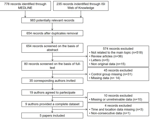
The Circannual pattern and TEMPerature-related incidence of Electrical Storm (TEMPEST) study was conducted following the current guidelines (39, 40), and registered in the PROSPERO International prospective register of systematic reviews of the University of York, United Kingdom (registration # CRD42013003744).
Two big medical databases (MEDLINE and Embase) were systematically searched in order to include all available papers. MEDLINE was searched using the following query: “electrical storm” [Mesh] OR “arrhythmic storm” [Mesh] OR “recurrent ventricular arrhythmias” [Mesh] OR “ventricular tachycardia clusters” [Mesh] OR “electrical instability” [Mesh]. ISI Web of Science was searched using the following query: title contains “electrical storm” OR “arrhythmic storm” OR “recurrent ventricular arrhythmia” OR “ventricular tachycardia clusters” OR “electrical instability”. The search was performed up until November 1st, 2015 and was limited to English-language
literature. Two authors independently screened all the records reviewed full-texts articles and determined their eligibility. Reviews and other meta-analyses on the subjects, as well as reference lists of all identified articles, were also searched for relevant studies. In order to be selected, a study has to meet all the following criteria:
a) diagnosis of ES as the occurrence of three or more episodes of VT/VF within 24 hours (each episode at least 5 minutes apart) or VT for more than 12 hours; b) the absence of acute coronary syndrome as the arrhythmic trigger;
c) ten or more patients included;
d) selection of the most recent publication when the same group reported on the same patients in separate publications.
Inter-observer concordance was optimal during the whole selection process (k = 0.97). At the end of the selection process, 35 papers were included in the data collection process.
3.2 Data collection
Corresponding authors of aforementioned papers were contacted by e-mail and asked to participate. Of those, 19 agreed and the study protocol was provided to them. All authors were asked for additional information on a patient-level basis regarding clinical characteristics, lab exams, ES characteristics, therapies delivered by the ICD, and pharmacological and non-pharmacological management. Nine authors replied back with a complete dataset. In order to ensure optimal data quality, datasets with no data available regarding ES time and geographical location of each patient were excluded, as were datasets collecting non-consecutive patients.
Figure 1 shows the study selection flow chart according to PRISMA statement. At the end of the selection and collection processes, five centres had the datasets from their papers (9–13) merged into the present registry.
3.3 Temperature data collection
Temperature data were retrieved from the “historical weather” section of Weather Underground (www.wunderground.com), the largest weather database available online. For each patient, average, maximum, and minimum temperatures during the event day were collected, as were average mean, maximum and minimum temperatures during the week and the month of the ES, and the week and the month before. If the weather almanac was not available for the exact geographical location at that time, the nearest forecast station with available data was used instead. Weekly
temperatures minus the average of minimum temperatures during a week or a month, respectively. The same database was used in order to retrieve sunrise and sunset hours. If ES started after sunrise and before sunset it was considered as happened during daytime; if it started after sunset and before sunrise it was considered as happened during night-time.
3.4 Statistical analysis
Quantitative variables were checked for normality by the Kolmogorov-Smirnov test. Normally distributed variables were described as mean ± standard deviation. Not-normally distributed variables were described as median and first-to-third interquartile range (1st-3rd IQR). Categorical variables were assessed by using χ2 analysis, and
described as absolute or relative prevalence. Bonferroni adjustment was used for multiple comparisons. ANOVA adjusted for age and sex was used to compare normally distributed quantitative variables. Kruskal-Wallis ANOVA was used to compare non-normally distributed quantitative variables. One-sample T-test was used to test the association between ES incidence and monthly temperature range variation. SPSS 21.0 for Windows (SPSS Inc., Chicago, IL, USA) was used for statistical analysis. Values of p<0.05 (two-tailed) were taken as statistically significant.
4. Results
4.1 Population characteristics
Total population included 246 patients presenting with ES (221 males, age 66±9 years). Clinical characteristics of the population and pharmacological therapy used to terminate ES are shown in Table 1.
Table 1. General characteristics of the population.
Variable Patients (n=246)
Male gender (n,%) 221 (89.8%)
Age (years) 66±9
Type of heart disease:
Ischemic cardiomyopathy (n,%) 155 (63.0%)
Non-ischemic cardiomyopathy (n,%) 51 (20.7%)
Other (n,%) 40 (16.3%)
Left ventricular ejection fraction (%) 31.6±10.4
Serum potassium (mEq/l) 4.3±0.6
Serum creatinine (mg/dl) 1.3±0.5
Triggering arrhythmia:
Monomorphic ventricular tachycardia (n,%) 192 (78.0%) Polymorphic ventricular tachycardia (n,%) 17 (6.9%) Ventricular fibrillation (n,%) 37 (15.1%) Acute treatment: Amiodarone (n,%) 172 (69.9%) Beta-blocker (n,%) 187 (76.0%) D-sotalol (n,%) 10 (4.1%) Lidocaine (n,%) 58 (23.6%) Magnesium sulphate (n,%) 39 (15.9%) Procainamide (n,%) 65 (26.4%) Propafenone (n,%) 40 (16.2%) Sedation (n,%) 25 (10.2%)
While mean serum potassium levels were within the normal ranges (4.3±0.6 mEq/l), 23 patients (9.3%) had hypokalemia and 6 patients (2.4%) had hyperkalemia on presentation. Mean creatinine levels were 1.3±0.5 mg/dl and 22 patients (8.9%) were admitted with creatinine serum levels ≥2 mg/dl.
Each ES episode was made up by a median of 7 VT/VF episodes (1st-3rd IQR 4-16).
On average, each patient experienced a median of 8 anti-tachycardia pacing (ATP) therapies (1st-3rd IQR 3-10) and 5 shocks (1st-3rd IQR 3-6) before ES termination.
4.2 Electrical storm incidence over time
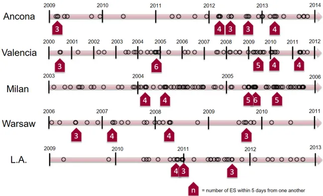
ES incidence over time followed a non-homogeneous distribution in all participating centres (Figure 2), with low-incidence periods alternating with clusters of many ES admitted within a few days from one another.
Figure 2. Distribution of electrical storm incidence over time in each participating centre showing a non-homogeneous, “hot spot” presentation.
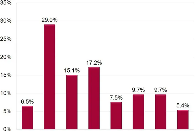
Regarding ES distribution over the day, a significantly higher number of ES happened during daytime hours rather than during night-time hours (58.7% vs. 41.3%; p<0.001). Plotting ES incidence over time shows that 29% of all ES started from 8 to 10 a.m., with a significant incidence reduction during the following hours (Figure 3; p<0.05).
Figure 3a. ES incidence over daytime and night-time hours.
Figure 3b. Circadian pattern of ES’s triggering arrhythmia.
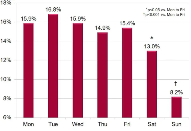
Weekly incidence of ES was similar from Mondays to Fridays, while both Saturdays and Sundays showed a lower incidence of ES when compared to other days (all p<0.05; Figure 4).
Figure 4. ES incidence over the week.
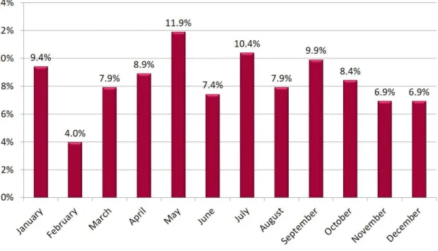
ES distribution over the year was homogeneous with no month significantly associated with a higher prevalence of ES (Figure 5). Similar incidences of ES have been found between winter, spring, summer and fall.
Figure 5. ES incidence over the year.
4.3 Temperature and electrical storm
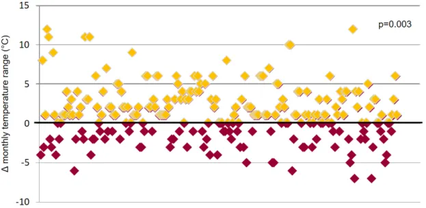
No linear association was found between ES incidence and average, minimum and maximum daily, weekly or monthly temperatures. However, ES incidence was significantly associated with an increase in monthly temperature range when compared to the month before, with 68.9% of ES happening after an increase in monthly temperature range (p=0.003; Figure 6).
5. Discussion
5.1 Temperature-related variations
From our data, ES presentation is not homogenous over time but seems to recognize a clustered or “hot-spot” pattern, alternating low-incidence with “high-intensity” periods, when many patients were admitted with ES within a few days. This pattern, while not superimposable for each center due to the different timeframes and geographical coordinates considered, was indeed quite evident for all of the five participating hospitals (Figure 2). The pattern described was not related to a specific circannual pattern neither to absolute temperatures, as centers varied widely in terms of latitude, longitude, and mean monthly averages (these latter ranging from 10°C in Warsaw to 20° C in Valencia and 21°C in Los Angeles). On the other hand, we found a significant association between ES incidence and variations in the temperature range (Figure 6), with nearly 70% of all ES happening after an increase in temperature ranges of their specific geographical locations. The mechanisms underlying the connection between ES and environmental triggers are unknown, but some hypothesis can be made. In favor of a direct causal relationship, we have many available data underlying an increased risk of ventricular arrhythmias in patients undergoing fast body temperature cooling and rewarming, as commonly experienced in therapeutic hypothermia (41). Animal models demonstrated that inducing hypothermia amplifies dispersion of repolarization (42) and increases myocardial vulnerability to VF (43), thus potentially eliciting multiple arrhythmic events in predisposed patients. Of course, our patients experienced a variation in external rather than body temperature, which was not as big in magnitude neither so concentrated in time. Therefore, other hypotheses must be sought. Recently, it has been suggested that the increased mortality and morbidity caused by hot and cold temperatures could
not be strictly related to hypothermia/hyperthermia but by other causes triggered by the human organism’s attempts to adapt to the external temperature (44). In this regard, increased mortality and hospitalization rates for cardiovascular diseases has been reported after both cold spells and heat waves in many different geographical locations (45). Hospitalization rates from a large population-based study showed a reverse J-shaped association between hospitalization for blood pressure, diabetes, and, with a minor extent, arrhythmias over a lag of 21 days, with higher risks at both temperatures extremes (46). Thermoregulation-mediated vasoreactivity (47), sympathetic nervous system activation (48), and sodium and volume retention through the renin-angiotensin-aldosterone system (49) could all be involved as underlying pathophysiological mechanisms. On a note, the changes in atmospheric pressure related to weather and temperature variations are also associated with the concentration of particulate and gaseous air pollutants, both of whose have been related to an increased risk of arrhythmia-related hospitalization or mortality (50).
5.2 Weekly and daily variations
The higher incidence of ES during working days in our population was not totally unexpected, as was already underlined on unclustered VT/VF(51). This behavior potentially highlights a predominant role of autonomic regulation in triggering the ES, similarly to the well-known circadian pattern in acute myocardial infarction (51). This hypothesis is supported by a recent sub-analysis of the SCD-HeFT trial, which demonstrated a significant weekly variation with an increase in VT/VF incidence on Mondays only in patients not treated with β-blockers (52).
Available analyses of VT/VF daily patterns report a higher incidence of ICD therapies in the early morning both in ischemic and non-ischemic cardiomyopathy (35), while
patients with hypertrophic cardiomyopathy and Brugada syndrome experience ICD therapy more often during the afternoon (36) and late-night (37), respectively. In our population, mainly composed of ischemic (63.0%) and idiopathic dilated cardiomyopathy (20.7%), ES starting hour distribution is similar to the circadian distribution of VT/VF described by Anand et al. (51), although the late rise described during the afternoon hours is much less evident in our population (Figure 3b). So, while it is feasible to hypothesize that the first VT/VF triggering the ES follows the same circadian pattern already described, some other factors must be held responsible for the coalescence of many VT/VF into an ES. Given the high prevalence of structural heart disease and severe left ventricular dysfunction in our population, it is feasible to identify heart failure (HF) as a major potential actor. Available data on a 5-year follow-up shows that ES patients share some striking similarities with patients admitted for HF exacerbation, and ES could be seen as a warning sign of impending pump failure rather than an independent event (45). In this setting, the many dysregulated biochemical pathways typical of HF could help in creating a substrate for arrhythmia recurrence, with ES as an epiphenomenon (57). Potentially overlapping as an arrhythmic risk factor in HF patients is sleep-disordered breathing (SDB). Recent observational data have shown that SDB is an independent predictor of appropriate therapy in ICD patients (58), and could explain, at least in part, the increased incidence of ES seen in the morning.
5.3 Limitations
The present paper shares all the limitation intrinsic to a retrospective, patient-level pooled analysis. Moreover, average temperatures were extracted from a web-based forecasting service, thus limiting the real assessment of external temperature to a wide geographical area.
Regarding collected data, chronic pharmacological therapy was not routinely collected, as the papers selected focused on the acute treatment of ES. Therefore, is not cautious to extend the present findings to specific subpopulations (i.e. patients previously treated with β-blockers or other anti-arrhythmic drugs). Moreover, we were not able to provide data regarding ICD programming, which has been showed as an important risk factor for ES and therefore a potential confounder (59).
6. Conclusions
ES incidence is not homogenous over time but seems to recognize a clustered or “hot-spot” pattern. While ES does not seem to be associated with absolute temperature values, most ES happened in association with an increase in monthly temperature range. Although a higher incidence of ES can be demonstrated also during working hours and working days, the present findings are observational in nature, and new hypotheses must be tested in order to explain this peculiar behaviour.
7. References
1. Pedersen CT, Kay GN, Kalman J, et al. EHRA/HRS/APHRS Expert Consensus on Ventricular Arrhythmias. Hear. Rhythm 2014;11:e166–e196.
2. Arribas F, Auricchio A, Boriani G, et al. Statistics on the use of cardiac electronic devices and electrophysiological procedures in 55 ESC countries: 2013 report from the European Heart Rhythm Association (EHRA). Europace 2014;16:i1–i78.
3. Emkanjoo Z, Alihasani N, Alizadeh A, Tayyebi M, Bonakdar H, Barakpour H, Sadr-Ameli MA. Electrical storm in patients with implantable cardioverter-defibrillators: can it be forecast? Tex Heart Inst J 2009;36(6):563-7
4. Sesselberg HW, Moss AJ, McNitt S, Zareba W, Daubert JP, Andrews ML, et al. Ventricular arrhythmia storms in postinfarction patients with implantable defibrillators for primary prevention indications: a MADIT-II substudy. Heart Rhythm 2007;4(11):1395-402.
5. Exner DV, Pinski SL, Wyse DG, Renfroe EG, Follmann D, Gold M, et al. Electrical storm presages nonsudden death: the antiarrhythmics versus implantable defibrillators (AVID) trial. Circulation 2001;103(16):2066-71.
6. Gatzoulis KA, Andrikopoulos GK, Apostolopoulos T, Sotiropoulos E, Zervopoulos G, Antoniou J, et al. Electrical storm is an independent predictor of adverse long-term outcome in the era of implantable defibrillator therapy. Europace 2005;7 (2):184-92.
7. Wellens HJ, Bar FW, Lie KI. The value of the electrocardiogram in the differential diagnosis of a tachycardia with a widened QRS complex. Am J Med 1978;64(1):27-33. 13.
tachycardia in wide complex left bundle branch block morphology tachycardias. Am J Cardiol 1988; 61(15):1279-83. 14.
9. Brugada P, Brugada J, Mont L, Smeets J, Andries EW. A new approach to the differential diagnosis of a regular tachycardia with a wide QRS complex. Circulation 1991;83(5):1649-59.
10. Baerman JM, Morady F, DiCarlo LA Jr, de Buitleir M. Differentiation of ventricular tachycardia from supraventricular tachycardia with aberration: value of the clinical history. Ann Emerg Med 1987;16(1):40-3.
11. Villacastin J, Almendral J, Arenal A, Albertos J, Ormaetxe J, Peinado R, et al. Incidence and clinical significance of multiple consecutive, appropriate, high-energy discharges in patients with implanted cardioverter-defibrillators. Circulation 1996;93(4):753-62.
12. Silvia G. Priori, Carina Blomström
Lundqvist, Andrea Mazzanti, Nico Blom, Martin Borggrefe, John Camm, Perry Mark Elliott, DonnaFitzsimons, Robert Hatala, Gerhard Hindricks, Paulus Kirchhof, K eld Kjeldsen, Karl-Heinz Kuck, Antonio Hernandez-Madrid, NikolaosNikolaou, Tone M. Norekvål, Christian Spaulding, Dirk J. Van Veldhuisen 2015 ESC Guidelines for the management of patients with ventricular arrhythmias and the prevention of sudden
cardiac death European Heart Journal Aug
2015, ehv316; DOI: 10.1093/eurheartj/ehv316
13. Saksena S, Ciccone JM, Craelius W, Pantopoulos D, Rothbart ST, Werres R. Studies on left ventricular function during sustained ventricular tachycardia. J Am Coll Cardiol 1984;4(3): 501-8.
14. Bigger JT Jr, Dresdale FJ, Heissenbuttel RH, Weld FM, Wit AL. Ventricular arrhythmias in ischemic heart disease: mechanism, prevalence, significance, and management. Prog Cardiovasc Dis 1977;19(4):255-300.
15. Herlitz J, Bang A, Holmberg M, Axelsson A, Lindkvist J, Holmberg S. Rhythm changes during resuscitation from ventricular fibrillation in relation to delay until defibrillation, number of shocks delivered and survival. Resuscitation 1997; 34(1):17-22.
16. Haissaguerre M, Shoda M, Jais P, Nogami A, Shah DC, Kautzner J, et al. Mapping and ablation of idiopathic ventricular fibrillation. Circulation 2002;106(8):962-7
17. Ohgo T, Okamura H, Noda T, Satomi K, Suyama K, Kurita T, et al. Acute and chronic management in patients with Brugada syndrome associated with electrical storm of ventricular fibrillation. Heart Rhythm 2007;4(6):695-700.
18. Bourque D, Daoust R, Huard V, Charneux M. beta-Blockers for the treatment of cardiac arrest from ventricular fibrillation? Resuscitation 2007;75(3):434-44
19. Tsagalou EP, Kanakakis J, Rokas S, Anastasiou-Nana MI. Suppression by propranolol and amiodarone of an electrical storm refractory to metoprolol and amiodarone. Int J Cardiol 2005;99(2):341-2.
20. Kudenchuk PJ, Cobb LA, Copass MK, Cummins RO, Doherty AM, Fahrenbruch CE, et al. Amiodarone for resuscitation after out-of-hospital cardiac arrest due to ventricular fibrillation. N Engl J Med 1999;341(12):871-8. 47.
21.Levine JH, Massumi A, Scheinman MM, Winkle RA, Platia EV, Chilson DA, et al. Intravenous amiodarone for recurrent sustained hypotensive ventricular
tachyarrhythmias. Intravenous Amiodarone Multicenter Trial Group. J Am Coll Cardiol 1996;27(1):67-75.
22. Dorian P, Cass D, Schwartz B, Cooper R, Gelaznikas R, Barr A. Amiodarone as compared with lidocaine for shock-resistant ventricular fibrillation [published erratum appears in N Engl J Med 2002;347(12):955]. N Engl J Med 2002;346(12):884- 90.
23. Eisenach JC, Tong CY. Site of hemodynamic effects of intrathecal alpha 2-adrenergic agonists. Anesthesiology 1991;74 (4):766-71. 54. Fotopoulos GD
24. Kuck KH, Schaumann A, Eckardt L, Willems S, Ventura R, Delacretaz E, et al. Catheter ablation of stable ventricular tachycardia before defibrillator implantation in patients with coronary heart disease (VTACH): a multicentre randomised controlled trial. Lancet 2010;375(9708):31-40
25. Poole JE, Johnson GW, Hellkamp AS, Anderson J, Callans DJ, Raitt MH, et al. Prognostic importance of defibrillator shocks in patients with heart failure. N Engl J Med 2008;359 (10):1009-17. 65. Sears SF, Todaro
26. Kuhlkamp V, Mewis C, Mermi J, Bosch RF, Seipel L. Suppression of sustained ventricular tachyarrhythmias: a comparison of d,l-sotalol with no antiarrhythmic drug treatment. J Am Coll Cardiol 1999;33(1):46-52.
27. Pacifico A, Hohnloser SH, Williams JH, Tao B, Saksena S, Henry PD, Prystowsky EN. Prevention of implantable-defibrillator shocks by treatment with sotalol. d,l-Sotalol Implantable Cardioverter-Defibrillator Study Group. N Engl J Med 1999;340(24):1855-62.
28. Dorian P, Borggrefe M, Al-Khalidi HR, Hohnloser SH, Brum JM, Tatla DS, Brachmann J, Myerburg RJ, Cannom DS, van der Laan M, Holroyde MJ, Singer I,
Pratt CM, Placebo-controlled, randomized clinical trial of azimilide for prevention of ventricular tachyarrhythmias in patients with an implantable cardioverter defibrillator.SHock Inhibition Evaluation with azimiLiDe (SHIELD) Investigators.Circulation. 2004 Dec 14; 110(24):3646-54.
29. Connolly SJ, Dorian P, Roberts RS, Gent M, Bailin S, Fain ES, et al. Comparison of beta-blockers, amiodarone plus beta-blockers, or sotalol for prevention of shocks from implantable cardioverter defibrillators: the OPTIC Study: a randomized trial.
JAMA 2006;295(2):165–71.
30. Wathen MS, DeGroot PJ, Sweeney MO, Stark AJ, Otterness MF, Adkisson WO, et al. Prospective randomized multicenter trial of empirical antitachycardia pacing versus shocks for spontaneous rapid ventricular tachycardia in patients with implantable cardioverter-defibrillators: Pacing Fast Ventricular Tachycardia Reduces Shock Therapies (PainFREE Rx II) trial results. Circulation 2004;110(17):2591–6.
31. Wilkoff BL, Williamson BD, Stern RS, Moore SL, Lu F, Lee SW, et al. Strategic programming of detection and therapy parameters in implantable cardioverter-defibrillators reduces shocks in primary prevention patients: results from the PREPARE (Primary Prevention Parameters Evaluation) study. J Am Coll Cardiol 2008;52(7):541–50.
32. Pacifico A, Ferlic LL, Cedillo-Salazar FR, Nasir N Jr, Doyle TK, Henry PD. Shocks as predictors of survival in patients with implantable cardioverter-defibrillators. J Am Coll Cardiol 1999;34(1):204–10.
33. Nordbeck P, Seidl B, Fey B, Bauer WR, Ritter O. Effect of cardiac resynchronization therapy on the incidence of electrical storm. Int J Cardiol 2010;143(3):330–6.
34. Vanderheyden M, Mullens W, Delrue L, Goethals M, de Bruyne B, Wijns W, et al. Myocardial gene expression in heart failure patients treated with cardiac resynchronization therapy responders versus nonresponders. J Am Coll Cardiol 2008;51 (2):129–36.
35. Englund a, Behrens S, Wegscheider K, Rowland E. Circadian variation of malignant ventricular arrhythmias in patients with ischemic and nonischemic heart disease after cardioverter defibrillator implantation. J. Am. Coll. Cardiol. 1999;34:1560–8.
36. Maron BJ, Semsarian C, Shen WK, et al. Circadian patterns in the occurrence of malignant ventricular tachyarrhythmias triggering defibrillator interventions in patients with hypertrophic cardiomyopathy. Hear. Rhythm 2009;6:599–602.
37. Kim S-H, Nam G, Baek S, et al. Circadian and seasonal variations of ventricular tachyarrhythmias in patients with early repolarization syndrome and Brugada syndrome: analysis of patients with implantable cardioverter defibrillator. J. Cardiovasc. Electrophysiol. 2012;23:757–63.
38. Guerra F, Shkoza M, Scappini L, Flori M, Capucci A. Role of electrical storm as a mortality and morbidity risk factor and its clinical predictors: a meta-analysis. Europace 2014;16:347–53.
39. Stroup DF, Berlin J a, Morton SC, et al. Meta-analysis of observational studies in epidemiology: a proposal for reporting. Meta-analysis Of Observational Studies in Epidemiology (MOOSE) group. JAMA 2000;283:2008–12.
40. Moher D, Liberati A, Tetzlaff J, Altman DG. Academia and Clinic Annals of Internal Medicine Preferred Reporting Items for Systematic Reviews and Meta-Analyses :
41. Carbucicchio C, Santamaria M, Trevisi N, et al. Catheter ablation for the treatment of electrical storm in patients with implantable cardioverter-defibrillators: short- and long-term outcomes in a prospective single-center study. Circulation 2008;117:462–9.
42. Koźluk E, Gaj S, Kiliszek M, Lodziński P, Piątkowska A, Opolski G. Efficacy of catheter ablation in patients with an electrical storm. Kardiol. Pol. 2011;69:665–70.
43. Izquierdo M, Ruiz-Granell R, Ferrero A, et al. Ablation or conservative management of electrical storm due to monomorphic ventricular tachycardia: differences in outcome. Europace 2012;14:1734–9.
44. Vaseghi M, Gima J, Kanaan C, et al. Cardiac Sympathetic Denervation In Patients With Refractory Ventricular Arrhythmias Or Electrical Storm: Intermediate And Long Term Follow Up. Heart Rhythm 2013.
45. Guerra F, Flori M, Bonelli P, Patani F, Capucci A. Electrical storm and heart failure worsening in implantable cardiac defibrillator patients. Europace 2014;17:247–254.
46. Lee WS, Nam G-B, Kim S-H, et al. ECG features and proarrhythmic potentials of therapeutic hypothermia. Heart 2016.
47. Piktel JS, Jeyaraj D, Said TH, Rosenbaum DS, Wilson LD. Enhanced dispersion of repolarization explains increased arrhythmogenesis in severe versus therapeutic hypothermia. Circ. Arrhythmia Electrophysiol. 2011;4:79–86.
48. Hsieh YC, Lin SF, Lin TC, Ting CT, Wu TJ. Therapeutic hypothermia (30 degrees C) enhances arrhythmogenic substrates, including spatially discordant alternans, and facilitates pacing-induced ventricular fibrillation in isolated rabbit hearts. Circ.J. 2009;73:2214–2222.
with cold spells in the Czech Republic. BMC Public Health 2009;9:19.
50. Liu C, Yavar Z, Sun Q. Cardiovascular response to thermoregulatory challenges. Am. J. Physiol. Heart Circ. Physiol. 2015;309:H1793–812.
51. Bai L, Li Q, Wang J, et al. Hospitalizations from Hypertensive Diseases, Diabetes, and Arrhythmia in Relation to Low and High Temperatures: Population-Based Study. Sci. Rep. 2016;6:30283.
52. Hong CH, Kuo TBJ, Huang BC, et al. Cold exposure can induce an exaggerated early-morning blood pressure surge in young prehypertensives. PLoS One 2016;11:1– 15.
53. Sarzani R, Guerra F, Mancinelli L, Buglioni A, Franchi E, Dessì-Fulgheri P. Plasma aldosterone is increased in class 2 and 3 obese essential hypertensive patients despite drug treatment. Am. J. Hypertens. 2012;25:818–26.
54. Song X, Liu Y, Hu Y, et al. Short-Term Exposure to Air Pollution and Cardiac Arrhythmia: A Meta-Analysis and Systematic Review. Int. J. Environ. Res. Public Health 2016;13:642.
55. Anand K, Aryana A, Cloutier D, et al. Circadian, Daily, and Seasonal Distributions of Ventricular Tachyarrhythmias in Patients With Implantable Cardioverter-Defibrillators. Am. J. Cardiol. 2007;100:1134–1138.
56. Patton KK, Hellkamp AS, Lee KL, et al. Unexpected Deviation in Circadian Variation of Ventricular Arrhythmias. J. Am. Coll. Cardiol. 2014;63:2702–2708.
57. Nayyar S, Ganesan AN, Brooks AG, Sullivan T, Roberts-Thomson KC, Sanders P. Venturing into ventricular arrhythmia storm: a systematic review and meta-analysis. Eur. Heart J. 2013;34:560–71.
58. Kreuz J, Skowasch D, Horlbeck F, et al. Usefulness of sleep-disordered breathing to predict occurrence of appropriate and inappropriate implantable-cardioverter defibrillator therapy in patients with implantable cardioverter-defibrillator for primary prevention of sudden cardiac death. Am. J. Cardiol. 2013;111:1319–1323.
59. Guerra F, Palmisano P, Dell’Era G, et al. Implantable cardioverter-defibrillator programming and electrical storm: Results of the OBSERVational registry On long-term outcome of ICD patients (OBSERVO-ICD). Hear. Rhythm 2016.