Università degli Studi di Ferrara
DOTTORATO DI RICERCA IN
"FARMACOLOGIA E ONCOLOGIA MOLECOLARE"
CICLO XXV
COORDINATORE Prof. Antonio Cuneo
CONSTRUCTION OF REPLICATION-DEFECTIVE HERPES
SIMPLEX VIRAL VECTORS FOR TARGETING THE LHX2
GENE IN THE CENTRAL NERVOUS SYSTEM
Settore Scientifico Disciplinare BIO/14
Dottorando Tutori
Dott. Verlengia Gianluca Prof.Simonato Michele
Prof. Glorioso Joseph
ABSTRACT
Epilepsy is a chronic disorder affecting about 65 million people worldwide and temporal lobe epilepsy (TLE) is among the most frequent types of intractable epilepsy. In most cases the causes of TLE are unknown but it is believed that it may take place after an initial precipitating injury (IPI) such as brain tumor, ictus, head trauma, meningitis, encephalitis, and febrile seizures during childhood. Despite the development of new
antiepileptic drugs (AEDs), about 35% of epileptic patients still suffer from
pharmacoresistant seizures, with surgical resection of the epileptic locus as possible last
option. In addition, AEDs don’t prevent the progression of the disease but they are designed only for treatment of patients with an already established syndrome. Hence, there is an unmet medical need for the prevention of seizures for the patients at high-risk of developing epilepsy. This clarify how urgent is the need to find novel therapeutic concepts to fill this gap.
Epilepsy could develop when the intracerebral balance between excitation and inhibitory neurotransmission is impaired. Experimental findings show that after an epileptogenic insult the brain react to the injury with an enhanced hippocampal neurogenesis as a homotypic response to the neuronal loss in an attempt to restore the pre-existing cellular network. However, this plastic remodeling that the brain goes through is usually aberrant, since the cells that undergo the replacement of degenerating neurons upon severe brain injury are mostly high proliferating reactive astrocytes. This leads to important alterations of brain signals and, consequently, high risk of seizure development.
Starting from this concept, we hypothesize that controlling the neural stem cells fate after an initial precipitating injury could prevent epileptogenesis or at least improve the clinical picture of the patient.
Based on the recent literature, we decided to test the effects of Lhx2 protein overexpression on cells of central nervous system. Lhx2 is a transcription factor that plays a crucial role since early stages in telencephalic patterning but its function is not limited to the early embryonic neuroepithelium: recent evidences have shown a unique role for this protein in the phase of active neurogenesis, when its overexpression may enhances and prolongs the neurogenesis to generate neurons from progenitors that would otherwise give rise to astrocytes.
The recent advances of gene therapy promise innovative and revolutionary new
treatments for neurological disorders. Various methods have been developed for gene
delivery to target cells. However, gene transfer by viral vectors is thus far the widest used approach. In particular, up today the most efficient systems to achieve a long term transgene expression is based upon retroviral and lentiviral vectors. Unfortunately both these viruses cannot be designed for clinical applications since their infections result in insertion of viral DNA into the host chromosomes at an unpredictable position, a dangerous event which can seriously disturbs cellular genes functions potentially leading to cancer transformation of infected cells. It is then important to set up novel tools to safely deliver genes. Herpes simplex virus-1 (HSV-1) offers unique features that support its development as a great candidate viral vector especially for targeting the nervous system: it is a highly infectious, naturally neurotropic virus able to establish life-long latency in neurons, along with the largest capacity for exogenous DNA cloning. Moreover, it doesn’t integrate into the host genome, avoiding any possibility of insertional activation or inactivation of cellular genes. However, some technical problems still need to be overcome, such as the efficient delivery of the vector to target cells, the maintenance and control of foreign gene expression, and the control of unwanted host immune responses.
This thesis describes the development of a highly efficient method for in vitro and in vivo targeting of the Lhx2 gene using novel replication-defective herpes simplex viral vectors, named JΔβββ4 and JΔΝΙ, opportunely engineered to reduce the innate toxicity of the virus and to allow a good expression of the transgene. HSV-mediated delivery of Lhx2 resulted in highly effective gene overexpression in several cell types in vitro, including mouse neuronal and non-neuronal cells, along with reduced or null toxicity and a differential transgene expression, depending on the viral backbones. These vectors have been additionally tested in vivo by injection into the hippocampus of naïve rats and of rat models of epilepsy. Ex vivo analyses of injected brains showed good infection pattern from both viruses along with no evident toxicity. Moreover, the hippocampal delivery of Lhx2 by JΔβββ4-based vector was associated with reductions of both astrocyte density and recurring seizures, giving rise to more favorable pathologic features and improved outcomes. Put together, we can finally assess that both the JΔβββ4 and JΔΝΙ-based vectors could represent useful tools for differential purposes: while for in vitro applications the JΔΝΙ vector is the best compromise between transgene expression and low toxicity
effects, for in vivo gene transfer it result almost ineffective. On the other hand, the JΔβββ4 vector displayed an opposite behavior, too toxic for in vitro approaches but much more effective for gene delivery in vivo.
ACKNOWLEDGMENTS
Laboratory research is undoubtedly a collaborative effort, and this thesis is no exception. I would like to acknowledge all the many peoples that helped me to accomplish this work. None of my personal improvement and scientific achievements could have happened without the many talented scientists I worked with in the past three years. Firstly, I would like to thank my tutor Prof. Michele Simonato which encouraged me and granted me the autonomy to generate my own hypotheses before deciding upon a dissertation topic. The encouragement and unconditional faith he had in me go beyond science. I will never forget the unique chance I had from him to spending all the last year of my PhD program abroad, working in Joseph Glorioso’s laboratory at University of Pittsburgh. Here I had the unique pleasure to work with my co-tutor Prof. Jospeh Glorioso, which supported me with his sense of humor, patience and preciouses suggestions throughout the journey to completion of this project.
Many thanks also to Dr. Paola Grandi, the living encyclopedia that shared with me her huge expertise in any field of science; thank you Paola for the willingness to persist in helping me carry out this project and your patience as I acquired insight into the subject matter.
And, most importantly, I want to thank the person who shared his huge knowledge about herpes virus and stem cell biology, but also a lot of material with me, Dr. Yoshitaka Miyagawa. Rarely I had the pleasure of working so closely with someone who brought so much collaboration. Without him I probably couldn’t write this thesis. What I have learned from Dr. Miyagawa is immeasurable. He taught me to be critical, precise, persistent and consistent in science. He is the best mentor and inspiring educator I could ever meet. Thank you, Yoshi!
I also give thanks to all the people of both my Italian and American laboratories I met during this years, all of them essential to achieve this goal.
I thank my family for their continuous, unconditional love and support throughout my education.And, last but not last, great thanks to my family, the most important people of my world.
ABSTRACT 1 AKNOWLEDGMENTS 4 1. INTRODUCTION 8 1.1 EPILEPSY 8 1.1.1 HISTORICAL OVERVIEW 8 1.1.2 EPIDEMIOLOGY 8 1.1.3 ETIOLOGY 9 1.1.4 DEFINITION 9
1.1.5 TREATMENTS AND DRUGS 11
1.1.6 TEMPORAL LOBE EPILEPSY 13
1.1.7 ANIMAL MODELS OF TLE 15
1.2 NEUROGENESIS AND ASTROGLIOSIS 20
1.2.1 NEUROGENESIS 20
1.2.3 NEUROGENESIS AND EPILEPSY 23
1.2.4 ASTROCYTES AND EPILEPSY 24
1.3 HERPES VIRUSES 27 1.3.1 HERPERSVIRIDAE CLASSIFICATION 27 1.3.2 HSV-‐1 OVERVIEW 28 1.3.3 HSV-‐1 DNA GENOME 29 1.3.4 HSV-‐1 NUCLEOCAPSID 30 1.3.5 HSV-‐1 TEGUMENT 30 1.3.6 HSV-‐1 ENVELOPE 31 1.3.7 HSV-‐1 INFECTION 31 1.3.8 IE GENE EXPRESSION 32 1.3.9 ICP0 33 1.3.10 ICP4 34 1.3.11 ICP22 34 1.3.12 ICP27 35 1.3.13 ICP47 35 1.3.14 VIRUS EGRESS 36 1.3.15 LATENCY 36 1.4 GENE THERAPY 38 1.4.1 OVERVIEW 38
1.4.2 VECTORS FOR GENE THERAPY 38
1.4.3 RETROVIRUSES & LENTIVIRUSES 40
1.4.4 ADENOVIRUS 41
1.4.5 HSV-‐BASED VECTORS 42
1.4.6 WILD-‐TYPE HSV-‐1 VECTORS 43
1.4.7 REPLICATION DEFECTIVE VECTORS 43
1.4.8 REPLICATION DEFECTIVE VECTORS 44
1.4.9 REPLICATION ATTENUATED VECTORS 45
1.4.10 AMPLICONS & BAC 45
1.5 LIM-‐HOMEODOMAIN PROTEINS 49
1.5.1 LHX-‐PROTEIN OVERVIEW 49
1.5.2 LIM-‐HOMEODOMAIN PROTEIN 2 (LHX2) 49
2. MATERIALS AND METHODS 51
2.1 CELL LINES 51
2.3 LIGATION OF DNA FRAGMENTS AND TRANSFORMATION OF COMPETENT BACTERIA 52
2.4 PCR ANALYSIS 52
2.5 CHEMICAL TRANSFORMATION OF COMPETENT BACTERIA CELLS 52
2.6 DNA EXTRACTION FOR SCREENING OF TRANSFORMED BACTERIA COLONIES 52
2.7 GATEWAY® RECOMBINATION 53
2.8 ELECTROPORATION 53
2.9 BAC MINIPREPS AND MIDIPREPS 53
2.10 TRANSFECTION OF BAC-‐DNA INTO U2OS-‐ICP4 TO GENERATE JΔβββ4 AND JΔΝΙ4 VIRUSES 54
2.11 VIRAL TITRATION IN PLAQUES FORMING UNITS (P.F.U/ML) 54
2.12 INFECTION TO TEST VIRAL GROWTH 55
2.13 HIGH SCALE VIRAL PRODUCTION 55
2.14 IMMUNOFLUORESCENCE OF ADHERENT CELLS 55
2.15 WESTERN BLOT 56
2.16 VIRUSES 56
2.17 PRIMARY CELL CULTURES 57
2.18 ANIMALS 58
2.19 PILOCARPINE MODEL AND HSV INFUSION 58
2.20 FIXATION PROCEDURE 58
2.21 HEMATOXILIN AND EOSIN STAINING 59
2.22 FLUORO-‐JADE C AND NEUROTRACE STAINING 59
2.23 GFAP IMMUNOFLUORESCENCE AND NEUROTRACE STAINING 59
3.RESULTS 60
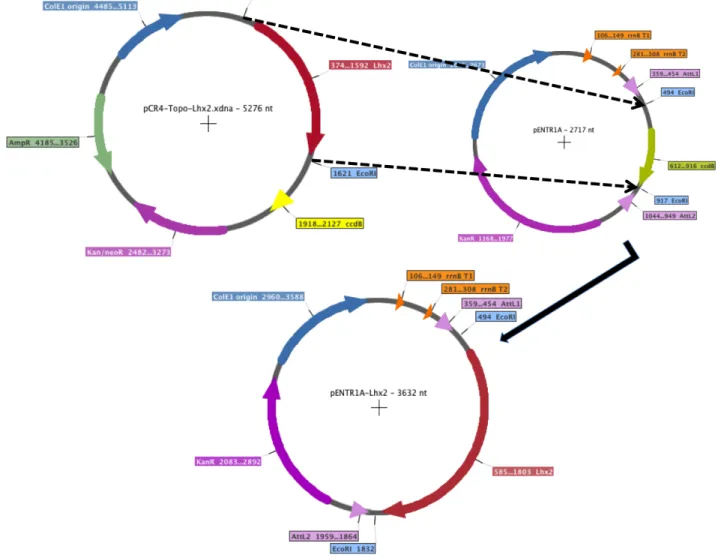
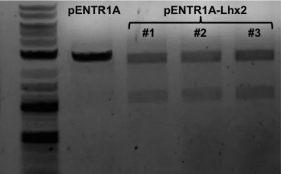
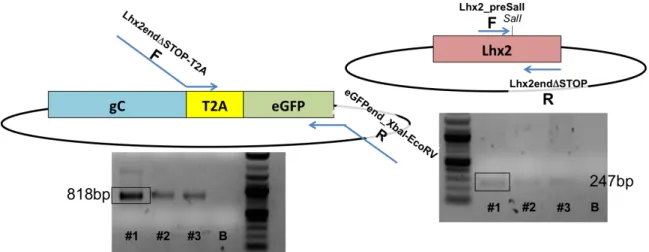
3.1 LHX2 CDNA CLONING INTO PENTR1A® PLASMID 60
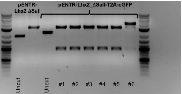
3.2 CONSTRUCTION OF THE FUSION GENE LHX2-‐T2A-‐EGFP 61
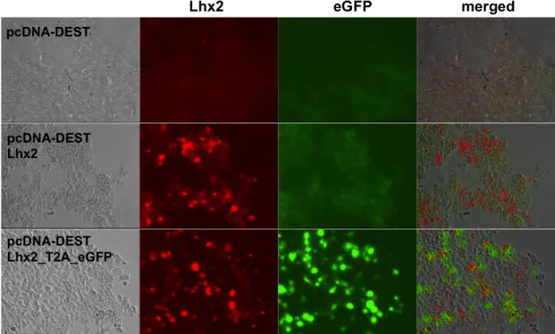
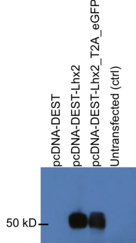
3.3 CONSTRUCTION OF THE EXPRESSION PLASMID PCDNA-‐DEST_LHX2 AND PCDNA-‐DEST_LHX2-‐T2A-‐
EGFP 63
3.4 GENERATION AND CHARACTERIZATION OF JΔβββ4-‐LHX2 AND JΔΝΙ-‐LHX2 VIRUSES 66
3.5 IN VITRO INFECTION OF PRIMARY CULTURES OF NEURONS AND ASTROCYTES FROM JΔβββ4-‐ AND JΔΝΙ-‐
BASED HSV-‐1 VECTORS 81
3.6 IN VIVO INOCULATION OF HSV-‐1 BASED VECTORS 83
3.7 NAÏVE GROUP: HEMATOXILIN-‐EOSIN STAINING OF CORONAL BRAIN SLICES 86
3.8 NAÏVE GROUP: FLURO-‐JADE STAINING OF CORONAL BRAIN SLICES 87
3.9 EFFECTS ON SE-‐INDUCED ASTROCYTOSIS AND OUTCOME OF PATHOLOGY 88
4. DISCUSSION 92
5. TABLE OF ARTICLES 97
5.1 INCREASED EXCITABILITY IN TAT-‐TRANSGENIC MICE: ROLE OF TAT IN HIV-‐RELATED NEUROLOGICAL
DISORDERS 97
5.2 BRADYKININ B2 RECEPTORS INCREASE HIPPOCAMPAL EXCITABILITY AND SUSCEPTIBILITY TO SEIZURES
IN MICE 98
5.3 CHANGES IN THE SENSITIVITY OF GABAA CURRENT RUNDOWN TO DRUG TREATMENTS IN A MODEL OF
TEMPORAL LOBE EPILEPSY 99
Alle mie nonne,
insostituibili maestre di vita.
1. INTRODUCTION
1.1 EPILEPSY
1.1.1 Historical overview
Epilepsy is a medical disorder old as human existence; few diseases have generated same interests and controversies as epilepsy.
The term epilepsy derives from the Greek verb epilambanem, meaning "to take hold on or to seize upon." The astonishing manifestation of the epileptic seizures inspired in people of prehistoric civilizations the belief of a supernatural punishing event.
Hippocrates, the Father of Medicine, made the first historical records of epilepsy in the fifth century BC referring to epilepsy as the “Sacred Disease”, an attack from demons. Later in the fifteenth century AD, it was believed to be a sign of witchcraft. It wasn't until the late nineteenth century when pioneering studies on human brain showed the true nature of epilepsy as a disease of the central nervous system, which could be identified by electroencephalography (Hughlings-Jackson, 1888; Berger, 1929).
1.1.2 Epidemiology
Epilepsy is the commonest neurological condition that affects people of all ages, race and social class. It is estimated that about 1% of the world’s population is affected by epilepsy and up to 5% may experience just a single unprovoked seizure (Adelöw et al., 2009). Across all ages worldwide, the incidence is approximately 50-100000/year in developed countries and 100-200000/year in developing countries (Theodore et al., 2006; Ngugi et al., 2010)
Incidence and prevalence studies are critical to provide measures of frequency and therefore the burden of disease, and allow for proper planning of services.
The epidemiology of epilepsy is subjected to several factors that vary worldwide as age, sex, comorbidities, overall health, etc. The age distribution of the incidence of epileptic seizures follows a bimodal trend, showing two peaks of frequency in childhood and seniority.
1.1.3 Etiology
It is a common misconception that epilepsy is a single homogeneous disease. In reality, epilepsies are highly heterogeneous disorders that can be categorized on the basis of several differentiating factors. Not even the origin of the disease is unique, since it may be acquired or inherent, pediatric or adult onset. There are many possible causes of epilepsy. The etiology of this syndrome is complex, arising from the contribution of multiple genetic and non-genetic factors. It is commonly accepted that epilepsy results from an abnormality in brain wiring and neurotransmitter imbalance. This atypical wiring can result from known brain pathology, traumatic brain injury, brain infections, prolonged febrile seizures, brain tumors, genetic propensity, neural developmental disorders or recreational drugs.
The International League Against Epilepsy (ILAE) has also classified epilepsies by etiology in:
-Familial epilepsies: inherited, these are identified in large families with an epileptic trait segregating in the absence of environmental factors.
-Idiopathic: usually age-dependent, these have a genetic or presumed genetic origin and do not involve underlying structural brain lesions or other signs of neurologic dysfunction. About 500 genes have been estimated to play a role in disease development, either by directly altering protein production or by increasing one’s resistance to pharmacological treatments.
-Symptomatic: caused by a specific systemic or environmental factor, without particular neuroanatomic abnormalities.
Additionally, we can include the “Cryptogenic” epilepsies, or rather those without any well identified origin. Even if the number of such cases is diminishing, this category still counts about 40% of adult-onset cases of epilepsy.
1.1.4 Definition
The wide heterogeneity of clinical features regarding epileptic syndromes makes particularly complex to draw up any classification.
Today is well known that epilepsy is not one condition, but a diverse family of disorders occurring when the electrical signals in the brain are disrupted, leading to changes in neuronal activity that give rise to seizures. This medical condition has always been controversial, even regarding its definition.
Several classifications have been proposed since 1970 but any attempts to get a consensus failed until 1997, when the ILAE and the International Bureau for Epilepsy (IBE) defined epilepsy as “a disorder of the brain characterized by an enduring predisposition to generate epileptic seizures and by neurobiological, cognitive, psychological and social consequences. The definition of epilepsy requires also the occurrence of at least one epileptic seizure” defined as “a transient occurrence of signs and symptoms due to abnormal, excessive or synchronous neuronal activity in brain” (Fisher et al., 2005).
Epilepsy occurs when electrical signals in the brain are disrupted, leading to changes in neuronal activity (neurons may fire up to 6 times as fast as the normal rate of about 80 times a second) that give rise to seizures. A seizure is a synchronized paroxymal event caused by an excessive electrical discharge of central nervous system neurons (Lowenstein et al. 2004), which can cause brief changes in sensations, emotions and behavior and, in worst cases, convulsions, muscle spams and loss of consciousness. Seizures can also be classified grounding on the evoked physical manifestations, which can range from wild swinging movements of arms and legs (tonic-clonic seizures) to brief losses of awareness appearing as staring spells (absence seizure) and from to the spatial extent of the brain involved at clinical onset: when localized within the brain at the onset of the seizure they are referred to as focal or partial seizures and are usually named by the origin of the epileptic focus as in temporal lobe epilepsy, while those that appear to involve the entire brain at once are called generalized seizures. Finally, seizures can be differentiated based on whether the patient loses consciousness during the seizure event (complex seizure) or not (simple seizure). These categorizations are not mutually exclusive, so one will often describe an epilepsy disorder as complex partial seizures of the frontal lobe, for instance. In 1997, the ILAE has classified human epilepsies by seizure type as self-limited, continuous, or reflex, and as focal or generalized (Engel, 2001). Generalized, self-limited seizures are exemplified by tonic-clonic seizures, which begin with tonic extension of the limbs and trunk, evolve into rhythmic movements (clonus) and terminate spontaneously within a few minutes. Absence seizures also are generalized and self limited, characterized by brief episodes (about 10 sec) of staring and unconsciousness that can occur more than 100 times per day (Panayiotopoulos, 1997). In contrast, focal seizures arise from a focal region of one cerebral hemisphere, and their manifestations depend on the extension of the brain
status epilepticus, sometimes occur. Reflex seizures are rare and are evoked by visual stimuli, somatosensory stimuli, thinking, reading, or tooth brushing. The precipitating stimulus is specific for each patient.
The human classification scheme can be used to classify seizures in other species, since self-limited, generalized tonic-clonic seizures and status epilepticus occur in many species.
1.1.5 Treatments and drugs
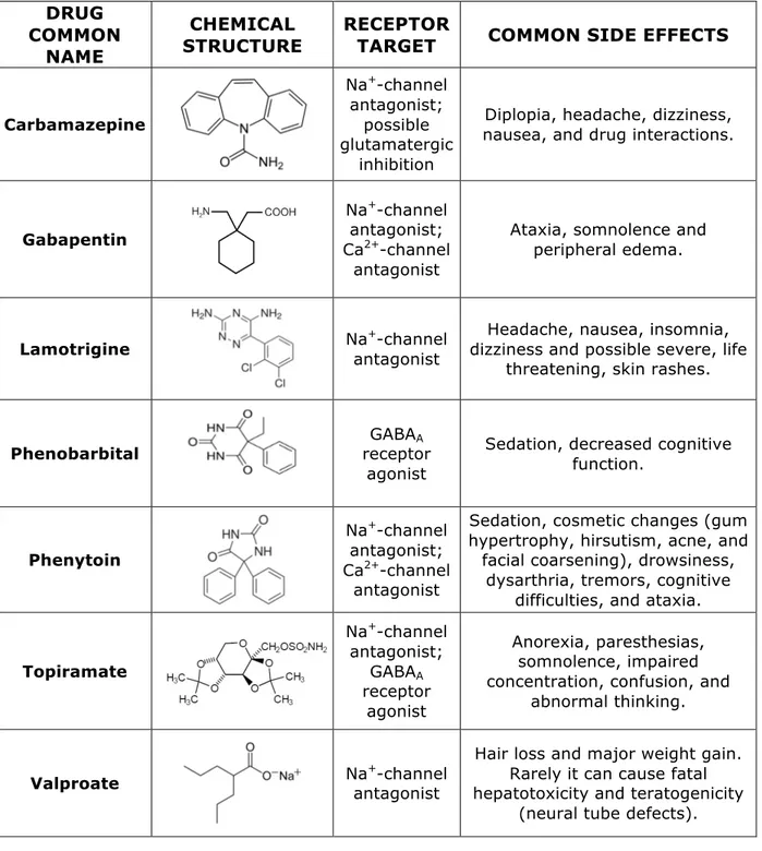
Upon diagnosis (EEG monitoring, video monitoring and magnetoencephalogram testing is a typical battery of examinations for diagnosis), the first-line treatment of epilepsy is pharmacological intervention. To date there is no cure for epilepsy and current treatment options only include lifelong drug therapy and, in a number of difficult cases, surgery may be necessary. While the overall goal of antiepileptic drugs is to completely eliminate seizures, this goal has not yet been achieved. Current antiepileptic drugs (AEDs) aim at symptomatically reduce the frequency and the duration of the seizures. Drug treatment of epilepsy began as early as 1857 and mainly involved the use of bromides. Phenobarbital was also a popular choice for epilepsy patients early in history. Since then, a large number of antiepileptic drugs have been developed for the therapy of various forms of epilepsy. No one treatment option works for all types of epilepsy disorders and, therefore, the preferred drug treatment of choice depends on a number of different factors such as the characteristic pharmacokinetic profile of the various AEDs and on the specific biochemical malfunction of the patient (Faught and Pollock, 2001). The most common antiepileptic drugs are summarized in Table 1.
Antiepileptic drugs are divided into a short list of classes according to their mechanisms of action (Howland et al., 2005), which aimed to:
-‐ reduce sodium current (i.e. carbamazepine, lamotrigine, phenytoin) -‐ reduce excitatory coupling between neurons (i.e. topiramate)
-‐ increase inhibitory coupling between neurons (i.e. clonazepam, divalproic acid, phenobarbital)
DRUG COMMON
NAME
CHEMICAL
STRUCTURE RECEPTOR TARGET COMMON SIDE EFFECTS
Carbamazepine Na+-channel antagonist; possible glutamatergic inhibition
Diplopia, headache, dizziness, nausea, and drug interactions.
Gabapentin
Na+-channel antagonist; Ca2+-channel
antagonist
Ataxia, somnolence and peripheral edema.
Lamotrigine Naantagonist +-channel dizziness and possible severe, life Headache, nausea, insomnia, threatening, skin rashes.
Phenobarbital receptor GABAA agonist
Sedation, decreased cognitive function. Phenytoin Na+-channel antagonist; Ca2+-channel antagonist
Sedation, cosmetic changes (gum hypertrophy, hirsutism, acne, and
facial coarsening), drowsiness, dysarthria, tremors, cognitive
difficulties, and ataxia.
Topiramate Na+-channel antagonist; GABAA receptor agonist Anorexia, paresthesias, somnolence, impaired concentration, confusion, and
abnormal thinking.
Valproate Naantagonist +-channel
Hair loss and major weight gain. Rarely it can cause fatal hepatotoxicity and teratogenicity
(neural tube defects).
Table 1: Most common used antiepileptic drugs in clinic.
These AEDs are clearly intended to reduce the abnormal excessive neural activity of epileptic patients. Their effects on neural synchrony, however, still remain unclear and more than 30% do not respond to current anticonvulsant drugs (Kwan and Brodie, 2000); this percentage has remained unchanged for over twenty years despite the discovery of several new anti-epileptic medications.
This lack of improvement in efficacy can be attributed to the fact that none of the recently discovered medications exhibit a novel mechanism of action. The most commonly prescribed medications are Carbamazepine, Valproate or Phenytoin.
When seizures cannot be controlled pharmacologically, surgery is the most considered alternative. Three broad categories of epilepsy can be treated successfully with surgery: partial seizures, seizures that begin as partial seizures before spreading to the rest of the brain and unilateral multifocal epilepsy with infantile hemiplegia (Bleck, 1987). The most common type of surgery for epilepsy is removal of a seizure focus (often called topectomy or lesionectomy). Usually, the more well defined the seizure focus the better is the post-surgical prognosis (Bleck, 1987). Other types of surgery include loboectomy (i.e. temporal lobe resection, reportedly having up to 70-90% success rate, Brodie & Schachter, 2000), subpial transection (Morrel et al., 1989), callosotomy, severing the bundle of neural connections between the right and left cerebral hemispheres, and similar procedures involving the anterior and posterior commissures (Brodie & Schacter, 2000). The most radical surgical procedure to treat epilepsy is hemispherectomy.
Other treatment strategies of epilepsy include vagus nerve stimulation, transcranial magnetic stimulation and biofeedback.
An additional line of research is currently investigating development of implantable devices that can deliver drugs to specific parts of the brain (Pathan at al., 2009). Furthermore, researchers are looking into altering certain environmental and life-style factors such as diet, which might alleviate seizure disorders (Korsholm & Law, 2013. Unfortunately many patients (about 20%) continue to experience seizures even after several treatment attempts, highlighting the need of new innovative therapies to improve their threatening condition.
1.1.6 Temporal Lobe Epilepsy
Temporal lobe epilepsy (TLE) is the most common type of epilepsy in adults, and many patients continue to be affected by uncontrolled seizures despite treatment with AEDs (Engel et al., 1997). This condition is associated with specific structural lesions in the hippocampus (which may be surgically resected in medically intractable cases), leading to progressive development of spontaneous recurrent seizures (SRS) from the medial or lateral temporal lobe (so called Medial temporal lobe epilepsy or Lateral temporal lobe epilepsy).
Patients with temporal lobe epilepsy have approximately 2 to 30 seizures per month (French et al., 1993). These seizures may be partial, complex or generalized (depending on the involvement of one or both emispheres) and are unfortunately unpredictable. They start with an aura (King, 1977), during which the patient is still conscious, that is a visceral sensation of nausea, pressure, “butterflies” and/or epigastric rising, followed by a focal motor seizure and loss of consciousness which begins with motor arrest and staring and evolves to oral-alimentary automatisms (e.g., lip smacking, chewing, tooth grinding). Focal motor seizures sometimes progress to generalized tonic-clonic seizures. The mechanisms that terminate seizures and determine whether focal seizures will become generalized are still unknown.
Most patients with temporal lobe epilepsy have a history of brain injury and up to 2/3 of them had febrile seizures. Age at time of the injury is quite variable, but it usually happens during childhood (Mathern et al., 2002). Other precipitating injuries include head trauma, brain infections or hypoxia/ischemia (Mathern et al., 1995). Not all these precipitating events involve seizures at the time: after recovery from the initial precipitating injury, patients usually begin a seizure-free latent period, ranging from months to years, when aberrant changes in structure and physiology of the brain tissue happen before the development of SRS (Mathern, et al., 2002.). After this time, spontaneous, recurrent seizures occur, which typically continue throughout life.
This seizure-free latent period has been matter of great interest. It has been proposed that it is attributable to the time necessary for synaptic reorganization to establish a sufficient degree of recurrent excitation to surpass the seizure threshold. During the latent period, the hippocampus is undoubtedly the brain region mainly affected by unique morphological alterations. The most common lesional abnormality observed in patients with TLE is the Hippocampal Sclerosis (HS) (Babb and Brown; 1987), consisting in a massive loss and widening of the neurons in the hilus of the dentate gyrus and in the CA1 and CA3 layers (Engel, 1989; Ben-Ari and Cossart, 2000), and the mossy fiber sprouting, that is the aberrant growth of granule cell axons into the inner molecular layer of the dentate gyrus (Sutula et al., 1989; Babb et al., 1991; Isokawa et al., 1993). Consequently, a prominent hypothesis states that hippocampal neuronal loss and mossy fiber sprouting play a critical role in the genesis and progression of TLE (Lothman and Bertram, 1993); nevertheless, lesions also may develop in subregions of the amygdala and entorhinal cortex (Bernasconi et al., 2003).
mechanisms of epileptogenesis are still unknown. Many hypotheses of temporal lobe epileptogenesis focus on the hippocampal dentate gyrus, which is thought to serve as a seizure-suppressing filter or gate (Lothman et al., 1991): the dentate gyrus displays dramatic lesions, such as loss of hilar neurons (Margerison and Corsellis. 1966) including excitatory mossy cells (Babb et al., 1984) and inhibitory interneurons (Maglóczk et al., 2000). Excitatory dentate granule cells survive but their inhibition is dramatically worsened due to loss of inhibitory GABAergic interneurons that normally inhibite the granule cells making them hyperexcitable and lowering the seizure threshold (Staley and Mody. 1992).
In addition, loss of mossy cells can also give rise to axon sprouting and synaptogenesis, inducing an aberrant positive-feedback circuit between dentate granule cells that generates seizures (Nadler et al., 1980). Mossy cells are the predominant neurons in the hilus and concentrate their glutamatergic axon terminals in the inner molecular layer of the dentate gyrus where they form excitatory synaptic contacts with granule cells (Wenzel et al., 1997). Since mossy cells are particularly sensitive to a wide range of insults (Buckmaster and Schwartzkroin, 1994), when they die their axon terminals degeneration leaves the postsynaptic sites vacant on the proximal dendrites of granule cells (Nadler et al., 1980), leading to abnormal granule cell axon reorganization (Laurberg and Zimmer, 1981).
Granule cells then ectopically sprout axon collaterals to invade the inner molecular layer, forming synapses that fill the empty synaptic sites. Anatomic evidence from patients with temporal lobe epilepsy showed that granule cell axons reorganize to form a positive-feedback circuit (Zhang and Houser. 1999) and the extent of granule cell axon sprouting correlates with the extent of hilar neuron loss (Babb, et al., 1991).
It would be extremely useful to develop methods to block the aberrant reorganization after an epileptogenic injury. Currently such treatments do not exist: prescribed epilepsy medications are only seizure-suppressing anti-convulsants and not anti-epileptogenic, temporarily treating the symptoms by reducing the probability of seizures, but they do not permanently block or reverse the development of epilepsy (Temkin, 2001). Creating antiepileptogenic treatments is an important goal of epilepsy research.
1.1.7 Animal models of TLE
spite of the large diffusion, the study of epilepsy cannot be performed on humans for disparate reasons, such as ethical issues, unavailability of controls and high costs of human research. Study of brain tissues obtained with surgery or autopsy can be helpful, but is limited in quantity, quality, and experimental versatility, and control tissue is frequently unavailable. Moreover, since TLE is the most common form of drug-refractory epilepsy, studies on human specimens are usually not very informative and consistent because the clinical history of the affected patients is never the same and the results could be altered by a prolonged pharmacology therapy. The need of animal models to pursue in vivo studies that cannot be done in humans is based on the belief that more extensive investigations will provide us a deep knowledge of epilepsy, from the basic mechanisms underlying the epileptogenesis to the consequences of the seizures. Therefore, laboratory animal models are essential to help identify causes of temporal lobe epilepsy and translate such findings into better treatments for patients.
In particular, animal models of seizures or epilepsy serve a variety of purposes, among which:
-Discover novel AEDs.
-Evaluate the possible specific efficacies of the compound against different types of seizures or epilepsy.
-Use of specific models of AED-resistant seizures to investigate whether the novel drug has advantages towards clinically established AEDs for therapy of difficult-to-treat types of seizures or epilepsies.
-Characterize the preclinical efficacy of novel compounds during chronic administration, to evaluate for instance whether drug efficacy changes during prolonged treatment and if epileptogenesis alters the adverse effect potential of a given drug.
-Estimate effective plasma concentrations of new AEDs for first clinical trials.
Since epilepsy is such a complex pathology, the attempts to accurately model all the human aspects of every single disease subtype not always succeeded. Hence, some experimental approaches can reproduce only some of the manifestation of epilepsy, allowing only the investigation on that symptom and not on the whole complex picture of a complete model. In any case, this gives the chance to study an aspect of the disease. Several animal models have been developed by application of chemical, electrical or damaging insults on a healthy brain to recapitulate the changes in human patients with TLE. Among them, electrical kindling of the limbic regions, systemic injections of
human ion channel mutations are expressed, or important regulatory genes for interneuron development have been knocked out. Febrile seizures and traumatic brain injury are additional seizure models that mimic early onset or acquired epilepsy in adult rodents. Electrical kindling creates abnormal brain electrical activities directly by providing high frequency electrical stimulation of the hippocampus and its afferent pathways (Loscher et al., 1998). KA is a glutamatergic neuron agonist and excitotoxic that is specific for AMPA/kainate receptors. When injected systemically or focally into the brain, it can cause excitotoxicity in the CA3 region (Neema et al., 2005; Carpentino et al., 2008). While rats subjected to KA-induced SE will develop spontaneous epileptic seizures, many strains of mice do not.
Pilocarpine is a parasympathomimetic alkaloid that binds to M1 muscarinic receptors, altering Ca2+ and K+ currents (Segal, 1988). The increased concentration of intracellular Ca2+ allows the release of glutamate from presynaptic termini that, in turn, provokes the SE. Once activated, seizures are subsequently maintained by activation of NMDA receptors. Glutamate promotes the entrance of Na+ and Ca2+ into the cells by interaction with on AMPA/KA receptors, removing the Mg2+ which blockades the NMDA receptor. Increased Ca2+ concentration into the postsynaptic cells induces excitotoxic effects and cell death. The dose of alkaloid necessary to evoke the SE ranges from 300 to 400 mg/Kg; this treatment evoke powerful limbic seizures by activating cholinergic neurons in the entorhinal cortex. Pilocapine induces seizures within the limbic circuit but is not directly neurotoxic. However, it has been shown to induce leakiness in the blood-brain barrier and the influx of albumin into the brain may be a cause of neuronal degeneration and astrogliosis in this model (Marchi et al., 2010). Due to the excessive excitation to the dentate gyrus, hilar inhibitory interneurons, whose function is to keep dentate gyrus activity under control, degenerate and lead to further excitation of the limbic circuit (Baraban et al., 2009). Moreover, after injection of pilocarpine have been detected high levels of serum IL-1β, known to cause sudden rapid changes in excitability of both inhibitory and excitatory neurons (Plata-Salamán and Ffrench-Mullen, 1992; Yang et al., 2005).
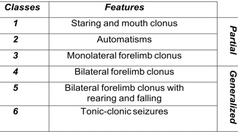
Pilocarpine model is considered one of the best models for severe human TLE. After administration of Pilo, rodents exhibit stereotypical behaviors that can be categorized into 6 classes according to Racine scale (Racine, 1972), recently revised (Veliskova et al., 2006). As schematized, it uses numbers from 1 to 6 to define seizure classes: the first three represent partial seizures, the last three the generalized ones. SRSs begin as
partial seizures and become secondary generalized (Table 2).
Classes Features
1 Staring and mouth clonus
P ar ti a l 2 Automatisms
3 Monolateral forelimb clonus
4 Bilateral forelimb clonus
G enera li ze d
5 Bilateral forelimb clonus with rearing and falling
6 Tonic-clonic seizures
Table 2: Classes of epileptic seizures according to Racine’s scale classification (Racine, 1972)
Some rodents develop prolonged severe seizures, named status epilepticus (SE), characterized by continuous head bobbing and body shake. SE can last from 2 hrs to 12 hrs. Following SE rodents have a latent seizure-free period, in which they then enter post-ictal coma, lasting 1-2 days, before showing spontaneous recurrent seizures (SRSs). The length of the latent period varies between animals and techniques utilized to detect seizures. By using video-EEG monitoring, a study in mice showed that the latent period is two weeks on average, spanning from 4 to 42 days after SE (Cavalheiro, 1997). A more recent study established the mean latent period in rats between 5 and 17 days, averaging one week (Goffin et al., 2007). During the seizure-free phase, brain networks rearrangements occur (Pitkänen and Sutula, 2002). Following the latent period, the recurrence of seizures become quite regular, usually clustered in a cycle peaking every 5-8 days or more with an higher frequency during the day (Arida et al., 1999). In 90% of the cases, EEG trace is characterized by a cerebral activity that starts from the hippocampus and spreads to the neocortex, usually lasting less than 60 seconds (Cavalheiro et al., 1991). Despite SE is routinely pharmacologically stopped by administration of anticonvulsant drugs such as diazepam and ketamine, only a percentage of animals (ranging from 60 to 70%) survive the treatment (Cavalheiro et al., 1991; Liu et al., 1994). During the very first days following Pilo-induced SE, treated animals might experience some occasional, self-limiting generalized seizures for 1-3 days (Mazzuferi et al., 2010).
1.2 NEUROGENESIS AND ASTROGLIOSIS
1.2.1 Neurogenesis
Neurogenesis is the process that gives rise to functional neurons from precursors. Although experimental studies put in evidence for many decades that neurogenesis occurs in the adult mammalian brain (Altman, 1962), the concept has become more widely accepted only upon introduction of the thymidine analog bromodeoxyuridine (BrdU) in these studies (Christie and Cameron, 2006; Gage, 2002). BrdU is incorporated into DNA during synthesis in the S-phase of the cell cycle, allowing the identification of cells within the cell cycle or their postmitotic progeny. The administration of BrdU, mostly conducted in rats or mice, have been extended also to humans, showing evidence of ongoing neurogenesis also in the brain of aged subjects (Eriksson et al., 1998). These results pointed out the attention on the opportunities offered from the modulation of neurogenesis on a clinical perspective view, from the repairing of neuronal loss due to aging, pathologies or injuries to the cell fate control of neural progenitors (Kozorovitskiy and Gould, 2003).
Neurogenesis takes place mostly during embryonic and perinatal stages of mammalian life. However, new neurons are born throughout life (at least under physiological conditions) into restricted two neurogenic regions: the subventricular zone (SVZ) lining the lateral ventricles and the subgranular zone of the dentate gyrus (DG) of the hippocampus (Gage, 2000). In humans and rodents, the hippocampus is located in the temporal lobes. Principal cells are confined to compacted layers called the dentate gyrus, composed of granule cells, and the CA regions composed of pyramidal neurons.
Even if neural stem cells (NSCs) both originate from SVZ and DG, distinct progenies arise from these two niches: cells generated in the SVZ migrate toward the olfactory bulb to differentiate into distinct types of olfactory neurons [with the majority being GABAergic or dopaminergic neurons and only a few glutamatergic cells (Nissant and Pallotto, 2011)] whereas in the DG, newborn neurons don’t migrate long distances and differentiate into one neuronal subtype, excitatory glutamatergic granule neurons (Young et al., 2011). It’s still unknown if this fate divergence is intrinsically predetermined or is due to external signals. Gene expression and protein profiles of NSCs isolated from distinct subregions
within the neurogenic areas indicate strong similarities among NSCs within (e.g. isolated from dorsal and ventral SVZ) and between neurogenic regions (e.g. comparing SVZ and DG). Such profiles could then be compared with the profiles of their progeny at different maturation stages. However, this analysis is incomplete because we still lack of reliable markers that are specific for different stages of neuronal differentiation; the absence of such markers continues to represent one of the major limitations in the field of adult neurogenesis (Suhonen et al., 1996).
To assess the role of extrinsic cues towards fate determination, several studies involving transplantation of NSCs from one region into another have been made, showing as newborn cells may change their “natural” fate when grafted in a different region, indicating a strong influence of extrinsic cues on fate determination (Suhonen et al., 1996). But these results are controversial since other experiments put in evidence how the fate of NSC progeny is critically affected by intrinsic cues (i.e. NSCs isolated along the dorsoventral axis of the SVZ generate cells retaining their original site-specific behavior even after heterotopic transplantation (Merkle et al., 2007). Certainly additional studies are required to analyze the instructive role of the microenvironment and the cell autonomous vs. cell non-autonomous determination of NSCs.
We can distinguish three layers in the dentate gyrus: the molecular layer is the closest to the cortex, mainly formed by dendrites and axons; just below there is a structure forming a characteristic “U” shaped structure named granule cell layer, formed by the bodies of the granule neurons; underneath the granule cell layer lays the hilus, composed of the axons of the granule cells and interneurons. Adult newborn neurons of the hippocampus develop from stem cells located in the subgranular zone lining the granule cell layer (Kriegstein & Alvarez-Buylla, 2009). Here the multipotent neural stem cells undergo intermittent cycles of division that originate proliferating precursor cells with a progressive limited renewal potential, which then differentiate into various lineages.
The primary multipotent stem cells of the neurogenic niche, called type 1 cells (or radial glia cells), are slowly dividing but they have an unlimited self-renewal capacity and are positive for some neural stem cell markers such as Nestin or Sox2 (Seri et al., 2001). They project a single process that crosses the granule cell layer and ramifies into the molecular layer. Upon division, type 1 cells differentiate into type 2 cells (or non-radial precursor cells), which are more proliferative intermediate neuronal progenitor cells with only short processes (Suh et al., 2007). We can consider type 2 cells as the transition phase between multipotency and lineage specialization. Actually, these cells are further
subdivide into type 2a cells, mitotic and multipotent cells which still express some neural stem cell markers, and type 2b cells, lineage-committed proliferative neuronal precursors which start to express immature neuronal markers (i.e. doublecortin) (Steiner et al., 2006). Type 2b cells then lead to type 3 cells (or neuroblasts), which also can proliferate but don’t express anymore any stem cell markers and give rise to mature neurons (Filippov et al., 2003; Fukuda et al., 2003; Kempermann et al., 2004; Zhao et al., 2008). Every single stages is finely modulated by signals within the niche: recent experimental evidences have enlightened the critical importance of key molecules involved in the regulation of neurogenesis both during embryonic development and in adult brain. For example, signaling mediated by Bone morphogenetic protein (BMP) and Notch is essential to regulate the balance between quiescent and proliferative neural stem cells, as well as specific growth factors (i.e. Fibroblast Growth Factors or Brain Derived neurotrophic factor) may also play a pivotal role in regulating NSC behavior in the adult brain (Palmer et al., 1995; Ables et al., 2010; Lugert et al., 2010; Mira et al., 2010, Paradiso et al., 2009). However, the mechanism to which these pathways act together and integrate into a common endpoint is still unknown.
Similar to embryonic and early postnatal development, adult-born neurons are generated in excess: about half of them fail to survive and to integrate into pre-existing neural networks. The continuous production and elimination of cells in the DG vouch for the dynamic ability of the brain to remodel discrete networks throughout the entire lifespan (Aasebø et al., 2011). New neurons are specifically selected for integration, a process that seems to be activity-dependent (Kempermann et al., 1997; Tashiro et al., 2006). However, we still do not know which types of signals are necessary and sufficient to enhance the survival and integration of new neurons. Similarly, it remains unclear why new neurons show an increased excitability compared with mature neurons.
In the rat, neurogenesis occurs in the subgranular zone, that lies within the first 50–100 µm of the granule cell layer, and new neurons are thought to derive from radial glia which in turn divide into more differentiated cells that ultimately become dentate gyrus granule cells (Seri et al., 2004). Newly born granule cells can functionally integrate into hippocampal circuitry (Jessberger and Kempermann, 2003), resembling other granule cells both for morphology and for electrophysiological properties (van Praag et al., 2002). Even though promising new data show that newborn neurons generated into the DG can integrate into the hilus and CA3 region of hippocampus (Toni et al., 2007), further
function of new neurons in adult brain behavior.
1.2.3 Neurogenesis and epilepsy
Even though there is to date no proof for the relevance of adult neurogenesis in brain diseases, the finding that NSCs persist in the adult brain and constantly generate new neurons may represent a novel therapeutic target in a number of neuropsychiatric and neurological disorders. Furthermore, NSCs may not only be disease-relevant or a therapeutic target in the two neurogenic areas: under certain conditions (such as ischemic stroke), it has been shown that new neurons are generated within normally non-neurogenic areas such as the striatum (Arvidsson et al., 2002).
The proliferation and integration of newborn cells in adult brain is highly sensitive to environmental stimuli (Kempermann et al., 1997) and, in particular, can be strongly affected by physiopathological events. For instance, neurogenesis drastically increases upon induced epileptic seizures in rodents (Parent et al., 1997; Scharfman et al., 2000). However, increasing evidence suggests that aberrant neurogenesis might concur for abnormalities both in human and experimental model of TLE, such as mossy fiber sprouting, dentate gyrus cells (DGCs) layer dispersion, and the appearance of DGCs in ectopic locations or with abnormal hilar basal dendrites (Kron et al., 2010). In contrast, other work suggests that newborn DGCs that integrate normally during epileptogenesis may serve a compensatory role to restore inhibition (Jakubs et al., 2006).
Experimental evidences on animal model of epilepsy showed that the most affected neural stem cell population by kainic acid-evoked seizures are cell type 1, 2a and 3; the administration of proconvulsant drugs wreaks drastic effects on adult neurogenesis and on morphology and localization of the newborn cells, with dispersion of granule cell layer (Steiner et al., 2008, Jessberger et al., 2005; Parent and Murphy, 2008; Scharfman et al., 2000, Steiner et al., 2008).
Interestingly, some attempts addressed to induce neuron production in neocortical areas or to reprogram glial cells into neurons have been successful carried out (Magavi et al., 2000; Heinrich et al., 2010).
Regarding epilepsy, the increased generation of new functional neurons to replace the neuronal loss due to epileptic seizures may exert a therapeutic effect, even though the achievement of this goal appears much more difficult than previously anticipated (Scharfman, 2004).
As discussed before, the dentate gyrus is the primary site in the temporal lobe where the majority of neurogenesis is thought to occur in normal adult brain, and the temporal lobe is the most epileptogenic region of the brain. The proliferation rate of neuronal progenitor cells is not steady but is modifiable by environmental and pathological conditions. In particular, neuronal activity exerts a strong influence on proliferation rate: prolonged neuronal depolarizations or repetitive discharge significantly increases the neurogenesis rate in the dentate gyrus, highlighting the strict relation between seizure activity and increased neurogenesis (Bengzon et al., 1997). Indeed, a bilateral increase of neurogenesis has been demonstrated in rodents following status epilepticus evoked by administration of chemoconvulsivant such as pilocarpine (Parent et al., 1997) or kainic acid (Gray and Sundstrom, 1998), but even after amygdala kindling (Scott et al., 1998) or electroconvulsive shock (Madsen et al., 2000). Seizures can also modify the survival of new neurons in a severity-dependent manner (Ekdahl et al., 2001).
1.2.4 Astrocytes and epilepsy
Astrocytes have a prominent role to ensure the central nervous system to work properly by regulation of critical transmitter-signaling pathways including c–aminobutyric acid (GABA), adenosine, and glutamate through regulation of extracellular neurotransmitter levels responsible for fast excitatory and inhibitory signaling in the central nervous system. Consequently, these cells have the potential to modulate synaptic transmission, neuronal excitability, and the generation of ictal discharges. Failing to maintain the right extracellular concentration of these amino acids may results in ruinous effects on neuronal survival and functionality (Rothstein et al., 1996). Although it is commonly accepted that the main cause of epilepsy is an over-excitation of a brain specific neuronal population, the primary involvement of astrocytes in causation of seizure activity becomes undeniable. Astrocytosis and microglial activation are indeed well described features of temporal lobe epilepsy (TLE), confirmed by several studies that suggest their decisive contribution to epileptogenesis (Briellmann et al., 2002; Vessal et al., 2005; Kang et al., 2006; Binder et al., 2006). Upon an epileptogenic insult occur, hypertrophied astrocytes in the dentate gyrus form an ectopic glial scaffold that promotes the aberrant growth of basal dendrites into the hilus (Shapiro et al., 2006). These basal dendrites are targeted for synaptogenesis by mossy fibers (Ribak et al., 2000) and contribute to a recurrent excitatory circuit that may facilitate seizures (Austin and Buckmaster, 2004).
Astrocytes are also involved in metabolism and regulation of neurotransmitter levels. Synaptically released glutamate is normally taken up by glial cells and converted in non-toxic glutamine, essential for neurons as a renewable source of neurotransmitter (Meldrum et al., 1999), by glial-specific enzyme glutamine synthetase (Rothstein et al., 1996). As found in the hippocampus of patients with mesial temporal sclerosis, the expression of the astrocyte-specific enzyme glutamine synthetase (GS) is strongly decreased, leading to a misregulation of glutamate/glutamine rate. GS converts glutamate to glutamine, an essential amino acid supplied to neurons as a renewable source of neurotransmitter (such as the chief inhibitory neurotransmitter GABA). Brain-slice studies showed that selective reactive astrocytosis and the loss of GS leads to a reduction of synaptic inhibition (Ortinski et al., 2010), raising the possibility that astrocytes can contribute significantly to the genesis of epilepsy. Astrocyte and microglial activation are well-described features of temporal lobe epilepsy (TLE), and studies have suggested that glial cells may contribute to epileptogenesis (Briellmann et al., 2002; Vessal et al., 2005; Kang et al., 2006; Binder et al., 2006). Other studies have shown that after seizures, hypertrophied astrocytes in the dentate gyrus form an ectopic glial scaffold that promotes the aberrant growth of basal dendrites into the hilus (Shapiro et al., 2006). These basal dendrites are targeted for synaptogenesis by mossy fibers (Ribak et al., 2000) and contribute to a recurrent excitatory circuit that may facilitate seizures (Austin and Buckmaster, 2004).
Postmortem studies of patients with temporal lobe epilepsy showed that loss of glutamine synthetase is accompanied by reactive astrocytosis with alteration of protein expression that eventually leads to a reduced synaptic inhibition and increased spread of excitation (Ortinski et al., 2010). When taken together with the observation that astrocytes release chemical transmitters, the idea that glial cells might contribute to the generation of seizures has got a foothold (Wetherington et al., 2008). There are no unique mechanisms for the contribution of astrocytes to epileptogenesis, since they can trigger the neuronal hyperactivity in previously normal neurons or just promote the epileptic discharge in abnormal neurons or just fail to arrest neuronal hyperactivity (Castiglioni et al., 1990). Significant increases of astrocytosis have been detected in several brain foci related with seizure generation, leading to the concept that gliotic scar formation is a key feature of human epilepsy and, therefore, suggesting a prominent role for glia in epileptogenesis (Harris, 1975; Mazzuferri et al., 2010; Bovolenta et al., 2010).
GABA, astrocytes regulate the extracellular concentration of adenosine, a powerful endogenous anticonvulsant. An increase in the expression of adenosine kinase, the enzyme involved in the conversion of adenosine to AMP, has been registered in presence of reactive astrocytes (Boison, 2008); thereupon, a reduction in this endogenous anti-seizure is strictly related to reactive astrocytosis.
Moreover, astrocytes are involved in modulation of neuronal N-methyl-D-aspartate (NMDA) receptors expression, contributing to their excitation through the release of glutamate and D-serine. Astrocytic Ca2+ signals, which are dampened by some anticonvulsants, stimulate the release of glial glutamate, leading to neuronal excitation (Tian et al., 2005). Considering the decrease in adenosine- and GABA-dependent inhibition that happen during reactive astrocytosis, becomes easy to conceive how these events combination might trigger epileptic seizures.
1.3 HERPES VIRUSES
1.3.1 Herpersviridae classification
The Herpesviridae family is a group of enveloped viruses highly dispersed in most animal species.
Members of this family with human tropism are classified in 8 different subtypes:
Herpes simplex virus Type-1 (HSV-1), Herpes simplex virus type 2 (HSV-2), Varicella zoster virus (VZV of HHV-3), Epstein-Barr virus (EBV or HHV-4), Human cytomegalovirus (HCMV or HHV-5), Human herpesvirus 6 (HHV-6), Human herpesvirus 7 (HHV-7), Human herpesvirus 8 (HHV-8), Kaposi’s sarcoma-associated herpesvirus (KSHV) (P. E. Pellett and B. Roizman, 2007).
All these subtypes share distinct biological properties: firstly, their genomes encode a large group of enzymes which participate in nucleic acid metabolism (such as thymidine kinase), DNA synthesis (i.e. DNA polymerase and helicase) and protein modification (i.e. protein kinase); secondly, viral DNA replication and capsid assembly take place in the nucleus, whereas the tegument association and envelope acquisition occur in the cytoplasm, as viruses exit the host cell; finally, infectious viruses can either go through a lytic lifecycle accompanied by destruction of the host cell or establish a latent state in the host cell. The latent genome can be reactivated by various stimuli, entering the lytic life cycle and causing disease upon reactivation. Humans are readily infected by these viruses and often are positive for five or more of these viruses in their lifetime. On the basis of host-cell range, length of replication cycle, cell type where latency is established and genomic analysis, Herpesviruses have been classified into three subfamilies: Alphaherpesviruses, Betaherpesviruses and Gammaherpesviruses (table 3).
FAMILY NAME AND CYTOPATHOLOGY GROWTH CYCLE INFECTION LATENT Alpha-herpesviruses HSV-1 HSV-2 VZV Short, Cytolytic Neurons Beta-herpesviruses CMV HHV-6 HHV-7 Long, Cytomegalic Long, Lymphoproliferative Glands, Kidneys, Lymphoid tissue Gamma- herpesviruses EBV
HHV-8 Lymphoproliferative Variable, Lymphoid tissue
Members of the Alphaherpesvirus subfamily are characterized by a broad host range, relatively short lytic lifecycle, rapid spread in culture, and efficient destruction of infected cells. In addition, they encode a similar set of homologous genes arranged in similar order and can efficiently invade the peripheral nervous system moving from the infected epithelial cells to infect neurons resident in the dorsal root ganglion or cervical ganglion via retrograde transport of viral capsids (Frampton et al., 2005). The virus will persists in a latent state in the nervous systems of the host for a lifetime where the viral genome remains in an epichromosomal state associated with histones without integrating into the host genome (Steiner and Kennedy, 1993).
The Betaherpevirinae have a narrow host range and long replication cycle in infected cells. They are able to establish latency in secretory glands, lymphoreticular cells, kidneys and other tissues.
The Gammaherpevirinae generally replicate in lymphoblastoid cells, especially in T or B lymphocytes, and also establish latency in lymphoid tissues.
1.3.2 HSV-1 overview
Viral particles mediate the transfer of the viral genome and accessory proteins from an infected host cell through the production of progeny virus or from cell-to-cell spread to a noninfected host cell. Since viruses with a DNA genome must find a way to get the DNA to the nucleus, viruses use a basic strategy in which the infected cell assists the virus. To infect a target cell, a virus particle proceeds through a multistep entry process, tightly regulated in time and space. In general, this mechanism occurs via a universal set of steps involving attachment, stable binding, and fusion.
The HSV virion is designed to protect the viral genome from adverse conditions in the extracellular environment and to permit cell invasion so that the viral genome can be released to the cell nucleus to efficiently express its genes.
The Herpes Simplex Virus Type-1 virion is composed of four main elements: a linear double-stranded DNA encompassed in an electron-opaque core, an icosahedral capsid, a large proteinacous space referred as the tegument, and an envelope with glycoprotein spikes at the outermost layer of the virion (Fig.1).
Fig. 1: Electron microscroscopy picture of HSV-1 virus
1.3.3 HSV-1 DNA genome
The genome of HSV-1 in a mature virion is a large linear and double stranded DNA (152 kilo base pair, of which 68% are G/C), and it carries at least 84 protein-encoding open reading frames (ORF) organized into a 126 Kb unique long segment (UL) and 26 Kb unique short segment (US), tightly packed in a linear form that become circularized after the virus reach the nucleus (McGeoch et al., 1988). These regions are flanked by inverted repeat sequences, a terminal repeat termed TRL and an internal repeat termed IRL (Perry, 1988) containing sequences required for cleavage and packaging of the HSV-1 genome, termed “a” sites (Deiss, 1986). The US also is flanked by inverted repeats termed TRS and IRS (Fig. 2).
It has been shown that the L and S units can invert relative to each other and the DNA genome produces four linear isomers of equal proportion in infected cells (Hayward et al., 1975). Moreover, the HSV-1 genome has three lytic origins of replication, two located within the US segment (oriS) and one in the UL segment (oriL).
The HSV-1 replication is strictly regulated in a sequential manner: three gene classes, known as the immediate early genes (α-genes), the early genes (β-genes), and the late genes (γ-genes), are coordinately expressed following the transcriptional regulation of viral and host cell proteins (Watson et al., 1981).
The α genes don’t need any viral protein synthesis for their expression but they just requires a cis-acting site (alpha TIC; with the consensus
5'-GyATGnTAATGArATTCyTTGnGGG-3') located in the promoter-regulatory domains of the alpha genes; the products of these genes are involved in the transcriptional regulation of β- and γ-genes and also can operate a positive and negative feedback regulation of α-genes (O’Hare and Hayward, 1985) though the mechanisms regulating these processes are not completely understood.
1.3.4 HSV-1 nucleocapsid
The HSV genome is packed within an icosahedral protein shell, called nucleocapsid or simply capsid, which total molecular mass is 0.2 billion daltons. The nucleocapsid displays 162 capsomers made up of four viral proteins: VP5, VP26, VP23, and VP19C (Zhou et al., 2000). Within the envelope, the nucleocapsid takes up about one third of the volume, while the tegument occupies the remaining rest two thirds of the volume. Interestingly, the encapsidation and release of viral DNA take place through a portal located within the capsid, made up by a dodecamer of the pUL6 protein (Cardone et al. 2007).
1.3.5 HSV-1 tegument
The capsid is coated with an amorphous proteinaceous filled space designated as the tegument, particularly important since is involved in transcriptional regulation of immediate early viral genes and in regulation of host-cell transcription (virion-host-shutoff protein) (Dargan et al., 1998). The tegument contains at least 20 viral proteins; some of these, such as VP16, are responsible for triggering the viral immediate early gene
expression (Campbell et al., 1984). Others contribute to creating a more suitable environment for viral replication in infected cells, such the virion-associated host shutoff protein (vhs) which degrades cellular mRNA to enhance the efficiency of viral protein translation (Kwong and Frenkel, 1989) and the protein encoded by US11, an RNA binding protein which inhibits the activation of protein kinase R inducing cellular translation arrest in host cells (McKnight et al., 1994). The tegument also includes the VP22 protein, that works like a stabilizer for important viral proteins such as gE, gD and ICP0 (Duffy et al., 2009) and is involved in viral spread during lytic infection.
1.3.6 HSV-1 envelope
Through the electron microscope, HSV-1 virions are visualized as pleiomorphic membrane-bound particles surrounded by an envelope obtained from the cytoplasmic membranes of previously infected cells. the average diameter of a spherical HSV-1 virion is 186 nm, which enlarges to 225 nm once spikes on the envelope surface are included. The lipid layer of the envelope is seen as a continuous silkily circular surface, around 5 nm thick (Grunewald et al., 2003).
The envelope consists of a trilaminar lipid membrane containing multiple viral membrane proteins with 9 different glycoproteins embedded on its surface: gB, gC, gD, gE, gG, gH, gI, gL and gM. Among these, only gB, gC, gD, gH, and gL are important for cellular attachment, fusion, and internalization of the virus (Cai, et al., 1988).
About 600-700 glycoprotein spikes have been counted on the surface of the envelope (Grunewald K, et al., 2003), non-randomly distributed since they are presented thinly at the proximal pole and compactly around the distal pole (Spear et al., 2000).
1.3.7 HSV-1 infection
HSV-1 enters host cells through a fusion event of the virion envelope with the plasma membrane. Fusion activity is triggered by receptor binding at neutral pH, inducing irreversible conformational changes that allows for viral fusion (Wittels and Spear, 1991). The virus can also spread from cell to cell by inducing the fusion of cellular membranes. Viral entry into cells requires at least four viral glycoproteins: gD, gB and the heterodimer gH-gL. Initially, gC (WuDunn et al., 1989) and gB (Herold et al., 1994) interact independently with glycosaminoglycan (GAG) moieties of cell surface proteoglycans to promote the attachment of the virion to the host cell. Following the attachment of the