UNIVERSITY OF CATANIA
DEPARTMENT OF CHEMICAL SCIENCES
INTERNATIONAL PhD IN CHEMICAL SCIENCES – XXXI CYCLE
NOEMI BELLASSAI
Surface Plasmon Resonance Imaging
Biosensors for Cancer Diagnosis: Detection of
Circulating Tumor DNA
PhD Thesis
Tutor:
PhD Coordinator:
Abstract
This doctoral thesis focused on the realization of Surface Plasmon Resonance Imaging (SPRI) biosensor for the rapid, simple and label-free detection of single point mutations in the KRAS gene, standard actionable cancer biomarkers for colorectal cancer, in human plasma samples. Initially, the SPRI assay included the immobilization of specific peptide nucleic acid (PNA) probes onto the gold sensor to ensure the hybridization reaction of PNA-DNA complexes. The spatially controlled immobilization of PNA probes has been obtained by injecting PNA wild-type and PNA mutated solutions into a microfluidic system coupled to SPR sensor chip. The extremely low concentration of genomic DNA required an improvement of SPRI detection capabilities, by using functionalized gold nanoparticles to amplify the hybridization signal between target analytes and corresponding PNA probes. Three representative single-point mutations, gDNA G12D, G12V and G13D, have been successfully detected.
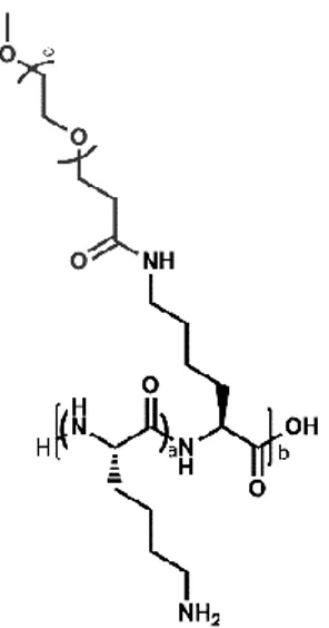
After preliminary results of nanoparticle-enhanced SPRI assay, a mixed-charge polymer based on Poly-L-lysine (PLL) polypeptide backbone modified with an anionic peptide, connected via a nonionic OEG spacer, has been synthesized in order to achieve control over the charge distribution of PLL-coated surfaces, and thus the antifouling property. The PLL backbone has been functionalized with different percentages (y%) of maleimide-OEG-NHS ester chains (PLL-mal(y%), from 13% to 26%), and the anionic oligopeptide CEEEEE, composed of one cysteine (C) and five glutamic acids (E), with a short sequence to limit the thickness of the mixed-charge polymer antifouling coating, has been attached to the maleimide units through the thiol–maleimide Michael-type addition. The grafting density has been varied to tune the balance of charged groups at polymer backbone. PLL-mal(y%)-CEEEEE surfaces have been characterized by water contact angle and polarization modulation infrared reflection-absorption spectroscopy (PM-IRRAS). Complementary acoustic (quartz crystal microbalance with dissipation, QCM-D) and plasmonic (surface plasmon resonance imaging, SPRI) techniques have been employed to monitor the adsorption of bovine serum albumin (BSA), used as standard protein solution, and diluted human plasma samples. Hence, a new nanoparticle-enhanced SPRI assay for circulating tumour DNA (ctDNA) detection in human plasma samples using PLL-mal(y%)-CEEEEE layer as the antifouling coating has been devised. The PNA probes and the anionic peptide have been attached to the maleimide units through the thiol–maleimide reaction using a microfluidic system
Abstract
II
coupled to SPR sensor chip. The analysis of ctDNA G12D target in diluted human plasma samples (5 pg μL-1), collected from cancer patients and healthy donors, has been carried out using the conjugated AuNPs system, with a minimal sampling handling to avoid any contamination and disruption of the antifouling activity of PLL-mal(y%)-CEEEEE layer. The combined use of PLL-mal(y%)-CEEEEE as the antifouling layer with functionalized gold nanoparticles for the amplification of target detection overcomes the limiting factors related to the biosensor in the clinical field and offers an excellent ctDNA discrimination in the bloodstream at attomolar level.
Abstract
Questo lavoro di tesi di dottorato si è concentrato sulla realizzazione di un biosensore per la rivelazione di mutazioni puntiformi della sequenza di DNA del gene KRAS, considerato un biomarcatore standard per il cancro al colon-retto, direttamente in campioni di plasma umano applicando una procedura rapida, semplice e senza l’uso di marcatori molecolari mediante la tecnica Surface Plasmon Resonance Imaging (SPRI). Inizialmente, il saggio SPRI ha previsto un primo step di immobilizzazione di sonde molecolari ad acido peptidonucleico (PNA) sul sensore di oro per garantire la reazione di ibridizzazione (complessazione) tra le molecole PNA-DNA. È stata ottenuta un’immobilizzazione delle sonde iniettando le soluzioni di PNA wild-type e PNA mutato spazialmente controllata mediante l’uso di un sistema microfluidico accoppiato al sensore SPR. La concentrazione estremamente bassa del DNA tumorale circolante nei fluidi biologici ha richiesto un aumento della sensibilità per la rivelazione di questo target con l’analisi SPRI, ottenuto mediante l’utilizzo di nanoparticelle di oro funzionalizzate per amplificare il segnale di ibridizzazione tra gli analiti target e le sonde PNA corrispondenti. In un primo momento, il saggio SPRI è stato testato in soluzione acquosa rivelando con successo tre mutazioni puntiformi del gene KRAS, gDNA G12D, G12V e G13D.
Dopo i primi risultati ottenuti con il saggio SPRI utilizzando le nanoparticelle di oro, è stato sintetizzato un polimero a carica mista costituito da Poli-L-lisina (PLL), un polimero con struttura cationica polipeptidica modificato con un oligopeptide a carica negativa legato al PLL mediante un gruppo oligo etilenglicole spaziatore, in modo da avere un maggior controllo sulla distribuzione di carica e quindi sulla proprietà antifouling della superficie funzionalizzata. Il polimero PLL è stato modificato con differenti percentuali (y%) di catene di maleimide-OEG-NHS estere e con un oligopeptide anionico CEEEEE, formato da un residuo cisteinico (C) e cinque acidi glutammici (E) con una lunghezza di sequenza piuttosto corta per minimizzare lo spessore del polimero antifouling sul sensore SPR. Il peptide CEEEEE è stato legato covalentemente alle unità maleimmidiche mediante la reazione di Michael tiolo-maleimmide. È stato investigato un ampio intervallo di densità di grafting (PLL-mal(y%), dal 13% al 26%) per regolare il bilancio di carica dei gruppi funzionali presenti nella struttura polimerica.
Le superfici funzionalizzate con PLL-mal(y%)-CEEEEE sono state caratterizzate mediante misure di angolo di contatto e di spettroscopia di assorbimento della riflessione nell'infrarosso con modulazione della polarizzazione (PM-IRRAS). Per monitorare l’assorbimento aspecifico delle proteine sulla superficie trattata con il
Abstract
IV
polimero antifouling, sono state utilizzate due tecniche complementari, quali la microbilancia a cristalli di quarzo con dissipazione (QCM-D) e la risonanza plasmonica di superficie ad immagini (SPRI) valutando l’interazione aspecifica sia dell’albumina siero bovina (BSA), usata come proteina standard, sia di campioni di plasma umano a diverse concentrazioni.
Infine, il saggio SPRI per la rivelazione di frammenti di DNA circolante con mutazioni puntiformi legate al tumore (ctDNA G12D) è stato ottimizzato combinando l’uso delle nanoparticelle di oro con un sensore pretrattato con il polimero PLL-mal(y%)-CEEEEE per assicurare le proprietà antifouling e quindi la migliore discriminazione dell’analita di interesse direttamente nel plasma umano. Le sonde PNA e il peptide CEEEEE sono stati legati covalentemente alle unità maleimmidiche mediante la reazione di Michael tiolo-maleimmide sfruttando un sistema microfluidico accoppiato al sensore SPR. L’analisi del target ctDNA G12D (5 pg μL-1) in campioni di plasma umano al 10%, provenienti sia da pazienti affetti dal tumore che da donatori sani, è stata eseguita attraverso un trattamento minimo del campione biologico, in modo da evitare qualunque tipo di contaminazione del campione e alterazione dell’attività antifouling del film PLL-mal(y%)-CEEEEE, utilizzando nanoparticelle di oro funzionalizzate per l’amplificazione del segnale SPR a seguito della reazione di ibridizzazione PNA-DNA. L’uso combinato del PLL-mal(y%)-CEEEEE, come polimero antifouling con nanoparticelle di oro funzionalizzate per la rivelazione del target di interesse supera i fattori limitanti legati all’applicazione dei biosensori in ambito clinico e diagnostico ed offre un’eccellente discriminazione per il DNA tumorale circolante nel flusso sanguigno con una sensibilità dell’ordine dell’attomolare.
Acknowledgements
The completion of this doctoral dissertation was possible with the support of several people. I would like to express my sincere gratitude to all of them.
Firstly, I am grateful to my supervisor Prof. Giuseppe Spoto for the continuous support of my PhD study and related research, for his patience, motivation, and immense knowledge. Prof. Spoto has always made himself available to clarify my doubts despite his busy schedules, and I personally consider it a great privilege to have attended my doctoral programme under his guidance and to have learnt from his research expertise.
My sincere thanks also go to Prof. dr. ir. Jurriaan Huskens, Professor of Molecular Nanofabrication group at the University of Twente in the Netherlands, who provided me with the opportunity to join his team as intern, and who gave access to the laboratory and research facilities. Without his precious support, it would not have been possible to conduct part of this PhD project. I have been delighted to have made his acquaintance, and his help supported me all the time of research and writing of this thesis.
Besides, I would like to thank Dr Roberta D’Agata for her insightful comments and encouragement which urged me to widen my research from various perspectives. With her experience and her love for the scientific research, she passed down to me the desire to persevere in my project during the doctoral studies.
A very special gratitude goes out to Prof. Carmelo Sgarlata for his valuable guidance, personal and scholarly inputs and consistent encouragement I received throughout all of my PhD experience.
I would like to thank the Horizon 2020 Health project “ULTRAPLACAD” (n. 633937) for financial support, and the Erasmus plus programme for the research stay at the University of Twente.
Acknowledgements
VI
I thank my lab mates and friends in the University of Twente, Daniele Di Iorio and Jacopo Movilli, for the stimulating discussions, for their support during the research work at the University, and for all the fun we had during the internship in The Netherlands.
Also, I thank my colleagues and friends at the University of Catania Rossella Migliore, Dr Giusy Grasso, Dr Valentina Oliveri, Vanessa Jungbluth, Dr Valentina Giglio, Dr Maria Chiara Giuffrida and Mrs Tosto (the PhD secretary), who have all extended their support in a very special way. I gained a lot from their personal and scholarly interactions, their suggestions on my research programme.
Last but not least, I would like to thank my parents because I owe it all to them. My mother, my life-coach, has always been keen to know what I was doing and how I was proceeding, although it is likely that she has not always grasped deeply what it was all about! She always joined with me whenever a significant momentous was reached but, obviously, she has not ever missed the opportunity to remind me never to neglect my health and my happiness before the academic job.
My father has always encouraged me in difficult situations during these years by passing me down his calm and patience. Many Thanks!
I am also grateful to my other family members and friends who have supported me along the way.
Ringraziamenti
Vorrei esprimere la mia più sincera gratitudine a tutti coloro che mi hanno sostenuto nella realizzazione di questa tesi di dottorato.
In primo luogo, vorrei ringraziare il mio tutor Prof. Giuseppe Spoto per aver continuamente incoraggiato i miei studi e le mie ricerche scientifiche all’interno del programma di dottorato, per la sua pazienza, motivazione e immensa conoscenza. Il Prof. Spoto si è sempre messo a disposizione per chiarire i miei dubbi nonostante i suoi impegni e, personalmente, considero un grande privilegio aver frequentato il mio programma di dottorato sotto la sua supervisione e aver imparato molto dalla sua esperienza di ricerca scientifica.
Un sincero ringraziamento va al Prof. dr. ir. Jurriaan Huskens, professore del gruppo di Molecular Nanofabrication presso l'Università di Twente nei Paesi Bassi, che mi ha dato l'opportunità di unirmi al suo team come studente con libero accesso al suo laboratorio e alle strutture di ricerca. Senza il suo prezioso supporto non sarebbe stata possibile la realizzazione di parte di questo progetto di dottorato. Per me è stato un onore aver fatto la sua conoscenza e il suo prezioso aiuto mi ha accompagnato per tutto il tempo della ricerca e di scrittura di questa tesi.
Inoltre, vorrei ringraziare la dott.ssa Roberta D'Agata per i suoi perspicaci commenti ed incoraggiamenti che mi hanno spinto ad ampliare i miei studi in diversi ambiti. Con la sua esperienza e il suo amore per la ricerca scientifica, mi ha trasmesso il desiderio di perseverare nel mio progetto di ricerca.
Una speciale ringraziamento va al Prof. Carmelo Sgarlata per la sua preziosa guida, per gli input personali e accademici e per il costante incoraggiamento che ho ricevuto durante tutta la mia esperienza di dottorato.
Desidero ringraziare il progetto Horizon 2020 “ULTRAPLACAD” (n. 633937) per il supporto finanziario e il programma Erasmus plus per il periodo di ricerca eseguito presso l'Università di Twente.
Ringraziamenti
VIII
Ringrazio i miei colleghi di laboratorio e amici dell'Università di Twente, Daniele Di Iorio e Jacopo Movilli, per le discussioni stimolanti, per il loro sostegno durante il lavoro di ricerca presso l'Università e per tutte le divertenti esperienze che abbiamo vissuto durante lo stage in Olanda.
Ringrazio anche le mie colleghe e amiche dell'Università di Catania, Rossella Migliore, la dott.ssa Giusy Grasso, la dott.ssa Valentina Oliveri, Vanessa Jungbluth, la dott.ssa Valentina Giglio, la dott.ssa Maria Chiara Giuffrida e la signora Tosto (la segretaria del dottorato), che mi hanno mostrato il loro sincero sostegno in modo molto speciale. Ho ricevuto molto da loro grazie ai continui confronti, sia personali che lavorativi, e ai loro costruttivi suggerimenti sul mio programma di ricerca.
Infine, ma non meno importante, vorrei ringraziare i miei genitori perché devo tutto a loro.
Mia madre, il mio maestro di vita, ha sempre voluto sapere cosa stavo facendo e come stavo procedendo, anche se probabilmente non sempre capiva fino in fondo di cosa mi stavo occupando! Ha sempre gioito con me ogni volta che è stato raggiunto un obiettivo importante, ma ovviamente non ha mai perso l'occasione di ricordarmi di non trascurare mai la mia salute e la mia felicità davanti al mio lavoro accademico.
Mio padre mi ha sempre incoraggiato in situazioni difficili durante questi anni trasmettendomi la sua calma e la sua pazienza. Grazie infinite!
Vorrei ringraziare tutti i miei familiari e amici che mi hanno sostenuto lungo la strada.
Table of Contents
Abstract ... I Acknowledgements ... V Ringraziamenti ... VII Introduction ... XIII Chapter I... 17I. Biosensors for Medical Diagnostics ... 19
I.2. Biosensors ... 21
I.2.1.Definition and Classification... 21
I.3. Surface Plasmon Resonance (SPR) ... 26
I.3.1.Surface Plasmon Resonance Imaging (SPRI) ... 30
I.3.2.Applications of SPR-based Biosensors ... 32
I.4. Quartz Crystal Microbalance (QCM) ... 35
I.4.1.Quartz Crystal Microbalance with Dissipation (QCM-D) ... 37
I.4.2.Applying QCM-D technique to Biomolecular Studies ... 38
I.4.3.An example of QCM-D application: protein adsorption ... 39
Chapter II ... 41
II. An Overview of Liquid Biopsy in Cancer Diagnosis ... 43
II.2. The Origins of ccfDNA and ctDNA ... 45
II.3. Approaches to ctDNA analysis ... 47
Table of Contents
X
Chapter III ... 57
III. Nonspecific Protein Adsorption ... 59
III.1.Protein – surface interaction ... 59
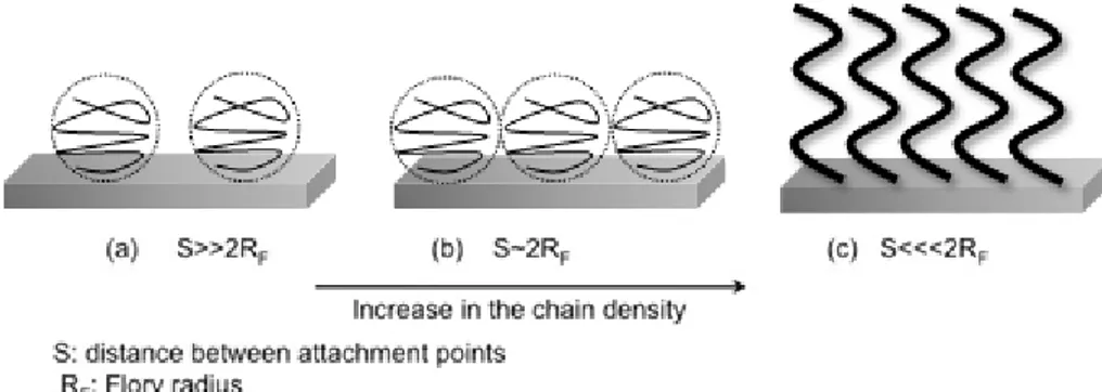
III.2. Mechanism of Antifouling Surfaces ... 63
III.2.1.Hydration ... 63
III.2.2.Steric hindrance ... 64
III.2.3.Surface charges ... 65
III.3. Antifouling Materials ... 67
III.3.1.HEMA – based systems ... 67
III.3.2.Polyethylene Glycol (PEG) polymer ... 68
III.3.3.Zwitterionic polymers ... 70
III.3.4.Special Zwitterionic Materials: Peptide and Peptoid ... 72
III.3.5.Hybrid, derivative and biomimetic materials ... 73
PhD Thesis Aim ... 77
Chapter IV ... 81
IV. Materials and Methods ... 83
IV.1.Materials and Reagents ... 83
IV.1.2.PNA probe synthesis and surface immobilization ... 83
IV.1.3.SPRI apparatus and measurements ... 84
IV.1.4.Synthesis and functionalization of gold nanoparticles ... 85
IV.1.5.Genomic DNA sample treatments ... 86
IV.1.6.Amplification of SPRI signals by functionalized gold nanoparticles ... 86
IV.2. Materials and Reagents ... 87
IV.2.1.Synthesis of oligopeptide CEEEEE ... 87
IV.2.2.Synthesis of poly-L-lysine-g-maleimide(y%) (PLL-mal(y%)) ... 88
Table of Contents
IV.2.4.Immobilization of PLL-mal(y%)-CEEEEE monolayer and antifouling
measurements by SPRI technique ... 89
IV.2.5.Immobilization of PLL-mal(y%)-CEEEEE and antifouling measurements by QCM-D technique ... 91
IV.3. Materials and Reagents ... 92
IV.3.1.PNA probe synthesis and PLL-mal(26%)-PNA surface immobilization... 92
IV.3.2.Genomic DNA sample treatments and discrimination ctDNA samples by functionalized gold nanoparticles ... 93
IV.3.3.Amplification of SPRI signals by functionalized gold nanoparticles ... 94
Chapter V ... 95
V. Results and Discussion – part I ... 97
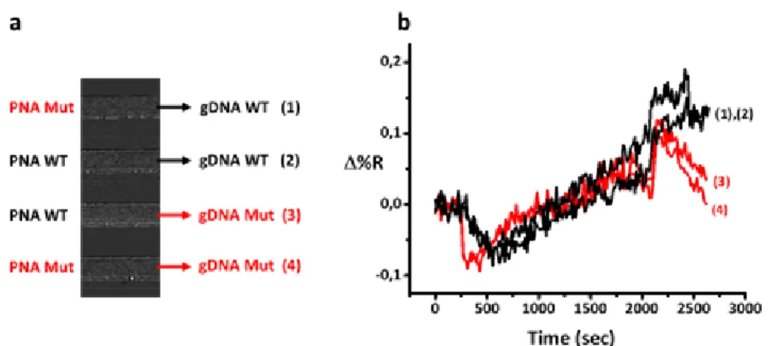
V.1. Development of nanoparticle-enhanced SPRI assay for gDNA KRAS mutations ... 97
V. Results and Discussion – part II ... 108
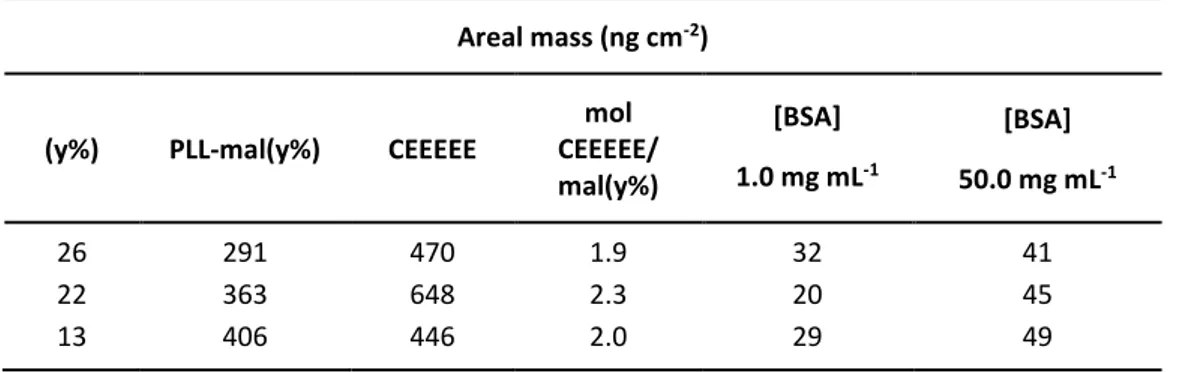
V.2. Antifouling layer based on peptide-PLL polymer ... 108
V. Results and Discussion – part III ... 119
V.3. Development of nanoparticle-enhanced SPRI assay for ctDNA mutations using PLL-mal(26%)-CEEEEE as antifouling layer ... 119
Chapter VI ... 129 VI. Conclusions ... 131 Chapter VII ... 133 VII. Bibliography ... 135 Publications ... 153 Supplementary Materials ... 157
Introduction
Nowadays, healthcare represents a crucial challenge for the scientific community around the world. According to the latest World Health Organization (WHO) assessments, cancer is the most frequent cause of mortality worldwide.1 Overall, there were 14.1 million new cases and 8.2 million deaths in 2012. The most commonly diagnosed cancers are lung (1.82 million), breast (1.67 million), and colorectal (1.36 million); while the most common causes of death are associated to lung cancer (1.6 million deaths), liver cancer (745,000 deaths), and stomach cancer (723,000 deaths), respectively.2
The capability to early diagnose a pathological condition and, at the same time, to improve the efficacy of therapeutic treatments is the main objective for biomedical research and pharmaceutical industries.
Nanotechnology promises an exceptional pathway for personalized medicine and for the enhancement of the person's health, through the development of molecular assay for the detection of specific genetic information, such as tumour-linked genetic alterations. Especially for cancer disorder, the real challenge of personalized medicine is to consider the evolution of the disease integrally by examining all primary tumour and metastatic sites, parting on the principle that tumours are heterogeneous at molecular level, including cellular morphology, gene expression, metabolism, motility, proliferation, and metastatic potential, and different samples of a tumour might yield different results. Heterogeneity can occur both between tumours of the same type in different patients (inter-tumour heterogeneity) as well as between cancer cells within a tumour (intra-tumour heterogeneity).3 The standard analysis based on tissue biopsy would underestimate the complexity of the genomic overview of the tumour, and it would provide discordant information about the efficacy and possible acquired resistance after pharmacological therapy.
Above all, optical biosensors have been profiled as reliable and efficient analytical tools offering rapid, simple and highly sensitive detection of disease-related molecular biomarkers.
In order to accelerate the technology-transfer process of biosensors to the medical field, considerable research efforts must be addressed to create and optimize the biofunctionalization and the assay procedures, which permit reliable and accurate detection of relevant biomarkers directly in biological fluids and, essentially, avoiding
Introduction
XIV
any pretreatment of the sample. The development of versatile and fully optimized biosensor methodologies might indicate a breakthrough for early cancer diagnosis and, thereby, for personalized medicine.
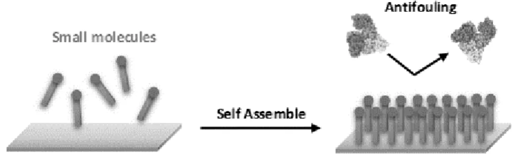
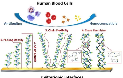
Biosensors and biomedical devices require antifouling surfaces to prevent the non-specific adhesion of proteins or cells, for example, when aiming to detect circulating cancer biomarkers in complex natural media (e.g., in blood plasma or serum). The study of protein–inorganic surface interactions is crucial to the rational design of new tools for biomaterials science, biosensing, nanobiotechnology and nanomedicine. Nonspecific adsorption of proteins, well-known as protein fouling effect, can affect surface properties of biomaterials and trigger the degradation of devices operating in contact with biological fluids. When a solid comes in contact with a body fluid such as blood, plasma or serum, a layer of proteins is formed on the surface of the solid material within a time frame of seconds to minutes. For sensors, this may cause functional device interference, possibly preventing the detection of biological targets available at low concentrations in complex media (down to the ng L-1 region, or a difference of nine orders of magnitude).4,5 This, unfortunately, may lead to the occurrence of “false positive” results due to the incorrect interpretation of the biosensor response as originating from an unaffected binding event.4,6 On the other hand, “false negative” results might also occur if analyte binding is prevented. Both these situations, understandably, create serious clinical dangers.4
To relieve this major technical difficulty to biosensor development in early cancer diagnosis using real clinical samples, more intensive research efforts have to be dedicated to engineer antifouling coatings capable of reducing or, ideally eliminate, non-specific adsorption of blood-borne proteins.4,5 It has become abundantly clear that the educated design and development of antifouling/biocompatible materials for bioanalytical and biomedical applications require a prior detailed understanding of how proteins adsorb onto surfaces.
From the abovementioned, this PhD Thesis has aimed at designing and evaluating an innovative biosensor methodology improving the performance of nanoplasmonic biosensors in the early clinical diagnosis of cancer disease. Specifically, the main objectives outlined for this work involve:
1) Assessment and optimization of biofunctionalization strategies that improve the analytical features of the assay in terms of selectivity and reproducibility; 2) Design of methodologies for prevention and minimization of protein fouling,
Introduction
3) Validation of the accuracy and reliability of the proposed strategies with the nanoplasmonic biosensors employing real clinical samples.
This research project was performed in collaboration with Prof. dr.ir. Jurriaan Huskens’ group, at the Molecular Nanofabrication group, University of Twente, Enschede (The Netherlands) as visiting Erasmus PhD student.
Chapter I
Biosensors for Medical Diagnostics
This chapter describes the role of biosensor devices as diagnostic tools in the clinical field. After a brief review of biosensor technologies, plasmonic and mechanical biosensors are defined, pointing out their advantages and limitations for the clinical practice. Special attention is given to the properties and requirements of the biorecognition layer to achieve optimal biosensor performance, especially SPR and QCM-D platforms, for biomarkers detection in early cancer diagnosis.I.
Biosensors for Medical Diagnostics
Diagnostics play a crucial role in medicine for the successful prevention and monitoring of disease progression. Modern medicine relies heavily on the biomolecular information for diagnosis process, which includes the detection and identification of the pathology, the definition of its burden and stage, and the choice of more suitable pharmacological treatment. Moreover, monitoring of the therapeutic response and continuous follow-up of pathologies or physiological states, during and after treatments, are critical aspects to improve patient’s health.
Nowadays, clinical diagnosis is based on highly sensitive and specific laboratory assays such as cell culture methods, polymerase chain reaction (PCR) or enzyme-linked immunosorbent assays (ELISA), which are broadly applicable to different classes of biomolecules and were designed to be highly efficient for processing relatively large numbers of samples. These conventional methods involve laborious, multi-step and time-consuming procedures such as 1) the complex sample protocol; 2) the slow turnaround time (days for DNA sequencing, hours for PCR-related approaches),7 3) relatively large volume input requirements (approximately 1 mL of biofluids),8 4) the introduction of potential sources of bias owing to sample contamination and PCR errors,9,10 5) relatively high costs per analysis. Additionally, these medical diagnostic technologies require fully equipped laboratories and specialized personnel to perform the analysis, curbing the accessibility of these techniques to large centres and often at the expense of lower speed of analysis.
More recently, microarray technology is offering a highly efficient option for simultaneous identification and determination of a broad range of biomolecules. Microarray substrate is composed of regular patterns of DNA sequences or proteins, attached to a solid support, able to bind complementary nucleotide sequences, to detect mutations or relevant protein biomarkers in a sample using fluorescence labels. Although this technology began in the 1980s, recent advances in nanomaterials and nanofabrication techniques improved the multiplexing capabilities as well as the detection sensitivity.11-14 Nanoarray technology proposes accurate, rapid and high-throughput screening, widely used to investigate and profile the fundamental causes of numerous human diseases and to design new therapeutic drugs.
Most promising alternative solutions for diagnosis or therapy monitoring of relevant diseases, such as allergy, celiac and diabetes diseases, neurological disorders or cancer, are expected from biosensor devices, which can offer rapid and reliable biomedical analysis,15 by employing low sample volumes with minimum pretreatment. Biosensors
Chapter I
20
would represent exceptional analytical tools for the effective clinical diagnosis as well as for better comprehension of the molecular mechanisms involved in the pathophysiology, by revealing new biomarkers useful for the evaluation of appropriate pharmaceutical treatments.
In this context, the analysis of tumor-linked genetic alterations has been used for diagnostic, prognostic and therapeutic purposes to obtain rapid and accurate cancer somatic mutation profiling. Different drugs have been used as interfering agents to slow down the proliferation of cancerous cells, and it has been demonstrated that their efficacy is related to the oncogenic mutation status of patients.16,17 The presence or absence of specific genomic alterations indicates if the pharmacological treatment will effectively limit the spread of cancer thus improving patient survival rate. A similar testing prior to treatment serves as a useful tool for precision medicine.18 The monitoring of oncogenic mutations is not only helpful before the therapy, but it is also relevant during and after the pharmaceutical treatment to assess the developed drug resistance and cancer recurrence.19-21
Initially, standard clinical protocols have been applied to evaluate the genetic tumour mutation status through the direct sampling of cancerous tissue with biopsy or surgical resection.22 However, that approach shows limiting factors such as its invasively, inherently clinical risks, potential surgical complications and high costs.21 Multiple or serial biopsies are often impractical and some tumors are not always available in the surgical section. Additionally, the analysis based on a single biopsy might increase the risk for seeding cancerous cells to new sites, and it would underestimate the complexity of the genomic overview of a tumour by providing discordant information about the efficacy and possible acquired resistance after pharmacological therapy.23
Biosensors for Medical Diagnostics
The molecular analysis would allow detecting de novo mutations which confer resistance, or mutations following the clinical treatment which may be sensitive to alternative targeted therapies. At the same time, the development of this molecular assay would enhance the monitoring of cancer patients during specific treatment, as well as the therapeutic resistance of tumour cell clones.
In light of this difficulty, early diagnosis and monitoring of pathological conditions, especially for cancer disease, through molecular biomarker analysis by biosensor platforms would significantly improve prognosis and survival rates, reducing disease burden and helping social development, opening the door to a global healthcare access. The next section offers a complete description and classification of the biosensors also used as clinical diagnostic tools for cancer biomarkers detection.
I.2.
Biosensors
I.2.1. Definition and Classification
According to the International Union of Pure and Applied Chemistry (IUPAC), a biosensor is a self-contained integrated device, able to provide specific quantitative or semi-quantitative analytical information using biosensing receptors and a transducer with a digital output.24 The biorecognition layer, typically comprised of enzymes, antibodies or nucleic acids (DNA or RNA sequences), cells or other biomaterials, is specifically projected to interact with the target in a sample, and it is responsible for the biosensor selectivity. Also, synthetic molecular recognition elements are used for analyte detection and analysis. Those include nanomaterials or membrane structures such as molecular imprinted polymers (MIPs), aptamers, phage display peptides, binding proteins and synthetic peptides as well as metal oxides materials. When the receptor selectively recognizes a particular biological molecule through specific reactions, physicochemical variations in the medium, or on the surface, are measured by the transducer and converted into discrete or continuous signals (Fig. 1).25
Chapter I
22
Figure 1.Schematic representation of a biosensor device. The specific biological receptor recognizes the specific analyte in the sample; the transducer measures the interaction analyte-receptor, the data processing system and the final signal can be detected. Adapted from Ref.26.
The design and integration of biosensors offer unique features to enhance the existing analysis. The combination of the bioreceptor layer with the transducer in a single device allows the rapid detection of the target analyte with high sensitivity and selectivity. Furthermore, biosensors could ideally overcome relevant disadvantages of conventional techniques, such as the need of analyte extraction or purification or the use of additional equipment for signal read-out (e.g. UV-VIS spectrometer, microscope, etc.) which requires specialized personnel.
Biosensors can also detect biological interactions in real time letting the evaluation of the affinity and kinetics of the interaction and, thereby, clarifying the biochemical mechanisms involved in the disease.27 Biosensors also benefit from notable versatility for the measurement of a wide range of analytes just by selecting the appropriate biological receptor. Recent advances in nanofabrication further give interesting opportunities for biosensor miniaturization, high-throughput and low-cost production.28,29 Biosensor platforms exhibited exceptional capabilities to develop into portable and user-friendly devices used at doctor’s office or patient’s home.30,31
Considering the nature and properties of the biochemical interaction, biosensors can be classified into two main categories: catalytic biosensors and affinity biosensors. In catalytic biosensors, the recognition event involves a (bio)chemical reaction, catalyzed by the biomolecular receptor, which converts a substrate in the sample in a product (Fig. 2 (a)).32 Most typical biocatalytic elements are enzymes, cellular organelles, microorganisms or tissues. The analyte is chemically transformed by the biorecognition element in a product detected by the transducer, such as proton concentration, light or heat emission, the release of ammonia or oxygen gasses. Catalytic biosensors have extremely high specificity and rapid response times, but they have problems related to the activity and stability of the biorecognition element.
Biosensors for Medical Diagnostics
Figure 2. Main examples of biosensors depending on the biorecognition element: (a) enzymatic biosensor (catalytic), (b) immunosensor (affinity) and (c) DNA biosensor (affinity).
Affinity biosensors work through the conformational recognition between the analyte and its specific receptor leading to an equilibrium reaction.33 The change of mass or variations in optical or electrical properties detected by the transducer can be used to estimate the interaction analyte-receptor. Usually, immunosensors are the representative example of affinity biosensors, where the interaction occurs between an antigen and its antibody (Fig. 2 (b)). DNA biosensors are also increasingly employed, and they are based on the specific interaction between complementary oligonucleotide chains (Fig. 2 (c)). In most systems, the high affinity between the analyte and the bioreceptor bestows the elevated sensitivity and specificity.
Biosensors can be also classified in relation to the transducer used in the detection for example, electrochemical, mechanical, optical, piezoelectric, thermometric or magnetic transducers.
The electrochemical biosensors are the most used ones in the clinical field owing to their excellent analytical features, simple and efficient production and unique miniaturization properties. In these systems, the transducer determines the electrochemical changes in the medium triggered by the biomolecular interaction.34 Normally, the electrochemical biosensors use catalytic bioreceptors (e.g. enzymes), which provide high sensitivity and selectivity through amplification or labeling step which entails extra pretreatment or processing. Furthermore, since the detection mechanism is based on variations of the electrochemical properties, the inherent changes in biological fluids, for example, pH or ionic strength, lead to relevant interferences by deteriorating the biosensor performance.35
In mechanical biosensors, the biochemical interaction is detected by changes of mass on the surface of the transducer.36 The acoustic-wave and nanomechanical biosensors are considered as the main examples of mechanical biosensors. In acoustic-wave devices, also indicated as piezoelectric biosensors, the transducer is a quartz crystal microbalance without a centre of symmetry, where the crystal is inserted
Chapter I
24
between two electrodes carrying an alternating electrical field.37 Changes of mass on the surface produce variations of the acoustic wave frequency which are transduced to measurable signals. Nanomechanical biosensors measure changes of mass by using micro or nanocantilevers as transducers.38 The biological interaction arising on the surface of the cantilevers may either produce a nanomechanical deflection or variations in the vibration frequency which can be measured and quantified. Mechanical biosensors have attractive features such as label-free detection, multiplexing capabilities and thermal stability.39 Nevertheless, the mechanical nature of the sensing mechanism is the main limitation for sample handling or when working in liquid environments.40
Optical biosensors measure the biological interactions by variations of the optical properties of the propagated light, such as intensity, wavelength, refractive index or polarization.41,42 Optical sensors can be classified into two categories: bio-optrodes and evanescent wave sensors. In the bio-optrodes, the light is addressed, generally with an optic fiber, to the evaluation chamber where the biomolecular interaction induces a variation of the light properties (absorption, fluorescence, refractive index, bioluminescence or dispersion). These sensors usually utilize optical labels, such as dyes or fluorescent molecules. Conversely, the evanescent wave biosensors are based on electromagnetic (EM) wave defined in dielectric and/or metals, which can result in either a localized or propagating EM mode (Fig. 3). A part of the EM mode enters into the external medium, developing a so-called evanescent field, which acts as a probe to measure the refractive index (RI) variations due to for example a biological interaction without any label for the sensing. Interferometers, resonators or plasmonic biosensors are typical examples of evanescent wave biosensors.43,44
Biosensors for Medical Diagnostics
Figure 3. Schematic representation of the sensing principle of an evanescent wave biosensor.
Optical detection has long been a powerful tool for biomedical applications. Optical sensors based on bio-recognition events pose some unique advantages over other analytical methods. For example, the light beam produces much less interference to biological events compared with electronic, electrochemical or magnetic signal sources. Optical signals are immune to electromagnetic interference, capable of performing remote sensing and providing multiplexed detection within a single device. Moreover, the selectivity of the biological sensing element offers the opportunity for the development of highly specific devices for real-time analysis in complex mixtures, without the need for extensive sample pre-treatment or large sample volumes.
The optical biosensor format may involve direct detection of the analyte of interest or indirect detection through optically labelled probes, and the optical transducer may detect changes in the absorbance, luminescence, polarization, or refractive index. Some optical techniques, such as fluorescence, have intrinsic amplification in which a single label can lead to a million photons. Surface-enhanced techniques such as surface-enhanced Raman scattering (SERS) from molecules located near metallic nanoparticles allow sensing of small concentrations and the ability to recognize specific analytes in the sample. Additionally, some optical techniques, such as Surface Plasmon Resonance (SPR) and null ellipsometry, are zero- or black-background techniques: the only source of the signal is due to the presence of the analyte species, thereby enabling high-sensitivity measurements. More recently, SPR-based methods have demonstrated to be a powerful tool for the simple, rapid and cheap nucleic acids and proteins detection.45-48 Large efforts have been paid during the last decade with the aim to develop even more sensitive and specific devices for the direct detection of genomic DNA. For example, surface plasmon resonance (SPR) biosensors have been employed
Chapter I
26
for highly sensitive detection of DNA sequences,49-51 revealing an attomolar concentration for non-amplified human genomic DNA with point mutations.52
In the following section, the physic-chemical properties of plasmonic and mechanical biosensors and their benefits for the development of potential devices for cancer biomarkers diagnosis in the clinical field will be described.53-57
I.3. Surface Plasmon Resonance (SPR)
SPR is a quantum optical-electrical phenomenon arising from the interaction of light with the free electrons at the metal surface.
Under certain specific resonance wavelength of light, the energy carried by photons is transferred to collective excitation of electrons, called plasmons.58 Surface plasmons are strongly localized electromagnetic waves that propagate along the interface between the metal and the ambient medium and decay exponentially with penetration distance into an emergent dielectric medium.
The concept of plasmon begins with Maxwell’s theory: the free electrons of a metal are treated as an electron liquid (density of about 1023 cm–3) called “plasma”. Electron density fluctuations propagate through the volume of a metal with a characteristic frequency given by:
e p m ne h 2 4 = Eq. 1
where p is the frequency associated to plasmon oscillations, n is the free electron density of the material, e is the electron charge, and me is the effective mass of an
electron. When the plasma is excited by an external source with a frequency equal to
p, electrons collectively and coherently oscillate in the metal.59
Electrons can generate coherent fluctuations called surface plasmons (SPs). They are confined between a metallic surface and a medium with dielectric constant ɛ2 and
then vanish both sides of the metal surface. Surface plasmon waves are p-polarized and are described by a wave vector Kx parallel to the x direction, with an energy equal to
2.28 eV for gold surface:
2 1 2 1 + = c Kx Eq. 2
where 𝜀1= 𝜀1′ + 𝑖𝜀2′′ is the complex dielectric constant of the metal, and ɛ2 is the
Biosensors for Medical Diagnostics
The wave vector (Kx) related to SPs always is higher than the wave vector of light
(Klight) having the same energy ħp and travelling through the medium ɛ2:
2
c
Klight= Eq. 3
For plasmon excitation by a photon to take place, the energy and the momentum must both be conserved during the photon-plasmon coupling. This condition is verified when the wave vector Klight and Kx are equal in magnitude and direction for the
frequency of both waves. The direction of the wave vector is the direction of the wave propagation, while its magnitude depends on the dielectric constant of media at the interface.
As can be deduced easily, even from the dispersion curve below (Fig. 4), the equations (2) and (3) can never equalize, thereby it is not possible to obtain a resonant coupling with surface plasmons by irradiation direct metal with electromagnetic radiation.
Figure 4. Plasmon dispersion curves (right) and straight dispersion of a radiation (left) that propagates in a medium. The vertical axis is scaled as ω (eV). The straight solid line in the figure shows the light line 𝐾𝑥=
𝜔⁄ √𝜀𝑐
2
Furthermore, from the analysis of the graph, it shows that for the same energy surface plasmons always have a wave vector with greater module than that of the light radiation. Therefore, the excitation of the surface plasmon needs particular coupling with the incident radiation.
The most commonly used methods for surface plasmon excitation exploit the attenuated total reflection (ATR) effect. Among the main experimental configurations, Kretschmann geometries (Fig. 5) represents a useful method for satisfying the resonance coupling.60
Chapter I
28
ATR configurations increase the wave vector of the radiation travelling through an optically denser medium. For example, Kretschmann configuration uses a prism (ɛpr)
with a thin (about 50 nm for gold) metallic layer (ɛ1) on one side.
Figure 5. Configuration of the Kretschmann geometry (left); the dispersion relation of photons in a coupling prism and SPR curve for SF10 (nD1.723) |gold (50 nm, 1D0.1726Ci3.4218)|air(nD1.0) for collimated white light source (830 nm) (right).
Through Snell’s law, it is possible to determine the angle of incidence of light beam: pr
1sin =
Eq. 5If angle is equal to Eq.5, an evanescent wave is produced in the reflection point. Its intensity diminishes exponentially with penetration distance. The module of vector wave is equal to:
sen
c
K
pr=
pr Eq. 6The resonance condition (Eq. 7) is satisfied when a p-polarized light beam hits the prism on a side opposite to the thin metal layer. This match produces plasmonic coupling between Kpr and SP wave Kx, and a drop in intensity of reflected light is
observed (ATR condition) (Fig. 5, right).
2 1 2 1 + = = c sen c K Kpr x pr Eq. 7
Any change of the ɛ2 value caused by chemical or physical effects modifies the
matching condition, and a shift in the energy position of the minimum of the reflected light (reflectance dip) is observed.
Biosensors for Medical Diagnostics
The magnitude of this shift is quantitatively related to the magnitude of the dielectric constant change (simply related to the refractive index) of the medium in contact with the metal surface.
Consequently, the SPR phenomenon can be applied to the study of processes that occur near the surface: for example, molecular interactions that occur at the interface between the metal film and the external medium. Such a distinguishing property is the basic principle which makes the surface plasmon resonance useful as a biosensor.61-63
The surface for SPR experiments is generally composed of a layer of gold (45 nm) deposited on a glass support with a chromium adhesive layer (5 nm).64 The choice of gold, a metal sensor, depends on many factors such as major resistance to oxidation and other atmospheric contaminants and is sufficiently reactive to establish a surface chemistry with a wide variety of biomolecules. Also, gold is noble metal with a low imaginary part iɛ2’’, that allows reducing the dissipation and, consequently, to obtain a
narrower reflectance dip.
A surface plasmon resonance biosensor can measure the binding between target analyte molecules and receptor molecules immobilized on the gold surface. During the receptor/analyte binding event, the shift of the dip in the spectrum of reflected light is monitored over time, and kinetics information about biomolecules interactions are collected.
SPR has the potential to investigate interactions between antigens and antibodies, nucleic acid sequences and their complementary strands, and substrates and enzymes with no need for labeling of the interacting components. The kinetic events at the metal surface, displayed as a sensorgram (Fig. 6), can be investigated by monitoring the SPR signal change as a function of time. Generally, three phases can be defined during SPR experiment: the association phase where the analyte interacts with the surface bound receptor; the dissociation phase in which the analyte interaction is termed. A steady-state condition is reached when the association rate equals the dissociation rate.
Chapter I
30
Figure 6. A pictorial description of the receptor-analyte interaction: the analyte is captured by receptors immobilized on the sensor surface. A sensorgram with the steps of an analysis cycle is also shown.
It also needs to observe how the system response is proportional to the concentration of the analyte in the solution. Therefore, the SPR technique can be used as a quantitative technique. The sensitivity of this method is such as to detect the amount of analyte up to the order of nanograms. Moreover, the interaction between analyte and receptor can be monitored through dynamic studies and binding forces. Thanks to the analysis of SPR experiments for the interaction of biomolecules, the association and dissociation rate constants (ka) and (kd) can be determined. Also, their
ratio allows defining the affinity constant (KA = ka/kd or KD = kd/ka) for the equilibrium
reaction. This information is acquired by different mathematic approaches, such as linear regression, for the measurements obtained by sensogram.65
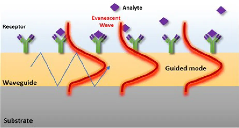
I.3.1.Surface Plasmon Resonance Imaging (SPRI)
The physical principle of Surface Plasmon Resonance Imaging (SPRI) is similar to the SPR methodology, but it takes another step forward. SPRI combines typical advantages of the traditional SPR in terms of real-time analysis, no label requirements and high sensitivity, with those associated to an imaging system such as the easy coupling with microfluidic devices, the high spatial resolution and the possibility to adopt an array-compatible detection approach. It was tested for the first time in 1988 by Knoll and Rothenhäusler; however, during the last two decades, it has been further developed for the study of interactions between systems such as DNA/DNA, RNA/DNA, protein/DNA, RNA/protein, antibody/antigen. 66-69
Biosensors for Medical Diagnostics
SPRI measures spatial differences in the intensity of the reflected light (expressed as percentage of reflectivity %R) at a fixed angle and wavelength.70 In detail, SPRI platforms based on a Kretschmann configuration employ a p-polarized light beam to hit a prism/thin gold film assembly at a fixed incident angle. The intensity of the reflected light is detected through a 2D detector (CCD), to generate an SPR image. The spatially resolved surface functionalization of the metallic SPRI surface allows detecting in real time parallel interaction events.71-73
Figure 7. The general principle of surface plasmon resonance imaging (SPRI). (Left) The analyte-ligand interaction shifts the SPR curve towards a higher angle (red to orange (Right)). Due to the measurement confinements (fixed wavelength and angle of incidence θ), changes in the reflectivity (Δ%R) at a single spot of the array can be simultaneously detected. Adapted from Ref.74.
The spatial contrast in an SPR image derives from the heterogeneity in the dielectric constant, owing to differences in refractive index or film thickness at different positions within the surface. If an analyte adsorbed has a refractive index different from that of the buffer solution, the phenomenon on the surface can be detected with a spatial resolution by controlling changes in the reflected light intensity.
For small modifications in the SPR angle, the variations in reflected intensity are proportional to changes in the effective refractive index, thickness or surface coverage of a monolayer on the SPR-active surface.75 The proportionality factor can be determined by determining SPRI intensity versus angle curves from different points of an SPRI surface. Either side of the minimum show nearly linear regions. These latter are referred to as “linear regions” or “high-contrast angles”, where the slope is high and remains constant. If the angle of incidence is set in correspondence with the linear region on the left side of the minimum, the reflectivity decreases when the minimum shifts to higher angles; if the intensity of reflected light is monitored at an angle in the linear region on the right side of the minimum, the measured reflectivity increases. The relationship remains linear for variations in reflectivity by 20%; for variations in the
Chapter I
32
larger the curve becomes non-linear indices of refraction and it could have a saturation of signal.
SPRI system allows simultaneous and independent measurements on different points of the same sensor surface. In addition, the sensor surface can be observed in real time, thereby highlighting the force of SPRI, along with other advantages such as inherent optical sensitivity and instrumentation simplicity.
Moreover, the possibility to monitor simultaneously the kinetic curves; the use of a minimum volume of analyte in contact with different areas of sensor surface at the same time; the measurement of multi-analytes makes SPRI-method a potential candidate for the optimal construction of biosensors.
However, to take full advantage of this approach, a precise control of both the patterning of biomolecules onto the SPRI sensor surface, as well as the control of the analyte flow is obliged. In this perspective, microfluidic devices offer an SPRI-compatible convenient mean for both using small amounts of sample solutions as well as for checking the patterning of different biomolecules.
Laminar flow of fluids is induced by microscale conditions,76 and the method with which liquids are put in contact with the SPRI sensor surface is optimized.77 The use of microfluidic devices significantly diminishes the experiment duration and the sample utilization. In addition, diffusion distances, nonspecific interactions, and instrument drifts by internal signal referencing are reduced, thus allowing a better control over reaction conditions. 78,79
I.3.2. Applications of SPR-based Biosensors
A wide range of applications has been developed for the use of SPR biosensors in the biomedical field. First of all, SPR has been used as a powerful tool to examine interactions between biomolecules based on affinity binding analysis of a variety of bonds, including antibody-antigen,80 ligand-receptor kinetics,81,82 enzyme-substrate reaction,83 and epitope mapping.
SPR technique is often used as a complementary method to analyse conformational changes study rather than as a primary technique. This application has been used to monitor structural transition in protein-small molecule interactions,84 proteins under diverse environmental conditions,85 or impacts on apoptosis inducers.69
Another extension of SPR-based detection applications is its use in point mutation detection of unamplified genomic DNA, evaluated for the first time by using plant, bovine and human genomic DNAs.86
Biosensors for Medical Diagnostics
The limits suffered by SPR for the parallel detection of different probe/target interactions are overcome by SPR imaging.87 The possibility to detect unamplified genomic DNA by using an SPRI-based multiplexed assay has been first shown by detecting the testis-specific protein, Y-encoded, (TSPY) gene located in the Y chromosome of the human genomic DNA.88
The enhanced sensitivity required, when unamplified genomic DNA is going to be detected, has encouraged efforts for innovative strategies for the amplification of transducer signals after the DNA detection. To achieve this aim, the use of nanoparticles has been widely investigated in combination with optical or electrochemical transducers.
Nanotechnology offers unique opportunities for creating highly sensitive innovative biosensing devices and ultrasensitive bioassays. Moreover, nano-sized devices generally allow for a faster response because mass transport occurs over smaller distances. Biointerfaces based on nanomaterials are particularly suitable for the development of improved the DNA detection assays.89 Among metallic nanostructures nanomaterials, gold nanoparticles (AuNPs) have been so far the most useful and extensively examined for improved DNA detection, thanks to their intriguing electronic and optical properties.77
Also, the rise of sensitivity by using gold nanoparticles depends on three main factors: (i) an increase of the absolute mass in each binding event, (ii) a rise in the bulk refractive index of the analyte, and (iii) coupling between the Localized Surface Plasmon (LSP) of metallic nanoparticles and SPR of the sensing film.
The resonant excitation of LSPs is determined by the size, the shape, and the surrounding dielectric environment of the plasmonic nanostructure. Moreover, to achieve a successful and reproducible detection of biological targets using nanomaterials, the as-synthesized nanomaterials must be well dispersed in aqueous solution, with minimal to no aggregation, and nonspecific binding to biomolecules or substrates.
There are many published works in the literature for the detection of cancer biomarkers using SPR-based techniques.90-92 Among the tumour-related genetic aberrations, DNA methylation represents an interesting biomarker for cancer diagnosis, because linked to tumour progression.93 Methylation level of circulating tumour DNA (ctDNA) has been detected by SPR using, in this case, a single-stranded oligonucleotide capable to hybridize methylated DNA target by two inverted recognition ends (molecular inversion probe, MIP).54
In 2006, Li et al. developed a combination of surface hybridization, surface ligation and nanoparticle amplification for single-nucleotide polymorphism (SNP) genotyping in
Chapter I
34
the BRCA1 gene,94 which is one of the two major genes (BRCA1 and BRCA2) reported to be connected to breast cancer susceptibility. These two tumour suppressor genes are involved in repairing the DNA double-strand breaks that are responsible for breast cancer. Therefore, the identification of BRCA mutations is of a great relevance for prospective interventions and treatments for breast cancer patients. By using nanoparticles with oligonucleotides complementary to the ligation probe DNA aiming at enhancing the SPR signal, single mismatches of BRCA1 were successfully detected at concentrations as low as 1 pM.
More recently, a powerful tool for biosensing has been achieved by localized SPR (LSPR) approach,95-97 where metallic nanostructures with plasmonic properties are associated with designed probes and surface architectures for ultrasensitive assays. In detail, LSPR platform has been employed for the ultrasensitive and simultaneous detection of different cancer biomarkers, namely tumour-specific mutations (E542K and E545K) and ctDNA methylation of PIK3CA gene (phosphatidylinositol-4,5-bisphosphate 3-kinase catalytic subunit alpha gene, PIK3CA).55 This biosensor offered considerable performances in relation to sensitivity, combining the use of PNA-probe sequences for ctDNA mutations with a specific anti-5-methylcytosine monoclonal antibody (mAb) immobilized onto gold nanoparticles for the targeting of the ctDNA methylation.
Another improvement for SPR technology has been given thanks to recent studies about surface chemistry of SPR-biosensor. For example, the selectivity of the hybridization reaction with the target complementary sequence has been increased by using the surface immobilized peptide nucleic acid (PNA) probes for the detection of a single point mutation in unamplified genomic DNA.98-100
The surface modification, for the hybridization step, with suitable synthetic probes such as PNAs demonstrated a high rise of the detection specificity and sensitivity. Unique structural and hybridization qualities of PNAs make them superior to DNA as sequence-specific hybridization probes. PNAs are DNA mimics, in which the negative phosphate-deoxyribose backbone is substituted by a neutral N-(2-aminoethyl) glycine linkage. These synthetic probes interact with their complementary DNA sequence with a stronger binding, more quickly and more specifically than the analogous DNA.101 The absence of the coulombic repulsion, established between the two negatively charged strands in DNA, lends the above mentioned special properties for PNA probes. PNA complexes are more thermally stable than DNA complexes and, through the nature of their backbone, less susceptible to biological degradation by nucleases, proteases, and peptidases.102
Biosensors for Medical Diagnostics
I.4.
Quartz Crystal Microbalance (QCM)
Quartz crystal microbalance (QCM) is a nanogram sensitive technique that utilizes acoustic waves produced by oscillating a crystal quartz plate to determine the mass absorbed onto the surface. The heart of QCM operation concerns quartz’s inherent property of piezoelectricity. This latter is generated by the electric charge that accumulates in certain solid materials (such as crystals, like quartz, certain ceramics, and biological matter such as bone, DNA and various proteins)103-105 in response to applied mechanical stress. By applying alternating electric fields to the crystal, a swapping expansion and contraction of the crystal structure are induced. In today’s most common QCMs a circular piece of quartz is sandwiched between two metal electrodes.
The quartz is generally handled using the so-called “ATcut” to give favourable properties relating to stability (low-temperature coefficients and a purely shear mode of oscillation). When a sufficient AC voltage is applied with a frequency close to the resonant frequency (f0), the resonance is excited of the particular crystal. The resonant
condition of the QCM occurs when the standing wave, produced by the alternating expansion and contraction, is an odd integer of the thickness of the quartz plate. Resonant frequencies of typical QCMs are on the order of MHz and the tradeoff between the frequency (relating to sensitivity) and the thickness (relating to usability) of QCMs is that the higher the resonant frequency the thinner the crystal. The common frequency (f0) of 5 MHz has a corresponding thickness of ≈ 330 µm. QCMs became
widely employed as mass balances only after the theory and experiments relating a frequency change of the oscillating crystal to the mass adsorbed on the surface had been demonstrated by Sauerbrey.106 This linear relationship between frequency change (∆f) and mass adsorbed (∆m) is given by:
∆m =C
n∆f
where n is the harmonic number and:
C =tqρq f0
with tq being the thickness of quartz, and ρq being the density of quartz and equals
Chapter I
36
There are three assumptions that must be satisfied for the Sauerbrey relationship to hold:
1. The adsorbed mass must be small relative to the mass of the quartz crystal; 2. The mass adsorbed is rigidly adsorbed;
3. The mass adsorbed is evenly distributed over the active area of the crystal. Liquid application of QCM technology multiplied the number of potential applications dramatically including biotechnology and, in particular, biosensor applications.107 The problem of the QCM to manage many liquid applications was that the liquid phase often incorporated viscous and elastic contributions to the frequency change and thus violated the assumption of the Sauerbrey equation, affirming that the mass adsorbed must be rigidly adsorbed. This difficulty encouraged the development of new methods for characterizing mass deposits with dissipative losses due to their viscoelastic property, and the elaboration of another theory for interpreting this new data.
Figure 8. Description of the main components in QCM-D. (a) Typical QCM-D sensor with Au electrodes. (b) Quartz crystal with alternating current applied across electrodes. (c) Short-circuiting the alternating current. (d) The oscillatory decay as the quartz disk comes to rest. The frequency of the oscillating crystal, shown in (b), is related to the total oscillating mass adsorbed on the surface, while the energy dissipation, shown in (c), is related to the viscoelastic properties of the oscillating mass. Thus, changes in adsorbed mass of, for example, a rigid protein provide a change in frequency, but for viscoelastic masses such as biomacromolecules, there is a change both in frequency and dissipation. Adapted from Ref.108.
Biosensors for Medical Diagnostics
I.4.1.Quartz Crystal Microbalance with Dissipation (QCM-D)
Two main processes for dealing with the dissipation (D), due to viscoelastic film adsorption, control the reduction of a crystal’s oscillation after a rapid excitation close to the resonant frequency (since the decay rate is proportional to the energy dissipation of the oscillator) or impedance analysis.109
QCM with dissipation monitoring (QCM-D) measures the voltage of oscillatory decay after a driving power is switched off in such a way as to ensure that the quartz oscillation has a value close to the series resonant mode.110 The amplitude declines over time depending on the properties of the oscillator and the contact medium. The decay voltage, i.e., the output voltage amplitude as a function of time, with a frequency given by f0 is blended with a reference frequency (fR) and filtered with a low-pass band filter.
This latter imparts an output frequency (f) based on the difference between fR and f0.
This frequency difference is fit to an exponentially damped sinusoidal, A(t), according to:
A(t) = A0𝑒
𝑡 𝜏
⁄sin (2πft+α)
where f = f0 – fR. The dissipation parameter is given by:
D = 1
πfτ
and is dimensionless, expressed as:
D = 1
Q=
Edissipated
2πEstored
with Q the quality factor, Edissipated the energy dissipated during one oscillatory cycle
and Estored the energy stored in the oscillating system. Resolution of frequency and
dissipation in liquids is on the order of ± 0.1 Hz and 1 × 10⁻⁷, respectively, with approximately one order of magnitude better in air or vacuum.
Typical f and D responses for protein, vesicle, or cell adsorption are on the order of 10 – 100 Hz and 1 – 10 (× 10–6) of dissipation units. For viscoelastic films greater than 100 nm thick, these responses are typically an order of magnitude higher. The QCM-D approach allows for determining f and D values at multiple harmonics (n = 3, 5, ...) of a resonant frequency in succession on the millisecond timescale.
The multiple harmonic data allows for treating the experimental data with a theory to extract meaningful parameters such as mass, thickness, density, viscosity, or storage modulus. The viscoelastic data permits a broader characterization of systems that do not hold the linear Sauerbrey relationship between Δf and Δm and makes QCM-D more