DEPARTMENT OF BIOLOGICAL AND ECOLOGICAL SCIENCES (DEB)
XXVII PhD COURSE IN GENETICS AND CELL BIOLOGY
BIO/11
Evaluation of biochemical effects of oxidative stress in
blood bank-stored red blood cells
PhD student Cristiana Mirasole
Coordinator Tutor
AIM OF THE STUDY
Blood banking underpins modern medical care, but blood storage, necessary for testing and inventory management, reduces the safety and efficacy of individual units of red blood cells (RBCs). Stored RBCs are damaged by the accumulation of their own waste products, by enzymatic and oxidative injury. These chemical activities lead to a complex RBC storage lesion that includes haemolysis, reduced in vivo recovery, energy and membrane loss, altered oxygen release, reduced adenosine triphosphate and nitric oxide secretion, and shedding of toxic products. The latter includes also free iron that can potentiate susceptibility to oxidative stress, which in turn promotes oxidative-lesions to proteins, lipids (peroxidation) and morphological changes (membrane blebbing, vesiculation). In particular, reactive oxygen species seem to be the eligible trigger for these lesions and the main contributor to the final quality loss (both at the macroscopic and microscopic levels) of RBCs.
In order to ensure safety and quality of blood products, the study of in vitro (storage conditions) ageing of red blood cells has recently taken advantage of the introduction of mass spectrometry-based “omics” disciplines, such as proteomics, metabolomics and lipidomics, which are characterized by the systematic determination and quantification of broad classes of molecules, such as proteins, metabolites and lipids. Data from multiple “omics” platforms can be then integrated through bioinformatic approaches and mathematical modeling to obtain a system biology level of understanding, which has been performed during our research.
During this work of thesis, we focused on the evaluation of irreparably protein oxidative modifications, in particular those related to non-enzymatic glycosylation of hemoglobin α and β-chains (HbA1c). The observation of apparently increased rates of HbA1c levels over storage progression might be also exacerbated by the excessive glucose found in anticoagulant and additive solutions. On the contrary, we followed how some of these oxidation products can be repaired (e.g. oxidations of sulfur-containing amino acid residues), representing an exceptional way to sense the redox state in cells. The glycolytic enzyme glyceraldehyde-3-phosphate dehydrogenase (GAPDH) has been found to regulate its activity in response to storage-induced oxidative stress through the formation of an intramolecular disulfide bond involving catalytic cysteines. This mechanism has been postulated to function as a metabolic switch to redirect
carbohydrate flux from glycolysis to the pentose phosphate shunt, allowing the red cell to maintain oxidation-reduction balance and protect itself against oxidative insults. Hereby, we describe alternative storage protocols which have been proposed in order to overpass these hurdles, such as RBC anaerobic storage. We could confirm previous evidences about long term anaerobiosis promoting glycolytic metabolism in RBCs and prolonging the conservation of high energy phosphate reservoirs and purine homeostasis. In parallel, we evidenced that, contrarily to aerobic storage, anaerobiosis impairs erythrocyte capacity to cope with oxidative stress by blocking metabolic divertion towards the pentose phosphate pathway, which negatively affects glutathione homeostasis. Therefore, although oxidative stress was less sustained than in aerobically stored counterparts, its markers still accumulate over anaerobic storage progression. Finally, we presented an HPLC-microTOF-Q approach to investigate the RBC lipidome. We could exploit this analytical workflow to consolidate existing knowledge on the RBC lipid composition and individuate statistically significant fluctuations of lipids throughout storage duration of RBC concentrates under blood bank conditions. While this field of research still warrants future investigations, our analysis indicated ceramides, glycerophospholipids and sterols as key targets of RBC storage lesions to the lipidome, which will deserve further targeted investigations in the future.
INDEX Page
CHAPTER 1: Introduction 1
1.1 Alterations to Red Blood Cells (RBCs) during storage 2 (“the storage lesions”)
1.1.1 Why Red Blood Cells? 2
1.1.2 History of Blood storage: its meaning 2
for Transfusion
1.2 Parameters of the oxidation/reduction balance in in vivo 8 Red Blood Cells
1.2.1. Reactive species generation 8
1.2.2. Reactive oxygen species 9
1.3 Oxidative stress during the formation of storage lesions in blood 12 banking RBCs: chemical modification of proteins by reactive
oxygen species
1.3.1 Oxidation of Sulfur-containing Amino Acid Residues 14
1.3.2 Protein Carbonylation: Generation of Protein 15 Carbonyl Derivatives
1.3.3 Protein-Protein Cross-Linkage 17
1.3.4 Hemoglobin oxidation 19
1.3.5 Hemoglobin and the functions of nitric oxide (NO) 21 correlated with the Red Cell Storage Lesion
1.3.6 Lipid Peroxidation 25
1.3.7 The effects of phosphorylation on integrity membrane 26 structure of stored RBCs under blood banking conditions
Index
CHAPTER 2: Haemoglobin glycation (Hb1Ac) increases 44
during red blood cell storage: a MALDI-TOF mass-spectrometry-based investigation
2.1 Aim of the study 45
2.2 Materials and Methods 45
2.2.1 Sample collection 45
2.2.2 Matrix and sample preparation for MALDI-TOF MS 46
2.2.3 Linear MALDI-TOF MS analysis and calculation 46
of glycated or glutathionylated hemoglobin
2.3 Results and discussion 47
2.4 Conclusion 50
References 51
CHAPTER 3: Red Blood Cell metabolism under prolonged 53 anaerobic storage
3.1 Aim of the study 54
3.2. Design and method 56
3.2.1 Sample collection 56
3.2.2. Materials 57
3.3 Untargeted metabolomics analyses 57
3.3.1 Metabolite extraction 57
3.3.2 Rapid resolution reversed-phase HPLC 58
3.3.3 Mass spectrometry: Q-TOF settings 58
3.3.4 Data elaboration and statistical analysis 59
3.4 Results and discussion 60
3.4.1 Glycolysis and energy metabolism are sustained 60 throughout anaerobic storage duration
Index
3.4.2 Purine salvage pathway is activated proportionally to 67 storage progression even under anaerobic conditions
3.4.3 Amino acid transport and lipid homeostasis are 68 affected to some extent under anaerobic storage
3.5 Conclusion 71
References 73
CHAPTER 4: Thiol-based regulation of glyceraldehyde- 85
3-phosphate dehydrogenase in blood bank–stored red blood cells: a strategy to counteract oxidative stress
4.1 Aim of the study 86
4.2 Materials and Methods 88
4.2.1 Storage of RBCs and sample preparation 88
4.2.2 GAPDH assay 89
4.2.3 Western blotting analysis 89
4.2.4 Statistical analysis 89
4.2.5 In-gel digestion and peptide sequencing 90
by nano-LC-ESI Q-TOF MS/MS
4.3 Results 90
4.3.1 Effects of RBC storage on GAPDH activity 90
4.3.2 Storage-dependent displacement of RBC 92
GAPDH from membrane to cytosol
4.3.3 Identification of posttranslational oxidative modifications 92 responsible for GAPDH inactivation
4.4 Discussion and Conclusion 93
Index
CHAPTER 5: Red Blood Cell Lipidomics analysis through 100 HPLC-ESI-qTOF: application to red blood cell storage
5.1 Aim of the study 101
5.2. Materials and Method 103
5.2.1. Sample collection 103
5.2.2. Untargeted Metabolomics Analyses 104
5.2.2.1. Metabolite extraction 104
5.2.3. Rapid Resolution Reversed-Phase HPLC 104
5.2.4. Mass spectrometry analysis through microTOF-Q 105
5.2.5. Data elaboration and statistical analysis 105
5.3. Results and Discussion 106
5.3.1. Fatty acids 111
5.3.2. Glycerolipids and glycerophospholipds 112
5.3.3. Sphingolipids, sterols and prenol lipids 115
5.3.4. Red blood cell Lipidomics: application to the 117 storage of erythrocyte concentrates
5.4. Conclusion 120
1
Chapter 1
2
1.1 ALTERATIONS TO RBCS DURING STORAGE (“THE STORAGE
LESIONS”)
1.1.1 Why Red Blood Cells?
Derived from pluripotent stem cells in bone marrow through a maturation process called erythropoiesis, mature Red Blood Cells (RBCs) are biconcave disks of approximately 7.2 µm in diameter, 1.5 to 2.5 µm thick, with a mean volume of 90 fL. Along the developmental process, there is a reduction in cell volume, condensation of chromatin, loss of nucleoli, decrease in the nucleus, RNA, mitochondria, and an increase in hemoglobin synthesis, resulting in a mature RBC, which lacks a nucleus and organelles. The primary function of RBCs is to transport oxygen from the lungs to the body tissues, where the exchange for carbon dioxide is facilitated through synergistic effects of hemoglobin, carbonic anhydrase, and band 3 protein, followed by carbon dioxide delivery to the lungs for release. Successful oxygen transport is dependent on efficacy of the three elements of RBC metabolism: the RBC membrane, hemoglobin, and cellular energy. Continuous research of oxygen transport is crucial for development of improved RBC storage and biopreservation technologies.
1.1.2 Hystory of Blood storage: its meaning for Transfusion
Whole blood storage was first demonstrated by Robertson in 1917. Acid-citrate-dextrose (ACD) and Citrate-phosphate-Acid-citrate-dextrose solution (CPD) were subsequently approved for 21-day storage of blood. CPD with adenine (CPDA-1) was later introduced and used for extending the shelf-life of stored blood for up to 5 weeks. Red blood cells stored in these solutions have shown steady deterioration after about 5 to 6 weeks as determined by the inability of such cells to survive in the circulation for 24 hours after reinfusion back into the human donor. It has been observed that during continued refrigerated storage, glucose is consumed at a decreasing rate, as the concentration of metabolic waste, i.e. lactic acid and hydrogen ions, increases. Such a decrease in the rate of glucose metabolism leads to depletion of adenosine triphosphate (ATP) which directly correlates to the recovery of RBCs when the cells are returned to the circulation. The development of additive solutions for the preservation of red blood cells after their separation from whole blood has allowed the design of formulations which are specifically tailored to the needs of RBCs. Additive solutions such as Adsol®
Chapter 1
3
(AS-1), Nutricel® (AS-3), Optisol® (AS-5), and Erythro-Sol® were designed to extend the storage of RBCs at 4°C.
In Italy, the “design” of the national blood system was changed through the "New discipline for blood transfusion activities and national production of blood derivatives" [Italian Parliament, October 21st, 2005]. In parallel, the 2002/98/EC directive of the Council of Europe and the 2004/33/EC, 2005/61/EC and 2005/62/EC directives of the European Commission have been transposed into national laws. Actually, new regulatory standards have been introduced, contributing to improve the ability to store RBCs and to rich the strategic goals of the Italian blood system.
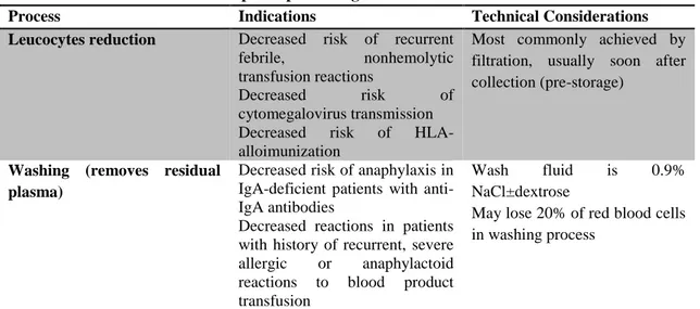
At present, transfusion of RBCs is a common therapeutic intervention for which recent developments suggest steps that might be taken to improve their quality, safety and their benefits for the transfused recipient. Most RBC products are derived by collection of 450-500 (±10%) mL of whole blood into anticoagulant solutions (typically citrate-dextrose-phosphate) from volunteer donors and removal of the plasma by centrifugation (see Table 1) and, in some cases, also leukocytes. Although in healthy subjects, most RBCs live for 110 to 120 days (with the standard deviation of their life span of about 6 days in normal subjects [Högman et al, 1999], after removal of the plasma, the resulting product is red blood cells (referred to informally as “packed red blood cells”) with 42-day blood bank shelf life, which are stored at 4 ± 2° C in a slightly hypertonic additive
sterilely solution, generally SAGM (sodium, adenine, glucose, mannitol, 376 mOsm/L).
Table 1- Special processing of RBC for transfusion
Process Indications Technical Considerations
Leucocytes reduction Decreased risk of recurrent
febrile, nonhemolytic
transfusion reactions
Decreased risk of
cytomegalovirus transmission
Decreased risk of
HLA-alloimunization
Most commonly achieved by filtration, usually soon after collection (pre-storage)
Washing (removes residual plasma)
Decreased risk of anaphylaxis in IgA-deficient patients with anti-IgA antibodies
Decreased reactions in patients with history of recurrent, severe allergic or anaphylactoid reactions to blood product transfusion
Wash fluid is 0.9%
NaCl±dextrose
May lose 20% of red blood cells in washing process
Chapter 1
4
The RBC is a highly specialized cell; because it lacks a nucleus and other cellular machinery, it has to rely on its original contents of enzymes and other proteins for the whole of its limited lifetime. In addition, because it lacks mitochondria, it cannot use oxidative phosphorylation for adenosine triphosphate regeneration.
In term of prolonged storage, this means that RBCs undergo a dramatic molecular changes, the so-called "red blood cell storage lesion"[Hess 2006, Hess 2007,Hess 2010]. The RBC storage lesions include:
i. Metabolic changes:
1. RBCs reduce concentrations of adenosine 5′-triphosphate (ATP) and 2,3-diphosphoglycerate (2,3-DPG). Glycolysis is the only source of energy for RBCs, through which they break down glucose to make ATP and produce lactic acid and protons as a result. The protons produced in glycolysis make the storage solution and the red cells increasingly acidotic. This acidosis in turn slows further glycolysis because the protons inhibit hexose kinase and phosphofructokinase, the first two activating enzymes in the main glycolytic pathway, so that less ATP is made as storage progresses [Hess 2006, Hess 2007, Hess 2010, Ian Chin-Yee et al., 1997].
2. The pH typically decreases from 7.0 to 6.5, while the rate of glucose consumption fall down 50%, starting from RBCs collection from venous blood at a pH of about 7.35, put into CPD at a pH of 5.5 to 5.8[Hess 2006].
3. The fluxes of sodium ions (massive entry into the cell) and potassium ions (exit from the cell) are involved in the storage lesion, because of loss of function (usually transient) of cation pumps , since the Na+/K+ pump is inactive at 4°C [Bennett-Guerrero et al., 2007] and ATP-dependent. This leads to loss of intracellular potassium and accumulation of sodium within the cytoplasm. The concentration of extracellular potassium in the blood bag typically accumulates at a rate of about 1 mEq/day, causing damages when the blood is used in cardiac bypass machines [Hall et al., 1993].
Chapter 1
5
ii. Enzymatic changes:
Enzymatic damages are related to the presence of white blood cells (WBCs) during storage period. Indeed, it has been demonstrated that WBCs break down and release enzymes such as proteases, lipases and glycosidases in the storage solution. These enzymes attach their target on the outer surface of erythrocytes [Riedner et al., 1990]. Proteases can contribute to the loss of protein antigen strength with storage and generally they are blocked by plasma antiproteases. Lipases such as phospholipase can dealkylate trialkyl-glycerols to produce lysophospholipids. An important example of this process is the production of platelet activating factor (PAF), thought to be responsible for some cases of transfusion-related acute lung injury (TRALI) [Gajic et al., 2007]. Glycosidases remove sugars from glycolipids and glycoproteins, and the loss of these sugars can expose underlying structures that increase the binding of stored red cells to endothelial cells [Sparrow et al., 2007]. The removal of the WBCs by filtration, the so called leukoreduction, can reduce enzymatic damage, thereby improving recovery and decreasing the hemolysis of stored red cells [Heaton et al., 1994]. The discovery of TRALI was the first and still the most important example of possible damages associated with stored blood. About glycosidases, Sparrow’s group [Sparrow et al., 2007] has shown that stored red cells adhere to endothelial cell monolayer proportionally with the loss of membrane sugars. Such adherence is assumed to be responsible of endothelial inflammation if the red cells subsequently break down and release iron and heme. For all these reasons, leukoreduction increases red cell recovery by about 2% and reduces the hemolysis by half at the end of the storage.
iii. Shape change and membrane vesiculation:
RBC storage, even for relatively short periods of time (between the 7th and the 14th day of storage [Tamara, Berezina et al, 2002]), affects RBC morphology and function (Table 2). The echinocyte-spheroechinocyte red blood cell shape transformation [Lockwood et al., 2003] is induced by loose of membrane through the blebbing of vesicles from the spicules of cells that have undergone echinocytic (in the early stage). After prolonged storage (beyond 21 days), when
Chapter 1
6
the ATP-content of stored RBC decreases rapidly, the total amount of released vesicular proteins increases exponentially, reaching 10 times the amount by 50 days as compared to 14 days of storage. The vesiculation contributes to irreversible cell shape and membrane changes, in order to eliminate proteins and lipids that have been altered by oxidative stress, as to protect the cell from a further chain reaction of stress and consequent removal from the circulation [Willekens et al., 2008]. It can be considered not only part of the physiological process of maturation and programmed cell-death mechanism unmasked by low concentrations of ATP, but also the result of a cell-death-independent step making vesicles that carry away oxidized lipids and damaged proteins [Hess 2010, Salzer et al., 2008]. Moreover, the shape change seen during RBC storage is associated with rheologic changes, increased viscosity and reduced flow in capillary systems [Relevy et al., 2008].
Table 2- Changes in RBC shape during storage [Berezina et al., 2002]
Shape RBC
Day of storage Discocyte
(%) Reversibly changed RBC (%) Irreversibly changed RBC (%) 5th 79.0±2.7 14.0±1.7 7.0±1.6 7th 78.0±3.0 13.6±1.7 8.4±1.6 14th 57.4±3.8*^ 27.9±1.9*^ 14.7±2.6*^ 21th 53.7±3.5* 30.6±3.0* 15.7±3.3* 28th 47.6±3.6* 35.2±1.6*^ 17.2±4.1*^ 35th 37.5±5.0*^ 40.6±3.4*^ 21.9±5.0* 42th 23.3±4.3*^ 46.8±6.7* 29.9±4.0*^ * P˂ 0.01 vs 5th day ^ P˂ 0.01 vs previous point iv. Oxidative damage:
RBC oxidant susceptibility during storage results in Reactive Oxygen Species (ROS) targeting protein and lipid fractions [Dumaswala et. al., 1999]. Lipid peroxidation and phospholipid reorganisation of the membrane occurred in the first 7-14 days of cold storage [Brad et al., 2012]. Banked RBCs show a time-dependent increase in (i) protein clustering [D'Amici et al., 2007, Walpurgis et
Chapter 1
7
al., 2012], especially on band 3 (early, in the first 7 days of storage); (ii) carbonyl modification of band 4.1 [D'Alessandro et al., 2012, Kriebardis et al., 2007]and changes in spectrin-protein 4.1-actin interactions [Card 1988, Wolfe et al., 1986]; (iii) malondialdehyde increase and (iv) a decline of erythrocyte glutathione (GSH) levels and glutathione-peroxidase (GSH-PX) activity [Umakant et al., 1999]. In contrast loss of acetylcholinesterase activity and haemolysis of RBC occurred relatively late (14-21 days) during the storage period [Brad et al., 2012]. Microvesicle generation from ATP-depleted RBC also correlates with the breakdown of polyphosphoinositides to diacylglycerol on the inner monolayer [Müller et al., 1981]. Membrane budding is dependent on both the diffusible diacylglycerol partitions into the outer monolayer and exoplasmic exposure of phosphatidylserine (PS), a negatively charged phospholipid. Microvesicles (160 nm diameter) from RBC stored for more than 20 days [Kriebardis et al., 2008] contain band 3, are enriched in acetylcholinesterase and in stomatin, but threefold depleted of flotillin 2 that remains in the residual RBC membrane [Salzer et al.,2008]. Microvesicles from long stored RBC contain heavily aggregated hemoglobin, band 3 aggregates and increasing amounts of autologous IgG [Kriebardis et al., 2008], while the residual RBCs reveal a decrease in band 3 content, but yet a substantial increase in bound IgG [Kriebardis et al., 2007].
The band 3, in addition of being an anion exchanger protein, is the site of macromolecular assembly for cytoskeleton anchorage, glycolytic enzymes (the N-terminal cytosolic domain of band 3 is an inhibitory docking site for glycolytic enzymes), and regulatory proteins. Indeed, band 3 is involved in the modulation of oxygen transport by influencing the intracellular pH (and thus oxygen off-loading) through its regulation of the chloride shift (exchange of Clˉ /HCO3ˉ anions).
Under oxidative stress, haemoglobin (Hb) could be oxidized to met-Hb (a protein unable to carry oxygen) which can be converted into low-spin ferric Hb compounds named hemichromes, a variant of Hb in which cystein thiol groups have been dangerously oxidized to form denatured Hb-aggregates precipitating in inclusion bodies within RBCs, also known as “Heinz bodies”. Normally,
Chapter 1
8
reversible hemichromes can be reduced back to deoxyHb [Telen et al., 2004]. Significant distortions in the tertiary structure of the Hb can lead to the so-called ‘irreversible hemichromes’. The latter cannot be converted back to normal Hb [Rachmilewitz et al., 1971]and are susceptible to further oxidative injury, during prolonged storage, which may result in their fragmentation and formation of aggregates.
Oxidation to proteins also results in the progressive accumulation of glycated forms of haemoglobin (Hb1Ac, non-enzymatic addition of sugar moieties to the proteins) during storage duration in the membrane and cytosol [Sparrow et al., 2007]. Notably, altered membrane protein glycosylation patterns might influence the rheological properties of RBCs [Berezina et al., 2002].
1.2 PARAMETERS OF THE OXIDATION/REDUCTION BALANCE IN in vivo RED BLOOD CELLS
Red blood cells are responsible in vivo, for oxygen transport from lung to tissues. Their function depends on reduced state of iron (Fe2+) in hemoglobin (Hb). Both element oxygen and iron are able to quickly shift their oxidative state in response to different emerging stimuli. Literature shows that many approaches for evaluation of RBC redox status have been established, including indirect approaches such as measurement of protein oxidation, that reflects an accumulated damage [Lee, Britz-McKibbin, 2009]. The bigger the reactive species release is the higher normal deformability and flexibility of erythrocytes is disturbed.
1.2.1. Reactive species generation
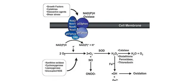
In erythrocyte, oxygen (O2), nitrogen oxide (NO•) and iron (Fe2+) are found together. All these elements act in normal conditions in established mechanisms but they may generate alone or together reactive species, named radicals, that damage red blood cells as well as vascular endothelium (Figure 1).
Chapter 1
9
Figure 1-Generation of superoxide (•O2ˉ) and H2O2 from O2 in vascular cells. Many enzyme systems,
including NAD(P)H oxidase, xanthine oxidase, and uncoupled nitric oxide synthase (NOS) among others, have the potential to generate reactive oxygen species (ROS). Superoxide acts either as an oxidizing agent, where it is reduced to H2O2 by superoxide dismutase (SOD), or as a reducing agent, where it
donates its extra electron (eˉ) to form ONOOˉ with NO. Hydrogen peroxide is scavenged by catalase, glutathione and thioredoxin systems, and can also be reduced to generate •OH in the presence of Fe2+ [Touyz et al., 2004].
A radical is a chemical species that possesses a single unpaired electron in outer orbitals, is able to independently exist and is also known as free radical. Radicals are highly reactive in extracting an electron from any neighbor molecule in order to complete their own orbitals.
There are two main groups of free radicals: ROS or reactive species of oxygen and RNS or reactive nitrogen species. ROS and RNS can act together damaging cells and causing nitrosactive stress. Therefore, these two species are often collectively referred to as ROS/RNS.
1.2.2. Reactive oxygen species
Reactive species of oxygen refers to a group of highly reactive O2 metabolites, including superoxide anion (O2•¯), hydrogen peroxide (H2O2), singlet oxygen (1O2), and hydroxyl radical (OH•), that can be formed within cells. Reactive oxygen species are constantly formed as byproducts in normal enzymatic reaction in all human cells through normal aerobic processes as mitochondrial oxidative phosphorylation or as necessary products in neutrophils, in order to kill invading pathogens. The above mentioned phenomena are consuming oxygen processes. Erythrocytes must save oxygen for delivering it to the cells; as a consequence red blood cells lack
Chapter 1
10
mitochondrion the main oxygen consumer within cell. In this particular condition the source of ROS in erythrocyte may be the carried oxygen itself.
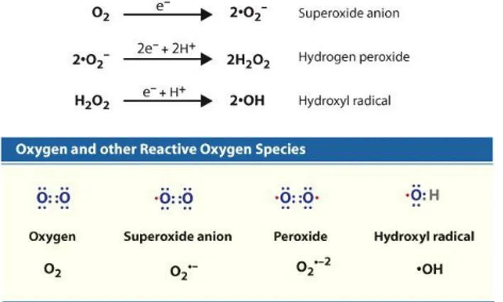
Within cells where transitional metals are bounded to proteins (in metal containing proteins or enzymes) oxidative attack of O2 tend to be slow, meaning that a first single electron is relatively difficult to add. As a consequence superoxid radical (O2•¯) will form very slow. In the presence of a free electron, the univalent reduction of oxygen yields reactive species of oxygen. (Figure 2).
Figure 2-Several reactive oxygen species that are generated in cells are shown. The most damaging
radical is the hydroxyl radical.
Superoxide has an unpaired electron, which imparts high reactivity and renders it unstable and short-lived. Superoxide is water soluble and acts either as a reducing agent, where it donates its extra electron to form ONOO¯ with NO [Darley-Usmar et al. 1995; Fridovich 1997], or as an oxidizing agent, where it is reduced to H2O2. The latest, in the next one-electron reduction step, generates water and the hydroxyl radical (OH•) which is probably the most reactive free radical. A final electron acceptance reduces hydroxyl radical to water.
Superoxide radical (O2•¯) is a reactive radical, however it cannot diffuse to far having limited lipid solubility. Instead it might react in the presence of ferric iron (Fe2+) with dehydrogen peroxide generating the most potent hydroxyl radical through a non-enzymatic reaction known as Haber-Weiss reaction. The reaction takes place in two steps that involve ferric iron and superoxide as follows:
O2•¯+ Fe3+ → O2 + Fe2+ Fe2+ + H2O2 → Fe3+ + OH − + OH•
Chapter 1
11
The general non-enzymatic reaction occurring in living cells is:
O2•¯+ H2O2 → OH • + OH − + O2
Hydrogen peroxide (H2O2) is not a free radical, but is produced mainly from dismutation of O2•¯. H2O2 is lipid soluble and as a consequence it can diffuse through lipid membranes.
Another reactive species (but not a radical) derived from molecular oxygen is singlet oxygen, designated as 1O2. Singlet oxygen (1O2), a highly excited state created when molecular oxygen absorbs sufficient energy to shift an unpaired electron to a higher orbital, can be formed from superoxide radical:
2O2•¯+ 2H + → H2O2 + 1O2
Singlet oxygen is even more reactive than the hydroxyl radical, although it is not a radical. As a conclusion the most reactive radical is hydroxyl (OH•) which indiscriminately extracts electrons from any other molecules around it, whereas superoxide (O2•¯) and hydrogen peroxide (H2O2) are more selective in their reactions with biological molecules.
All the above reactions and processes take place in all human cells including erythrocytes. These pro-oxidants are tightly regulated by anti-oxidants such as superoxide dismutase, catalase, thioredoxin, glutathione, anti-oxidant vitamins, and other small molecules [Stralin et al., 1995; Halliwell 1999; Channon and Guzik, 2002; Yamawaki et al., 2003].
Under normal conditions, the rate of ROS production is balanced by the rate of elimination. However, a mismatch between ROS formation and the ability to defend against them by antioxidants results in increased bioavailability of ROS leading to a state of oxidative stress [Griendling et al., 2000; Landmesser and Harrison, 2001; Zalba et al., 2001].
Chapter 1
12
1.3 OXIDATIVE STRESS DURING THE FORMATION OF STORAGE LESIONS IN BLOOD BANKING RBCs: CHEMICAL MODIFICATION OF PROTEINS BY REACTIVE OXYGEN SPECIES
Protein oxidation is the covalent modification of a protein induced either directly by reactive oxygen species (ROS) or indirectly by reaction with secondary by-products of oxidative stress [Shacter, 2000, Dean et al., 1997]. Circulating RBCs are equipped, in vivo, with effective anti-oxidative systems that make them mobile free radical scavengers, providing antioxidant protection not only to themselves but also to other tissues and organs in the body [Siems, 2000, Arbos et al., 2008]. A condition of oxidative stress develops when the critical balance between oxidants and antioxidants is disrupted due to depletion of antioxidants and/or excess accumulation of ROS [Dalton et al., 1999]. Indeed, the irreversible events occurring during the storage process, exemplified by the haemolysis in the second half of the actual maximal blood bank storage period, are mostly activated by radical species generated by prolonged, continuous oxidative stress [Wolfe 1986, Racek et al., 2001].
Because there are so many mechanisms for induction of protein oxidation and because all of the amino-acyl side chains can become oxidatively modified, there are numerous different types of protein oxidative modifications (Table 3).
The demonstration that oxidatively modified forms of proteins accumulate during storage and oxidative stress, has focused attention on physiological and non-physiological mechanisms for the generation of ROS and on the modification of biological molecules by various kinds of ROS.
Table 3-Oxidative Modifications of Proteins [Shacter 2000]
Modification Amino acids involved Oxidizing sourcea
Disulfides, glutathiolation Cys All ONOO¯
Methionine sulfoxide Met All, ONOOˉ
Carbonyls (aldehydes,
ketones) All (Lys, Arg, Pro, Thr) All
Oxo-histidine His -Ray, MCO, 1O
2
Dityrosine Tyr -Ray, MCO, 1O
2
Chlorotyrosine Tyr HOCl
Chapter 1
13
Tryptophanyl modifications,
(N-formyl)kynurenine Trp
-Ray
Hydro(pero)xy derivatives Val, Leu, Tyr, Trp -Ray
Chloramines, deamination Lys HOCl
Lipid peroxidation adducts
(MDA, HNE, acrolein) Lys, Cys, His -Ray, MCO (not HOCl)
Amino acid oxidation adducts Lys, Cys, His HOCl
Glycoxidation adducts Lys Glucose
Cross-links, aggregates,
fragments Several All
a MCO-metal catalyzed oxidation; All = -ray, MCO, HOCl, ozone, 1O2.
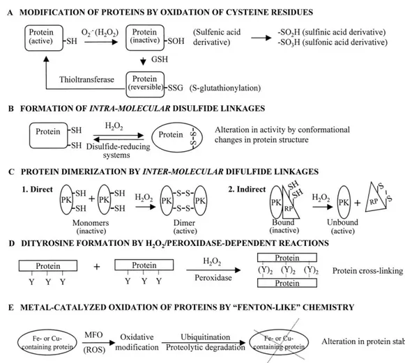
The pioneering studies of Swallow, Garrison, and Scheussler and Schilling demonstrated that reactions with OH• mainly initiated the modification of proteins; however, the course of the oxidation process is determined by the availability of O2 and O2ˉ• or its protonated form (HO2•). Collectively, the chemical effects of oxidative changes on membrane proteins can lead to diverse functional consequences, such as oxidation of amino acid residue side chains, formation of protein-protein cross-linkages, and oxidation of the protein backbone resulting in protein fragmentation (Figure 3). Current concepts of ROS signaling can be divided into two general mechanisms of action: 1) alterations in the intracellular redox state, and 2) oxidative modifications of proteins. The first is due to the alteration of the “redox-buffering” capacity of intracellular thiols, primarily GSH and thioredoxin (TRX), maintained by the activity of GSH reductase and TRX reductase, respectively.
Both of these thiol redox systems counteract intracellular oxidative stress by reducing both H2O2 and lipid peroxides, reactions that are catalyzed by peroxidases (e.g., GSH peroxidase catalyzes the reaction H2O2 + 2GSH → 2H2O + GSSG). The second is the subject of discussion below.
Chapter 1
14
Figure 3-Oxidative modification of proteins. A: the sulfhydryl (-SH) group of cysteine residues in
proteins may be modified by O2•ˉ (or H2O2) to form various oxidized derivatives. S-glutathionylation
appears to confer reversibility to such modifications by the action of thioltransferases . B: formation of intramolecular disulfide bridges can alter protein activity by changes in conformation. C: intermolecular disulfide linkages can mediate protein dimerization. D: H2O2- or peroxidase-catalyzed dityrosine
formation can induce protein cross-linking. E: transitional metal-containing proteins may be targets of site-directed, metal-catalyzed oxidation by ROS produced by certain mixed-function oxidases (MFO), which “target” them for ubiquitination and degradation by proteases. This is a potential mechanism for ROS-mediated alterations in protein stability [Thannickal et al., 2000].
1.3.1 Oxidation of Sulfur-containing Amino Acid Residues
Although all amino acid residues of proteins are susceptible to oxidation, Cysteine (Cys) [Shacter 2000; Radi et al., 1991] and methionine (Met) [Shacter 2000, Vogt 1995] are those especially prone to oxidative attack, both of which contain susceptible sulfur containing groups (thiols). Under even mild conditions, in the case of cysteine residues, oxidation leads to the formation of sulfenic acid, inter- or intra-molecular disulfides, protein mixed disulfides with low molecular weight thiols (e.g., GSH), and S-nitrosothiols; methionine residues generate Met sulfoxide (MeSOX), the major product
Chapter 1
15
of conversion under biological conditions. Most biological systems contain disulfide reductases and MeSOX reductases that can convert the oxidized forms of cysteine and methionine residues back to their unmodified forms. These reversible modifications can be part of regulatory processes of protein functions, in which cysteines can cycle between the oxidized and reduced state [Ahmed 2007].
However, Cys residues can be irreversibly oxidized by strong oxidative agents to sulfinic and sulfonic acids, which cause loss of protein function and cannot be reversed by metabolic processes [Woo et al., 2003].In the case of prolonged storage, the glycolytic enzyme glyceraldehyde-3-phosphate dehydrogenase (GAPDH) has been found to undergoes temporary change of its catalytic center in response to storage-induced oxidative stress through the formation of an intramolecular disulfide bond involving catalytic cysteines [Rinalducci et al. 2011].
1.3.2 Protein Carbonylation: Generation of Protein Carbonyl Derivatives
Protein carbonylation is considered a major form of protein oxidation and aging, not only because it is a sign of oxidative stress, but also of disease-derived protein dysfunction. Formation of protein carbonyls can occur either by the α-amidation pathway or by oxidation of glutamyl side chains which leads to formation of a peptide in which the N-terminal amino acid is blocked by α-ketoacyl derivative. However, direct oxidative attack on lysine, arginine, proline and threonine residues may also generate carbonyl derivatives [Berlett et al. 1997].In addition, carbonyl groups may be introduced into proteins by Michael addition reactions with α,β-unsatured aldehydes (4-hydroxy-2-nonenal, malondialdehyde) produced during lipid peroxidation [Schuenstein 1979, Esterbauer 1991, Uchida 1993] or with reactive carbonyl derivatives (ketoamines, ketoaldehydes, deoxyosones) generated as a consequence of glycation and glycoxidation reactions, in which reducing sugars or their oxidation products react with lysine residues of proteins [Monnier et al. 1995](Figure 4).
Chapter 1
16
Figure 4-A general scheme for the different routes for protein carbonylation [Ashraf Madian, 2010]
The higher levels of cytoskeletal protein carbonyl groups are observed after 35 days of prolonged storage of RBCs, especially in non-leukodepleted CPDA units, and this phenomenon is correlated with the extent of the oxidized Hb accumulation in the membrane and the cytoskeleton systematically after 10 days of storage [Annis et al., 2005; Kriebardis et al., 2007] (Figure 5). However, the observation of a decrease of cytoskeleton-bound Hb and protein carbonylation levels by the end of the storage period can be explained in part by the microvesiculation and the hemolysis of the most severely damaged cells, or by the proteasome activity, two recognized self-protective/ age-dependent mechanisms [Kriebardis et al., 2006, Delobel et al., 2012]. Strictly related to the protein carbonyl production is the progressive accumulation of advanced glycation end-products, where glycated forms of haemoglobin (Hb1Ac) accumulate proportionally to storage duration. Notably, alterations to Hb1Ac levels in units that have been stored for longer periods might cause problems when treating/monitoring certain categories of patients, such as those suffering from diabetes. In parallel, altered membrane protein glycosylation patterns might influence the rheological properties of RBC.
Chapter 1
17
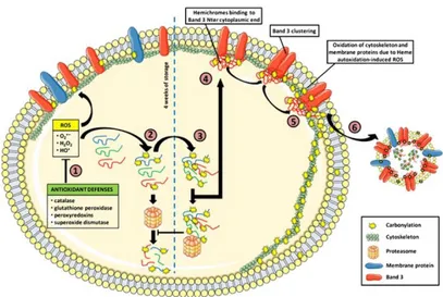
Figure 5-Carbonylation pathway in stored RBCs (1) RBC antioxidant defenses prevent protein
carbonylation by reducing ROS into less reactive intermediates. After a certain storage period, these defenses are exceeded by the oxidative stress and protein carbonylation occurs on both soluble and membrane proteins. (2) Carbonylated proteins are recognized and degraded by the 20S proteasome. (3) Over-oxidized proteins crosslink and cannot be degraded by proteasome anymore, inhibiting degradation of fairly oxidized proteins as well as other damaged proteins. (4) Oxidized Hb aggregates as hemichromes and binds to and alters band 3 conformation. (5) Hemichromes heme autoxidation produces ROS which induces cytoskeleton and membrane protein carbonylation. (6) Membrane vesiculation allows elimination of altered band 3 and oxidized proteins [Delobel 2012].
1.3.3 Protein-Protein Cross-Linkage
Cross-linking is often seen in ROS induced protein oxidation, occurring in at least six different ways [Stadtman 1997, Berlett 1997, Mirzaei 2007]. Among the more frequent are a) formation of disulphide bonds between proteins through cysteine oxidation, b) Schiff base formation between a carbonyl group on an oxidized amino acid side chain of one protein and a lysine residue on another, c) Schiff base formation between the carbonyl group on an 4-hydroxynonenallysine (HNE) adduct of one protein and a lysine residue on another, d) the same process as with HNE but involving a malondialdehyde adduct on a protein, e) Schiff base formation again but with a carbonylated advanced glycation end products (AGE) product and another protein or f) by free radical cross-linking involving carbon centered radicals.
The red cell membrane skeleton is composed predominantly of four peripheral membrane proteins: spectrin, actin, protein 4.1, and ankyrin. Spectrin, the major red cell membrane protein, is a long, unusually flexible heterodimer that participates in the three major protein-protein interactions in the membrane skeleton [Shotton et al., 1979]: (i)
Chapter 1
18
Spectrin associates with itself, head to head, to form tetramers [Shotton et al., 1979]and perhaps higher order oligomers [Morrow et al., 1981]; (ii) The opposite end of the molecule binds short filaments of actin, thus completing lateral connections within the skeleton; finally, (iii) this spectrin-actin association is greatly enhanced by the interaction of protein 4.1 with spectrin, near the spectrin-actin binding site. Several studies [Ohanian et al., 1984, Cohen et al., 1984, Platt et al., 1984] have demonstrated a spectrin injury (which interferes with spectrin-protein 4.1 interaction) remarkably similar to the storage lesion in a patient with hemoglobin Nottingham, that results from oxidation by breakdown products of stored red cells accumulated hemoglobin on the membrane. Moreover, oxidation of membrane proteins, due to treatment with 2mM periodate (known to oxidize vicinal hydroxyl groups in carbohydrates), results in a total cross-linking of the spectrin polypeptides [Gahmberg et al., 1978]. Moreover, during RBC storage, β-chain of hemoglobin becomes fused to the membrane fraction, mainly with intracellular portion of the membrane band 3 [Zhang et al., 2000], partially in a non-reducible, cross-linked form [Wolfe et al., 1986]. The formation of spectrin-Hb crosslinking represents a kind of widespread oxidative damage in RBC membrane that has been reported to normally occur in the more senescent cells in vivo [Snyder et al., 1983].
Another example emerges from studies about oxidative stress-dependent oligomeric status of erythrocyte peroxiredoxin II (PrxII) during storage under standard blood banking conditions [Rinalducci et al. 2011]. Its major functions comprise cellular protection against oxidative stress, modulation of intracellular signaling cascades that apply hydrogen peroxide as a second messenger molecule, and regulation of cell proliferation. In particular, human RBC PrxII, a 2-cys peroxiredoxin with a thiol-dependent H2O2 scavenger activity [Wood 2003], exhibits four different oligomeric states in cytosol, in which it exists as variable copies of its monomeric subunits, except that with the highest molecular weight (440kDa), containing both reduced and oxidized (disulphide-linked dimers) PrxII decamers. Although PrxII is a preferential cytosolic protein, its association to RBC membrane has been previously reported [Moore et al., 1991; Plishker et al., 1992; Moore et al., 1997; Murphy et al., 2004; Rocha et al., 2008; 2009; Antonelou et al., 2010]. In particular, the increase of intracellular reactive oxygen species, such as during storage, leads to the PrxII accumulation as dimer in the
Chapter 1
19
membrane; oligomeric complex at 440 kDa is converted to a higher molecular weight structure (480 kDa) due to the presence therein of cross-linked species of PrxII and hemoglobin, with the presence of both disulphide-linked PrxII dimers and monomer subunits, with a larger percentage of dimers.
Erythrocyte band 3 is a major multifunctional transmembrane glycoprotein divided into two rather different domains of similar size, the hydrophobic 52 kDa C-terminal domain and the water-soluble 43 kDa N-terminal domain (CDB3). The latter plays a crucial structural role in linking the bilayer with the spectrin-based skeletal network. Immunochemical data show immune recognition with the generation of aging-specific anti-band 3 neoantigens in the oldest erythrocytes in concomitant with an apparent aging associated increase in aggregation of band 3 [Kriebardis et al., 2007].
1.3.4 Hemoglobin oxidation
Hemoglobin, being the most abundant (98% of total protein content) protein in RBC, is the main generator of reactive oxygen species, the main target of oxidative damage and also a scavenger of free radicals.
In this way, the binding of oxygen to Hb, often denoted in the literature as oxygenated Hb (oxyHb) or HbFe(II)O2, is accompanied by a migration of charge from the heme iron to the oxygen, such that the structure of oxyHb is a ferric-superoxide anion complex (Fe3+O2ˉ) [Misra et al., 1972] (Figure 6). Oxy-Hb is a fairly stable molecule but does slowly auto-oxidize to metHb. The autoxidation of oxyHb involves the dissociation of the oxygen without electron transfer to yield superoxide (O2ˉ) and metHb [HbFe(III)] [Misra et al., 1972].
HbFe(II)O2 ↔ HbFe(III) + O2ˉ
Hydrogen peroxide (H2O2) is produced during Hb autoxidation by the spontaneous and enzyme driven dismutation of superoxide.
Chapter 1
20
Under normal unstressed conditions, the red cell reducing capacity is greater than 250 times its oxidizing potential: metHb is normally reduced back to deoxygenated Hb (deoxyHb) by NADH–cytochrome b5–metHb reductase [Moore et al., 2004] and the superoxide is dismutated without consequences. However, MetHb can react with hydrogen peroxide to generate two oxidizing species: oxoferrylHb and a protein radical centered on the globin chain [Winterbourn 1985, Sztiller et al., 2006]. This short-lived ferryl radical reacts rapidly with oxygen to yield peroxyl radicals, which can degrade to irreversibly green Hb derivatives including choleglobin and sulfhemoglobin [Winterbourn 1985, Moxness et al., 1996]. Both derivatives are similar in having covalently modified porphyrin rings and a hydrophobic affinity that enables them to bind lipids. This feature has been linked to membrane damage through the extraction of membrane components, such as phosphatidylserine, leading to shape change and phospholipid redistribution [Moxness et al., 1996, Brunauer et al., 1994].
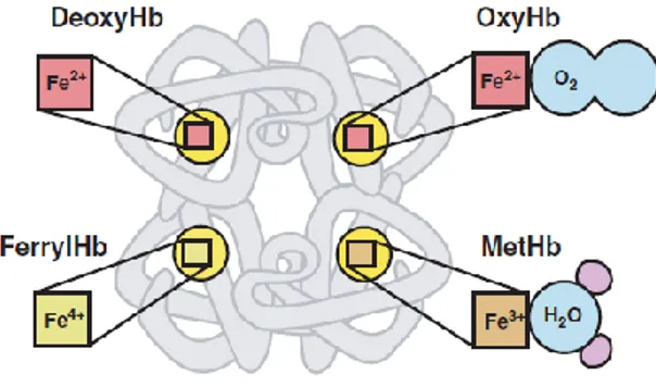
Figure 6-Different oxidative states of the heme iron. Each of the four globin chains of the hemoglobin
(Hb) molecule contains a heme group with an iron atom at its center. Hb oxidation can involve changes in the iron’s oxidative state, and, consequently, modifications in ligand binding. The reduced ferrous state (Fe2+, red) allows oxygen binding to deoxygenated Hb (deoxyHb) at physiological conditions. The resultant oxygenated Hb (oxyHb) can undergo oxidation to methemoglobin (metHb), in which the heme iron is in the ferric state (Fe3+, brown). MetHb can bind water, but not oxygen under physiological conditions. The ferryl state (Fe4+, green) can be obtained during Hb oxidative injury by hydrogen peroxide. This state can lead to Hb denaturation [Misra et al., 1972].
Nevertheless, several erythrocyte abnormalities that circumvent or overwhelm the erythrocyte oxidant defense system have been identified, such for example, those related to hypothermic storage. In fact, although cooling below normal physiological temperatures partially contrasts the accumulation of oxidative injuries, (I) the rate of met-Hb reduction by cytochrome b5 reductase is slowered; (II) met-Hb may be more
Chapter 1
21
prone to denaturation as suggested by lower thermodynamic stability of met-myoglobin at 4°C; and (III) the solubility of oxygen is doubled at 4°C. As a result, oxidative damage can accumulate with refrigerated red cell liquid storage [Zolla and D’Alessandro, 2011]. Moreover, the storage means (e.g. plastic bag and additive solutions) contribute to exacerbate this phenomenon, by promoting an increased concentrations of intracellular ROS during blood storage [Deepa Devi et al., 1998]. Under certain conditions, including heat stress, changes in pH, senescence [Sugawara et al., 2003] and also increased Hb autoxidation, MetHb can be converted into low-spin ferric Hb compounds named hemichromes (haemoglobin whose cysteine residues have been oxidised, leading to the formation of aggregates). The release of the heme moiety by denatured hemichromes has been shown to initiate oxidative damage to adjacent membrane components [Atamna et al., 1995, Li et al., 2006]. Free heme has been shown to inhibit a number of cytosolic enzymes [Scott et al., 1993], as well as to react nonspecifically with membrane proteins leading to abnormal thiol oxidation and cross-linking [Atamna et al., 1995] (especially at the level of the cytoplasmatic domain of band 3), with consequent reduction of its deformability. Although free heme is degraded in vivo through the heme oxygenase system [Kapitulnik 2004], during prolonged storage, when the flux of glycolytic pathway decreases and NADH concentrations fall, release of the heme iron has been shown to be catalyzed by intracellular GSH [Atamna et al., 1995] or hydrogen peroxide [Nagababu et al.,1998]. As previously stated, free iron is potentially toxic capable of acting as a Fenton reagent in the Haber–Weiss cycle, which produces the deleterious hydroxyl radicals (OH•) and finally results in the formation of Heinz bodies [Chiu et al., 1989] (Figure 7).
1.3.5 Hemoglobin and the functions of nitric oxide (NO) correlated with the Red Cell Storage Lesion
Although hemoglobin can induce significant oxidative stress and inflammation, a central mechanism for toxicity is likely the direct effect of free hemoglobin on nitric oxide (NO) catabolism. Nitric oxide has a myriad of functions in human physiology including neurotransmitter, host-defense molecule, inhibitor of platelet aggregation, regulator of endothelial adhesion molecule expression, and antioxidant. It is perhaps most widely recognized for its role in metHb formation. In fact, during this reaction,
Chapter 1
22
that is rate limited by diffusion to the heme group within Hb [Doyle et al., 1981, Eich et al., 1996, Herold et al., 2001], oxygenated Hb reacts with NO to form metHb (FeIII) and nitrate (NO3ˉ), and effectively destroys NO activity.
HbFe(II) O2 + NO → HbFe(III) + NO3ˉ
This rapid binding means a reduced NO bioavailability for vasodilation, although the erythrocyte can be considered a potent NO sink [Kevil et al., 2010, Ferrari et al., 2009]. However, as noted above, cell-free Hb reacts with NO much faster (1000 times) than RBC-encapsulated Hb. With increasing length of storage, the concentration of free hemoglobin (in the form of ferrous oxyhemoglobin) in RBCs increases, with a direct, proportional consumption of NO to the concentration of free heme. In addition, after a dramatic increase in the number of microparticles in stored RBCs, cell-free Hb and Hb-containing microparticles scavenge NO very fast. It is easy to see how disruption of the RBC membrane, as in hemolysis (with a range of extracellular Hb levels reported in the literature from 28 mmol/L - in heme - after 35 days of storage to 80 mmol/L after 50 days of storage [Bonaventura et al., 1997]), will effectively contribute to affect NO bioavailability upon transfusion. Nevertheless, several mechanisms exist to prevent the depletion of NO through interaction with hemoglobin. The first is that hemoglobin located within the RBC membrane (RBC-encapsulated Hb) reacts with NO much more slowly (1000 times) than does cell-free hemoglobin [Coin et al., 1979], primarily due to the limitation of NO diffusion to erythrocyte through “an unstirred layer” [Azarov et al., 2005]. A cell-free zone at the blood-endothelial interface is created by laminar blood flow, which pushes RBCs to the center of the vessel and away from endothelial cell, where NO is produced. In addition, the RBC membrane has a finite permeability to NO. Indeed, any NO at greater distances from the external surface of the red cell membrane has the rate limited by NO diffusion to the red cell. It is of great biological significance that the Stamler laboratory has demonstrated another relevant reaction of NO with Hb: S-nitrosylation of a cysteine residue, conserved in β-globins of all birds and mammals (βCys93 of human Hb), which yields S-nitrosohemoglobin (SNO-Hb) [Jia et al., 1996, Gow et al., 1998, Bonaventura et al., 1997].
Chapter 1
23
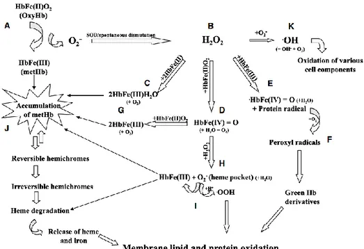
Figure 7-Summary of the major hemoglobin (Hb) oxidative pathways and their link to membrane
damage. (A) Autoxidation of oxygenated Hb [oxyHb, HbFe(II)O2] generates methemoglobin [metHb,
HbFe(III)] and superoxide anions (O2ˉ). Spontaneous or enzymatic dismutation of O2ˉ generates hydrogen
peroxide (H2O2).(B) Accumulation of H2O2 caused by the loss of vital antioxidants (catalase, glutathione)
may cause irreversible oxidative damage to Hb molecules. H2O2 can react with deoxygenated Hb
[HbFe(II)], oxyHb and metHb to generate metHb (C), ferrylHb [HbFe(IV)=O] (D) and oxoferrylHb [·HbFe(IV)=O] (E), respectively. The reaction between H2O2 and metHb (E) also generates a protein
radical that can further react with oxygen to yield peroxyl radicals (F) and, eventually, decompose to lipophilic green Hb derivatives, which bind to membrane lipids and induce oxidative damage. FerrylHb (D) can react with oxyHb to generate two molecules of metHb (G), or with H2O2 (H) to generate metHb
and O2ˉ. The formation of O2ˉ within the heme pocket of the Hb has been shown to result in heme
degradation. Alternatively, a protonation of O2ˉ generates perhydroxyl radicals (•OOH) (I) that are known
to initiate membrane lipid peroxidation. Accumulation of intracellular metHb (J) may result in the formation of hemichromes. Irreversible hemichromes undergo further denaturation leading to heme degradation and release of the heme moiety and iron atoms. Both degradation products have been shown to initiate membrane oxidative damage. The presence of O2ˉ and H2O2 may result in the formation of
hydroxyl radicals (•OH) (K) through the Haber–Weiss reaction. Heme degradation releases iron atoms, which can catalyse this reaction. •OH is highly reactive and can cause oxidative damage to various cell components. Note that all three major oxygen radicals (O2ˉ, H2O2 and •OH) can take place in the
oxidative denaturation of Hb and membrane damage. Remark: In order to simplify this model, reaction products that do not participate in oxidative pathways (i.e. H2O, O2) are denoted by smaller fonts in
parentheses [Kanias et al., 2009].
Importantly, under hypoxic conditions, SNO-Hb retains endothelium-derived relaxing factor (EDRF/NO) like bioactivity and is capable of transferring NO to low molecular weight thiol-containing molecules (e.g. the membrane anion exchange protein, glutathione). Notably, allosteric structural transitions of Hb, triggered by changes in oxygen tension in vivo, contribute to the molecular gymnastics that dictate NO addition and release from Hb-βCys93 [Jia et al., 1996, Gow et al., 1998, Bonaventura et al., 1997].
Chapter 1
24
Although mechanistic details continue to be debated, recently it has been observed that levels of SNO-Hb are altered in several disease states, characterized by disorders in tissue oxygenation [Reynolds et al., 2007], like these affecting banked blood. The storage-related deficiency of SNO-Hb, with associated impaired vasoregulation hinders to improve oxygen delivery by RBCs in the microcirculation, predisposing excess mortality and morbidity accompanying RBC transfusion. However, under conditions of exacerbated Hb autoxidation (like ex-vivo storage of RBCs), when produced in excess, a significant amount of O2ˉ• reacts with NO to produce peroxynitrite (ONOOˉ), a strong oxidant formed intravascularly [Darley-Usmar et al., 1995], with a very short half-life (g.e. milliseconds). Peroxynitrite can promote the nitration of aromatic compounds such as protein tyrosine residues, a process that can profoundly alter protein structure and function, and requires the intermediate formation of secondary nitrating species. Generally, peroxynitrite can behave as either a one- or two-electron oxidant; in this particular case, one-electron oxidants derived from peroxynitrite attack the phenolic ring, leading to formation of a tyrosyl radical, followed by a coupling of the amino acid-derived radical with •NO2 to yield 3-nitrotyrosine (3-NT) [Romero et al., 2003]. The biological significance of tyrosine nitration supports the formation of 3-NT in vivo in diverse pathologic conditions and the formation of 3-NT is thought to be both a relatively specific marker of oxidative damage mediated by peroxynitrite and other nitrogen free radical species [Radi 2013] and a post-translational modification with important pathophysiological consequences, that disrupts NO signaling and skews metabolism towards pro-oxidant processes. Concerning RBCs, recent studies have shown that peroxynitrite induces morphological alterations, phosphatidylserine externalization, activation of caspase cascades, increased amount of membrane-bound Hb (presumably oxidized/denatured Hb), band 3 phosphorylation and a burst of both the glycolytic pathway (with lactate accumulation) and the pentose phosphate shunt [Matarrese et al. 2005, Metere et al., 2009]. Interestingly, all of these are also features of storage lesions, thus the investigation of protein nitration in stored RBCs would be beneficial and of great interest.
Chapter 1
25
1.3.6 Lipid Peroxidation
It is generally believed that membrane lipids are major targets for cellular damage induced by free radicals. Red blood cells offer a number of advantages studies of effects of oxidants on protein and lipid breakdown and oxidation respectively. As previously stated, oxygen radicals are produced continually in red cells and may cause and damage macromolcules and cellular structure of the organism, endothelium erythrocytes and lipid peroxidation (the level of lipid peroxidation expressed as malondialdehyde). The latter initiates when ROS remove a hydrogen atom from methylene carbons of fatty acid side chains of Polyunsaturated fatty acids (PUFAs), resulting in lipid radical which reacts with molecular oxygen to yield peroxyl radicals. Peroxyl radicals propagate the oxidative process by removing hydrogen atoms from adjacent fatty acids, and consequentially create various lipid hydroperoxides which further decompose to secondary lipid peroxidation products such as malondialdehydes (MDAs) [Boaz et al., 1999; Fiorillo et al., 1998]. MDAs have been widely used as lipid peroxidation indicators. MDAs have been shown to have adverse effects on cell integrity as shown by membrane damage and hemolysis in RBCs. The lipid peroxidation of polyunsaturated fatty acids may be enzymatic and non-enzymatic. Enzymatic lipid peroxidation is catalyzed by the lipoxygenases family, a family of lipid peroxidation enzymes that oxygenates free and esterified PUFA generating as a consequence, peroxy radicals. Researchers have focused their attention on lipid peroxidation by non-enzymatic, non-radical mechanisms and formation of lipidperoxides, which is initiated by the presence of molecular oxygen and is facilitated by Fe2+ ions [Repetto et al., 2010]. Singlet oxygen and ozone are examples of molecules that induce such oxidation The presence of cholesterol in cell surface membranes influences their susceptibility to peroxidation, probably both by intercepting some of the radicals present and by affecting the internal structure of the membrane by interaction of its large hydrophobic ring structure with fatty acid-side-chains. As lipid peroxidation procedes in any membrane and several of the products, including hydroperoxides, cleavage products such as aldehydes, and polymeric materials produced, have a detergent-like activity, or exert cytotoxic and genotoxic effects. This will contribute to increased membrane disruption and further peroxidation.
Chapter 1
26
The onset of lipid peroxidation within biological membranes is associated with changes in their physicochemical properties and with alteration of biological function of lipids and proteins [Catala 2006].
1.3.7 The effects of phosphorylation on integrity membrane structure of stored RBCs under blood banking conditions
It has been known for decades that phosphorylation plays an important role in cell cycle, apoptosis, metabolism, receptor function and stress responses, through the activation of signaling cascades [Dephoure et al., 2008, Macek et al., 2009, Beltran et al., 2012]. The identification of the storage-associated alterations in the interaction between the cytoskeleton and the lipid bilayer that determine membrane elasticity has been widely documented [Berezina et al., 2002, Blasi et al., 2012, Cluitmans et al., 2012]. Protein phosphorylation/dephosphorylation is one of the physiological processes possibly controlling the membrane stability. In fact, recent data indicate that phosphorylation is involved in molecular interactions at several levels and thereby contributes to the integrity of the general membrane structure [Manno et al., 2005, De Oliveira et al., 2008]. In particular, a central position on the functionally relevant changes in the membrane composition of stored RBCs is occupied by alterations of phosphorylation status of band 3.Cluitmans and collegues have postulated that altered phosphorylation of band 3 on tyrosine residues, due to oxidative stress, may be responsible for the metabolic changes, such as the described increase in glycolytic intermediates within the first two weeks of storage [D'Alessandro et al., 2012, Gevi et al., 2012] (in fact, the cytoplasmic domain of band 3 has a high affinity for key enzymes of the glycolysis, and binding is regulated by phosphorylation). The oxidative stress-dependent increase in band 3 tyr-phosphorylation is a trigger for activation of src tyrosine kinases (Syk and Lyn) [Mallozzi et al., 2001] and down-regulation of tyrosine phosphatases (PTP1B, SHP-1 and SHP-2) [Zipser et al., 2002]. The src kinases phosphorylate tyrosines 8 and 21 of the cytosolic domain of the band 3 protein, thereby facilitating formation of high-molecular-mass band 3 aggregates [Pantaleo et al., 2009]. Both srk kinases syk and lyn and the tyrosine phosphatase PTP1B are redox-sensitive, indirectly the first, because their activity is controlled by the redox- and calcium-sensitive phosphorylation steps mediated in particular by protein kinase C alpha. On the
Chapter 1
27
contrary, PTP1B possesses cysteine residues in the kinase domains, whose oxidation is direct induced by oxidative stress [Bordin et al., 2005, Knock and Ward, 2011]. The resulting cumulative tyrosine hyperphosphorylation, during last weeks of storage, decreases the affinity of band 3 to ankyrin, causing destabilization of the band 3-cytoskeleton interaction, increases the lateral mobility of band 3 within the membrane and induces a progressive vesiculation [Ferru et al., 2011]. In addition, recent researches have demonstrated the association between casein kinase II-catalyzed phosphorylation of beta spectrin serine and protein kinase C- catalyzed phosphorylation of protein 4.1 and complement receptor- mediated increase in deformability [Glodek et al., 2010]. Phosphorylation of band 4.1 promotes dissociation of actin from the cytoskeleton, also contributing to an increase in deformability [Manno et al., 2005, De Oliveira et al., 2008].
References
28
References
Hess J. An update on solutions for red cell storage. Vox Sanguinis (2006) 91, 13–19.
Hess J. Red cell storage. Journal of Proteomics, Volume 73, Issue 3, 3 January 2010, Pages 368–373.
Hess J. Red cell changes during storage. Transfusion and Apheresis Science 43 (2010) 51–59.
Högman et al. Storage parameters affecting red blood cell survival and function after transfusion. Transfusion Medicine Reviews Volume 13, Issue 4, October 1999, Pages 275–296.
Ian Chin-Yee et al. The Red Cell Storage Lesion and its Implication for Transfusion. Transfus. Sci. Vol. 18, No. 3, pp. 447±458, 1997.
Bennett-Guerrero E, Veldman TH, Doctor A, Telen MJ, Ortel TL, Reid TS, Mulherin MA, Zhu H, Buck RD, Califf RM, McMahon TJ. Evolution of adverse changes in stored RBCs. Proc Natl Acad Sci U S A. 2007 Oct 23; 104(43):17063-8.
Tamara L. Berezina et al. Influence of Storage on Red Blood Cell Rheological Properties. Journal of Surgical Research 102, 6–12 (2002) doi:10.1006/jsre.2001.6306.
Lockwood WB, Hudgens RW, Szymanski IO, Teno RA, Gray AD. Effects of rejuvenation and frozen storage on 42-day-old AS-3 RBCs. Transfusion 2003;43:1527– 32.
Salzer U, Zhu R, Luten M, Isobe H, Pastushenko V, Perkmann T. et al. Vesicles generated during storage of red cells are rich in the lipid raft marker stomatin. Transfusion 2008;48:451–62.
References
29
Dumaswala UJ, Zhuo L, Jacobsen DW. et al. Protein and lipid oxidation of banked human erythrocytes: role of glutathione. Free Radic Biol Med 1999; 27: 1041-9.
Umakant j. Dumaswala, Limei Zhuo, Donald W. Jacobsen, Sushil K. Jain, Katherine A. Sukalski Protein and lipid oxidation of banked human erythrocytes: Role of glutathione. Free Radical Biology & Medicine, Vol. 27, Nos. 9/10, pp. 1041–1049, 1999.
D'Alessandro A, D'Amici GM, Vaglio S, Zolla L. Time-course investigation of SAGM-stored leukocyte-filtered red bood cell concentrates: from metabolism to proteomics. Haematologica 2012; 97: 107-15.
Kriebardis AG, Antonelou MH, Stamoulis KE, et al. Progressive oxidation of cytoskeletal proteins and accumulation of denatured hemoglobin in stored red cells. J Cell Mol Med 2007; 11: 148-55.
D'Amici GM, Rinalducci S, Zolla L. Proteomic analysis of RBC membrane protein degradation during blood storage. J Proteome Res 2007; 6: 3242-55.
Walpurgis K, Kohler M, Thomas A, et al. Storage-induced changes of the cytosolic red blood cell proteome analyzed by 2D DIGE and high-resolution/high-accuracy MS. Proteomics 2012; 12: 3263-72.
Card R. Red cell membrane changes during storage. Transfus Med Rev 1988; 2: 40-7.
Wolfe L, Byrne A, Lux S., Molecular defect in the membrane skeleton of blood bank-stored red cells. Abnormal spectrin-protein 4.1-actin complex formation. J Clin Invest 1986; 78: 1681-6.
Brad S. Karon, Camille M. van Buskirk, Elizabeth A. Jaben, James D. Hoyer, David D. Thomas Temporal sequence of major biochemical events during Blood Bank storage of packed red blood cells. Blood Transfus 2012; 10: 453-61.
![Table 3-Oxidative Modifications of Proteins [Shacter 2000]](https://thumb-eu.123doks.com/thumbv2/123dokorg/2777080.1408/20.892.123.771.872.1095/table-oxidative-modifications-of-proteins-shacter.webp)
![Figure 4-A general scheme for the different routes for protein carbonylation [Ashraf Madian, 2010]](https://thumb-eu.123doks.com/thumbv2/123dokorg/2777080.1408/24.892.151.746.120.549/figure-general-scheme-different-protein-carbonylation-ashraf-madian.webp)