Archives • 2017 • vol.1 • 46-54
EFFECTS ON IN VIVO ANGIOGENESIS OF EXTRACTS FROM ASTRONIUM URUNDEUVA (FR. ALL.)
ENGL. (ANACARDIACEAE)
Negri, K.M.S
1.; Malafronte, N.
2; Certo, G
3.; D’Angelo, V.
3; Rapisarda, A.
3; Bauab, T.M.
1; Germanò,
M.P.
3*
1Department of Biological Sciences, Univ Estadual Paulista, Araraquara, São Paulo, Brazil 2Department of Pharmacy, University of Salerno, Via Giovanni Paolo II 132, 84084 Fisciano (SA), Italy 3Department of Chemical, Biological, Pharmaceutical and Environmental Sciences, University of Messina, Italy
Abstract
Astronium urundeuva (Fr. All.) Engl. (aroeira-do-sertão) (Anacardiaceae) is an arboreal species typical of
northeast brazilian savanna popularly used for its anti-inflammatory, antimicrobial, anti-ulcerogenic and healing properties.
In this study the effects of A. urundeuva on in vivo angiogenesis are evaluated. Stem bark (AUS) and leaves (AUL) extracts (EtOH 70%) are investigated on zebrafish embryos and chick chorioallantoic membrane (CAM).
Results of the biological screening show that AUS extracts induce a weak anti-angiogenic response in both experimental models. Conversely, AUL extracts at 25 and 50 µg/mL enhance vessel formation in zebrafish embryos submitted to the endogenous alkaline phosphatase (EAP) assay. Moreover, an increased blood vessel development and an higher level of total hemoglobin content is observed in the CAMs exposed to AUL at 80 µg/egg as compared to control.
The proangiogenic effects of AUL in two in vivo models suggest its potential use as a source of new therapeutic agents able to modulate angiogenesis.
Keywords: Astronium urundeuva, angiogenesis, zebrafish, endogenous alkaline phosphatase staining, chick
Introduction
Angiogenesis is a fundamental process in many physiological conditions such as the embryonic development, ovulation and wound repair involving the growth of new blood vessels from preexisting vessels [1]. On the other hand, the balance of activators and inhibitors of angiogenesis contributes to numerous pathologies, such as cancer, age-related macular degeneration, rheumatoid arthritis, and inflammation [2]. Thus, the identification of new substances having angiogenic properties is actually of a great interest [3].
Astronium urundeuva (sin. Myracrodruon urundeuva) is a
broadleaf deciduous tree confined to Seasonally Dry Tropical Forests (SDTF) in South America. The trunk is straight and cylindrical. The characteristic outer bark is dark brown and scaly, with a rough surface and longitudinal depressions. The inner bark is fibrous and pinkish. The stem bark is collected intensively and it is used by indigenous people particularly in Caatinga (Brazil) to treat metrorragias, gastric ulcers, inflamed sores, respiratory and urinary diseases, microbial infections. The compound imparipinnate leaves are oblong or ovate, subcoriaceous. They are used in popular medicine for wound healing, painful, ulcerative and inflammatory conditions [4]. Reports on previous studies described anti-inflammatory [5,6]; analgesic and antiulcer [7,8,9], antibacterial and antifungal activities [10,11,12], healing properties [13]. According to the traditional use of this plant neuroproptective effects in an in vivo model have been also described [14,15]. A previous study reported that the methanol extract of A. graveolens leaves shows anti-angiogenic properties by regulating the placental growth factor (PIGF), an angiogenesis modulator and allowed identification of 1,2,3,4,6-penta-O-galloyl-D-glucopyranose as the most active compound [16]. Basing on these scientific evidences and considering that the discover of new therapeutic agents able to module angiogenesis has recently generated much attention, in this study we evaluate the effects of A. urundeuva stem bark and leaves extracts on angiogenesis using two in vivo models: the zebrafish embryos and the chick chorioallantoic membrane (CAM).
Methods
General experimental procedures
NMR experiments were performed on a Bruker DRX-600 spectrometer (Bruker BioSpin Germany) equipped with a Bruker 5 mm TCI CryoProbeat 300 K. HR-ESIMS were acquired on a Q-TOF premier spectrometer (Waters-Milford). [17].
Plant material
A. urundeuva stem bark and leaves were collected in
Bálsamo and Votuporanga (Brazil). The authenticity of the samples was compared with Herbarium files from the State University of Campinas (Unicamp). A voucher specimen of A. urundeuva is deposited under the registration no. 1446.
Extraction
The dried and powdered stem bark (100 g) and leaves (100 g) were submitted to exhaustive extraction (EtOH 70%) by percolation according to the Brazialian Pharmacopeia [18]. The obtained extracts were concentrated under reduced pressure at 40°C. The yields were 2% for stem bark (AUS) and 3% for leaves (AUL) extracts, respectively [19].
Chemicals and reagents
2-methoxyestradiol, retinoic acid, β-sitosterol, endogenous alkaline phosphatase (EAP) staining, propylthiouracil (PTU), nitroblue tetrazolium chloride/5-bromo-4-chloro-3-indolyl phosphate, toluidine salt (NBT/BCIP) ready-to-use tablets, Drabkin’s reagent, standard hemoglobin were obtained from Sigma-Aldrich (Milan, Italy).
Zebrafish assay
Zebrafish embryos were obtained from wild type fishes bought from local pet stores and maintained in flow through aquaria at 28.5°C on a 14/10 h (light/dark) photoperiod.
Embryos were generated by natural mating as described by Westerfield [20] and they were cultured in embryo water (0.2 g/L Instant Ocean® Salt, Aquarium Systems, USA) at 28.5°C. Developmental age of the embryos was correspondent to hours post fertilization (hpf) [21]. The ethical guidelines, described by the National Institutes of Health Guide for care and Use of Laboratory Animals were followed throughout the experiments.
Healthy and regular zebrafish embryos were selected at 24 hpf, distributed in 96 single-well microplates (one embryo per well) and incubated with 100 µL of embryo water containing AUS or AUL extracts (25, 50 and 100 µg/mL), 2-methoxyestradiol (2-ME) (30 µg/mL) or β-sitosterol (10 µg/mL), employed as antiangiogenic/proangiogenic reference compounds. DMSO (0.2%) was used as vehicle for those treatments. Control group received only DMSO. Control and treated embryos (10 for each group) were incubated until 72 hpf. Quantitative Endogenous Alkaline Phosphatase (EAP) assay was performed as described previously [22]. Treated embryos at 72 hpf were dehydrated by increasing concentration of ethanol and then, they were washed
three times with diethanolamine buffer and stained according to the protocol described in phosphatase substrate kit. After staining, 50 µL NaOH (2 M) was added to stop the reaction. The optical density of the soluble end product was measured at 405 nm, using a microplate reader (Bio-Rad Model 2550 EIA READER). Vessel growth was determined as % optical density (OD) compared with control using the following equation: [% vessel formation
= (OD treated day 3 − OD control day 1)/(OD control day 3
− OD control day 1) × 100%]. Each assay was repeated at least three times.
Endogenous Alkaline Phosphatase (EAP) staining for visual inspection NBT/BCIP substrate was used to stain the blood vessels. Embryos at 24 hpf were preliminarily incubated with embryo water containing PTU (final concentration 0.2 mM) before sample administration in order to prevent pigment development to facilitate blood vessel observation. At 72 hpf, embryos were fixed with paraformaldehyde (4%) in PBST (phosphate-buffered saline + 0.1% tween-20) for 30 min. Then, the embryos were dehydrated with ethanol, rinsed with PBST and equilibrated with NTMT (Tris buffer 100 mM pH 9.5; NaCl 100 mM; MgCl 250 mM; 0.1% Tween-20). The staining reaction was started by incubating embryos with NBT/BCIP solution for about 15–30 min according to the protocol described for NBT/BCIP kit (Sigma –Aldrich, Milan, Italy). After staining was completed, the embryos were washed with PBST and they were examined by an optical microscope (Leica DMLB).
Chick chorioallantoic membrane (CAM) assay
The chick chorioallantoic membrane (CAM) assay was performed following the method of Song [23] modified [24]. Fertilized eggs of Gallus gallus were previously maintained in a humidified incubator at 37°C and, after four days of incubation, a small window was created on the broad side of the eggs to apply different doses of AUS and AUL extracts (30, 50, and 80 μg/egg) directly on the CAM surface, previously suspended in albumen. Retinoic acid (2 μg/egg), vascular endothelial growth factor (VEGF) (0.25 μg/egg) were used as antiangiogenic/proangiogenic reference compounds. After treatment, the eggs were reincubated for two days, then they were observed under a steromicroscope (Zeiss Stemi 2000-c) equipped with a digital camera (Axiocam MRc 5 Zeiss) and photographed. The antiangiogenic effects on the CAMs were quantified by counting the number of blood vessel branch points in a standardized area using an artistic software (Paint). To evaluate the hemoglobin content, CAMs were isolated and homogenized for 1 min in KCl (1.15%) with an ULTRA-TURRAX® homogenizer. The CAM homogenate (20 µL) was mixed with Drabkin’s reagent (5 mL) containing ferricyanide which reacts with hemoglobin to form
met-Hb and then cyan met-met-Hb [25]. After 5 min of incubation in the dark at room temperature, the absorbance values were read with a spectrophotometer (Shimadzu UV1601) at 540 nm. The hemoglobin content was extrapolated using a calibration curve (10 - 90 mg /mL) and finally expressed as mg/g of CAM.
Statistical Analysis
All values obtained were calculated as mean ± standard deviation of minimum three replicates. Statistical differences between control and treated groups were tested by Student’s t-test. Variances with p<0.05 were considered statistically significant.
Results
EAP assay on zebrafish embryos
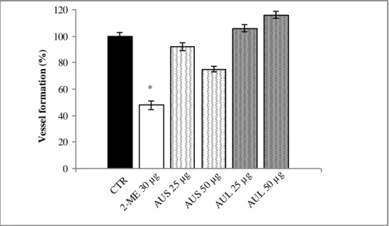
The EAP assay was used to assess the effects of AUS and AUL extracts on angiogenesis. AUS treatment produced a weak anti-angiogenic activity. The vessel formation in zebrafish embryos treated with 25 and 50 μg/mL is 92% and 72% respectively, as compared to control (Fig. 1). Treatment with the highest dose (100 μg//mL) induced the death over to 50% of embryos (data not shown). The vessel formation is inhibited in zebrafish embryos treated with 2-methoxyestradiol (2-ME) used as reference substance (48% at 30 μg/mL). Conversely, AUL exhibited pro-angiogenic effects: vessel formation is 104% and 112% at 25 and 50 μg/mL, respectively. Even in this case, the highest dose of AUL (100 μg//mL) induced 50% death of embryos (data not shown).
The EAP staining was also performed to visualize the subintestinal vessels (SIVs) in zebrafish embryos treated with AUL extracts. Fig. 2a shows microscopic images of SIVs in control group extented on the dorsolateral surface of embryo yolk in the shape of a basket. Fig. 2b represents treatment with AUL at 50 μg/mL showing the increased number of SIVs comparable to β-sitosterol (10 µg/mL) used as pro-angiogenic reference compound (Fig. 2c).
CAM assay
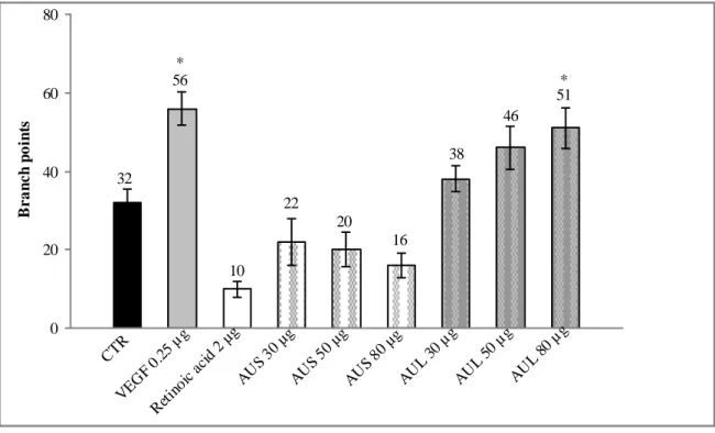
In the CAM assay, the results, expressed as numbers of blood vessel branch points, are reported in Fig. 3. It is evident that while AUS extracts inhibit vessel formation comparable to the anti-angiogenic reference compound retinoic acid, AUL extracts induce opposite effects due to the increased number of blood vessel branch points respect to control. The pro-angiogenic effects of AUL were dose dependent and they were compared to VEGF treatment. Representative microscopic images of the CAM in control eggs show the presence of a clear
vascular net-work with large vessels converging towards the embryo (Fig. 4a). AUL extract at the dose of 80 µg/egg is able to promote angiogenesis as comparable to VEGF treatment at 0.25 µg/egg (Figs b and c).
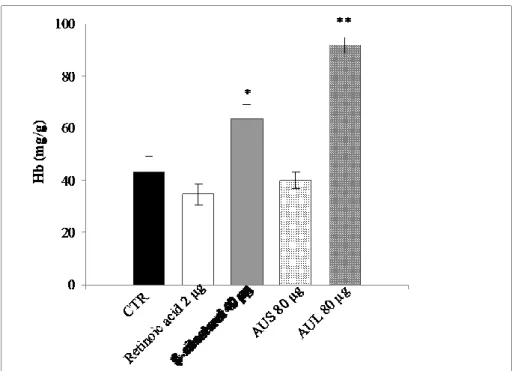
This pattern of angiogenesis modulation is also confirmed quantifying hemoglobin content in the CAMs. Results evidence that the hemoglobin content is significantly increased after treatment with AUL extract at the highest dose (91.85 mg/g at 80 µg/egg) respect to control (43.42 mg/g) confirming the good pro-angiogenic response, better than β sitosterol (63.42 mg/g at 40 µg/egg), used as reference compound. The hemoglobin content in the CAMs treated with AUS at the same dose is almost comparable to control (42.54 mg/g). The standard anti-angiogenic compound, retinoic acid, induces a lower hemoglobin content in the CAMs (34.61 mg/g at 2 µg/egg).
Discussion
Medicinal plants of Caatinga, a type of dry-savannah predominant in the semi-arid region of northeastern Brazil, are extensively used for various therapeutic treatments and represent a rich source of compounds that might be useful for the development of new pharmaceutical agents [26]. A. urundeuva, popularly known as “aroeira” is used to treat various illness for its medicinal properties that have been proven scientifically [4]. In this study the effectiveness of A. urundeuva extracts (AUS and AUL) to modulate angiogenesis was evaluated considering its essential role both in many physiological events and also in pathological conditions. The results demonstrate that AUS and AUL extracts are able to interact with the 'targets' of angiogenesis in an opposite way. This experimental evidence was achieved employing two in vivo models: the zebrafish embryo and the chick chorioallantoic membrane.
The zebrafish model is widely used as a new drug screening platform for the ease of embryo maintenance and the simplicity of experimental techniques, since the development of blood vessels in early stages is well characterized and easily monitored [27]. In this study zebrafish embryos were exposed to AUS and AUL extracts from 24 to 72 hours post-fertilization (hpf). This window exposure is employed because vessel development begins at about 24 hpf and the whole vasculature is completed by 72 hpf. After this time of incubation, the number of dead and affected embryos was scored and the endogenous alkaline phosphatase (EAP) activity of vascular endothelial cells was quantified to assess the effect on angiogenesis. As EAP activity was slightly decreased in zebrafish treated with AUS demonstrating a weak antiangiogenic response, AUL induced opposite effects increasing vessel formation in
the treated embryos. The EAP staining was performed to visualize the developing subintestinal vessels (SIVs) which are an easily visible vascular bed to screen for molecules that influence angiogenesis [28] Although previous studies [29,30] described toxic effects mainly when A.
urundeuva ethanol extracts were used except with higher
dilutions (< 10 μg/mL), our results showed that doses lower than 100 μg//mL showed no sign of mortality. The CAM model offers advantages that include the comparative ease of culture, low cost and easy observation of the neovascularization while the embryo is still alive, without the need to sacrifice the animals at each timepoint [31]. The CAM assay is also a versatile tool for in vivo evaluation both antiangiogenic and proangiogenic natural products [23]. Additionally, possible side effects, like membrane irritation and toxic properties, can be observed in the same system. Our results suggest no pronounced antiangiogenic effects for AUS extracts while an increased vessel formation after treatment of the CAMs with AUL at the tested concentrations being significant at the highest dose (80 µg/egg) on the hemoglobin content as an index of vascular density. It is important to underline that both the extracts, even at the highest dose, showed no sign of irritation in our system.
Relating to the chemical composition, a previous study reported the presence of tannins in A. urundeuva leaves and stem bark [32]. In addition, chalcones having analgesic and anti-inflammatory activities [5] and neuroprotectrive effects [14] were isolated. The phytochemical prospecting of the essential oil from the leaves has been recently reported [11]. Furthermore, chemical investigations on Astronium species, revealed the presence of a series of gallotannins with a degree of polymerization (DP) from four to eight galloyl units in the stems and a DP extended up to tridecagalloylglucose in the leaves [33].The same authors reported additional informations on the different composition of stems and leaves due to detection of flavonoid constituents only in the leaves, while gallotannins both in stems and leaves. It may be hypothized that the chemical composition may influence the different biological response of AUS and AUL extracts in the two experimental models of angiogenesis. Anyway, it is known that the occurrence of pentagalloylglucose in Astronium species is pharmacologically interesting due to their capability to exhibit multiple biological activities [34].
In conclusion, Astronium urundeuva is known in popular medicine of Brazil and it has demonstrated many interesting biological activities. Recently, angiogenesis modulation becomes a promising strategy for the treatment of many human diseases. The positive
angiogenic response of extracts from A. urundeuva leaves in two in vivo models suggests their use as a source of new angiogenesis-promoting therapeutic agents.
Acknowledgments
The authors acknowledge “Prof. Imbesi Foundation” for its support
.
References
1. Auerbach R, Lewis R, Shinners B, Kubai L, Akhtar N. Angiogenesis Assay: A Critical Overview. Clinical Chemistry 2003; 49(1):32-40.
2. Folkmann J. Angiogenesis and apoptosis. Semin Cancer Biol. 2003;13:159-167.
3. Majewska I, Gendaszewska-Darmach E. Proangiogenic activity of plant extracts in accelerating wound healing - a new face of old phytomedicines. Acta Biochim Pol. 2011;58(4):449-460.
4. Cartaxo SL, Souza MMA, Albuquerque UP. Medicinal plants with bioprospecting potential used in semi-arid northeastern Brazil. Journal of Ethnopharmacology 2010;131:326-342.
5. Viana GSB, Bandeira MAM, Matos FJA. Analgesic and antiinflammatory effects of chalcones isolated from
Myracrodruon urundeuva Allemão. Phytomedicine
2003;10(2-3):189-195.
6. Rastrelli L, De Tommasi N, Berger I, et al. Glycolipids from Byrsonima crassifolia, Phytochemistry 1997; 45: 647-650.
7. Menezes AMS, Rao VSN, Fonteles MC. Antiulcerogenic activity of Astronium urundeuva. Fitoterapia 1986;57:253-256.
8. Rao VS, Viana GS, Menezes AM, Gadelha MG. Studies on the anti-ulcerogenic activity of Astronium urundeuva Engl. II. aqueous extract. Braz J Med Biol Res. 1987;20(6):803-805.
9. Carlini EA, Duarte-Almeida JM, Rodrigues E, Tabach R. Antiulcer effect of the pepper trees Schinus
terebinthifolius Raddi (aroeira-da-praia) and
Myracrodruon urundeuva Allemão, Anacardiaceae
(aroeira-do-sertão). Rev Bras Farmacogn. 2010;20(2):140-146.
10. Sá RA, Gomes FS, Napoleão TH, et al. Antibacterial and antifungal activities of Myracrodruon urundeuva
heartwood. Wood Sci. Technol. 2009;43:85-95.
11. Figueredo FG, Lucena BFF, Tintino SR, et al. Chemical composition and evaluation of modulatory of the antibiotic activity from extract and essential oil of
Myracrodruon urundeuva. Pharmaceutical Biology
2014;52(5):560-565.
12. Bonifácio BV, dos Santos Ramos MA, da Silva PB, et al. Nanostructured lipid system as a strategy to improve the anti-Candida albicans activity of Astronium sp. International Journal of Nanomedicine 2015;10:5081-5092. 13. Rodrigues LV, Ferreira FV, Regadas FSP, Matos D, Viana GSB. Morphologic and morphometric analyses of acetic acid-induced colitis in rats after treatment with enemas from Myracrodruon urundeuva Fr. All.(Aroeirado Sertão). Phytotherapy Research 2002;16:267-272.
14. Nobre-Júnior HV, Oliveira RA, Maia FD, et al. Neuroprotective effects of chalcones from Myracrodruon
urundeuva on 6-hydroxydopamine-induced cytotoxicity in
rat mesencephalic cells. Neurochem Res. 2009;34:1066-1075.
15. Calou I, Bandeira MA, Aguiar-Galvão W, et al. Neuroprotective properties of a standardized extract from Myracrodruon urundeuva Fr. All. (aroeira-do-sertão), as evaluated by a Parkinson’s disease model in rats. Parkinsons Dis. 2014;2014:519615-519626.
16. Hernàndez V, Malafronte N, Mora F, et al. Antioxidant and antiangiogenic activity of Astronium graveolens Jacq. leaves. Natural Product Research 2014;28(12):917-922. 17. BisioA, De MieriM, MilellaL, et al. Antibacterial and Hypoglycemic Diterpenoids from Salvia chamaedryoides. Journal of Natural Products 2017;80:503-514.
18. Brazilian Pharmacopoeia. Brasília: Fiocruz.
19. De Tommasi N, Autore G, Bellino A, et al. Antiproliferative triterpene saponins from Trevesia
palmata. Journal of Natural Products 2000;63: 308-314.
20. Westerfield M. 1993. The Zebrafish Book: A Guide for the Laboratory use of zebrafish, University of Oregon Press, Eugene (OR).
21. Kimmel CB, Ballard WW, Kimmel SR, Wullmann B, Schilling TF. Stages of embryonic development of zebrafish. Dev. Dyn. 1995;203: 253-310.
22. Costa R, Ragusa S, Russo M, et al. Phytochemical screening of Artemisia arborescens L. by means of advanced chromatographic techniques for identification of health-promoting compounds. J. Pharmaceut. Biomed. Anal. 2016;117:499-509.
23. Song YS, Kim JH, Sa JH, et al. Antiangiogenic , antioxidant and xanthine oxidase inhibition activities of the mushroom Phellinus Linteus. J. of Ethnopharmacol. 2003;88:113-116.
24. Germanò MP, Certo G, D'Angelo V, et al. Anti-angiogenic activity of Entada africana root. Nat Prod Res. 2015;29(16):1551-6.
25. Nooris M, Aparna D, Radha S. Synthesis and characterization of MFe2O4 (M = Co, Ni, Mn) magnetic nanoparticles for modulation of angiogenesis in chick chorioallantoic membrane (CAM). Eur. Biophys. J. 2016;45:139-48.
26. Albuquerque UP, Andrade LHC. Conhecimento botˆanico tradicional e conservac¸ ˜ao em uma ´area de caatinga no Estado de Pernambuco, Nordeste do Brasil. Acta Botanica Brasilica 2002;16: 273-285.
27. Zon LI, Peterson RT. In vivo drug discovery in the zebrafish. Nature Reviews Drug Discovery 2005;4: 35-44. 28. Goi M, Childs SJ. Patterning mechanisms of the sub-intestinal venous plexus in zebrafish. Developmental Biology 2016;409(1):114-128.
29. Machado AC, Dezan Junior E, Gomes-Filho JE, et al. Evaluation of tissue reaction to Aroeira (Myracrodruon
urundeuva) extracts: a histologic and edemogenic study.
J. Appl Oral Sci. 2012;20:414-418.
30. Machado AC, Souza PL, Saldanha LL, et al. “Aroeira” (Myracrodruon urundeuva) methanol extract: the relationship between chimica compounds and cellular effects. Pharmaceutical Biology 2016;54(11):2737-2741. 31. Ribatti D, Vacca A, Ranieri G, Sorino S, Roncali L. The chick embryo chorioallantoic membrane as an in vivo wound healing model. Pathol. Res. Pract. 1996;192:1068-1076.
32. Monteiro JM, Albuquerque UP, Lins Neto EMF, Araujo EL, Amorim ELC. Use patterns and knowledge of medicinal species among two rural communities in Brazil’s semi-arid northeastern region. Journal of Ethnopharmacology 2006;105:173-186.
33. da Silva VC, Napolitano A, Eletto D, et al. Characterization of gallotannins from Astronium species by flow injection analysis-electrospray ionization-ion trap-tandem mass spectrometry and matrix-assisted laser desorption/ionization time-of-flight mass spectrometry. Eur. J. Mass Spectrom. 2011;17:365-375.
34. Zhang J, Li l, Kim S-H, Hagerman AE, Lü J. Anti-cancer, anti-diabetic and other pharmacologic and biological activities of penta-galloyl-glucose. Pharm. Res. 2009;26(9):2066-80.
Figure 1. Effects of A. urundeuva stem bark (AUS) and A. urundeuva leaves (AUL) extracts on angiogenesis as determined in
the quantitative Endogenous Alkaline Phosphatase (EAP) assay.
Each point represents mean ± standard deviation. (n = 3). *p < 0.05 vs control in Student’s t-test.
Figure 2. Lateral view of Endogenous Alkaline Phosphatase (EAP) stained zebrafish embryos at 72 hours post-fertilization
(hpf). (a) control; (b); A. urundeuva leaves (AUL) (50 μg/mL); (c) β-sitosterol (10 μg/mL) The images were obtained using an optical microscope (Leica DMLB) (magnification 20×) equipped with a digital camera. Subintestinal vessels (SIVs) are
indicated by arrows. CTR 2-M E 30 µg AU S 25 µg AU S 50 µg AU L 25 µg AU L 50 µg 0 20 40 60 80 100 120 V e ss e l fo r m a ti o n ( %) *
Figure 3. Effects of A. urundeuva stem bark (AUS) and A. urundeuva leaves (AUL) extracts on angiogenesis as determined in
the CAM assay, expressed as number of blood vessel branch points. Each point represents mean ± standard deviation (n = 3). *p < 0.05 vs control in Student’s t-test.
Figure 4. Microscopical observations of chorioallantoic membranes (CAMs): (a) control; (b) A. urundeuva leaves (AUL 80
µg/egg); (c) vascular endothelial growth factor (VEGF, 0.25 µg/egg). The images of CAMs were captured using a Zeiss Stemi 2000-c microscope equipped with Axiocam MRc 5 Zeiss. Development of blood vessels is indicated by arrows.
CTR VEG F 0. 25 µ g Ret inoi c ac id 2 µg AU S 30 µg AU S 50 µg AU S 80 µg AU L 30 µg AU L 50 µg AU L 80 µg 0 20 40 60 80 B r a n c h p o in ts * * 32 56 10 38 20 22 16 46 51 a b c
Figure 5. Evaluation of the hemoglobin content in the chorioallantoic membranes (CAMs) treated with A. urundeuva stem
bark (AUS) and leaves (AUL), expressed as mg/g. Each point represents mean ± standard deviation (n = 3). *p < 0.05 and **p < 0.01 vs control in Student’s t-test.