Morphofunctional
compensation of masseter
muscles in unilateral posterior
crossbite patients
G. Cutroneo,1G. Vermiglio,1 A. Centofanti,1G. Rizzo,1M. Runci,1 A. Favaloro,1M.G. Piancino,2P. Bracco,2 G. Ramieri,2F. Bianchi,2F. Speciale,1 A. Arco,1F. Trimarchi1
1Department of Biomedical and Dental Sciences and Morphofunctional Imaging, University of Messina
2Orthodontic division, Department of Surgical Sciences, PhD School, C.I.R. Dental School, University of Turin, Italy
Abstract
Unilateral posterior crossbite is a wide-spread, asymmetric malocclusion character-ized by an inverse relationship of the upper and lower buccal dental cusps, in the molar and premolar regions, on one side only of the dental arch. Patients with unilateral posterior crossbite exhibit an altered chewing cycles and the crossbite side masseter results to be less active with respect to the contralateral one. Few studies about morphological features of masticatory muscle in malocclusion disorders exist and most of these have been performed on animal models. The aim of the present study was to evaluate morphological and pro-tein expression characteristics of masseter muscles in patients affected by unilateral pos-terior crossbite, by histological and immuno-fluorescence techniques. We have used anti-body against PAX-7, marker of satellite cells, and against a-, b-, g-, d-, e- and z-sarcoglycans which are transmembrane glycoproteins involved in sarcolemma stabilization. By sta-tistical analysis we have evaluated differences in amount of myonucley between contralateral and ipsilateral side. Results have shown: i) altered fibers morphology and atrophy of ipsi-lateral muscle if compared to the contraipsi-lateral one; ii) higher number of myonuclei and PAX-7 positive cells in contralateral side than ipsilat-eral one; iii) higher pattern of fluorescence for all tested sarcoglycans in contralateral side than ipsilateral one. Results show that in uni-lateral posterior crossbite hypertrophic response of contralateral masseter and atroph-ic events in ipsilateral masseter take place; by that, in unilateral posterior crossbite malocclu-sion masticatory muscles modify their mor-phology depending on the function. That could be relevant in understanding and healing of malocclusion disorders; in fact, the altered
bal-ance about structure and function between ipsilateral and contralateral muscles could, long-term, lead and/ or worsen skeletal asym-metries.
Introduction
Interactions between cell and extracellular matrix play a key role in mechanotransduction transmitting forces across the plasma mem-brane of muscle fibers and they are important for muscular development during somitogene-sis, cell migration from somites, correct inner-vation, and muscle patterning.1-4Several
stud-ies have shown that integrins play a key role in myoblast migration from the somite and in myotube formation.5,6In the adult muscle, an
intact basement membrane-cytoskeletal link-age is important for skeletal muscle stability and integrity. In agreement with several reports,7,8we have supported a role of integrins
in the function of human adult skeletal mus-cle9) and they have demonstrated a
bidirec-tional signaling between sarcoglycans and integrins;10this reciprocal control may
deter-mine the prevalence of one system over anoth-er with a consequent transmission of diffanoth-erent messages to the sarcolemma-associated cytoskeleton.9-11We have shown the correlation
between sarcoglycans and integrins and force of contraction in masticatory muscle of pri-mates.12,13 Sarcoglycans are transmembrane
glycoproteins which connect extracellular matrix to cytoskeleton; these proteins have been long investigated in muscle tissue because of their role in sarcolemma stabiliza-tion during muscle activity; they are consid-ered as important as dystrophin because a mutation in genes encoding a-, b-, g- and d-sarcoglycans determines limb girdle muscu-lar dystrophy (LGMD).14-17 Adult muscle fibers
are provided with quiescent mononucleated myogenic cells, Satellite cells, that are located between the sarcolemma and basement mem-brane of terminally-differentiated muscle fibers. These are normally quiescent in adult muscle, but act as a reserve population of cells able to proliferate in response to injury or exer-cises, playing a role in routine maintenance, repair and hypertrophy of adult muscle; thus, satellite cells could give informations about muscle plasticity in response to different work load. Quiescent satellite cells express the paired box transcription factor family member Pax7.18 When activated, they coexpress Pax7
with MyoD,19-21a key transcription factor for
myogenic differentiation and a member of the myogenic regulatory factor (MRFs) family, comprising MyoD, Myf5, myogenin and Mrf4.22
Most activated satellite cells then proliferate, downregulate Pax7 and differentiate. By
con-trast, other proliferating cells maintain Pax7 but lose MyoD.
Unilateral posterior crossbite is a wide-spread, asymmetric malocclusion character-ized by an inverse relationship of the upper and lower buccal dental cusps, in the molar and premolar regions, on one side only of the dental arch. Patients with unilateral posterior crossbite exhibit an altered kinematics of the mandible – reverse chewing cycles, and altered coordination of the masseter muscles, during mastication on the affected side, being the masseter of the crossbite side less active with respect to the contralateral and to controls.23,24
Few studies have observed morphological features of masticatory muscle in malocclu-sion conditions; some of these have been per-formed on rat models showing an increment in extracellular matrix remodeling in contralater-al side, probably in response to masticatory muscle remodeling and the presence of atroph-ic events in crossbite side25 in addition to
Correspondence: Giovanna Vermiglio,
Department of Biomedical and Dental Sciences and Morphofunctional Imaging, University of Messina, Policlinico Universitario, Torre Biologica, Via Consolare Valeria 1, 98125 Messina, Italy.
Tel: +39.090.2213361 - Fax: +39.090.692449. E-mail: [email protected] Key words: Sarcoglycans; satellite cells; skeletal muscle; fluorescence antibody technique; histo-logical techniques; confocal microscopy. Contributions: GC, FT, PB, MGP, RG, study design; GC, GV, AC, FS, GR, experimental procedures, immunohistochemical processing; AF, MR, AA, BF, collection of biopsies; AF, GV, FT, GC, MR, microscopic analysis, data collection and/or preparation of figures. All authors have reviewed and contributed to the different draft versions of the manuscript and have read and approved the final manuscript.
Conflict of interest: the authors declare no con-flicts of interests.
Acknowledgments: the present work has been supported by the Department of Biomedical and Dental Sciences and Morphofunctional Imaging, University of Messina, Italy.
Received for publication: 11 December 2015. Accepted for publication: 13 March 2016. This work is licensed under a Creative Commons Attribution-NonCommercial 4.0 International License (CC BY-NC 4.0).
©Copyright G. Cutroneo et al., 2016 Licensee PAGEPress, Italy
European Journal of Histochemistry 2016; 60:2605 doi:10.4081/ejh.2016.2605
another study that has observed altered fiber morphology in ipsilateral masseter after occlusal wear.26In human it has been shown
that types of malocclusion may have different masseter lengths and orientations and these differences may have implications for the mechanical advantage in bite force.27Previous
results, carried out on masseter muscle of uni-lateral posterior crossbite patients, have shown that the expression of integrins was significantly lower in the crossbite side muscle than contralateral masseter suggesting that integrins could play a key role regulating the functional activity of muscle and allowing the optimization of contractile forces.28
Considering the important functional role of the masseter during chewing and the serious alterations of the muscular activation in condi-tions of posterior crossbite malocclusion, we planned, on the basis of the clinical findings, an histological and immunohistochemical study of the masseter to evaluate the morpho-logical and protein expression aspects.
Materials and Methods
The work has been conducted in accordance with the Declaration of Helsinki (1964) and it has been performed with the understanding and the consent of the human subjects. The work was also approved by local ethics commit-tee, the Institutional Review Board of the University Hospital Health and Science Complex Turin-Italy.
Patients
Ten patients, 5 male and 5 females, 22.5±7 years old (mean ± standard deviation), with severe skeletal class III (point-Nasion-B point angle = – 2.6±1.5 mean ± standard deviation) and unilateral posterior crossbites were recruited to participate in this study. Patients showed left (6 patients) and right (4 patients) unilateral, posterior crossbite involving the molar teeth; five of them were open bite and five were deep bite. Inclusion criteria were as follows: skeletal and dental severe, surgical class III; posterior crossbite and presence of all teeth (with the exception of the third molars). Exclusion criteria were as follows: presence of fixed or removable dental prosthesis; peri-odontal disease; presence of craniofacial syn-dromes or clefts; presence of inflammatory or neuromuscular disorders.
The patients received bilateral sagittal split (BSSO) to reduce the mandibular excess. Biopsies of masseter muscle have been with-drawn from left and right sides for each patients
Histological procedures
After withdrawal, the specimens were post-fixed in 2% glutaraldehyde12.5% formalde-hyde, buffered in 0.1 M sodium cacodylate, pH 7.4 at room temperature for 4 h. After rinses in phosphate buffer 0.13 mol/L, pH 7.3, the speci-mens dehydrated in ethanol and embedded in paraffin. Five-μm-thick sections were obtained in a LEICA microtome and stained with hema-toxylin and eosin (H&E).
Using H&E stained sections we have per-formed a count of myonuclei within 200 fibers both of ipsi- and contralateral side.
Immunofluorescence
The muscle biopsies were extracted and were fixed in 3% paraformaldehyde in 0.2 mol/L phosphate buffer, pH 7.4. After numer-ous rinses in 0.2 mol/L phosphate buffer and phosphate buffered saline, 0.2 mol/L, pH 7.6 with 0.9% NaCl (PBS), they were infiltrated with 12% and 18% saccharose and then they were frozen in liquid nitrogen and were stored at -20°C. By cryotomy, specimens have been cut in 30 μm sections collected on glass slides coated with 0.5% gelatine and 0.005% chromi-um potassichromi-um sulphate. Two series of single localization reactions have been performed: 1) single localization for a-, β-, g-, d-, e- and z-sarcoglycans; 2) single localization for PAX-7 positive cells (satellite cells). To block non-spe-cific sites and to make the membranes perme-able, the sections were pre-incubated with 1% bovine serum albumin (BSA) and 0.3% Triton X-100 in PBS at room temperature for 15 min. Finally the sections were incubated with pri-mary antibodies at room temperature for 2 h. The following primary antibodies obtained from Santa Cruz Biotechnology, Inc. (Santa Cruz, CA, USA) were used: goat polyclonal anti a-sarcoglycan (diluted 1:100); goat polyclonal anti β-sarcoglycan (diluted 1:100); goat poly-clonal anti g-sarcoglycan (diluted 1:100); goat polyclonal anti d-sarcoglycan (diluted 1:100); goat polyclonal anti e-sarcoglycan (diluted 1:100); goat polyclonal anti z-sarcoglycan (diluted 1:100); mouse monoclonal anti PAX-7 (diluted 1:100). All primary antibodies were demonstrated with Texas-Red-conjugated IgG anti goat (1:100 dilution; Jackson Immuno -Research Lab., West Grove, PA, USA). The flu-orochrome was applied for 1 h at room temper-ature. After numerous rinses in phosphate buffer and PBS the sections were incubated with DAPI, diluted 1:1000 in PBS (Sigma Aldrich, St. Louis, USA), for 10 min at room temperature to label nuclei. Finally, the slides were washes in PBS and sealed with mounting medium.
Confocal microscopy observations
Samples were observed with a Zeiss LSM 510 confocal microscope equipped with Argonlaser (458 nm and 488 nm) and two HeNe laser (543 nm and 633 nm). All images were digi-tized at a resolution of 8 bits into an array of 2048 x 2048 pixels. Optical sections of fluores-cence specimens were obtained at 488 nm, at 62/s scanning shipped with up to 8 repetitions on average. The phinole was set for optimal resolution. Contrast and brightness were established by examining the most brightly labeled pixels and choosing settings that allowed clear visualization of structural details while keeping the highest pixel intensities near 200. Digital images were cropped and fig-ure montages prepared using Adobe Photoshop 7.0.
Results
Contralateral masseter
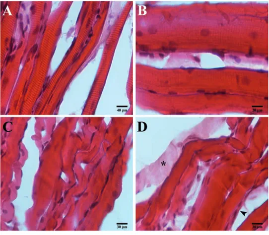
Hematoxilin-eosinIn masseter muscle of contralateral side it is possible to observe a normal structure of mus-cle tissue. The fibers present a healthy recti-linear morphology (Figure 1 A,B), with a con-sistent number of nuclei arranged at the periphery and several myonuclei within the fibers (Figure 1B).
Immunofluorescence
The single localization reaction for PAX-7 show that in contralateral masseter muscle there is an high number of satellite cells per fiber which are located between the muscle fiber sarcolemma and the basal lamina (Figure 2 A,B). The transmitted light shows healthy morphology and orientation of fibers (Figure 2 A,B). Single localization reactions for a-sarco-glycans show the presence of an uniform staining pattern along the fibers (Figure 2C); the same result was observed for β-, g-, d-, e-and z-sarcoglycans (images no shown)
Ipsilateral masseter
Hematoxilin-eosin
The masseter muscle of crossbite side show an altered structure as evidenced by the pres-ence of fibers which show to have a convolute morphology and loss of contractile elements. By that, fibers appear chopped in small ele-ments and each of them seem to have different orientation (Figure 1 C,D). Moreover, it is pos-sible to observe signs of atrophy, as evidenced by the presence of very small fibers if com-pared to the average size of the other fibers, and low number of myonuclei within the fibers (Figure 1C).
Immunofluorescence
In the masseter muscle of crossbite side few PAX-7 positive cells on the periphery of the
fibers have been observed, if compared to con-tralateral side (Figure 3 A,B); transmitted light confirm the convolute morphology of fibers (Figure 3). Moreover, images show a strong reduction of fluorescence pattern along the fibers for a-sarcoglycans (Figure 3C); the same result was observed for β-, g-, d-, e- and z-sarcoglycans (images not shown).
Discussion
In this report we have observed morphologi-cal and protein expression aspects of the mas-seter muscles of both sides of unilateral poste-rior crossbite patients. Our results, have shown relevant differences in muscle tissue morphology and protein compositions between ipsilateral and contralateral side.
In ipsilateral side we found convolute and chopped muscle fibers which appear as small elements not oriented in the same direction; serious signs of atrophy have been detected as the loss of contractile elements in several fibers and strong reduction of fiber’s size. In our opinion, the morphological characteristics of ipsilateral muscle tissue could depend on reduction of muscle tension exerted by less mechanical loading. By contrast, in contralat-eral side we found a normal muscle tissue’s structure.
These results are in accordance with Piancino et al.,18who have demonstrated, by
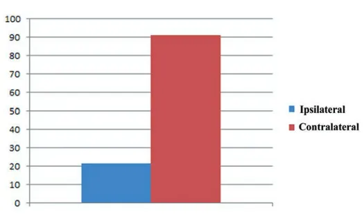
electrognatography and electromyography techniques, that when chewing on the cross-bite side, the chewing patterns show a reverse direction of closure and a reduced activity of the masseter of the same side, while on the non-affected side the chewing pattern and the masseter amplitude are unaltered or increased. Moreover, we have found that between the ipsilateral and contralateral mas-seters there is a difference in numbers of myonuclei per fibers; in detail we found a greater number of myonuclei in contralateral muscle than ipsilateral muscle. Different stud-ies have suggested that the myonuclei number in the myofiber play an important role during the skeletal muscle size adaptation, since dur-ing muscle hypertrophy the myonuclei number are increased (probably through fusion of myo-genic cells) while during muscle atrophy the total myonuclei number within muscle fibers are significantly reduced, hypothetically through apoptosis/necrosis.29,30
On this basis, our results show that the increase of myonuclei’s number in contralater-al masseter could be an adaptive hypertrophic response to the higher work load exercised on this muscle. That is also supported by results which show higher number of PAX-7 positive cells in contralateral side than ipsilateral side.
It is well known that satellite cells are muscle stem cells located between the sarcolemma and the basement membrane of the muscle fiber.31 In humans, satellite cell nuclei are
slightly smaller than muscle nuclei and are mostly in a quiescent state.32Satellite cells are
responsible for myonuclear addition during post-natal muscle growth. In addition, their proliferation can be evoked following acute injury33and in response to muscle overuse and
increased tension.34Our results suggest that
the higher mechanical loading exercised on
Figure 1. Compound panel of hematoxylin-eosin images of contralateral (A, B) and ipsi-lateral (C, D) masseter muscles. A) Low magnification of contraipsi-lateral muscle showing normal and rectilinear fibers morphology. B) High magnification of contralateral muscle showing high number of nuclei within the fibers and along sarcolemma. C) Low magni-fication of ispilateral muscle showing non-healthy convolute fibers morphology, some atrophic fibers (arrowhead). D) Image of ipsilateral muscle showing the loss of contrac-tile elements within the fiber (asterisk).
Figure 2. Compound panel of three images of immunofluorescence reactions performed in contralateral masseter muscle. A) Longitudinal section of masseter where it is possible to observe rectilinear morphology of fibers and the presence of high number of satellite cells, PAX-7 positive (red channel) which are located between sarcolemma and basal lam-ina; nuclei are evidenced by DAPI (blue channel); it is also possible to observe the pres-ence of some PAX-7 positive cells within the fibers (arrowhead). B) Transversal section of masseter muscle: presence of high number of PAX-7 positive cells around sarcolemma (red channel). C) Image of single localization reaction for alpha-sarcoglycan: the protein has been detected uniformly along the fibers (red channel) and also in the satellite cells (arrowhead). Bars: 20 μm
contralateral masseter muscle could determine the satellite cells’ proliferation and myonuclei donation to existing fibers. The satellite cells proliferation in contralateral side could be evi-denced by the increase of PAX-7 positive cells if compared to the ipsilateral side and myonu-clei donation is evidenced by the higher num-ber of myonuclei detected in contralateral side than ispilateral.
Immunofluorescence results also show that all tested sarcoglycans are more expressed in contralateral muscle than ipsilateral; that is in accordance with previous study which demon-strated an increase of muscle specific inte-grins expression in contralateral muscle of unilateral posterior crossbite patients where we have hypothesized that integrins could play as markers of muscle strength.23Since a
bidi-rectional signaling between sarcoglycans and
integrins has been found,11even sarcoglycans
could play as markers of muscle strength. On these basis we suggest that in unilateral poste-rior crossbite an hypertrophic response of con-tralateral masseter takes place; we explain that by the following events: we hypothesize that the major work load exercised on the non-crossbite side may determine the activation of satellite cells that would proliferate and give nuclei to existent fibers; the increase of myonuclei would determine an improvement of transcription, according to the myonuclear
domain theory, with a consequent increase of
sarcoglycans which play a key role in sarcolem-ma stabilization during muscle activity. At the same time ipsilateral side show morphological and protein expression characteristics typical of atrophic or dystrophic muscle, probably for the absence of an adequate stimuli. These
results show that in unilateral posterior cross-bite malocclusion masticatory muscles modify their morphology depending on the function. That could be relevant in understanding and healing of malocclusion disorders. In fact, masseter of crossbite side seems to have mor-phological feature which could anticipate atrophic conditions and the mechanical load exercised by contralateral muscle on skeletal structures could long-term worsen skeletal asymmetries. These results highlight the importance of early therapeutic or surgeon intervention in malocclusion disorders, espe-cially in young patients with unilateral posteri-or crossbite.
References
1. Ervasti JM, Ohlendieck K, Kahl SD, Gaver MG, Campbell KP. Deficiency of a glycopro-tein component of the dystrophin complex in dystrophic muscle. Nature 1990;345: 315-9.
2. Yoshida M, Suzuki A, Yamamoto H, Noguchi S, Mizuno Y, Ozawa E. Dissociation of the complex of dystrophin and its associ-ated proteins into several unique groups by n-octyl β-D-glucoside. Eur J Biochem 1994;222:1055-61. 3. Yoshida M, Ozawa E. Glycoprotein complex
anchoring dystrophin to sarcolemma. J Biochem. 1990;108:748-52.
4. Ervasti JM, Campbell KP. A role for the dys-trophin-glyco-protein complex as a trans-membrane linker between laminin and actin. J Cell Biol 1993;122:809-23. 5. Jaffredo T, Horwitz AF, Buck CA, Rong PM,
Dieterlen-Lievre F. Myoblast migration specifically inhibited in the chick embryo by grafted CSAT hybridoma cells secreting an anti- integrin antibody. Development 1988;103:431-46.
6. Menko AS, Boettiger D. Occupation of the extracellular matrix receptor, integrin, is a control point for myogenic differentiation. Cell 1987;51:51-7.
7. Song WK, Wang W, Foster RF, Bielser DA, Kaufman SJ. H36-α7 is a novel integrin alpha chain that is developmentally regu-lated during skeletal myogenesis. J Cell Biol 1992;117:643-57.
8. Monemi M, Kadi F, Liu JX, Thornell LE, Eriksson PO. Adverse changes in fibre type and myosin heavy chain composition of human jaw. Acta Physiol Scand 1999;167: 339-45.
9. Anastasi G, Cutroneo, G, Rizzo G, Arco A, Santoro G, Bramanti P, et al. Sarcoglycan and integrin localization in normal human skeletal muscle: a confocal laser scanning microscope study. Eur J Histochem
Figure 3. Compound panel of three images of immunofluorescence reactions performed in ipsilateral masseter muscle. A) Longitudinal section of masseter where it is possible to observe an altered, non-rectilinear morphology of fibers and the presence of a lower number of satellite cells PAX-7 positive (red channel) along sarcolemma; nuclei are evi-denced by DAPI (blue channel). B) Transversal section of masseter muscle: presence of a very low number of PAX-7 positive cells around sarcolemma (red channel). C) Image of single localization reaction for alpha-sarcoglycan: the protein has not been detected uni-formly along the fibers and its fluorescence pattern is almost absent (red channel); satel-lite cells were positive for alpha-sarcoglycan. Bars: 20 μm
Figure 4. The graphic based on the results obtained by a count of myonuclei within 200 fibers both ipsilateral and contralateral muscle revealed an higher number of myonuclei in contralateral side than ipsilateral one.
2004;48: 245-52.
10. Anastasi G, Amato A, Tarone G, Vita G, Monici MC, Magaudda L et al. Distribution and localization of vinculin-talin-integrin system and dystrophin-glycoprotein com-plex in human skeletal muscle. Cells Tissues Organs 2003;175:151-64. 11. Yoshida T, Pan Y, Hanada H, Iwata Y,
Shigekawa M. Bidirectional signaling between sarcoglycans and the integrin adhesion system in cultured L6 myocytes. J Biol Chem 1998;273:1583-90.
12. Favaloro A, Speranza G, Rezza S, Gatta V, Vaccarino G, Stuppia L, et al. Muscle-spe-cific integrins in masseter muscle fibers of chimpanzees: an immunohistochemical study. Folia Histochem Cytobiol 2009;47: 551-8.
13. Cutroneo G, Centofanti A, Speciale F, Rizzo G, Favaloro A, Santoro G, et al. Sarcoglycan complex in masseter and sternocleidomas-toid muscles of baboons: an immunohisto-chemical study. Eur J Histochem 2015;59: 2509.
14. Bönnemann CG, Modi R, Noguchi S, Mizuno Y, Yoshida M, Gussoni E, et al. Beta-sarcoglycan (A3b) mutations cause autosomal recessive muscular dystrophy with loss of the sarcoglycan complex. Nat Genet 1995;11:266-73.
15. Noguchi S, McNally EM, Ben Othmane K, Hagiwara Y, Mizuno Y, Yoshida M, et al. Mutations in the dystrophin-associated protein gamma-sarcoglycan in chromo-some 13 muscular dystrophy. Science 1995;270:819-22.
16. Duggan DJ, Manchester D, Stears KP, Mathews DJ, Hart C, Hoffman EP. Mutations in the delta-sarcoglycan gene are a rare cause of autosomal recessive limb-girdle muscular dystrophy (LGMD2). Neurogenetics 1997;1:49-58.
17. Endo T, Kawai H. [Adhalin(alpha-sarcogly-can) gene mutations in patients with malignant limb-girdle muscular dystrophy (MLGMD) (Miyoshi)]. [Article in Japanese]. Nihon Rinsho 1997;55:3159-64. 18. Seale P, Sabourin LA, Girgis-Gabardo A, Mansouri A, Gruss P, Rudnicki MA. Pax7 is required for the specification of myogenic satellite cells. Cell 2000;102:777-86. 19. Grounds MD, Garrett KL, Lai MC, Wright
WE, Beilharz MW. Identification of skeletal muscle precursor cells in vivo by use of MyoD1 and myogenin probes. Cell Tissue Res 1992;267:99-104.
20. Yablonka-Reuveni Z, Rivera AJ. Temporal expression of regulatory and structural muscle proteins during myogenesis of satellite cells on isolated adult rat fibers. Dev Biol 1994;164:588-603.
21. Zammit PS, Heslop L, Hudon V, Rosenblatt JD, Tajbakhsh S, Buckingham ME, Beauchamp JR, Partridge TA. Kinetics of myoblast proliferation show that resident satellite cells are competent to fully regen-erate skeletal muscle fibers. Exp Cell Res 2002;281:39-49.
22. Tajbakhsh S, Buckingham M. The birth of muscle progenitor cells in the mouse: spa-tiotemporal considerations. Curr Top Dev Biol 2000;48:225-68.
23. Piancino MG, Farina D, Talpone F, Merlo A, Bracco P. Muscular activation during reverse and non-reverse chewing cycles in unilateral posterior crossbite. Eur J Oral Sci 2009;117:122-8.
24. Piancino MG, Comino E, Talpone F, Vallelonga T, Frongia G, Bracco P. Reverse-sequencing chewing patterns evaluation in anterior versus posterior unilateral crossbite patients. Eur J Orthod 2012;34: 536-41.
25. Guerra C de S, Carla Lara Pereira Y, Issa
JP, Luiz KG, Guimarães EA, Gerlach RF et al. Histological, histochemical, and protein changes after induced malocclusion by occlusion alteration of Wistar rats. Biomed Res Int 2014;2014:563463.
26. Bani D, Bani T, Bergamin M. Morphologic and biochemical changes of the masseter muscles induced by occlusal wear: studies in a rat model. J Dent Res 1999;78:1735-44. 27. Becht MP, Mah J, Martin C, Razmus T, Gunel E, Ngan P. Evaluation of masseter muscle morphology in different types of malocclusions using cone beam computed tomography. Int Orthod 2014;2:32-48. 28. Cutroneo G, Piancino MG, Ramieri G,
Bracco P, Vita G, Isola G et al. Expression of muscle-specific integrins in masseter muscle fibers during malocclusion dis-ease. Int J Mol Med 2012;30:235-42. 29. Allen DL, Roy RR, Edgerton VR. Myonuclear
domains in muscle adaptation and disease. Muscle Nerve 1999;22:1350-60.
30. Roy RR, Monke SR, Allen DL, Edgerton VR. Modulation of myonuclear number in func-tionally overloaded and exercised rat plan-taris fibers. J Appl Physiol 1999;87: 634-42. 31. Mauro A. Satellite cell of skeletal muscle
fibers. J Biophys Biochem Cytol 1961;9: 493-5.
32. Watkins SC, Cullen MJ. A quantitative comparison of satellite cell ultrastructure in Duchenne muscular dystrophy, polymyositis, and normal controls. Muscle Nerve 1986;9:724-30.
33. Bischoff R, Heintz C. Enhancement of skeletal muscle regeneration. Dev Dyn 1994;201:41-54.
34. Hawke TJ, Garry DJ. Myogenic satellite cells: physiology to molecular biology. J Appl Physiol 1985;91:534-51.