UNIVERSITÀ DEGLI STUDI DELLA TUSCIA DI VITERBO DIPARTIMENTO DI
Innovazione dei sistemi biologici, agroalimentari e forestali Innovazione dei sistemi biologici, agroalimentari e forestali in convenzione con UNIVERSITA` DEGLI STUDI MEDITERRANEA
(REGGIO CALABRIA, ATENEO PARTNER)
Corso di Dottorato di Ricerca in
Scienze, Tecnologie e Biotecnologie per la sostenibilita' - XXIX Ciclo Scienze, Tecnologie e Biotecnologie per la sostenibilita' - XXIX Ciclo.
RECOVERY AND VALORIZATION OF LAVANDULA MULTIFIDA L. RECOVERY AND VALORIZATION OF LAVANDULA MULTIFIDA L. FOR THE BIODIVERSITY PRESERVATION IN CALABRIA REGION FOR THE BIODIVERSITY PRESERVATION IN CALABRIA REGION
(s.s.d. BIO/15) Tesi di dottorato di:
Dott.ssa Angela Fazio
Coordinatore del corso Tutore
Data della discussione 24 luglio 2018
SHORT ABSTRACT
SHORT ABSTRACT
Lavandula genus, belonging to Lamiaceae family, possesses a remarkable importance since it includes numerous species and hybrids that have been widely cultivated since ancient times. Among Lavandula genus, Lavandula multifida L. is a plant of great interest which has been traditionally used in folk medicine due to its multiple beneficial properties.
Taking in consideration the condition of high extinction risk of this species in Southern Italy, and the need to preserve the biodiversity and revalorize this plant in this European coastal region, the environmental adaptability and also some beneficial and therapeutic properties of this plant were evaluated. In particular, the antioxidant pattern and flavonoid profile, the cytotoxic and cytoprotective effects of leaf-extracts, and the effects of leaf-extracts of L. multifida on innate immune system are analysed and discussed in the present thesis. The aim is to revalorize this important species which is critically endangered in the Southern regions of Italy, contributing to its preservation in the Mediterranean region.
KEYWORDS
KEYWORDS
Lavandula multifida L., leaf extracts, antioxidant pattern, flavonoids, cytotoxicity, cytoprotection, immunostimulation.
EXTENDED ABSTRACT
EXTENDED ABSTRACT
This PhD thesis dealt with the analysis of environmental adaptability and germination strategies, as well as with the study of antioxidant, cytotoxic, cytoprotective and immunostimulant properties of the plant L. multifida L. growing in Calabria region. The aim is to emphasize the economic and biological value of L. multifida which is at high extinction risk in Calabria and Sicily regions, in order to promote the preservation of biodiversity in Mediterranean regions, and also to evaluate the possibility of therapeutical and pharmaceuticals applications.
Nella presente tesi di Dottorato vengono affrontati alcuni aspetti sulle proprietà germinative e sull' adattamento ambientale di L. multifida L. che cresce in Calabria, valutandone anche le proprietà antiossidanti, citotossiche, citoprotettive e immunostimolanti dell'estratto fogliare. Trattandosi di una specie a rischio erosione genetica nelle regioni del sud Italia, l'obiettivo e' quello di contribuire alla salvaguardia della biodiversità nel Mediterraneo e di valutare le possibilità di applicazione in campo terapeutico e farmaceutico di L. multifida L., al fine di enfatizzarne l'importanza economica e biologica.
In accordance with the aim of this PhD thesis, the main results of the following analysis are reported:
• evaluation of the germination capacity of Calabrian L. multifida under different environmental conditions (temperature and photoperiod), in comparison with Spanish L. multifida, a very widespread plant in different Spanish regions
• activity of some hydrolyzing enzymes involved in germination and the antioxidative pathway in seeds of Calabrian L. multifida, compared to Spanish L. multifida
• analysis of some enzymatic and non enzymatic antioxidants and phenolic compositions in leaf extract of Calabrian L. multifida, in comparison with the commercial species Lavandula angustifolia (L. officinalis)
• determination of cytotoxic and cytoprotective effects of water and ethanolic leaf extracts of Calabrian L. multifida (between 10 µg/mL and 1000 µg/mL) on HeLa cell line, compared to extracts of Sicilian L. multifida and L. angustifolia
• assessment of the effects of water and ethanolic leaf extracts (between 10 µg/mL and 1000 µg/mL) of Calabrian L. multifida on innate immune system of Sparus aurata (phagocytosis, respiratory burst activity, peroxidase activity), in comparison with Sicilian L. multifida and L. angustifolia
• determination of cytotoxic effects of water and ethanolic leaf extracts (between 1 µg/mL and 1000 µg/mL) of Calabrian L. multifida, Sicilian L. multifida and L. angustifolia on SAF-1 cell line.
• assessment of bactericidal effects of water and ethanolic leaf extracts (between 10 µg/mL and 500 µg/mL) of Calabrian L. multifida on some fish pathogens (Vibrio harveyi, Vibrio anguillarum, Aeromonas salmonicida), in comparison with Sicilian L. multifida and L. angustifolia.
Results showed a lower germination percentage in seeds of Calabrian L. multifida if compared to Spanish ones, being Calabrian L. multifida more sensitive to temperature and photoperiod. Accordingly to this, a less efficient mobilization and use of reserves in germinating seeds of Calabrian L. multifida can also explain the diverse germination speed and germination percentages between the two populations. In addition, to prevent oxidative damage during the metabolic reactivation that characterizes the germination process, the two population of L. multifida have been demonstrated to possess a battery of antioxidant enzymes and antioxidant compounds which are differently involved in seed germination. The different activation of these antioxidant systems during germination can be considered a metabolic adaptation and may explain the diverse ability to respond to external environmental conditions. These results support the possibility of utilization of propagation from seeds under controlled environmental conditions as a viable method for the ex-situ conservation of Calabrian L. multifida.
Calabrian L. multifida leaves contain relevant quantity of enzymatic and non enzymatic antioxidants, suggesting a remarkable ability to optimize survival strategies in its natural habitat and to withstand environmental stress, in comparison with the commercial species L. angustifolia. Furthermore, Calabrian L. multifida leaves may be a good potential source of antioxidant substances for human health, as revealed by the flavonoid composition in its leaves.
Water and ethanolic extracts of Calabrian and Sicilian L. multifida exert a significant cytoprotective effect against a chemically-induced condition of oxidative stress on HeLa cells, so that such extracts may be considered a potential source of natural antioxidants to protect cells against oxidative damage.
Extracts from Calabrian and Sicilian L. multifida positively affect phagocytosis and respiratory burst activity of gilthead seabream head-kidney leucocytes, but have no bactericidal activity against the fish pathogens tested, thus supporting the possibility to use them as immunostimulant in aquaculture production of Sparus aurata, in order to achieve protection against pathogenic infections through enhancement of the innate immunity.
Results reported in this PhD thesis aim to support the biological value of the threatened species L. multifida, demonstrating the possibility of applying conservation programs for its population reinforcement and reintroduction in Southern Italy, and also highlighting its potential utilization as source of new active principles for new antioxidant formulations and as immunostimulant in aquaculture production.
INTRODUCTION
INTRODUCTION
Gradual declining in plant biodiversity represents a worldwide problem. The rise in the global population, the rapid and sometimes unplanned industrialization, the indiscriminate deforestation, the overexploitation of natural resources, the climate change and the pollution have led to the degradation and fragmentation of natural habitats (Sen and Samanta, 2014). Since ancient times, people have gathered plant resources for their needs. Therefore, the necessity of preserving biodiversity is connected with the multiple uses of plants as source of food, fibers, building materials, as well as for medication and cultural needs (Shippmann et al, 2002). The natural diversity of plants is also considered to be a source of important traits, e.g., resistance to diseases and pests, resistance to drought, salting and to other abiotic stresses and also source of traits that allow to improve the quality, and nutritive value of cultivated plants.
Biodiversity contributes significantly towards human livelihood and development, and thus plays a predominant role in the well being of the global population. Around 80 % of the global population still relies on vegetal drugs, according to World Health Organization reports, and today several medicines owe their origin to medicinal plants (Sen and Samanta, 2014).
Many plants have been used for centuries as remedies for human diseases, because they contain natural bioactive compounds with therapeutic value. Lamiaceae (Labiatae) represents a very important medicinal plant family which stands for about more than 3000 species of plants. Plants belonging to this family are herbs or shrubs often with an aromatic smell and rich in medicinal properties of great worth in natural medicine and pharmacopoeia. Some examples of this family include mints, tulsi, thyme, spearmint, coleus and lavender. These plants are widely distributed in the Maltese Islands and other
Mediterranean countries because some of them produce a high amount of essential oils that enables them to survive to the hot summer season (Ramasubramania Raja, 2012). Plants belonging to this family are widely cultivated for medicinal, perfumery, culinary and ornamental purposes.
Among plants belonging to the Lamiaceae family, Lavandula (lavender, Lamiaceae) is a genus of 39 species of particular importance, since it includes numerous species and hybrids that have been widely cultivated since ancient times, and have been applied in food, pharmaceutical and agroindustries (Lis-Balchin, 2003). Lavender is native to the Mediterranean regions including France, Spain, Andorra and Italy, anyway it grows in many other countries of the world. The name lavender comes from the Latin verb lavo, lavare and means “to wash” or “to clean”. In fact, Lavender was used by the Romans as a bath additive, and it was the most used soap making during the Middle Ages. It was also used as food additive and as laxative (Prusinowska and Śmigielski, 2014). Its essential oil is of great economic importance in the perfumery and fragrance industry, and possess multidirectional biological activity. The material used for herbal purposes includes flowers and flowering aerial parts containing essential oil, anthocyanins, phytosterols, sugars, minerals and tannins. Its essential oil contains over 300 chemical compounds, and its qualitative and quantitative composition is variable depending on the genotype, climatic conditions, growing location, and morphological feature (Prusinowska and Śmigielski, 2014). This essential oil has significant antioxidant and antimicrobial activities, and it exerts a significant positive effect on the digestive and nervous system (Buchbauer et al, 1991; Dapkevicius et al, 1998; Guillemain et al, 1989; Mayaud et al, 2008; Wolfe et al, 1996). During the last few years, the exploitation of native lavender species has increased, due to the necessity of preserving their genetic heritage and also for a renewed interest in the use of naturally derived compounds. Among Lavandula genus, Lavandula multifida L. is a rare and short living plant, 30 to 100 cm high, with a diploid genetic pool, equipped with triangular pinnatisect leaves , and with blue or white purple flowers which give off a strong smell (P hoto 1). The species reproduces by seeds, with an outcrossing mating system (Larsen, 1960). It spontaneously grows along the Mediterranean coast in Egypt, Tunisia, Morocco, Algeria, Spain and Portugal. In Italy, it has been found in Calabria (Capo dell’Armi,
Photo 2), and in Sicily (Monte Pellegrino, Brucoli, Capo Sant’Alessio), where its populations are reduced and fragmented due to the human impact on its natural habitat (Galesi et al, 2005). The species has completely disappeared in Capo Scaletta and Taormina (Sicily) (Galesi et al, 2005). Its natural habitat is represented by coastal areas on poorly evolved limestone soils, and hot arid climatic conditions. Due to the rarity and the threats of this plant, L. multifida is included in the “Regional Red Lists of Italian Plants” under the status IUCN of "critically endangered” in Calabria and "endangered” in Sicily (Conti et al, 1997). For this reason, L. multifida has been involved in regional projects aimed to the recovery, the preservation of biodiversity and the revalorization of this plant species in Mediterranean region.
Photo 1
Photo 1. Leaves (on the left) and flower (on the right) of. Leaves (on the left) and flower (on the right) of Lavandula multifida.Lavandula multifida.
Photo 2
Calabrian L. multifida L. (Lamiaceae) spontaneously grows on the poorly evolved limestone soils in Capo dell’Armi (Reggio Calabria, Italy). The needs to preserve its genetic heritage and to revalorize L. multifida growing in Southern Italy led to the necessity of studying its environmental adaptability. Therefore, in the first part of the present thesis, the evaluation of the germination capacity, the activity of some hydrolyzing enzymes involved in germination and the antioxidative pathway in seeds of Calabrian L. multifida L. were investigated. The aim was the understanding of the environmental adaptability and of the germination strategies of this plant, with the objective of defining possible conservation programs for the population reinforcement and reintroduction in Southern Italy. The analysis were carried out in comparison with a Spanish core population of L. multifida growing in Almeria, as the Spanish species is very widespread on different types of substrates. The research of biological traits associated with reproduction is considered essential for the development of guidelines for conservation and management of endangered species (Evans et al, 2003).
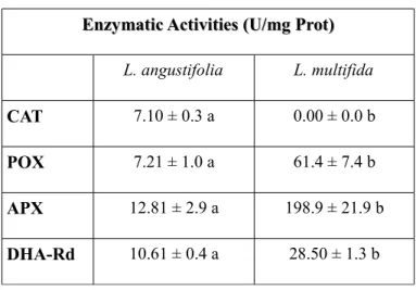
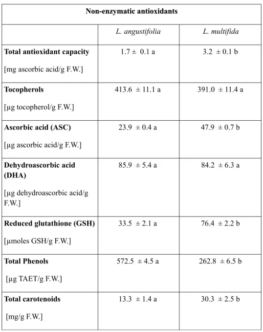
The natural habitat of L. multifida is not favorable to the growth of many crop species because of stressful conditions that are potential sources of reactive oxygen species (ROS). ROS are able to cause oxidative damage by reacting with biomolecules. It is known that a complex network of enzymatic and small antioxidant molecules controls the concentration of ROS and repairs oxidative damage, contributing to the control of plant growth, development and response to the environment (Smirnoff, 2005). Therefore, it is thought that a fine regulation of antioxidant resources exists in L. multifida in order to enable the plant to face ROS overproduction. This mechanism is included in plant's physiological conditions and responses to different environmental stimuli. For this reason, in the present thesis some enzymatic and non enzymatic antioxidants and phenolic composition of Calabrian L. multifida L. leaves are evaluated and discussed. Among enzymatic antioxidants, catalase, peroxidase, dehydroascorbate reductase, and ascorbate reductase activities were assessed, while total phenols, reduced glutathione, ascorbic and dehydroascorbic acid, total antioxidant capacity, tocopherols, total carotenoids and anthocyanins were determined as non enzymatic antioxidants. Furthermore the presence of carvacrol, carvacrol methyl-ether, thymol and flavonoid
antioxidant components were performed in comparison with the widely distributed species Lavandula angustifolia Miller (Lavandula officinalis), in order to show possible differences in antioxidant contents and to improve the knowledge of bioactive compounds present in leaves which could be helpful for possible pharmaceutical and therapeutical applications.
Besides the need to preserve the biodiversity in the Mediterranean coastal regions, L. multifida L. needs to be revalorized, contributing to the knowledge of its possibilities of applications in pharmaceutical and cosmetical fields.
Recently there is a growing interest in searching for antioxidants of natural origin. Due to their high content in polyphenolic compounds, many herbs belonging to Lamiaceae family represent a rich source of health-beneficial antioxidant components (Dragland et al, 2003).
It is well known that living cells and organims are exposed to reactive oxygen species (ROS) as natural bioproducts of the normal metabolism of the oxygen, which play an important role in cell signaling and homeostasis (Hayyan et al, 2016). Under normal conditions, ROS levels are low enough to be neutralized and removed by the natural endogenous antioxidant cellular defence mechanisms (Halliwell and Gutteridge, 1985). However, several stress conditions, including UV radiation, exposure to different toxic compounds or xenobiotics, may enhance ROS production and free radicals to such an extent that cellular defence mechanisms are unable to cope. Oxidative stress arises as consequence of an imbalance between ROS formation and degradation. The clinical implications of these alterations can be severe; in fact, the accumulation of ROS in several cellular components is thought to be a major cause of molecular injury leading to cell aging and to age-related degenerative diseases such as cancer, brain dysfunction and coronary heart disease (Kehrer, 1993; Duthie et al, 2001; Valko et al, 2006). Recently there is a growing interest in finding antioxidants as well as in identifying natural products for use in the treatment of several oxidative stress-related diseases. Antioxidants, as molecules that inhibit the oxidation of other molecules, help to prevent or reverse the negative effects caused by oxidative stress due to their capability to quench free radicals and ROS. They can be natural or synthetic. However, the
widespread use of synthetic antioxidants used in food, drugs and cosmetics has led to concerns about their safety and toxicity (Kahl and Kappus, 1993). Therefore, a growing interest in finding antioxidants of natural origin exists. Besides this, many plants belonging to Lavandula genus have been chemically characterized and are known to present large amounts of secondary metabolites such as coumarins, terpenoids, triterpenes and sesquiterpenes (Areias et al, 2000). Especially abundant in these plants are flavonoids, a class of natural polyphenolic compounds known for their important antioxidant and free radical scavenging activities (Procházková et al, 2011), but also for their ability to inhibit the activity of several enzymes that regulate cell proliferation (Formica and Regelson, 1995), arrest the cell-cycle progression (Yoshida et al, 1990; Ranelletti et al, 1992; Lepley et al, 1996; Plaumann et al, 1996) and induce cell death by apoptosis (Wei et al, 1994; Plaumann et al, 1996; Ahmad et al, 1997). In our study,in order to demonstrate the possibilities of industrial applications of this plant species, the cytotoxic and antioxidant cytoprotective effects of extracts obtained from leaves of Calabrian L. multifida L. were assayed on the human cervical cancer cell line Hela. These assays were performed in comparison with extracts obtained from leaves of Sicilian L. multifida L. and from leaves of the commercial species L. angustifolia. These assays allowed to test the ability of L. multifida L. leaf extract to offer cytoprotection on Hela cells against oxidative stress, considering the possibility of these extracts to be included in antioxidant formulation for application in dermopharmaceutical and cosmetic fields. Beside this, HeLa cell line was used to carry on the cytotoxic assays as it represents the first continuous cell line used worldwide for scientific and medical research, because of its steady growth making it an excellent model for almost every process that occurs in human cells (Masters, 2002). Thanks to their rapid growing and stability in cell culture, Hela cells have been globally used by researcher not only to study cancer, viral infections, gene mapping, radiation effects, but also to investigate the effect of toxic substances and phytochemicals, as well as to test the human sensitivity to tape, glue and cosmetics (Masters, 2002).
Currently, there is a growing interest in screening medicinal plants extracts for their bactericidal, fungicidal and even immuno-stimulant properties, in order to exploit new biocompounds of natural origin which could be employed in the prevention and/or control of fish diseases in aquaculture (Reverter et al, 2014). The importance of using immunostimulants in aquaculture is known since many years (Sakai, 1999). The use of immunostimulants as an alternative to drugs, chemicals and antibiotics currently being used to control fish diseases in fish culture is attracting the attention of many researchers, and the use of medicinal plants-originated products as potential therapeutic measures for modulating the immune response and to prevent and control fish diseases represents a topic of great interest in research (Bairwa et al, 2012). In the intensive aquaculture system, application of antibiotics and chemotherapeutics as prophylactic measures has been widely criticized for their negative impacts on the environment and also on fish, such as immunodepression or residue accumulation in tissues, and besides this leads to the development of drug resistant pathogens (Harikrishnan et al, 2009; FAO, 2003; Rijkers et al, 1980; Smith et al, 1994). In general, immunostimulants represent a group of biological and synthetic compounds that enhance the non-specific defence mechanisms in animals, thus imparting generalized protection. This kind of protection is particularly important for fish living in environments where the nature of pathogens is unknown and the immunization by specific vaccine may be inappropriate. Immunostimulants promote a greater and more effective sustained immune response to those infectious agents such as viruses, bacteria, fungi, and parasites, producing subclinical disease without risks of toxicity, carcinogenicity or tissue residues (Bairwa et al, 2012).
Herbal extracts-originated products have a potential application as immunostimulants in fish culture, primarily because they can be easily obtained, are not expensive and act against a broad spectrum of pathogens. Most of the herbs and herbal extracts can be given orally, which is the most convenient method of immunostimulation (Bairwa et al, 2012). The use of plant extracts as immunostimulants has recently received increasing attention not only because they combine lower costs with their high level of biodegradability, but also because they may have additional physiological effects since they contain many nutrients, micronutrients as well as other immunostimulant substances (Cuesta et al, 2005). Looking for new applications of L. multifida extracts,
and taking in considerations the need of searching for new immunostimulants of natural origin, and also considering the importance of gilthead seabream (Sparus aurata) in Mediterranean diet and in marine fish aquaculture, the in vitro effects of Calabrian L. multifida leaf extracts on head kidney leucocytes activities (viability, phagocytosis, respiratory burst and peroxidase activity) were tested, making a comparison with similar extracts obtained from L. multifida collected from Sicily and from the widely-distributed species L. angustifolia. Moreover, the cytotoxic activity on SAF-1 cells (a cell line obtained from gilthead seabream) and the bactericidal activity of the extracts on Vibrio harveyi, Vibrio anguillarum and Aeromonas salmonicida were also checked. All experiments performed and described in the present thesis aim to demonstrate the possibilities of applications of L. multifida extracts in pharmaceutical and cosmetic fields, and also in aquaculture system as immunostimulants, while promoting the preservation and conservation of this critically endangered plant species growing in Southern Italy.
STATE OF THE ART
STATE OF THE ART
The genus Lavandula L. comprises some of the most promising plants that have been used in screening assays to assess biological properties, such as antioxidant (Matos et al, 2009), antimicrobial (Moon et al, 2007; Roller et al, 2009), insecticidal (Pavela, 2005), antifeedant (González-Coloma et al, 2011), parasiticidal (Moon et al, 2006), and herbicidal (Haig et al, 2009) effects.
Among the plants belonging to Lamiaceae family, Lavandula multifida L. represents a plant species of remarkable importance because of its wide utilization in traditional and folk medicine to treat headaches, diabetes, depression and for its sedative properties (Gamez et al, 1987; Gilani et al, 2000), to prepare decoctions against rheumatism, chill and as digestive system benefic agent (El-Hilaly et al, 2003). Anyhow not all these therapeutic properties has been demonstrated and supported by experimental studies, and only two documents on pharmacological investigation were released to support the hypoglycemic action and the antinflammatory activity of L. multifida. The topical antinflammatory activity of both the ethanolic and acqueous extracts of L. multifida was evaluated by Sosa et al. ( 2005), as inhibition of the croton oil-induced ear edema in mice, supporting the traditional use of the plant as a remedy against inflammatory disorders such as rheumatism, while the hypoglycemic action was evaluated without evidencing any effect (Gamez et al, 1987).
Polyphenol-rich extracts of L. multifida L. collected from Morocco were tested for their antioxidant, hypocholesterolaemic and hypotriglyceridaemic activities, showing no significant effect on both plasma total cholesterol and triglycerides profiles in rat; on the contrary, the antioxidant activity was very high, being the half time hemolisis of erythrocytes increased by 479% in the test of inhibition of the AAPH (2, 2'-azobis
(2-amidinopropane) hydrochloride)-induced oxidative erythrocyte hemolysis (Ramchoun et al, 2009).
Many studies on L. multifida have been carried out on its essential oils. The antifungal activity of L. multifida L. essential oil collected from Portugal was determined against several pathogenic fungi responsible for candidosis, meningitis, dermatophytosis, and aspergillosis (Zuzarte et al, 2012), demonstrating the effectiveness of its essential oil in complementary therapy to treat disseminated candidosis, and suggesting a mechanism of action leading to cytoplasmic membrane disruption and cell death. Furthermore the chemical composition of its essential oils obtained from dried aerial parts was analyzed by gas chromatography/mass spectrometry (GC/MS), showing high contents of monoterpenes, with carvacrol and cis-β-ocimene being the main constituents, and predominance of linalool, camphene, linalyl acetate, α-thujene, bornyl acetate, β-caryophellene, nerol, and terpinolene (Msaada et al, 2012). Antibacterial activity of essential oil of L. multifida L. collected from Morocco was tested against human pathogenic gram-positive and gram-negative bacteria by the agar diffusion method, showing a significant inhibitory activity against Staphylococcus aureus, Bacillus subtilis, Proteus mirabilis, Proteus vulgaris, Listeria innocua, Listeria monocytogenes, Escherichia coli CECT and Escherichia coli K12 (Douhri et al, 2014).
The in vitro cytotoxic effect of various Moroccan L. multifida extracts (obtained through extraction with hexane, dichloromethane and methanol using Soxhlet) was evaluated on Embryonal Rhabdomyosarcoma cancerous cell lines (RD), Kidney adenocarcinoma of hamster (BSR), and on Monkey kidney cancerous cell lines (Vero). Results from MTT cytotoxicity test indicated different levels of cytotoxic activities towards the three cancer cell lines investigated, and in general a dose-dependent decrease in the survival of of the cancer cells was observed (M’hamed et al, 2016). Furthermore, the antibacterial activity of these extracts was evaluated against three bacterial strains of Genus Rhodococcus, showing a strong growth inhibition effect (M’hamed et al, 2016).
At present, L. multifida L. populations are being highly disturbed by increasing clearance, overgrazing and overexplotation, consequently leading to habitat fragmentation into small populations (less than 200 individuals for the most
encountered populations) and to a rapid reduction of genetic diversity (Franklin, 1980; Hamrich and Godt, 1996). The isolation of populations and the decrease of their size caused an increase in genetic drift and the gene fixation, leading to fitness loss (Pearson and Dawson, 2005; Byers et al, 2005; Korner and Jeltsch, 2008). Moreover, the lack of genetic variability caused the reduction of the ability of the plant to adapt to environmental changes (Chograni and Boussaid, 2010).
MATERIALS AND METHODS
MATERIALS AND METHODS
In order to perform the analysis described in the present PhD thesis, the following materials and methods have been used, as described below.
Germination experiments
Germination experiments
Seeds were collected in 2014, immediately after maturation from two different sites, where the plant spontaneously grows: Almeria (Southern Spain) and Capo dell’Armi (Reggio Calabria, Southern Italy). The sampling involved randomly throughout the subpopulation examined. Clean seeds were stored dry at 4°C. Germination experiments were carried out after 6 months of collection to ensure that the seeds were not dormant. First of all, steps were taken to test for the viability of the seeds; 50 seeds of each lot were selected and treated with a 1% solution of 2,3,5-triphenyltetrazolium chloride (ISTA, 2006). Results of this viability test showed that 80% of the selected seeds was viable. Seeds were selected and surface-sterilized with freshly-prepared 30% (v/v) commercial bleach solution for 15 minutes, followed by washing for several times with distilled and autoclaved water. Twenty seeds were placed on filter papers in Petri dishes (9 cm diameter) and then stratified at dark at 4°C for 72 hours, in order to synchronize germination process. Stratification procedure allows the break of dormancy, so improving the germination process (Budvytyte, 2001). After the stratification process, Petri dishes were placed in a growth chamber at different temperatures (15°C, 20°C, 25°C and 30°C), under a photoperiod of 12 hours light/12 hours-dark, to assess possible differences in germination percentage due to different temperature conditions. Some experiments were also performed at dark, at 20 °C and at 25°C, to evaluate the importance of the light on germination process. For all experimental conditions, five replicates were prepared and the number of germinated seeds was assessed every day over a period of 14 days. Seeds were considered germinated when the radicle was at
least 0.5 cm long. At a temperature of 15 ° C no seed germinated, or this reason, this temperature value was not included in the graphs.
Seed sample extraction
Seeds at the end of stratification (Time 0) and 4 days after sown (4 days) were homogenized using chilled mortar and pestle, with 0.1 M K-phosphate buffer (pH 7.0) (1:5 w/v), 1 mM Na2-EDTA, 10 mM MgCl2, and 1% (w/v) PVPP. The extracts were centrifuged at 12.000 rpm for 20 minutes at 4°C. The resulting supernatant was used for all assays. All enzyme activities were measured at 25◦C on a UV, visible light spectrophotometer.
Determination of total proteins
Total proteins were determined according to method of Bradford (1976) using bovine serum albumin as standard. The concentration of protein was obtained by reading the absorbance at 595 nm against blank and related to the calibration curve. Total proteins were expressed as mg proteins/g fresh weight.
Seed enzyme assay
Alpha-Amilase (EC 3.2.1.1) was determined at 546 nm as described by Coombe et al. (1967). At 0.3 ml of 1% starch solution was added 0.3 ml of seed-extract; the mixture was incubated in water bath at 30°C for 30 minutes. After 0.6 ml of colour reagent was added and the mixture was boiled for 5 minutes. At the end, 2.5 ml of distillate water were added and the final mixture was cooled for 30 minutes.
Isocytrate-lyase activity (ICL EC 4.1.3.1) was determined according to Bajracharya and Schopfer (1979). At 0.8 ml of buffer K-phosphate 0.1M (pH 7.6) were added: 0.15 ml of phenylhydrazine 33 mM, 0.15 ml of dithiothreitol 50mM, 0.15 ml of magnesium chloride 220 mM and 0.1 ml of seed extract. The mixture was incubated at 25°C for 5 minutes, and after 0.15 ml of isocytrate 175 mM was added. The kinetic was recorded at 334 nm for 180 seconds.
Glucose 6-Phosphate Dehydrogenase (G6P-DH EC.1.1.1.49) activity was determined at 340 nm as reduction of NADP to NADPH. At 0.8 ml of Tris Hcl 86mM were added 0.1
ml of seed-extract, and 0.1 ml of NADP 0,4 mM (freshly prepared) and of 0.1 ml of glucose 6-phosphate 1,2 mM (De Meillon et al, 1990).
Superoxide dismutase (SOD EC 1.15.1.1) activity was estimated by recording the decrease in absorbance of formazan produced from NBT, at 560 nm (Dhindsa et al., 1981). The mixture reaction contained:1.5 ml of 0.1 M potassium-phosphate buffer (pH 7.5) containing EDTA, 15 μl of 13 mM methionine, 15 μl of 50 μM nitroblue tetrazolium, 50 μl extract. Reaction was started by adding 150 μl of 2 mM riboflavin and placing the tubes under two fluorescent lamps for 15 minutes. A complete reaction mixture without extract gave the maximal color after the irradiation, and served as control; while a non-irradiated complete reaction mixture served as 151 a blank. Reaction was stopped by switching off the light and putting the tubes into dark at room temperature.
Catalase activity (CAT, EC 1.11.1.6) was determined according to Beaumont et al. (1990) by monitoring the disappearance of H2O2 at 240 nm and calculated by using its extinction coefficient (ε) = 0.036 mM−1 cm−1. The reaction mixture contained 1 ml potassium phosphate buffer (50 mM, pH 7.0), 40μl enzyme extract and 5 μl H2O2. Peroxidase activity (POX, EC 1.11.1.7) was determined as reduction in guaiacol concentration by reading the absorbance at 436 nm continuously for 90 seconds. The reaction mixture contained 1 ml potassium phosphate buffer (0.1 M, pH 7.0), 20 μl guaiacol, 40 μl enzyme extract and 15 μl H2O2. POX activity was quantified by the amount of tetraguaiacol formed using its extinction coefficient (ε) = 25.5 mM−1 cm−1 (Panda et al., 2003).
Dehydroascorbate reductase (DHA-Rd, EC 1.8.5.1) activity was assayed following the increase in absorbance at 265 nm owing to the reduced glutathione (GSH) dependent production of ASC (Doulis et al., 1997). The reaction mixture contained 0.1 M K-phosphate buffer pH 6.5, 1 mM GSH and 1 mM DHA (ε =14 mM-1cm-1).
Ascorbate peroxidase (APX, EC 1.11.1.11) activity was determined from the decrease in absorbance at 290 nm, due to oxidation of ascorbate. The reaction mixture was 0.1 M
K-phosphate buffer pH 6.5, 90 mM H2O2 and 50 mM ascorbate (Amako et al., 1994). The decrease in absorbance was recorded continuously for 90 seconds (extinction coefficient 14 mM-1cm-1).
Glutathione Reductase (GR, EC 1.6.4.2) activity was assayed following the oxidation rate of NADPH at 340 nm (ε = 6.2 mM−1 cm−1). The reaction mixture contained 0.1 M potassium phosphate buffer (pH 7.0), 20 mM GSSG, 2 mM NADPH2, 350 μl H2O and 50 μl enzyme extract.
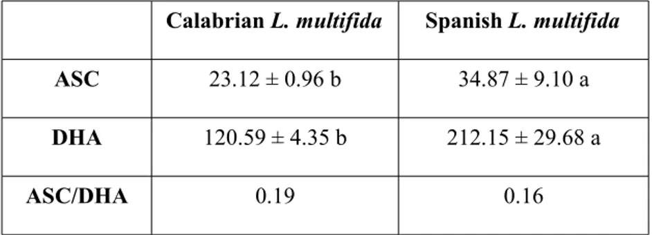
Ascorbate (ASC) and dehydroascorbate (DHA) were determined by Kampfenkel (1995). Seeds (1 g) were homogenized in 5% metaphosphoric acid. The homogenate was centrifuged at 13 000 g and the supernatant was used for ASC and DHA analysis. The effects of treatments on final germination were analysed by fitting factorial general-ized linear model (GLM) to the germination data. A statistical analysis was performed with the software SYSTAT v. 8.0 software (SPSS Inc.) using one-way Anova, followed by LSD test to evaluate significant differences in enzymatic activities and antioxidant pathway within species.
Antioxidant and flavonoid profile experiments
Antioxidant and flavonoid profile experiments
Leaf collection and extraction
The plants of L. multifida were collected between March and May 2014 from Capo dell’Armi (Reggio Calabria, Italy). Fresh leaves (0.5 g) were ground using a chilled mortar and pestle, and homogenized in 0.1M phosphate buffer solution (pH 7.0) containing 100 mg soluble polyvinylpolypyrrolidone (PVPP) and 0.1 mM EDTA. The homogenate was filtered through two layers of muslin cloth and centrifuged at 12,000g for 20 min at 4 °C. The same extraction procedures have been performed with fresh leaves of Lavandula angustifolia MILLER. The resulting supernatant was used for all assays. All enzyme activities were measured at 25 °C on a UV/VIS spectrophotometer.
Determination of total proteins and enzyme assays
Determinations of total proteins and the activities of catalase, peroxidase, dehydroascorbate reductase and ascorbate peroxidase were carried out as previously reported for seed analysis.
Total Phenols
Total phenols were measured by the Box method (Singleton et al, 1999), using Folin– Ciocalteau reagent. The extract from fresh leaves was obtained in H2O (1:4) and the absorbance was recorded against blank at 765 nm. Tannic acid was used as a reference standard and the results were expressed as microgram tannic acid equivalent (µg TAET/g fresh weight).
Reduced Glutathione
The reduced glutathione (GSH) was assayed using the method of Jollow et al (1974). Fresh leaves were homogenized in 3% CCl3COOH at 4 °C and the homogenate was centrifuged at 1000g for 10 min at 4 °C. The absorbance was measured at 412 nm and related to a calibration curve of GSH solutions (0 – 500 µg/ml).
Ascorbic and Dehydroascorbic acid
For ascorbic and dehydroascorbic acid (ASC and DHA), fresh leaves (0.5 g) were homogenized using a chilled mortar in 5% metaphosphoric acid at 4 °C. The homogenate was centrifuged at 39,200g at 4°C and the supernate was used for the determination of ASC and DHA using the method of Law et al (1983).
Total Antioxidant Capacity
Total antioxidant capacity was measured by the method of Prieto et al (1999). Fresh leaves were homogenized in 50% MeOH and were centrifuged at 4 °C at 12,000g for 20 min; the supernatant was used to measure the absorbance at 546 nm to express the total antioxidant capacity as µg ascorbic acid/g fresh weight.
Tocopherols
Tocopherols were assayed using the method of Prieto et al (1999). Fresh leaves were homogenized in hexane, the homogenate was centrifuged at 4 °C at 12000g for 20 min; the supernatants were used as described by the method. The absorbance was recorded at 695 nm against an appropriate blank.
Total Carotenoids
About 50 mg of fresh leaves were incubated in 2.5 ml of 100% EtOH at dark for 24 h at 4 °C; after incubation, the samples were centrifuged for 10 min at 6000g. For carotenoid determination, the absorbance of supernatants was recorded at 649, 665, and 470 nm, and total carotenoid concentrations were obtained using Lichtenthaler’s equations (Lichtenthaler, 1987).
Anthocyanins
For anthocyanin assay, 20 mg of fresh leaves were incubated in 0.5 ml of a MeOH:HCl solution (99:1); samples were centrifuged at 4 °C for 10 min at 6000g and the supernatant was used. The absorbance was recorded at 530 and 657 nm, and anthocyanin content was calculated using the following equation:
[A530nm - (0.025 x A657nm) X ml extract]/g fresh weight. Tartaric Acid Ester
Tartaric acid esters were assayed according to Romani et al (1996): the absorbance was recorded at 320 nm against a blank and caffeic acid was used as standard.
RP-DAD-HPLC identification of Flavonoid Components
Reverse phase-diode array detector-high performance liquid chromatography (RP-DAD-HPLC): Shimadzu system (Shimadzu Ltd., Canby, Oregon, USA), consisting of a LC-10AD pomp system, a vacuum degasser, a quaternary solvent mixing, a SPD-M10A diode array detector, a Rheodyne 7725i injector. Separation of each compound was performed on a 250 mm 9 4.6 mm i.d., 5 lm Discovery C18 column (Supelco, Bellefonte, PA, USA), equipped with a 20 mm 9 4.0 mm guard column. The column was placed in a column oven set at 25 °C. The injection loop was 20 µl and the flow rate was 1.0 ml/min. The spectroscopic determinations have been carried out with UV-1800 CE spectrophotometer (Shimadzu).
The identification of flavonoid components have been carried out with the HPLC system. The mobile phase consisted of a linear gradient of solvent A (MeOH/AcOH/ H2O 18:1:1) in 2% acidified H2O (AcOH/H2O 2:98) as follows: 25 – 100% (0 – 20 min), 100% (20 – 25 min), and 100 – 25% (25 – 35 min). UV-Vis spectra were recorded between 200 and 600 nm, and simultaneous detection by diode array was performed at 278 and 325 nm.
Determination of Carvacrol, Carvacrol Methyl-ether and Thymol
HPLC experiments were performed by a Shimazu system. The mobile phase was an isocratic combination of MeCN/ H2O (50:50) with a flow rate of 1.0 ml/min. The temperature of the column was 25 °C. Before injection, the samples were filtered with a 0.45 µm filter. UV/VIS spectra were recorded between 200 and 600 nm, and simultaneous detection by diode array was performed at 278 nm for identification of monoterpene phenol derivatives.
Acid Hydrolysis
Hydrolysis was carried out on the samples according to the procedure reported by Hertog et al (1992). About 6M HCl (10 ml) in a MeOH (25 ml)/ H2O (10 ml) solution was added to 5 ml of extract to give a solution of 1.2M HCl in 50% aqueous MeOH. Ascorbic acid (50 mg) was added as antioxidant. After refluxing at 90°C for 20 h under stirring, the solution was allowed to cool at room temperature, the solvents were evaporated under reduced pressure, and the residue was suspended in 10 ml H2O/DMF (1:1). The mixture was filtered through an Iso-Disc P-34 membrane (Sigma-Aldrich, St. Louis, Missouri, USA) and analyzed by HPLC. UV/VIS spectra were recorded between 200 and 600 nm, and simultaneous detection by diode array was performed at 278 and 325 nm.
Cytotoxicity and cytoprotection experiments
Cytotoxicity and cytoprotection experiments
Plant collection and preparation of the extracts
L. multifida leaves from 1 to 1.5 cm long were collected from Capo dell'Armi (Reggio Calabria, Calabria), during spring-summer time. The experiments described in this section were carried out in parallel with leaves of L. multifida collected from Siracusa (Sicily) and with leaves of the commercial and widespread species Lavandula angustifolia (L. officinalis).
The leaves were air-dried at 50ºC until constant weight and processed to obtain aqueous and ethanolic extracts. For the preparation of aqueous extracts, air-dry leaves were macerated and soaked in boiling water for 4 h at 25°C, then the supernatant was collected and the remaining pellet was resuspended in boiling water and reincubated (24 h, 25°C). Thus, both supernatants were pooled, and the resultant mixture was filtered twice as described above, and then evaporated in a rotary evaporator (Buchi Rotavapor R-215) until dryness.
To prepare the ethanolic extracts, air-dried leaves were macerated and shaken with pure ethanol (1:40) for 48 h at 25°C. The resulting mixture was then filtered twice using a nylon net filter with a 100μm mesh size, and concentrated in a rotary evaporator (Buchi Rotavapor R-215).
Prior to being used in the assays, the extracts were filtered using sterile filters of 0.22 µm of diameter.
HeLa cells culture
Mammalian cancer cell line, HeLa, was grown from 1 mL aliquots of stock culture that had been previously frozen at -170°C in liquid nitrogen. The cells were grown at 37°C in a 95% air/5% CO2 humidified atmosphere, in EMEM culture medium, supplemented with 10% foetal bovine serum (FBS, Life Technologies), 2 mM L-glutamine (Life Technologies), 100 i.u. ml-1 penicillin (Life Technologies) and 100 µg ml-1 streptomycin (Life Technologies). Exponentially growing cells were detached from culture flasks by brief exposure to 0.25% trypsin in PBS, pH 7.2-7.4, according to the standard trypsinization methods. Cell viability was determined by the Trypan blue exclusion test.
HeLa cells cytotoxicity
Cytotoxicity assays were performed in quadruplicate in 96-well tissue culture plates (Nunc). When cell cultures were confluent, the cells were detached and aliquots of 100 µL containing 1.0 x 10 ^ 4 cells well-1 were dispensed and incubated at 37°C and 5% CO2 for 24 h. Afterwards, the culture medium was replaced by 100µL well-1 of the assayed extracts at the different tested concentrations: 1, 10, 25, 50, 100, 250, 500 and 1000 µg mL-1. Controls received the same volume of culture medium (for aquaeous extracts) or of dimethyl sulfoxide 0.1% (DMSO for ethanolic extracts). Cells were incubated with the extracts for 2 and 24 h at 37°C and 5% CO2, and cell viability was determined by using the MTT assay.
The MTT assay is based on the reduction of the yellow soluble tetrazolium salt (MTT, Sigma) to a blue, insoluble formazan product by the mitochondrial succinate dehydrogenase (Berridge and Tan, 1993; Denizot and Lang, 1986). After exposure of the leaf-extracts, cells were washed with PBS solution and 200µL/well containing 1 mg mL-1 of MTT (3-(4, 5-dimethylthiazol-2-yl)-2,5-diphenyltetrazolium bromide) were added. After 4h of incubation at 37°C, cells were washed again and the formazan crystals solubilized with 100µL/well of dymethil sulfoxide (DMSO, Sigma). Plates were shacked for 5 min in dark conditions and the absorbance at 570 nm and 690 nm was determined in a microplate reader (BMG Fluostar Omega, USA).
Antioxidant cytoprotection of extracts on HeLa cell line
The antioxidant cytoprotective assay was performed as described below, according to the method of Botta et al (2014) with some modifications. Firstly, HeLa cells were exposed to different concentrations of H2O2 (Merck) to establish the concentration that decreased the cell viability by 50% (IC50). When cell cultures were confluent, cells were detached and aliquots of 100 µL containing 1.0 x 10 4 cells well-1 were dispensed into 96-well tissue culture plates (Nunc) and incubated at 37°C and 5% CO2 for 24 h. Then, cells were incubated for 24h with extract concentrations below IC50 of each extract before being treated for 2h with IC50 of H2O2. This experiment allowed assessment of the ability of the extracts to offer cytoprotection before a stressing event such as exposure to hydrogen peroxide.
The antioxidant cytoprotective assay described above was also performed after pretreating cells for 2h with IC50 of H2O2, followed by incubation for 24 h with extract concentrations below the IC50. In this way it was possible to evaluate the ability of the extracts to offer protection after the oxidative stress (post-oxidative stress cytoprotection).
The cytotoxicity results are given as the percentage of viability of treated cells, compared with that of the control consisting of untreated cells (taken to be 100%). The protective effect of the extracts against the oxidative stress induced by hydrogen peroxide was calculated as the increase in viability expressed as a percentage compared with the hydrogen peroxide-treated controls. Results are expressed as Mean ± Standard Error of at least 3 independent experiments.
Kinetic assay for catalase of the extract
The CAT kinetic was determined by the degradation of H2O2 using the method described by Aebi (1983). The reaction mixture in a quartz cuvette contained 50 mM phosphate buffer (pH 7.0), 40 mM of H2O2 and 50 μl of leaf-extract. The H2O2 degradation was monitored at 240 nm for 30 seconds at 25°C and expressed as Abs/min.
Determination of total phenolic content and flavonoid content
Total phenol content was determined using the Folin–Ciocalteu method (Singleton et al, 1999). Briefly, 0.1 mL of leaf extract was mixed with 0,5 mL Folin–Ciocalteu reagent (diluted 10 times with distilled water) and 1 ml of distilled water. After 5 min, 1 mL of sodium carbonate solution (7.5%) was added and the mixture was shaken and then incubated for 1 h in the dark at room temperature. The absorbance of the resulting solution was measured at 765 nm. The phenol content was expressed in terms of milligrams of gallic acid equivalents per gram of dry weight (mg GAEg-1 DW).
Flavonoid content in leaf extract was estimated according to the aluminium chloride colorimetric method of Djeridane et al (2006). In this method, 1 mL of diluted leaf extract was mixed with 1 mL of 2% AlCl3 methanolic solution. After incubation at room temperature for 15 min, the absorbance was measured at 430 nm. Flavonoids were calculated from a calibration curve of rutin and expressed as milligrams of rutin equivalents per gram of dry weight (mg REg-1 DW).
Immunostimulant and bactericidal assays
Immunostimulant and bactericidal assays
Plant extracts
Water and ethanolic leaf extracts from Calabrian L. multifida have been prepared as indicated in the above section “Cytotoxicity and cytoprotection experiments”. The experiments have been performed in parallel with leaves of L. multifida collected from Siracusa (Sicily) and with leaves of the commercial and widespread species Lavandula angustifolia (L. officinalis).
Animals
Thirty specimens (40.51 ± 1.47 gr weight) of the seawater teleost gilthead seabream (S. aurata L.), obtained from a local farm (Murcia Spain), were kept in re-circulating seawater aquaria (250 L) in the Marine Fish Facility at the University of Murcia. The water temperature was maintained at 20 ± 2 °C with a flow rate of 900 L h-1 and 28% salinity. The photoperiod was 12 h light: 12 h dark. Fish were allowed to acclimatise for 15 days before the start of the trial, where they were fed with a commercial pellet diet (Skretting, Spain) at a rate of 2% body weight day-1. The fish were killed after starving for 24 h by using an overdose of MS-222 (Sandoz, 100 mg mL-1 water). All experimental protocols were approved by the Ethical Committee of the University of Murcia (Permit Number A13150104).
Head-kidney leucocyte isolation and incubation with extracts
Before the dissection of the head-kidney (HK), the specimens were bled. Blood was collected from the caudal vein and afterwards fish were dissected to obtain HK fragments. For isolation of leucocytes to carry out the assays, HK fragments were transferred to 8mL of sRPMI [RPMI-1640 culture medium (Gibco) supplemented with 0.35% sodium chloride (to adjust the medium's osmolarity to gilthead seabream plasma osmolarity of 353.33 mOs), 3% fetal calf serum (FCS, Gibco),100 i.u. mL-1 penicillin (Flow) and 100mg mL-1 streptomycin (Flow)] (Esteban et al, 1998). Cell suspensions were obtained by forcing fragments of the organ through a nylon mesh (mesh size 100 mm), washed twice (400 g,10 min), counted (Z2 Coulter Particle Counter) and adjusted to 2 * 107
cells mL-1 in sRPMI. Cell viability was higher than 98%, as determined by the trypan blue exclusion test. To study the possible effects of water and ethanolic extracts on leucocyte activities, aliquots of 50 µL of the obtained leucocytes suspension containing 2 * 107 cells mL-1 were dispensed into flatbottomed 96-well microtitre plates (Nunc). Then aliquots of 50 µL well-1 of water or ethanolic extracts ranging from 10, 100, 500, to 1000 µg mL-1 prepared in sRPMI were added. The extract aliquot was replaced by sRPMI for control samples for assays with water extracts, or by 0,1% dymethil sulfoxide (DMSO, Sigma) in sRPMI in case of control samples for assays with ethanolic extracts. Cells were incubated in presence of the extracts for 24 h at 20 ° C in an incubator with 5% CO2 and 85% humidity. After incubation, leucocyte viability and phagocytic, respiratory burst and peroxidase activities were determined as described below.
Leucocyte viability
Aliquots of 100 µL of leucocytes previously incubated for 24h without (control) or with the plant extracts were placed in 5 mL glass tubes (Falcon, BectoneDickinson) and 40 µL of propidium iodide (PI) (400 µg mL-1, Sigma) were added to each sample. The tubes were gently mixed before analysis in a FACScan (BectoneDickinson, Madrid, Spain) flow cytometer with an argon-ion laser adjusted to 488 nm. Analyses were performed on 5000 cells, which were acquired at a rate of 300 cells s-1. Data were collected in the form of two-parameter side scatter (granularity, SSC) and forward scatter (size, FSC), and green fluorescence (FL1) and red fluorescence (FL2) dot plots or histograms were made on a computerized system. Dead cells were estimated as the percentage of cells with propidium iodide (red-PI fluorescent cells).
Phagocytic activity
The phagocytic activity of gilthead seabream HK leucocytes was studied by flow cytometry according to Esteban et al (1998). Heat killed (30 min, 60 °C) lyophilized S. cerevisiae, strain S288C, were washed twice, counted and adjusted to 108 yeast cells mL-1 in sRPMI-1640. To label yeast cells with fluorescein isothiocyanate (FITC, Sigma) yeast cells were incubated with 5 µg mL-1 FITC at 22 °C with constant stirring (40 cycles min-1) and in darkness for 15 min (Rodriguez et al, 2003). After labeling, free FITC was removed bywashing twice in phosphate buffer saline (PBS) and the yeast cells were resuspended in sRPMI-1640. FITC-labeled yeast cells were acquired for flow cytometric study. The staining uniformity was examined and then the yeast cell suspensions were aliquoted and stored at 4 °C. Samples of 125 µL of medium containing FITC-labeled-yeast cells and 100 µL of HK leucocytes in sRPMI were mixed, centrifuged (400 g, 5 min, 25 °C), resuspended and incubated at 25 °C for 60 min in dark conditions. At the end of the incubation time, the samples were placed on ice to stop phagocytosis and 400 µL of cold PBS was added to each sample. The fluorescence of the extracellular yeast cells (i.e. free yeast cells and yeast cells adhered to phagocytes but not ingested) was quenched by adding 50 µL of cold Trypan Blue (0.4% in PBS) per sample. Standard samples of FITC-labeled S. cerevisiae yeast cells or
HK leucocytes were included in each phagocytosis assay. Immediately, the samples were mixed gently, acquired, and analyzed in a FACScan. Data were collected in the form of two-parameter side scatter (SSC) and forward scatter (FSC), and green fluorescence (FL1) dot plots or histograms were made on a computerized system. Fluorescence histograms represented relative fluorescence on a logarithmic scale. Flow cytometer was set to analyze the phagocytic cells gated from all the leucocytes because of their higher SSC and FSC values. Phagocytic ability was defined as the percentage of cells with one or more ingested yeast cells (green-FITC fluorescent cells) within the phagocytic cell population. The relative number of ingested yeast cells per cell (phagocytic capacity) was assessed in arbitrary units from the mean fluorescence intensity of the phagocytic cells.
Respiratory burst activity
The respiratory burst activity of seabream HK leucocytes was studied by a chemiluminescence method (Bayne and Levy, 1991). Samples of 100 mL of a PMA/luminol solution [1 ng mL-1phorbol myristate acetate (PMA, Sigma) and 10-4 M luminol (Sigma) in HBSS with Ca2+ and Mg2+] were added to the HK leucocytes (previously incubated as described above). The plate was shaken and immediately read in a chemiluminometer (BMG, Fluoro Star Galaxy). Measurements were performed in 30 cycles of 2 min each. The kinetics of the reactions were analyzed and the maximum slope of each curve calculated. Control samples containing leucocytes that had not been incubated with the extracts were also analyzed.
Peroxidase content
The total peroxidase content of HK leucocytes was measured according to Quade and Roth (1997). To do this, 5 µL of HK leucocytes (previously incubated as described above) were incubated for 10 min with 0.02% cetyltrimethylammonium bromide (CTAB, Sigma) at 60 rpm. Afterwards, 100 µL of 10 mM 3,3',5,5'-tetramethylbenzidine hydrochloride (TMB, Sigma) and 5 mM H2O2 (both substrates prepared daily) were added and after 2 min, 50 µL of 2 M sulfuric acid was also added to stop the reaction.
The absorbance of the samples was measured at 450 nm in a microplate reader (BMG Fluostar Omega, USA). Control samples containing leucocytes that had not been incubated with extracts were also analyzed.
SAF-1 cell culture
The established cell line SAF-1 (ECACC n 00122301) was seeded in 25 cm2 plastic tissue culture flasks (Nunc, Germany) cultured in L-15 Leibowitz medium (Life Technologies, UK), supplemented with 10% fetal bovine serum (FBS, Life Technologies), 2 mM Lglutamine (Life Technologies), 100 i.u. mL-1 penicillin (Life Technologies) and 100 µg mL-1 streptomycin (Life Technologies). Cells were grown at 25 °C under humidified atmosphere (85% humidity). Exponentially growing cells were detached from culture flasks by brief exposure to 0.25% of trypsin in PBS, pH 7.2-7.4, according to the standard trypsinization methods. The detached cells were collected by centrifugation (1000 rpm, 5 min, 25 °C) and the cell viability was determined by the trypan blue exclusion test.
Cytotoxicity assay on SAF-1 cell line
Cytotoxicity assay was performed in quadruplicates. When SAF-1 cell lines were approximately 80% confluent, they were detached from flasks culture with trypsin (as described before), and aliquots of 100 µL containing 50000 cells well-1 were dispensed in 96-well tissue culture plates and incubated (24 h, 25 °C). This cell concentration was previously determined in order to obtain satisfactory absorbance values in the cytotoxic assay and avoided cell overgrowth. After that, the culture medium was replaced by 100 µL well-1 of the extracts to be tested at the appropriate dilution. Tested concentrations of water and ethanol extracts ranged from 1 to 1000 mg mL-1 (1, 10, 100, 1000). Cells were then incubated for 24 h. Control samples received the same volume of culture medium (for water extracts) or of DMSO 0.1% (for ethanolic extracts). Cells were incubated for 24 h at 25 °C and then their viability determined using the MTT assay.
The MTT assay is based on the reduction of the yellow soluble tetrazolium salt (3-(4,5-dimethylthiazol-2-yl)-2,5- diphenyltetrazolium bromide) (MTT, Sigma) into a blue,
insoluble formazan product by the mitochondrial succinate dehydrogenase (Berridge and Tan, 1993; Denizot and Lang, 1986). After incubation with the leaf-extracts, SAF-1 cells were washed with phosphate buffer saline solution (PBS) and 200 µL well-1 of MTT (1 mg mL-1) were added. After 4 h of incubation, cells were washed again and the formazan crystals solubilized with 100 µL well-1 of DMSO. Plates were shacked (5 min, 100 rpm) in dark conditions and the absorbance at 570 nm and 690 nm determined in a microplate reader.
Bacteria
Three pathogenic bacteria for fish (V. harveyi, V. anguillarum and A. salmonicida) and Escherichia coli, as control, were used in the bactericidal assays. All bacterial strains were grown from 1 mL of stock culture that had been previously frozen at 80 ° C. V. harveyi, V. anguillarum and A. salmonicida were cultured for 48 h at 25 ° C in Triptic Soy Agar (TSA, Difco Laboratories), and then inoculated in Triptic Soy Broth (TSB, Difco Laboratories), both supplemented with NaCl to a final concentration of 1% (w/v). Bacteria in TSB medium were then cultured at the same temperature, with continuous shaking (100 rpm) during 24 h. E. coli was cultured in Luria Bertani Agar (LB Agar, Difco) for 48h at 37 °C and then inoculated in Luria Bertani Broth (LB Broth, Difco). E. coli bacteria in LB broth medium were then cultured for 24h at 37°C with continuous shaking (100 rpm). Exponentially growing bacteria were resuspended in sterile Hank's balanced salt solution (HBSS) and adjusted to 1 * 108 colony forming units (c.f.u.) mL-1.
Bactericidal assay
Bactericidal activity was determined following the method of Stevens et al (1991) with some modifications. Samples of 20 µL of water or ethanolic leaf-extracts previously adjusted to 20, 100, 200, 500, 1000 µg mL-1 were added in quadruplicate wells of a U-shaped 96-well plate (Nunc). Hank's balanced solution was added to some wells instead of the extracts and served as positive control. Aliquots of 20 µL of the bacteria previously cultured were added and the plates were incubated for 2.5 h at 25 ° C (in case of V. harveyi, V. anguillarum and A. salmonicida), or at 37 °C (in case of E. coli). After that, 25 µL of MTT (1 mg mL-1) were added to each well and the plates were newly incubated for 2h (at the appropriate temperature taken into account the assayed bacteria) to allow the formation of formazan. Plates were then centrifuged (2000g, 10 min), being the precipitates dissolved in 200 µL of DMSO and transferred to a flat-bottom 96 well-plate. The absorbance of the dissolved formazan was measured at 560 nm. Bactericidal activity was expressed as percentage of no viable bacteria, calculated as the difference between absorbance of bacteria surviving compared to the absorbance of bacteria from positive controls (100%).
Statistical analysis
Results showed are representative of at least three independent experiments and are expressed as Mean ± Standard Error. All assays related to leucocyte activities were performed in duplicate and results were expressed as Mean ± Standard Error for each group (three fish per group). Data were analyzed by one-way analysis of variance (ANOVA), and Tukey post-hoc test was performed in order to make a multiple comparison between experimental groups. Differences were considered statistically significant when P < 0.05.
RESULTS AND DISCUSSION
RESULTS AND DISCUSSION
Germination experiments
Germination experiments
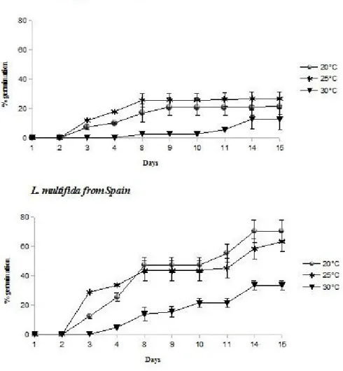
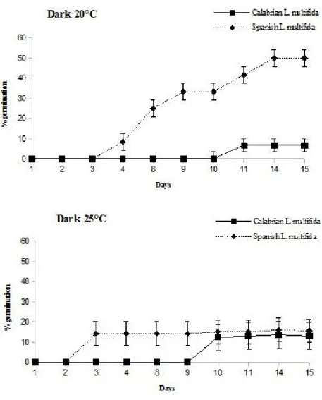
In this paragraph, the evaluation of the germination capacity of Calabrian Lavandula multifida under different environmental conditions (temperature and photoperiod) is described, in comparison with Spanish L. multifida, a very widespread plant in different Spanish regions. Germination capacity was assessed at 15°C, 20°C, 25°C and 30°C, under a photoperiod of 12 hours light/dark. Experiments were also performed at dark, at 20°C and 25°C, to evaluate the importance of the light on germination process.
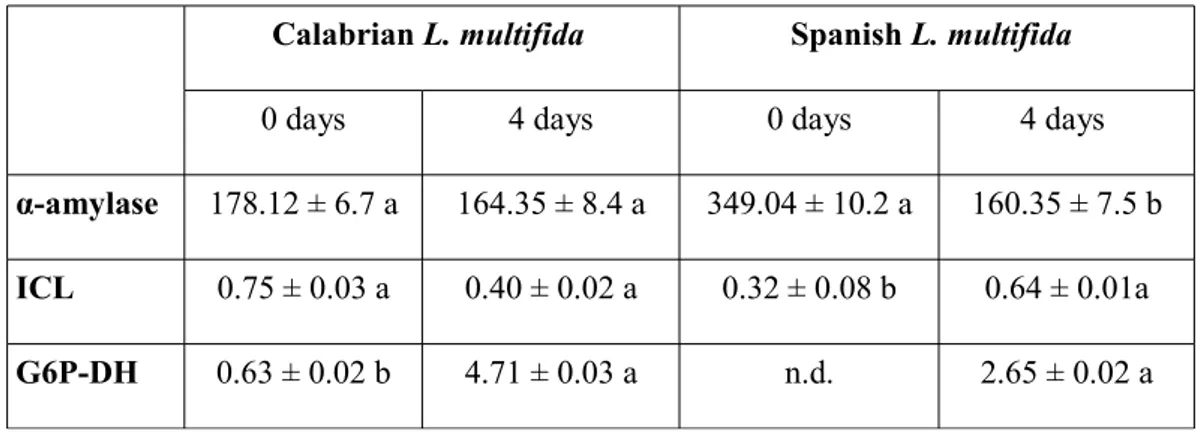
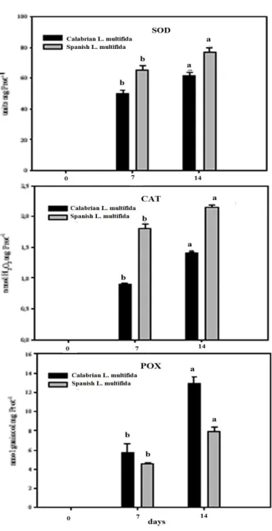
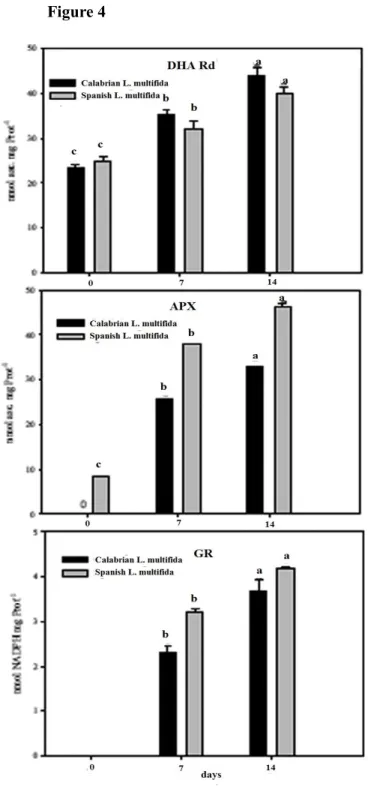
Also, the activity of some hydrolyzing enzymes involved in germination and the antioxidative pathway in seeds of Calabrian L. multifida are analysed and discussed, in comparison with Spanish L. multifida. Among hydrolyzing enzymes, alpha-amylase, isocytrate lyase, and glucose 6-phosphate dehydrogenate activities are analyzed and discussed. Regarding the antioxidative pathway in seeds, superoxide dismutase and catalase activities, as well as peroxidase, dehydroascorbate reductase, ascorbate peroxidase and gluthatione reductase activities are discussed. Also, ascorbic acid and dehydroascorbate content in dry seeds is presented.
The aim is the understanding of the environmental adaptability and of the germination strategies of Calabrian L. multifida, with the objective of defining possible conservation programs for the population reinforcement and reintroduction in Southern Italy.
Germination percentage of Calabrian L. multifida seeds did not show significant differences between 20°C and 25°C; but it strongly decreased at 30°C (Figure 1). Spanish L. multifida seeds showed a similar trend under the same temperature conditions (Figure 1), even if the maximum percentage of germination of Calabrian L. multifida (about 25%) was significantly lower than that of L. multifida from Spain (about 70%), and also the speed of germination was significantly lesser in Calabrian L. multifida seeds if compared to Spanish L. multifida, in all treatments.
Figure 1. Germination percentages of Calabrian L. multifida and Spanish L. multifida seeds incubated at 20°C, 25°C and 30°C under a photoperiod of 12 hours light/12 hours dark. Data were expressed as mean ± standard error.
Figure 2. Germination percentages of Calabrian L. multifida and Spanish L. multifida seeds incubated at 20°C and 25°C under dark conditions. Data were expressed as mean ± standard error.
Germination in both species was strongly affected when the seeds were incubated in the dark. Particularly, a delay in germination process until the 9th day of incubation was observed in Calabrian L. multifida. (Figure 2) and also total germination was reduced, with a maximum germination percentage of about 13% for the seeds incubated at 25°C and about 6% for the seeds incubated at 20°C. Conversely, no delay in the germination process was observed in Spanish L. multifida seeds (Figure 2), and germination percentage was significantly higher than that of Calabrian L. multifida seeds at 20°C, in the dark.
These results showed that temperature and photoperiod differently affect germination process in the two populations of L. multifida. Particularly, Calabrian L. multifida was more sensitive than Spanish and this different resistance may be important for the environmental adaptability and the distribution of this species. Both L. multifida provenances have the maximum germination at 20 and 25°C, with 12 h light/dark alternating, and these findings are in agreement with what has been observed for the Spanish populations of this species (Estrelles et al. 2004). Instead, there are significant differences in germination percentages. This different germination performance can be linked to specific characteristics of the populations, as reported by Menges (1991) who pointed out that seeds of small populations, at limit of their distribution area, have fewer germinative capacity.
The activities of some hydrolyzing enzymes like alpha-amylase and isocytrate lyase (ICL) were assayed on seed extracts of both L. multifida populations at the end of stratification (0 day) and four days after sown (4 days). The activity of α-amylase declined during four days in Spanish L. multifida while it did not change in the Calabrian lavandula seeds (Table I).