Homo-PROTACs: bivalent small-molecule
dimerizers of the VHL E3 ubiquitin ligase to induce
self-degradation
Chiara Maniaci
1,2
, Scott J. Hughes
1
, Andrea Testa
1
, Wenzhang Chen
1
, Douglas J. Lamont
1
, Sonia Rocha
3
,
Dario R. Alessi
2
, Roberto Romeo
4
& Alessio Ciulli
1
E3 ubiquitin ligases are key enzymes within the ubiquitin proteasome system which catalyze
the ubiquitination of proteins, targeting them for proteasomal degradation. E3 ligases are
gaining importance as targets to small molecules, both for direct inhibition and to be hijacked
to induce the degradation of non-native neo-substrates using bivalent compounds known as
PROTACs (for
‘proteolysis-targeting chimeras’). We describe Homo-PROTACs as an
approach to dimerize an E3 ligase to trigger its suicide-type chemical knockdown inside cells.
We provide proof-of-concept of Homo-PROTACs using diverse molecules composed of two
instances of a ligand for the von Hippel-Lindau (VHL) E3 ligase. The most active compound,
CM11, dimerizes VHL with high avidity in vitro and induces potent, rapid and
proteasome-dependent self-degradation of VHL in different cell lines, in a highly isoform-selective fashion
and without triggering a hypoxic response. This approach offers a novel chemical probe for
selective VHL knockdown, and demonstrates the potential for a new modality of chemical
intervention on E3 ligases.
DOI: 10.1038/s41467-017-00954-1
OPEN
1Division of Biological Chemistry and Drug Discovery, School of Life Sciences, University of Dundee, James Black Centre, Dow Street, Dundee, Scotland DD1 5EH, UK.2Medical Research Council Protein Phosphorylation and Ubiquitylation Unit, School of Life Sciences, University of Dundee, James Black Centre, Dow Street, Dundee, Scotland DD1 5EH, UK.3Centre for Gene Regulation and Expression, School of Life Sciences, University of Dundee, James Black Centre, Dow Street, Dundee, DD1 5EH Scotland, UK.4Dipartimento di Scienze Chimiche, Biologiche, Farmaceutiche ed Ambientali, University of Messina, Polo Universitario Viale SS. Annunziata SNC, Messina, 98168, Italy. Correspondence and requests for materials should be addressed to
E
3 ubiquitin ligases are emerging as attractive targets for
small-molecule modulation and drug discovery
1–3. E3s
bring a substrate protein and ubiquitin in close proximity to
each other to catalyze the transfer of a ubiquitin molecule to the
substrate
4,5. Substrate ubiquitination can trigger different cellular
outcomes
6, of which one of the best characterized is
poly-ubiquitination and subsequent proteasomal degradation
7,8.
The human genome comprises
> 600 predicted E3 ligases that
play important roles in normal cellular physiology and disease
states, making them attractive targets for inhibitor discovery
9.
However, E3 ligases do not comprise deep and
‘druggable’ active
sites for binding to small molecules
2. Blockade of E3 ligase
activity therefore requires targeting of protein–protein
interac-tions (PPIs), and the often extended,
flat and solvent-exposed PPI
surfaces make it a challenge for drug design
10. Only few potent
inhibitors have been developed to date, mostly compounds that
bind to the E3 substrate recognition site
2, 11, for example
MDM2
12,13, inhibitor of apoptosis proteins (IAPs)
14–16, the von
Hippel-Lindau (VHL) ligase
17–19and KEAP1
20,21. Inhibitors of
E3:substrate interaction can exhibit a discrepancy in effective
concentrations between biophysical binding and cellular
effi-cacy
19, 22, due to competition from high-affinity endogenous
substrates that markedly increase their cellular concentration as a
consequence of the inhibition. This poses limitations, such as
incomplete blockade of enzyme activity and the need to use high
inhibitor concentrations, which can lead to off-target effects and
cytotoxicity. Moreover, E3 ligases are domain and
multi-subunit enzymes, and targeting an individual binding site leaves
other scaffolding regions untouched and other interactions
functional. As a result, E3 ligase inhibition may be ineffective or
fail to recapitulate genetic knockout or knockdown. New
che-mical modalities to target E3 ligases are therefore demanded.
E3 ligases are not merely targets for inhibition. Compounds of
natural or synthetic origin have been discovered that bind to E3
ligases and promote target recruitment. These interfacial
com-pounds induce de novo formation of ligase-target PPIs, effectively
hijacking E3 activity towards the neo-substrates, for targeted
protein degradation
23, 24. One class of hijackers of E3 ligase
activity comprises monovalent compounds. These so-called
‘molecular glues’ include the plant hormone auxin, which binds
to the Cullin RING ligase (CRL) CRL1-TIR1 to target
tran-scriptional repressor proteins of the Aux/IAA family;
25the
immunomodulatory drugs (IMiDs) thalidomide, lenalidomide,
pomalidomide and analog CC-885, that all share binding to
cereblon (CRBN), a subunit of the CRL4-CRBN ligase, and
redirect CRBN activity to different substrates
26–31. More recently,
sulfonamide anti-cancer drug indisulam was found to induce
degradation of the splicing factor RBM39 via recruiting
CRL4-DCAF15 activity
32,33. Another class of degrader compounds that
display a similar mechanism of action comprises bivalent
mole-cules known as Proteolysis-Targeting Chimeras (PROTACs).
PROTACs comprise two warheads—one for ligase recruitment
and a second one for target-binding—joined by a linker
34.
For-mation of a ternary complex between the PROTAC, the ligase
and the target triggers proximity-induced target ubiquitination
and degradation. Potent and cell-active PROTACs have been
developed
for
recruiting
different
ligases,
including
CRL2-VHL
35–38, CRL4-CRBN
39–42, and IAPs
43, 44. Targets
successfully degraded by PROTACs include BET proteins Brd2,
Brd3 and Brd4
35,37–40, FKBP
39, protein kinases
36, 41, amongst
others
36,43. An attractive feature of bivalent degrader molecules is
their sub-stoichiometric catalytic activity
36, which does not
require full occupancy of the target-binding site as with
con-ventional inhibitors, leading to degrading concentrations that can
be orders of magnitude lower than the inhibitory concentrations
of their constitutive parts alone. Furthermore, induced target
depletion can have a more sustained cellular effect compared to
target inhibition, and can overcome compensatory cellular
feed-back mechanisms, such as increase in target levels
45. Crucially,
work from us and others have shown that PROTAC molecules
can exhibit an added layer of selectivity for protein degradation
beyond the intrinsic binding selectivity of the warhead ligand
35,38,41
. Our recent structural work with Brd4-selective PROTACs
targeting CRL2-VHL revealed the importance of specific
ligand-induced PPIs between the ligase and the target, which contribute
to cooperative formation of stable and highly populated ternary
complexes
38.
We hypothesized that it could be possible to trigger an E3
ligase to induce its own degradation, by designing tailored
homo-bivalent PROTACs that recruit two molecules of the same
E3 ligase. The idea was simple, namely that this compound class
could act as chemical inducers of dimerization (CID)
46–48,
forming a ternary complex in which the E3 acts as the enzyme
and the neo-substrate at the same time. To provide
proof-of-concept for the approach, we designed, synthesized and tested
homo-bivalent molecules aiming to target CRL2-VHL, the E3
ligase that targets for ubiquitination and degradation the hypoxia
inducible factor alpha subunit (HIF-α) under normoxic
condi-tions
49. We show the most active compound, CM11, made of two
instances of a potent hydroxyproline (Hyp) containing VHL
ligand
19, avidly forms a 1:2 complex with VHL, and induces
potent and preferential isoform-selective degradation of VHL. We
call these Homo-PROTACs, as a new modality to induce
che-mical knockdown of E3 ligases.
Results
Rational design. Design of VHL Homo-PROTACs began with
careful consideration of the position of derivatization on two
potent VHL ligands recently characterized by our group, VH032
and VH298 (Fig.
1
a, b)
18, 19. To retain the strong binding
affinity that characterizes the ligand, co-crystal structures were
analyzed to identify solvent-exposed regions from where the
ligands could be derivatized without perturbing their binding
modes (Fig.
1
a). This analysis and consideration of previous
VHL-targeting PROTACs pointed to the methyl group of the
left-hand site (LHS) terminal acetyl group of VH032 as a suitable
point of connection for a linker
35,36. A second solvent-exposed
position available for derivatization was the phenyl group on the
right-hand side (RHS), as previously employed with PROTACs
targeting the Halotag
50. To investigate the impact of
derivatiza-tion, we designed three classes of Homo-PROTACS: (a)
sym-metric via the LHS acetyl group of each ligand (Fig.
1
c); (b)
symmetric
via
the
RHS
phenyl
group
(Fig.
1
d);
and
(c) asymmetric via the acetyl group in one warhead and the
phenyl in the other (Fig.
1
e). In the cases b and c, at the
underivatized terminal LHS we decided to retain either an acetyl
(as in VH032) or a cyano-cyclopropyl moiety (as in VH298), a
modification that led to increased binding affinities, cell
perme-ability and cellular activities in the context of the VHL inhibitor
alone
19. To evaluate the potential impact of linker length, linkers
comprised of polyethylene glycol chains with either three, four or
five ethylene glycol units were chosen to connect the two VHL
ligands.
It is known that the trans epimer of Hyp is an absolute
requirement for VHL binding, and that the corresponding cis
epimer abrogates binding to VHL, both within the context of a
native HIF substrate peptide
51, and VHL ligands
19, 36. We
therefore designed two different PROTACs based on the structure
of the
first series (Fig.
1
c), with the aim to use them as controls: a
cis-cis epimer, expected to be completely inactive, and a cis-trans
epimer compound, expected to retain binding to a single VHL
molecule in a 1:1 fashion, thus potentially acting as inhibitor but
not as degrader.
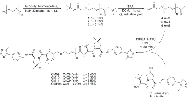
Synthesis. For the synthesis of the
first class of Homo-PROTACs
(Fig.
1
c), symmetric PEG linkers 4, 5 and 6 bearing free
carboxylate groups at either ends were obtained by reaction of
tert-butyl bromoacetate with tri-, tetra- and penta-ethylene glycol
in the presence of NaH in dioxane and followed, after
purifica-tion, by treatment with 50% TFA in DCM (Fig.
2
).
The
final compounds CM9, CM10 and CM11 were obtained by
amide coupling of the VHL ligand 7 (prepared as previously
described)
35with linkers 4, 5 and 6, in a 2:1 ratio, respectively, in
the presence of HATU as the coupling agent and DIPEA as the
base (Fig.
2
). For the synthesis of the symmetric cis-cis compound
CMP98, compound 8
35was coupled with linker 6 to afford the
desired product (Fig.
2
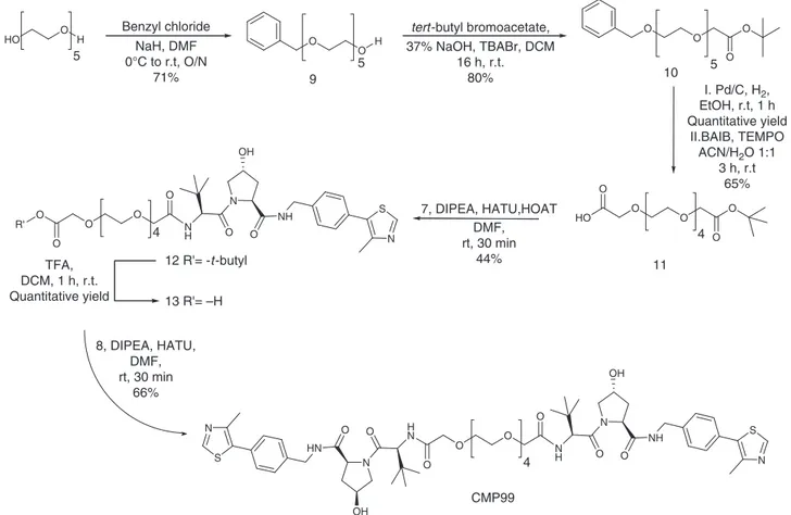
). For the preparation of the asymmetric
cis-trans compound CMP99, a synthetic route toward the
synthesis of the monoprotected di-carboxylate linker was
estab-lished. Pentaethylene glycol was the linker of choice because of
ease of purification compared to longer PEGs, and at the same
time yielding a control compound of average linker length
(PEG-4 in this case). Pentaethylene glycol was converted in monobenzyl
ether 9 in 71% yield, which was reacted with tert-butyl
bro-moacetic acid under biphasic conditions (DCM / 37% aq. NaOH
and stoichiometric tetrabutyl ammonium bromide). After
deprotection of the benzyl group by catalytic hydrogenation,
formation of the carboxylic acid moiety was achieved by
oxida-tion with TEMPO and bis-acetoxy iodobenzene (BAIB),
deliver-ing compound 11 in 65% yield (Fig.
3
). Compound 7 was then
coupled with linker 11 using the condition described above,
affording compound 12. Deprotection of the tert-butyl group
using TFA and subsequently coupling with 8 afforded CMP99 in
66% yield (Fig.
3
).
For the synthesis of the second class of symmetric
Homo-PROTACs (Fig.
1
d), it was decided to utilize compounds
17
and 18 as VHL warheads. Common precursor 16 was synthesized
following a previously reported procedure
50, with minor modification
that led to yield and purity improvements (see Supplementary
Notes
1
–
3
). Indeed, we observed that the use of HATU in
combination with HOAT for the coupling steps of both
Boc-L-Hyp and Boc-tert-leucine led to the formation of only the desired
products, avoiding the formation of a bis-acylate secondary
product
50, instead prominent when HATU was used alone.
Compound 17 or 18 were obtained by treatment of compound 16
with 1-cyanocyclopropanecarboxylic acid in presence of HATU,
HOAT and DIPEA or acetylimidazole and TEA (see Supplementary
Methods). Synthesis of 17 was also performed using acetic anhydride,
but during this reaction it was observed the formation of a secondary
product di-acetylated, not only at the desired position but also at the
hydroxyl group of the phenyl ring, which could however be
separated. The PEG linkers for this class of compound were designed
to contain a methanesulfonate group at either end, which could be
coupled in a single step with the phenol of the VHL ligand. Linker 19
was prepared by mesylation of pentaethylene glycol and reacted with
either compounds 17 or 18 in a 1:2 ratio in the presence of K
2CO
3to
afford CMP106 and CMP108, respectively, in good yield (Scheme 4).
For the synthesis of asymmetric Homo-PROTACs, PEG 10 was
converted in to the mesylated derivative 20 and reacted with 17 or 18
to obtain 21 and 22, respectively in good yield (see Supplementary
Notes
1
–
3
). Final compounds CMP112 and CMP113 were obtained
in good yield upon deprotection of the tert-butyl group and amide
coupling with compound 7 (see Supplementary Notes
1
–
3
).
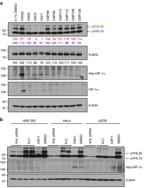
Biological evaluation. We next tested all our Homo-PROTACs
by monitoring protein levels after 10 h of compound treatment at
1
µM concentration in HeLa cells (Fig.
4
a). We observed the
a
b
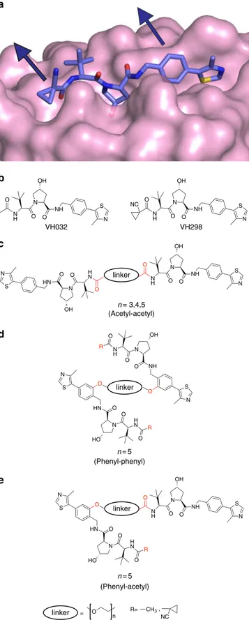
N O NH N S O N H O OH VH032 N O NH N S O N H O OH NC VH298 N O NH N S O N H N O HN N S O HN O O O HN N S N S O N HO O H N O O NH O N O N H O OH R R OH OH O HN N S O N HO O H N O R R= NC CH3 linker linker linker N O NH N S O N H O OH , (Phenyl-phenyl) (Phenyl-acetyl) linker = O n n = 3,4,5 (Acetyl-acetyl) n = 5 n = 5c
d
e
Fig. 1 Structure-guided design of Homo-PROTACs to induce VHL dimerization.a Crystal structures of VHL in complex with VH298 (PDB code 5LLI)19. VHL is shown as a pink surface and the bound ligand as sticks representation with purple carbons, nitrogen atoms in blue, oxygen in red and sulphur in dark yellow.b Chemical structure of VHL inhibitors VH032 and VH298.c–e General chemical structures and design of Homo-PROTACs compounds. Linkage sites at the acetyl and phenyl groups are indicated in red
striking effectiveness of CM09, CM10 and CM11 in inducing
VHL depletion in cells (Fig.
4
a), and a remarkably selective
degradation for the closely migrating bands corresponding to the
long isoform of VHL
52,53, preferentially over the short isoform.
The VHL gene includes three exons and it encodes two major
isoforms of VHL: a 213 amino-acid long, 30 kDa form (pVHL30)
and a 160 amino-acid long, 19 kDa form (pVHL19). pVHL19
lacks a 53 amino-acid long amino-terminal domain or N-terminal
tail (pVHL-N), which is instead present in pVHL30. Although
both isoforms are expressed in human cells, pVHL19 is the more
prominent form in human tissues
54. The most active compounds
are symmetrically linked from the terminal LHS acetyl group of
VH032. Linkage at different positions proved ineffective,
sug-gesting a critical role played by the linking pattern. Control
compounds CMP98 and CMP99 were unable to induce
degra-dation of VHL (Fig.
4
a), demonstrating that Homo-PROTAC
activity is dependent on productive bivalent recruitment of VHL
by the trans epimer. The length of the linker also seemed to affect
cellular potency. Indeed, a decrease in effectiveness was observed
at shorter linker lengths, with CM10 and CM11 being the most
active compounds achieving total knockdown of pVHL30,
fol-lowed by CM09 depleting 82% of the target protein. Interestingly,
some degradation of the short isoform pVHL19 was also
observed, albeit low (around 10% depletion). Levels of Cullin2,
the central subunit of the CRL2-VHL complex
55, were also
reduced upon treatment with CM10 and CM11 by up to 22%
(Fig.
4
a). Treatments with CM10 and CM11 also showed
detectable albeit low increase in protein levels of the hydroxylated
form of HIF-1α (Hdy-HIF-1α, Fig.
4
a). As the parent inhibitor
VH032 is completely ineffective at the same concentration of 1
µM (see ref.
19and vide infra, Fig.
5
), this effect cannot be due to
VHL inhibition and is thought to be the result of
compound-induced protein degradation. Levels of HIF-1α were, however,
significantly lower than observed when VH032 was used at
concentrations
> 100 µM (Fig.
4
a, see also ref.
19). VHL
knock-down by siRNA experiments in three different cell lines was
consistent with CM11-induced knockdown, and confirmed that
the bands observed indeed correspond to VHL (Fig.
4
b).
It is known that VHL RNAi is insufficient to induce significant
HIF stabilization (see also Fig.
4
b) and does not cause detectable
upregulation in HIF activity
56. On the basis of these
considera-tions, and the relatively low HIF stabilization observed with active
Homo-PROTACs CM09–11 (Fig.
4
a), it was presumed that
compound treatment would not induce a HIF-dependent hypoxic
response. To confirm this, we first used a luciferase reporter
assay
57. Hypoxia response element (HRE) -luciferase reporter
HeLa-HRE and U2OS-HRE cells were treated with different
concentrations of CM11 and at different times, and no increase in
luciferase activity was detected relative to DMSO control
treatment (Supplementary Fig.
1
). These results were confirmed
in a quantitative PCR with reverse transcription assay, where no
upregulation of mRNA levels of the known HIF-target genes CA9
was detected (Supplementary Fig.
2
). Together the data suggest
that un-degraded pVHL19 is sufficient to maintain low levels of
HIF-1α.
We next turned our attention to further characterize the mode
of action of the protein degradation induced by the active
Homo-PROTACs CM09–11. To interrogate their relative cellular
potency, dose-dependent treatments were performed at two
different time points, 4 and 24 h before harvesting. All
compounds confirmed preferential degradation of pVHL30 in a
concentration-dependent manner, relative to the corresponding
DMSO control (see Fig.
5
a for CM11, and Supplementary Fig.
3
for CM09 and CM10). CM11 proved the most potent compound,
inducing complete depletion of pVHL30 after 4 h already at 10
nM (DC
99= 10 nM, Fig.
5
a). Selective pVHL30 knockdown was
retained after 24 h, with half-degrading concentration (DC
50)
between 10 and 100 nM. The effective degrading concentrations
of CM11 are
> 3 orders of magnitude lower than the inhibitory
concentrations of the constitutive ligand VH032 alone, which is
only active in cells at ~ 100
µM, underscoring the difference in
cellular efficacy between the two mode of actions. Cellular levels
of Cullin2 decreased by up to 73% upon treatment with CM11
(Fig.
5
a). As previously observed, selective pVHL30 knockdown
by Homo-PROTACs resulted in only minor increase in levels of
HIF-1α, relative to hypoxia-inducing controls CoCl
2, PHD
O O O O O O HO O O O OH O N O NH N S O N H N O HN N S O HN CM09 CM10 CM11 X=OH Y=H CMP98 O O O O 1 n = 3 19% 2 n = 4 10% 3 n = 5 14% 4 n = 3 5 n = 4 6 n = 5 n n n tert -butyl bromoacetate,
NaH ,Dioxane, 16 h, r.t. TFA, DCM, 1 h, r.t. Quantitative yield DIPEA, HATU, DMF, rt, 30 min N O NH N S O H2N 7 trans -Hyp 8 cis -Hyp HO O H 3-5 OH X X Y Y n = 3 40% n = 4 30% n = 5 50% n = 5 45% X=OH Y=H X=OH Y=H X=H Y=OH
Fig. 2 Synthesis of symmetric homo-PROTAC compounds derivatized from the terminal acetyl group. The shown route yielded symmetrictrans-trans CM09, CM10 and CM11 and negative control symmetriccis-cis compound CMP98
inhibitor IOX2 and VH032 (Fig.
5
a). However, when tested at
high micromolar concentrations, Homo-PROTACs acted
prefer-entially as VHL inhibitors over VHL degraders, consistent with
the so-called
‘hook-effect’ whereby formation of binary
1:1 complexes competes with and eventually supersedes the
formation of the productive catalytic 2:1 complex
58. Stabilization
of Hdy-HIF-1α upon treatment with all three compounds at
100
µM was indeed comparable with the effect obtained with
VH032 alone (Fig.
5
a for CM11, and Supplementary Fig.
3
for
CM09 and CM10). To confirm the cellular activities of
Homo-PROTACs in a different cell line, U2OS cells were treated
for 10 h with CM09, CM10 and CM11 using the same range of
concentrations (1 nM–100 µM). A consistent profile of cellular
activity was observed, confirming that the effects observed are
independent from cell type (Supplementary Fig.
4
).
We
next
interrogated
the
time-dependent
activity
of
Homo-PROTACs. Progressive removal of VHL protein over
time was observed, confirming selective depletion of pVHL30
over the short isoform (Fig.
5
b for CM11 and Supplementary
Fig.
5
for CM09 and CM10). In particular, CM11 was confirmed
to be the most effective compounds, decreasing pVHL30 level by
more that 70% already after 2 h of treatment, and essentially to
completion after 8 h. The depletion effect was retained up to 12 h;
however, interestingly, pVHL30 levels up to 11% were detected
after
24–36 h treatment, to then decrease again after
48 h. Incomplete degradation of pVHL was observed upon
treatment with CM09, even in the longer time points
(Supple-mentary Fig.
5
). The results obtained treating U2OS cells were
consistent with what observed in the previous experiment.
However, in this cell line all three compounds induced complete
degradation of pVHL30 over time (Supplementary Fig.
4
). We
hypothesize that this could be due to the different expression
levels of VHL in the two cell lines. CM09 and CM10 achieved
complete degradation of the target protein after 2 h of treatment.
CM11 confirmed to be the most potent compound also in U2OS,
achieving complete degradation of pVHL30 already after 1 h.
Interestingly CM09 lost its cellular efficacy after 36 h. In contrast,
both CM10 and CM11 retained their efficacy even at these longer
time points (Supplementary Fig.
4
).
To gain mechanistic insights in the mode of action of
Homo-PROTACs, the dependency on CRL2-VHL and
protea-some
activities
was
examined.
The
reliance
of
the
Homo-PROTAC-induced protein degradation on CRL2-VHL
was assessed by inhibiting neddylation of Cullin2 using the NAE1
inhibitor MLN4924, which blocks the activity of CRLs, including
CRL2-VHL
59. Proteasome-dependency was interrogated by
treating cells with the proteasome inhibitor MG132. To limit
the known cytotoxicity of MLN4924 and MG132, HeLa cells
where pre-treated with MLN4924 for 3 h followed by MG132 for
30 min before adding CM11 to the media, and cells were
incubated for further 4 h before collecting. Single treatments with
DMSO, MLN4924, MG132 and CM11 and combinations thereof
were performed to disentangle the individual and combined
effects of compound treatments. Degradation of pVHL30 induced
by CM11 was completely abrogated when cells were pre-treated
with MG132, establishing the expected proteasome-dependence
of the chemical intervention (Fig.
6
). CM11-induced degradation
was also prevented by pre-treatment with MLN4924, confirming
the dependency on the activity of CRL2
VHL(Fig.
6
). The same
effect was observed when cells where co-treated with MLN4924
and MG132 before CM11 (Fig.
6
). Immunoblots of Cullin2 levels
confirmed the effective blockade of Cul2 neddylation by
MLN4924 (Fig.
6
). To assess if CM11 degrading activity was
dependent on VHL binding, a competition experiment was
O O O O O O HO O H 5 Benzyl chloride NaH, DMF 0°C to r.t, O/N 71% H 5tert -butyl bromoacetate, 37% NaOH, TBABr, DCM 16 h, r.t. 80% I. Pd/C, H2, EtOH, r.t, 1 h Quantitative yield II.BAIB, TEMPO ACN/H2O 1:1 3 h, r.t 65% 5 HO O O O O O 4 9 10 11 7, DIPEA, HATU,HOAT DMF, rt, 30 min 44% N O OH NH N S O N H O O O O 4 O 12 R'= -t -butyl TFA, DCM, 1 h, r.t. Quantitative yield 13 R'= –H R' N O OH NH N S O N H N O HN N S O HN CMP99 O O O O 4 8, DIPEA, HATU, DMF, rt, 30 min 66% OH
performed using the VHL inhibitor VH032
19. HeLa cells were
pre-treated with VH032 at 150
µM for 30 min before adding
CM11 into the media. The plates were incubated for further
4 h before harvesting. As expected, VH032 blocked pVHL
degradation (Fig.
6
) consistent with the hypothesis that VHL
induces degradation of itself. In contrast, pre-treatment with
IOX4, a PHD2 inhibitor, did not impact CM11 activity (Fig.
6
).
To evaluate the specificity of Homo-PROTAC-induced
degra-dation, and identify potential off-targets, we performed isobaric
tagging mass spectrometry proteomics to quantify degradation at
the proteome level in an unbiased fashion. Amongst the 6,450
detected proteins that passed
filtering criteria, no proteins other
than Cul2 were substantially depleted by Homo-PROTACs
CM09, CM10 or CM11 (1
µM for 10 h) compared to DMSO or
VHL inhibitor treatment (Supplementary Data
1
). Crucially, no
effect on protein levels of other Cullins (Cul1, Cul3, Cul4A,
Cul4B, Cul5 and Cul7) or CRL-associated subunits was observed
(Supplementary Fig.
6
). Together, the data demonstrate that
CM11 mainly induces depletion of pVHL30 and Cul2 but not
other proteins. The proteomics data further evidenced no increase
in protein levels of HIF-1α with Homo-PROTACs (but a small
decrease with CM09) relative to DMSO (Supplementary Fig.
6
).
In contrast, treatment with 150
µM VH032 led to increased
HIF-1α levels, as expected (Supplementary Fig.
6
).
Biophysical evaluation. Key to the catalytic mode of action of
PROTACs is the formation of a ternary complex
36, 38. With
Homo-PROTACs, VHL is presumed to act as both the E3 ligase
and the substrate. Therefore, we next sought to monitor and
biophysically characterize the ternary complex
VHL:Homo-PROTAC:VHL that is thought to underlie cellular activity. To
assess the formation of this ternary complex species in solution,
isothermal titration calorimetry (ITC), size-exclusion
chromato-graphy (SEC) and AlphaLISA proximity assays were performed
(Fig.
7
). In ITC titration of CM11 against the VCB complex (VHL
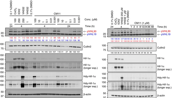
0.1% DMSO VH032 CM09 CM10 CM1 1 CMP98 CMP99 CMP1 13 CMP1 12 CMP108 CMP106 <-- pVHL30 <-- pVHL19 β-actin Cullin2 Hdy-HIF-1α HIF-1α 100 127 19 3 1 108 62 121 110 126 114 100 113 102 92 91 104 104 111 89 97 93 100 126 112 96 78 123 112 123 117 105 104 VHL siRNA N.C. CM1 1 DMSO VHL siRNA N.C. CM1 1 DMSO VHL siRNA N.C. CM1 1 DMSOHEK 293 HeLa U2OS
Hdy-HIF-1α <-- pVHL30 <-- pVHL19 β-actin
a
b
15 37 75 100 50 150 100 150 100 20 15 20 100 150 37 50Fig. 4 Homo-PROTACs CM09, CM10 and CM11 induce selective knockdown of the long isoform of VHL. a HeLa cells were treated with 0.1% DMSO, VH032 (150µM) and 1 µM of the indicated compounds for 10 h. Abundance of individual proteins was analyzed by western blotting using corresponding specific antibodies accordingly after SDS-PAGE. The data demonstrated is a representative example of experiments performed in biological duplicates. b Different cells lines were treated with siRNA targeting VHL proteins or negative control siRNA (for 48 h), as well as with CM11 (1µM) or 0.1% v/v DMSO for 10 h. The bands observed to selectively disappear in the presence of CM09–11 are closely migrating isoforms of pVHL30 (see refs52,53). The data demonstrated is a representative example of experiments performed as four biological replicates
with Elongin B and Elongin C)
60the stoichiometry of binding (n
value) was found to be 0.6, compared to n
= 1 with VH032
(Fig.
7
a, Table
1
). This result is consistent with CM11 binding to
VHL in a 1:2 molar ratio, in contrast to VH032 that binds to VHL
in a 1:1 ratio
18. Notably, the K
dvalue measured for CM11 was 11
nM (Table
1
). Closer examination of the titration curve revealed
that only one point features during the inflection of the curve.
Indeed, because the protein concentration used in the experiment
was 20
µM, the c value (defined as [P]
tot/K
d) calculated for this
experiment is 2500, which is above the upper limit of c
(~500–1000) that is a prerequisite for precise measurement of
binding affinity. Consequently, this analysis suggests that we may
be underestimating the binding affinity of CM11, i.e., we can
conclude that K
dis
≤11 nM. This corresponds to an avidity (also
known as cooperativity
α) of > 18-fold when compared to
VH032. Such large avidity of homo-bivalent molecules has been
observed previously with other systems, for example the BET
inhibitor MT1
48. The binding interaction between CM11 and
VHL was driven by a large apparent binding enthalpy (ΔΗ
= –12.3 kcal mol
−1), whereas the entropic term was slightly
unfavorable (–TΔS = 1.4 kcal mol
−1). This observation underlines
how the thermodynamic signature of CM11 is also very different
when compared with that of VH032, in which case the binding
ΔH was around half that observed with CM11, and both the
enthalpic and entropic term contributed favorably to the
ΔG of
binding (Table
1
). By contrast, the thermodynamic values
obtained for CMP99 binding were entirely consistent with the
ones of VH032 (Table
1
). Specifically, CMP99 bound to VHL in a
1:1 ratio, as expected due to the presence of the cis-Hyp in one of
the two moieties, and it exhibited comparable
ΔH and K
dvalues
to VH032. As anticipated, binding was not detected with CMP98,
the inactive cis-cis epimer. Superposition of integrated heat curves
of CM11, CMP98 and CMP99 is shown in Fig.
7
a and visually
highlights the different behaviors of the three compounds.
CM10 showed similar thermodynamic binding parameters
rela-tive to CM11, with n value equal to 0.7 and a low K
dof 32 nM. A
stoichiometry close to 1 was instead found for CM09, suggesting
that at the end of the titration this system was primarily
popu-lated by 1:1 complexes (Supplementary Figs
7
and
8
), consistent
with its lower avidity (Table
1
)
58,61. To compare and contrast
Homo-PROTACs binding to the long VHL isoform, we expressed
and purified pVHL30 in complex with Elongin C and Elongin B
(V
30CB) and performed ITC titrations (Table
1
and
Supple-mentary Fig.
9
). The thermodynamic parameters of binding of
CMP99 to V
30CB were comparable to those to V
19CB, suggesting
0.1% DMSO CoCl 2 IOX2 VH032 250 μ M VH032 1 μ M 0.1% DMSO 4 24 CM11 (1 μM) 24 4 Time (h) <--pVHL30 <--pVHL19 Cullin2 Hif-1α Hif-1α (longer exp.) Hdy-Hif-1α (longer exp.) Hdy-Hif-1α β-actin 1 2 8 12 36 48 100 106 84 127128100 85 26 10 2 1 4 9 2 100 120 91 91 108100 94 129 90 105110 93 81 79 100 126 96 81 106 100 80 90 70 96 87 66 55 69 1 0.1 0.01 0.001 100 10 100 10 1 0.1 0.01 0.001 0.1 %DMSO CoCl 2 IOX2 VH032 VH032 0.1% DMSO 4 24 CM11 Conc. (μM) 24 4 Time (h) 100 250 250 1 <--pVHL30 <--pVHL19 Cullin2 Hif-1α Hif-1α (longer exp.) Hdy-Hif-1α (longer exp.) Hdy-Hif-1α β-actin 100 82 71 112 138 100 88 102 2 11 2 89 106 0 0 15 80 77 100 71 72 92 104 100 102 100 92 91 112 87 146 127 148 121 126 96 100 101 100 130 120 100 86 72 76 105 124 107 118 41 27 98 132 101 15 20 75 100 100 150 100 150 100 150 37 50 100 150 100 150 100 150 100 100 37 50 15 20 75 100
Fig. 5 CM11 induces pVHL30 depletion in a concentration and time-dependent fashion. (left) Dose-response profile of HeLa cells treated with increasing concentration of CM11 for 4 or 24 h. (right) Time-course immunoblots of lysates from HeLa cells treated 1µM of CM11 up to 48 h. Control treatments of 0.1% DMSO, CoCl2(100µM), IOX2 (150 µM), and VH032 (250 µM or 1 µM) are included. All data demonstrated is a representative example of
experiments performed in biological duplicates
MLN4924 MG132 CM11 VH032 IOX4 DMSO Time (h) μM 7.5 4.5 4 4.5 4.5 7.5 3 50 1 150 50 – + – – – + + + + – – – –+ + + +– – – – + + + +++ + + + + + + + + + + – – – – – – – – – – – – + – – – – – – – – – – – – – – + – – – + – – – – – – – – – + + + + + + – – – – + + – – – – – – + + + + – – – – + + + + – – – – – – + + – – + + Hdy-HIF-1α pVHL30 --> pVHL19 --> β−actin Cullin2 15 37 75 50 100 150 100 20
Fig. 6 CM11 activity is CRL2VHLand proteasome-dependent. HeLa cells treated with CM11 in the absence or presence of proteasome inhibitor MG132, neddylation inhibitor MLN4924, VHL inhibitor VH032 or PHD2 inhibitor IOX4 as negative control. The data demonstrated here is representative of one biological replicate
identical binary target engagement in vitro between the two
iso-forms. CM11 also exhibited large
ΔH of binding and n < 1, albeit
interestingly lower avidity for pVHL30 compared to pVHL19
(Table
1
and Supplementary Fig.
9
).
SEC experiments showed that V
19CB migrates more quickly in
the presence of the active compound CM11 (2:1 protein:ligand
ratio), relative to vehicle control (Fig.
7
b). The shifted peak eluted
at a volume corresponding to a species of ~ 90 kDa molecular
weight, based on a calibration run with globular proteins of
known molecular weight (see Methods), suggesting the peak
corresponds to the ternary complex (VCB)
2:CM11. Formation of
a 2:1 complex was also observed with V
30CB (Supplementary
Fig.
10
). Mixing V
19CB, V
30CB and CM11 resulted in a mixed
population of the three species (V
19CB)
2:CM11, (V
19CB):CM11:
(V
30CB) and (V
30CB)
2:CM11, suggesting that both VHL isoforms
can form ternary complexes with CM11. No shift in VCB was
observed following incubation with inactive CMP98, CMP99 or
ligand VH032. Only in the sample containing CMP99 a small
peak eluted at 13.5 ml (green curve, Fig.
7
b). It is possible that
such peak could be due to the formation of a lowly populated
ternary complex. It is interesting that Schofield and colleagues
observed weak binding of a cis-hydroxyprolyl containing HIF-1α
peptide to VHL
51. This weak binding, potentially enhanced by
high avidity in the ternary complex, could be responsible for the
small decrease of VHL levels observed during biological tests in
cells (Fig.
3
a). CM10 and CM09 showed formation of a ternary
complex eluting at identical retention volume when compared to
CM11 (Supplementary Fig.
11
). No evidence of aggregation was
seen with any of the compounds evaluated, as all observed peaks
eluted well after the void volume.
Lastly, we employed an AlphaLISA proximity assay to compare
ternary complex formation by CM09, CM10 and CM11. The
assay showed the highest intensity signal for CM11, whereas
negligible levels of complex formation were detected for CM09
and CM10 (Fig.
7
c). Since SEC detected ternary species with all
three compounds, the minimal intensity detected in the
AlphaLISA likely reflects the inability of CM09 and CM10 to
yield a significant ternary population at the low concentrations
required for the assay. These results indicate that CM11 is the
most effective Homo-PROTAC at driving ternary complex
formation, consistent with CM11 exhibiting the highest avidity
and full 2:1 stoichiometry in ITC. Together, the biophysical data
supports CM11 as the most cooperative Homo-PROTAC in vitro,
and provides a molecular rationale explaining its potent
VHL-degrading activity inside cells.
Discussion
We describe Homo-PROTACs, a small-molecule approach to
effectively dimerize an E3 ubiquitin ligase to induce its own
self-destruction. Using potent ligands for the E3 ligase VHL, we
developed a series of homo-bivalent molecules that induce
remarkably rapid, profound and selective degradation of the long
isoform of pVHL at nanomolar concentrations.
Compound-induced degradation was exquisitely dependent on the linkage
pattern on the VHL ligand. The most active Homo-PROTAC,
CM11, induces complete depletion of pVHL30 after 4 h already at
10 nM. Potent and selective degradation of pVHL30 was
long-lasting, with a half-degrading concentration (DC
50) of
<100 nM,
a remarkable increase in cellular activity of
>1000-fold compared
to the parent inhibitor VH032. Mechanistically, we show that
CM11 activity is strictly dependent on proteasome activity, Cul2
neddylation, and on VHL binding, and specifically on the
for-mation of an avid 2:1 complex with VHL. Our data therefore
supports a model in which a highly cooperative ternary complex
(VHL)
2:CM11 functions as the key species responsible for the
induced degradation of VHL itself (Fig.
8
). Future structural
studies of this ternary species are warranted. Interestingly, CM11
also led to a decrease in cellular levels of Cullin2, which we
hypothesize to be the result of direct ubiquitination and
degra-dation of Cullin2 as part of the CRL2
VHLcomplex. To our
knowledge, it is unprecedented that a PROTAC can induce the
degradation of a protein forming part of the same complex with
the protein targeted directly.
The preferential induced degradation of pVHL30 over the
short VHL isoform was unanticipated and is an intriguing result
of this work. This observation adds to recent evidence from us
and others that chemical degraders designed from inhibitors
recruiting more than a single protein can add a layer of target
degradation selectivity independently of target engagement
35,38,41
. Biophysically, the VHL warhead was found not to distinguish
between the two VHL isoforms at the level of binary target
engagement. It was also found that CM11 does not considerably
distinguish between pVHL19 and pVHL30 within ternary
com-plexes. We therefore view it unlikely that the remarkable
selec-tivity of VHL degradation is due to differences in molecular
recognition. We also consider unlikely that preferential and more
efficient lysine ubiquitination could play a role, because the extra
region present in the long isoform (1–53) does not contain a
single lysine residue. On the other hand, this region is predicted
as intrinsically disordered
62, and it has been shown that proteins
containing disordered N-terminal regions are more prone to
b
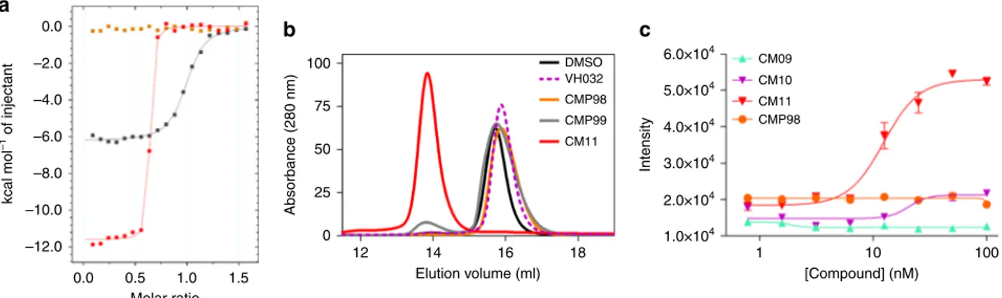
100 75 50 25 Absorbance (280 nm) 0 12 14 16 Elution volume (ml) 18 DMSO VH032 CMP98 CMP99 CM11a
0.0 kcal mol –1 of injectant –2.0 –4.0 –6.0 –8.0 –10.0 –12.0 0.0 0.5 1.0 1.5 Molar ratioc
Intensity [Compound] (nM) 1 CM09 CM10 CM11 CMP98 10 100 6.0×10⁴ 5.0×10⁴ 4.0×10⁴ 3.0×10⁴ 2.0×10⁴ 1.0×10⁴Fig. 7 Biophysical studies of Homo-PROTACs binding to VHL. a Superposition of the integrated ITC heat curves of CM11 (blue), CMP99 (black) or CMP98 (green) titrations against VCB.b SEC assay of complex formation after incubation of CM11 (red), CMP98 (pale green), CMP99 (purple), VH032 (dotted purple) or DMSO (black) with VCB.c AlphaLISA: intensity values titrating CM09, CM10, CM11 and CMP98 against VCB. Each point is mean (± SEM) intensity of four technical replicates
proteasomal degradation
63. It is also known that VHL is resistant
to proteasomal degradation when in complex with ElonginB and
ElonginC
64, so the form observed to be preferentially depleted
may be free VHL, i.e., not bound to Elongins, or other
proteasome-sensitive forms
65. Previous work revealed different
cellular localization patterns of pVHL19 and pVHL30, the former
being primarily nuclear, while the latter predominantly
cyto-plasmic
56. Crucially, pVHL30 was found to associate to
micro-tubules, a function that is thought to be independent from its
ligase activity
66. We therefore speculate that the selective
degra-dation of pVHL30 observed in cells could be the result of
pVHL19 acting preferentially as part of CRL2-VHL, and pVHL30
as the
‘neo-substrate’. Obtaining a more detailed biochemical and
mechanistic understanding of the cellular mode of action of VHL
Homo-PROTACs is of clear importance for future investigation.
Selective degradation of pVHL30 by CM11 led to minimal
stabilization of HIF-α in cells, and as a result did not trigger
HIF-dependent activity. This is consistent with complete
knock-down of all VHL isoforms being required to achieve effective HIF
stabilization in cells, as observed in vhl
–/–cells such as
VHL-deficient renal carcinoma cells
49. Our results underscore the
potential benefit of using CM11 to interrogate the biological
function of specific VHL isoforms, without the masking
down-stream effects of a hypoxic response. Not much is known about
the individual roles of VHL isoforms. Studies have highlighted
how the 53-residue extra region of pVHL30 is not needed for
tumor suppression
53, and how both isoforms can have
HIF-dependent tumor suppressor functions in vivo
67,68. Other
HIF-independent roles of pVHL have been proposed
69, including a
role for pVHL in collagen assembly
70. However, the individual
roles of the different isoforms in these biological functions remain
elusive. Moreover, many HIF-independent roles are thought to be
independent upon Hyp recognition
71, and thus cannot be probed
chemically using the current Hyp-based VHL inhibitors
19.
Selective and acute knockdown of pVHL30 by CM11 therefore
offers a new opportunity to address these questions.
In summary, we present CM11, a chemical probe for rapid
and selective pVHL30 knockdown. CM11 provides a chemical
tool alternative to conventional knockdown RNAi approaches
and gene editing knockout technologies such as CRISPR-Cas9.
Relevant information to the use of CM11 will be made available
in the newly established
‘Chemical Probes Portal’ (
http://www.
chemicalprobes.org/
)
72. We anticipate CM11 will
find wide use
amongst chemical and cell biologists alike interested in
inves-tigating and dissecting the pleiotropic biological functions of
pVHL. More generally, we provide proof-of-concept that
bivalent molecules can be designed to induce an E3 ligase to
destroy itself. This strategy opens powerful new avenues to
drugging E3 ligases in ways that are not possible with inhibitors
alone.
Methods
Synthesis of CM09, CM10, CM11 and controls CMP98 and CMP99. General method A. PEG (1 eq.) was solubilised in dioxane anhydrous and NaH (2 eq.) was added under stirring. The resulting mixture was stirred at r.t. for 3h. The mixture was cooled down to 0 °C using ice bath and tert-butylbromo acetate (2 eq.) was added drop by drop. The resulting mixture was stirred at r.t O/N. The precipitate wasfiltered off and the organic phase evaporated to dryness. The resulting oil was taken up with ethyl acetate, washed with water, dried over MgSO4and evaporated
to dryness. The resulting oil was purified by column chromatography using a gradient of ethyl acetate from 50% to 100% v/v in heptane.
General method B. tert-butyl esters 1, 2, 3 or 12 were dissolved in a solution of 50% v/v trifluroacetic acid in DCM. The resulting solution was stirred for 1 h or until complete conversion of starting material. The solvent was removed under high vacuum. The resulting carboxylic acid was used as crude in the next step without any further purification. To a solution of carboxylic acid in 1 ml DMF were added HATU (1 eq.) and HOBT (1 eq.) and the pH of the reaction mixture was adjusted to> 9 by addition of DIPEA (3 eq.). The resulting solution was stirred at room temperature for 5 min and then amine 7 or 8 was added. The mixture was stirred at room temperature until no presence of the starting materials was detected by LC-MS. Water was added and the mixture was extracted with ethyl acetate (×3). The combined organic phases were washed with brine (×2), dried over MgSO4and evaporated under reduced pressure to
give the corresponding crude, which was purified by HPLC using a gradient of 20% to 95% v/v acetonitrile in 0.1% aqueous solution of ammonia to yield the desired compound.
di‐tert‐butyl 3, 6, 9, 12 – tetraoxatetradecanedioate (1)
Following general method A, from triethylene glycol (1.125 g, 1 ml, 7.49 mmol, 1 eq.) in 10 ml of dioxane, NaH 60% in mineral oil (595.75 mg, 14.9 mmol, 2 eq.) and tert-Butyl bromoacetate (2.905 g, 2.19 ml, 14.9 mmol, 2 eq.), compound 1 was obtained as an oil after high vacuum. Yield: 538 mg, 1.42 mmol (19%). ¹H NMR
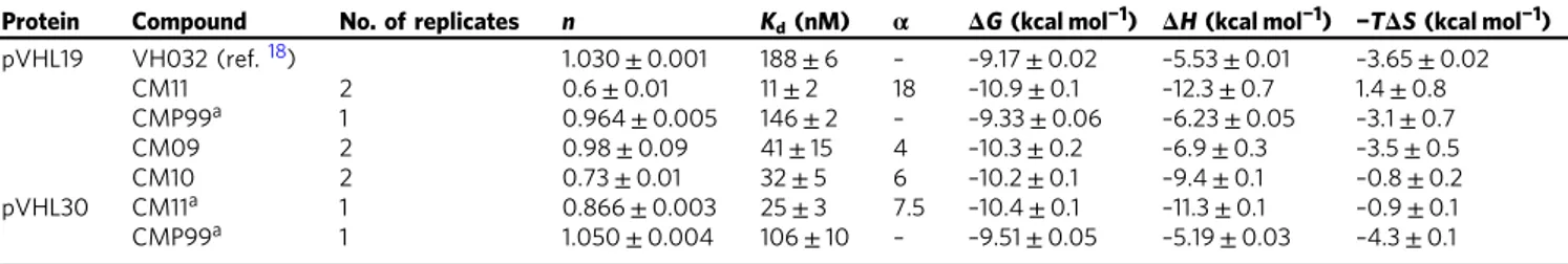
Table 1 Thermodynamic binding parameters of complex formation of Homo-PROTACs measured by isothermal titration
calorimetry
Protein Compound No. of replicates n Kd(nM) α ΔG (kcal mol−1) ΔH (kcal mol−1) −TΔS (kcal mol−1)
pVHL19 VH032 (ref.18) 1.030± 0.001 188± 6 – –9.17 ± 0.02 –5.53 ± 0.01 –3.65 ± 0.02 CM11 2 0.6± 0.01 11± 2 18 –10.9 ± 0.1 –12.3 ± 0.7 1.4± 0.8 CMP99a 1 0.964± 0.005 146± 2 – –9.33 ± 0.06 –6.23 ± 0.05 –3.1 ± 0.7 CM09 2 0.98± 0.09 41± 15 4 –10.3 ± 0.2 –6.9 ± 0.3 –3.5 ± 0.5 CM10 2 0.73± 0.01 32± 5 6 –10.2 ± 0.1 –9.4 ± 0.1 –0.8 ± 0.2 pVHL30 CM11a 1 0.866± 0.003 25± 3 7.5 –10.4 ± 0.1 –11.3 ± 0.1 –0.9 ± 0.1 CMP99a 1 1.050± 0.004 106± 10 – –9.51 ± 0.05 –5.19 ± 0.03 –4.3 ± 0.1 All titrations were performed at 25 °C. Error values reported are the means± 1 s.e.m., unless otherwise specified. Raw ITC data are shown for each titration in Fig.7a and Supplementary Figs7–9.
aErrors are generated by the Origin program and reflect the quality of the fit between the nonlinear least-squares curve and the experimental data.
VHL Target VHL Homo-PROTAC Uncomplexed/binary 1:1 complexes No target degradation avid 2:1 Ternary complex VHL VHL VHL self-degrades
Fig. 8 Proposed model for the mechanism of action of Homo-PROTAC CM11
(500 MHz, CDCl3):δ 3.81 (s, 4H), 3.51–3.46 (m, 12H), 1.26 (s,18H).13C NMR
(126 MHz, CDCl3):δ 169.1, 80.9, 70.1, 70.0, 68.5, 27.5. Analytical data matched
those previously reported73.
di‐tert‐butyl 3,6,9,12,15-pentaoxaheptadecanedioate (2)
Following general method A, from tetrathylene glycol (1.125 g, 1 ml, 5.49 mmol, 1 eq.) in 10 ml of dioxane, NaH 60% in mineral oil (463 mg, 11.5 mmol, 2 eq.) and tert-Butyl bromoacetate (2.25 g, 1.7 ml, 11.5 mmol, 2 eq.), compound 2 was obtained as an oil after high vacuum. Yield: 500 mg, 1.18 mmol (10%). ¹H NMR (500 MHz, CDCl3):δ 3.86 (s, 4H), 3.55–3.49 (m, 16H), 1.31 (s, 9H). Analytical data
matched those previously reported73.
di‐tert‐butyl 3,6,9,12,15,18-hexaoxaicosanedioate (3)
Following general method A, from pentaethylene glycol (1.126 g, 1 ml, 4.72 mmol, 1 eq.) in 10 ml of dioxane, NaH 60% in mineral oil (377 mg, 9.45 mmol, 2 eq.) and tert-Butyl bromoacetate (1.872 g, 1.7 ml, 11.5 mmol, 2 eq.), compound 3 was obtained as an oil after high vacuum. Yield: 300 mg, 0,641 mmol (14%).1H NMR(400 MHz, CDCl3):δ 3.94 (s, 4H), 3.66–3.56 (m, 20H), 1.40 (s, 18H).
Analytical data matched those previously reported73.
N1,N14-bis((S)-1-((2S,4R)-4-hydroxy-2-((4-(4-methylthiazol-5-yl)benzyl)carbamoyl) pyrrolidin-1-yl)-3,3-dimethyl-1-oxobutan-2-yl)-3,6,9,12-tetraoxatetradecanediamide (CM09)
Following general method B, from compound 1 (6.80 mg, 0.018 mmol, 1 eq.), compound 7 (20 mg, 0.045 mmol, 2.5 eq.), HATU (17 mg, 0.045 mmol, 2.5 eq), HOAT (6.12, 0.045 mmol, 2.5 mmol) and DIPEA (6.98 mg, 0.054 mmol, 3 eq) compound CM09 was obtained as a white solid. Yield: 8 mg, 0.007 mmol (40%). ¹H NMR(400 MHz, CDCl3):δ 8.61 (s, 2H), 7.48–7.45 (m, 2H), 7.31–7.27 (m, 8H), 7.23 (d, J= 10.2 Hz, 2H), 4.64–4.59 (m, 2H), 4.52–4.46 (m, 4H), 4.41–4.38 (m, 2H), 4.31–4.25 (m, 2H), 4.01–3.94 (m, 4H), 3.82 (d, J = 15.7 Hz, 2H), 3.62–3.52 (m, 12H), 2.45 (s, 6H), 2.42–2.34 (m, 2H), 2.12–2.06 (m, 2H), 1.19 (s, 2H), 0.89 (s, 18H);13C NMR(101 MHz, CDCl 3):δ 170.2, 169.9, 169.6, 149.3, 147.5, 137.3, 130.6, 129.9, 128.4, 127.1, 69.9, 69.5, 69.3, 69.1, 57.6, 56.1, 55.9, 42.2, 35.5, 34.6, 25.4, 15.1. HRMS (ESI) m/z: [M + H]+calculated for: C
54H74N8O12S2: 1090.49;
observed: 1091.5093.
N1,N17-bis((S)-1-((2S,4R)-4-hydroxy-2-((4-(4-methylthiazol-5-yl)benzyl)carbamoyl)
pyrrolidin-1-yl)-3,3-dimethyl-1-oxobutan-2-yl)-3,6,9,12,15-pentaoxaheptadecanediamide (CM10)
Following general method B, from compound 2 (7.60 mg, 0.018 mmol, 1 eq.), compound 7 (20 mg, 0.045 mmol, 2.5 eq.), HATU (17 mg, 0.045 mmol, 2.5 eq), HOAT (6.12, 0.045 mmol, 2.5 mmol) and DIPEA (6.98 mg, 0.054 mmol, 3 eq) compound CM10 was obtained as a white solid. Yield: 6 mg, 0.005 mmol (30%). ¹H NMR (500 MHz, CDCl3):δ p.p.m., 7.58–7.55 (m, 2H), 7.35–7.30 (m, 10H), 4.72–4.67 (m, 2H), 4.57–4.47 (m, 6H), 4.38–4.33 (m, 2H), 4.15–3.91 (m, 8H), 3.68–3.56 (m, 20H), 2.51 (s, 6H), 2.45–2.38 (m, 2H), 2.20–2.14 (m, 2H), 0.95 (s, 18H).13C NMR(126 MHz, CDCl 3):δ 170.3, 169.8, 169.6, 149.6, 146.9, 137.5, 129.5, 128.47, 128.40, 127.1, 70.0, 69.56, 69.52, 69.5, 69.2, 69.1, 57.7, 56.03, 55.98, 42.1, 35.6, 34.6, 25.4, 14.8. HRMS (ESI) m/z: [M + H]+ calculated for: C56H78N8O13S2: 1134.51; observed: 1135.5538.
N1,N20-bis((S)-1-((2S,4R)-4-hydroxy-2-((4-(4-methylthiazol-5-yl)benzyl)carbamoyl)
pyrrolidin-1-yl)-3,3-dimethyl-1-oxobutan-2-yl)-3,6,9,12,15,18-hexaoxaicosanediamide (CM11)
Following general method B, from compound 3 (8.39 mg, 0.018 mmol, 1 eq.), compound 7 (20 mg, 0.045 mmol, 2.5 eq.), HATU (17 mg, 0.045 mmol, 2.5 eq), HOAT (6.12, 0.045 mmol, 2.5 mmol), DIPEA (6.98 mg, 0.054 mmol, 3 eq) compound CM11 was obtained as a white solid. Yield: 11.74 mg, 0.0099 mmol (55%). ¹H NMR (400 MHz, CDCl3):δ 8.61 (s, 2H), 7.41–7.38 (m, 2H), 7.29 (t, J= 7.6 Hz, 10H), 4.66–4.61 (m, 2H), 4.49–4.41 (m, 6H), 4.35–4.29 (m, 2H), 3.98–3.91 (m, 6H), 3.62–3.50 (m, 24H), 2.45 (s, 6H), 2.42–2.35 (m, 2H), 2.11–2.06 (m, 2H), 0.88 (s, 18H);13C NMR(101 MHz, CDCl 3): δ 171.2, 170.9, 170.4, 150.3, 148.5, 138.3, 131.6, 130.9, 129.5, 128.1, 71.2, 70.61, 70.59, 70.5, 70.4, 70.3, 58.6, 57.0, 43.2, 36.5, 35.6, 26.4, 16.1. HRMS (ESI)m/z: [M + H]+calculated for: C
58H82N8O14S2: 1178.54; observed:
1179.6015.
N1,N20-bis((S)-1-((2S,4S)-4-hydroxy-2-((4-(4-methylthiazol-5-yl)benzyl)carbamoyl)
pyrrolidin-1-yl)-3,3-dimethyl-1-oxobutan-2-yl)-3,6,9,12,15,18-hexaoxaicosanediamide (CMP98)
Following general method B, from compound 3 (7.12 mg, 0.028 mmol, 1 eq.), compound 8 (18.06, 0.040 mmol, 2.1 eq.), HATU (15.2 mg, 0.040 mmol, 2 eq.), HOAT (5.44 mg, 0.040 mmol, 2 eq.), DIPEA (7.45 mg, 0.0010 ml, 3 eq.), compound CMP98 was obtained as a white solid. Yield: 10.58 mg, 0.0089 mmol (45%). ¹H NMR(400 MHz, CDCl3):δ 9.09 (s, 2H), 8.02 (s, 2H), 7.31 (d, J = 8.5 Hz, 4H), 7.22 (d, J= 8.0 Hz, 4H), 7.16 (d, J = 9.2 Hz, 2H), 4.75–4.64 (m, 4H), 4.51 (d, J = 8.9 Hz, 2H,), 4.41–4.37 (m, 2H), 4.24–4.17 (m, 2H), 3.94 (d, J = 3.2 Hz, 4H), 3.84–3.81 (m, 4H), 3.62–3.54 (m, 20H), 2.49–2.47 (m, 2H), 2.44 (s, 6H), 2.26–2.17 (m, 4H), 0.93 (s, 18H);13C NMR(101 MHz, CDCl 3):δ 173.2, 171.5, 169.7, 151.8, 138.8, 132.9, 129.5, 129.2, 128.3, 71.2, 71.1, 70.6, 70.48, 70.45, 70.4, 70.3, 59.9, 58.5, 56.5, 43.2, 35.6, 35.2, 26.4, 15.0. HRMS (ESI) m/z: [M + H]+calculated for:
C58H82N8O14S2: 1178.54; observed: 1179.6087.
1‐phenyl‐2,5,8,11,14-pentaoxahexadecan-16-ol (9)
Pentaethylene glycol (9.53 g, 50 mmol, 5 eq.) was added dropwise to a suspension of NaH 60% in mineral oil (800 mg, 20 mmol, 2.5 eq.) in 20 ml of DMF at 0 °C. The resulting mixture was stirred at r.t for 1 h. The reaction mixture was cooled to 0oC, benzyl chloride (1 ml, 1.1 g, 8.72 mmol, 1 eq.) was added. The resulting mixture was stirred O/N at r.t. The reaction was quenched with a saturated solution of NH4Cl and the aqueous phase was extracted with ethyl acetate
(×3). The combined organic phases were dried over MgSO4and evaporated to
dryness. The resulting oil was purified by column chromatography (from 0 to 60% of ethyl acetate in heptane) to afford the title compound as a oil. Yield: 2.055 g, 6.25 mmol (71%). ¹H NMR (400 MHz, CDCl3):δ 7.28–7.19 (m, 5H), 4.50 (s, 2H),
3.66–3.52 (m, 20H), 2.50 (s, 1H).13C NMR(101 MHz, CDCl
3):δ 138.2, 128.3,
127.8, 127.6, 73.2, 72.7, 70.61, 70.58, 70.53, 70.51, 70.2, 69.4, 61.7 tert-butyl 1-phenyl-2,5,8,11,14,17-hexaoxanonadecan-19-oate (10)
To a stirred solution of 9 (2.055 g, 6.25 mmol, 1 eq.) in 12.8 ml of DCM was added 37% solution of NaOH (12.8 ml), followed by tert-butylbromo acetate (4.882 g, 25 mmol, 4 eq.) and TBABr (2118 mg, 6.37 mmol, 1.02 eq.). The resulting solution was stired O/N at r.t. The reaction mixture was extracted with ethyl acetate (×3). The organic phases were combined and washed with brine (×1), dried over MgSO4and concentrate in vacuo. The resulting brow oil was purified by column
chromatography (from 0 to 30% of ethyl acetate in petroleum) to afford the titled compound as colorless oil. Yield: 2.216 g, 5 mmol (80%). ¹H NMR (500 MHz, CDCl3):δ 7.28–7.20 (m, 5H), 4.50 (s, 2H), 3.95 (s, 2H), 3.65–3.55 (m, 20H), 1.40
(s, 9H).13C NMR(126 MHz, CDCl
3):δ169.7, 128.4, 127.7, 127.6, 81.5, 73.2, 70.7,
70.7, 70.6, 70.6, 69.4, 69.1, 28.1. MS (ESI) m/z: [(M-tBu) + H]+calculated for: C23H38O8: 442.26; observed: 387.20.
19,19-dimethyl-17-oxo-3,6,9,12,15,18-hexaoxaicosanoic acid (11)
10(2.216 g, 5 mmol, 1 eq.) was dissolved in 75 ml of ethanol, Pd/C (10 wt%) was added and the resulting mixture was place under hydrogen and stirred at r.t. until complete conversion of the starting material. The reaction mixture was filtered through celite, the celite pad was washed few times using ethanol. The filtrate was concentrated in vacuum to give an oil that was used for the next step without further purification. Yield: 1764 g, 5 mmol (quantitative). BAIB (3.546 g, 11.01 mmol, 2.2 eq.) and TEMPO (171.87 mg, 1.10 mmol, 0.22 eq.) were added to a solution of ACN/H2O 1:1 containing previous obtained oil (1.764 g, 5 mmol, 1 eq.).
The resulting mixture was stirred at r.t until complete conversion of the starting material. The crude was purified using ISOLUTE® PE-AX anion exchange column. The column was equilibrate with methanol, the reaction mixture poured in the column and let it adsorbed in the pad. The column was washed with methanol (×3) to elute all the unbound material. Then, the titled product was eluted using a 50% solution of formic acid in methanol. The organic phase was evaporated to dryness to afford the title compound as oil. Yield: 1.200 g, 3.27 mmol (65%). ¹H NMR (400 MHz, CDCl3):δ 4.12 (s, 2H), 3.98 (s, 2H), 3.72–3.60 (m, 16H), 1.43 (s, 9H). 13C NMR(101 MHz, CDCl 3):δ 172.6, 169.7, 81.6, 71.0, 70.59, 70.56, 70.54, 70.46, 70.38, 70.35, 70.30, 68.9, 68.8, 28.1. tert-butyl (S)-19-((2S,4R)-4-hydroxy-2-((4-(4-methylthiazol-5-yl)benzyl)carbamoyl) pyrrolidine-1-carbonyl)-20,20-dimethyl-17-oxo-3,6,9,12,15-pentaoxa-18-azahenicosanoate (12)
To a solution of PEG linker 11 (78.8 mg, 0.215 mmol, 1 eq.) in 1.5 ml DMF was added HATU (81.74 mg, 0.215 mmol, 1 eq.), HOAT (29.26 mg, 0.215 mmol, 1 eq.), DIPEA (80.13 mg, 0.106 ml, 0.645 mmol, 3 eq.) and the solution was stirred at room temperature for 5 min. Compound 7 (100 mg, 0.215 mmol, 1 eq.) was added and the pH of the reaction mixture was adjusted to> 9 by addition of DIPEA (80.13 mg, 0.106 ml, 0.645 mmol, 3 eq.). The mixture was stirred at room temperature until no presence of the starting materials was detected by LC-MS. The solvent was evaporated under reduced pressure to give the corresponding crude, which was purified by HPLC using a gradient of 20–95% v/v acetonitrile in 0.1% aqueous solution of ammonia to yield the titled compound as white solid. Yield: 75.6 mg, 0.094 mmol (44%). ¹H NMR (400 MHz, CDCl3):δ 9.00 (s, 1H), 7.45 (t, J= 5.9 Hz, 1H), 7.39–7.33 (m, 4H), 7.29 (d, J = 8.9 Hz, 1H), 4.71 (t, J = 8.0 Hz, 1H), 4.59–4.48 (m, 3H), 4.34 (dd, J = 5.2, 14.6 Hz, 1H), 4.08–3.92 (m, 5H), 3.69–3.61 (m, 18H), 2.52 (s, 3H), 2.47–2.41 (m, 1H), 2.19–2.11 (m, 1H), 1.46 (s, 9H), 0.97 (s, 9H).13C NMR(101 MHz, CDCl 3):δ 171.3, 171.1, 170.5, 170.0, 151.7, 139.1, 129.4, 128.3, 82.0, 71.1, 70.6, 70.4, 70.4, 70.3, 70.3, 70.2, 70.2, 68.9, 58.7, 57.3, 56.8, 43.1, 36.3, 35.1, 28.1, 26.4, 15.1. MS (ESI) m/z: [M + H]+calculated for: C38H58N4O11S2: 778.38; observed: 779.4.
N1-((R)-1-((2R,4R)-4-hydroxy-2-((4-(4-methylthiazol-5-yl)benzyl)carbamoyl) pyrrolidin-1-yl)-3,3-dimethyl-1-oxobutan-2-yl)-N17
-((S)-1-((2S,4R)-4-hydroxy-2- ((4-(4-methylthiazol-5-yl)benzyl)carbamoyl)pyrrolidin-1-yl)-3,3-dimethyl-1-oxobutan-2-yl)-3,6,9,12,15-pentaoxaheptadecanediamide (CMP99)
Following general method B, from compound 12 (75.6 mg, 0.094 mmol, 1 eq.) and trifluoroacetic acid (1 ml in 1 ml of DCM), the carboxylic acid derivative was obtained as oil. The crude was used for the next step without further purification. Yield: 70 mg, 0.094 mmol (quantitative). MS (ESI) m/z: [M + H]+calculated for: C34H50N4O11S: 722.32; observed: 723.3. Following general method B, from
compound 13 (5.5 mg, 0.006 mmol, 1 eq.), compound 8 (2.77 mg, 0.006 mmol, 1 eq.), HATU (2.28 mg, 0.0.006 mmol, 1 eq.), HOAT (1 mg, 0.0.006 mmol, 1 eq.), DIPEA (2.23 mg, 0.002 ml, 0.018 mmol, 3 eq.), CMP99 was obtained as a white solid. Yield: 4.5 mg, 0.004 mmol (66%). ¹H NMR (400 MHz, CDCl3):δ 8.74 (s, 1H), 8.73 (s, 1H), 7.37–7.34 (m, 9H), 7.18 (d, J = 8.9 Hz, 1H), 4.76–4.64 (m, 3H), 4.59–4.44 (m, 5H), 4.37–4.26 (m, 2H), 4.05–3.59 (m, 27H), 2.52 (s, 6H), 2.31–2.11 (m, 4H), 0.96 (s, 9H), 0.95 (s, 9H).13C NMR(101 MHz, CDCl3):δ 173.0, 171.3,
170.0, 150.7, 145.4, 138.4, 129.53, 129.49, 128.2, 128.16, 71.2, 71.0, 70.54, 70.48, 70.4, 70.3, 58.4, 57.0, 56.7, 43.4, 35.2, 35.0, 26.4, 26.35, 15.9. HRMS (ESI) m/z: [M + H]+calculated for: C56H78N8O13S2: 1134.51; observed: 1135.5814.
Biological and biophysical assays. Cell culture. Human cell lines HeLa, U2OS and HEK 293, purchased from ATCC, were propagated in DMEM supplemented with 10% fetal bovine serum (FBS), L- glutamine, 100μg ml−1of penicillin/strep-tomycin at 37 °C and 5% CO2. Cells were maintained for no more than 30 passages.
All cell lines were routinely tested for mycoplasma contamination using MycoAlert kit from Lonza. For compound treatment experiments, cells were transferred in six-well plates with either 3 × 10⁵or 5 × 10⁵cells per well in 2 ml of media. At 80% confluence, cells were treated with compounds at the desired concentration, reachingfinal DMSO concentration of 0.1% v/v. Cells were incubated at 37 °C and 5% CO2for the desired time before harvesting.
Small interfering RNA. For siRNA knockdown experiments, 3 × 10⁵cells were seeded into each well of a six-well plate in order to achieve 70% of confluence on the day of transfection. siRNA (SMARTpool: ON-TARGETplus VHL siRNA L-003936-00-0005) was prepared as a 20μM solution in RNase-free 1× siRNA buffer. Negative control siRNA (siRNA from Life Technologies, cat. # 4390843) was used as negative control. Medium was replaced on the day of transfection. siRNA solution (5μL) of both VHL-targeting siRNA and negative control were added to 250µl of Opti-mem in 1.5 ml tube. This solution was prepared in duplicate. Lipofectamine RNAiMax (5µl) was added to 250 µl of Opti-mem in another 1.5 ml tube, also prepared in duplicate. The two solutions were combined and mixed by vortex and incubated at r.t. for 20 min. The whole volume of transfection mix was added to the six-well plate. Plates were incubated at 37 °C and 5% CO2for 48 h before harvesting.
ML4924 and MG132 co-treatment. Cells were transferred in six-well plates with 5 × 10⁵cells per well in 2 ml media in order to achieve 80% confluence the day after. At t= 0, MLN4924 was added into the desired wells at 3 μM final concentration and 0.1% v/v of DMSO. DMSO (0.1% v/vfinal conc.) was added to the remaining wells in order to match identical conc. of vehicle in all wells. At t= 3 h, MG 132 was added into the desired wells at 50μM final conc. and 0.1% v/v of DMSO. DMSO (0.1% v/vfinal conc.) was added to the remaining wells in order to achieve the same conc. of vehicle in all the wells. At t= 3.5 h, the desired wells were treated with 1μM of CM11 in 0.1% v/v DMSO final concentration. DMSO (0.1% v/ vfinal conc.) was added to the remaining wells to obtain the same conc. of vehicle in all the wells. The totalfinal concentration of DMSO was therefore 0.3% v/v. Plates were incubated for 4 h at 37 °C and 5% CO2before harvesting. For
competition experiments with VH032, cells were treated with VH032 at afinal concentration of 150μM (or IOX4 at 50 μM) for 30 min before treatment with CM11 at 1μM final concentration for 4 h. Plates were incubated for the desired time at 37 °C and 5% CO2before harvesting.
Immunoblotting. Cells were lysed in lysis buffer (20 mM Tris pH 8, 150 mM NaCl, 1% Triton × 100) and a protease inhibitor cocktail (Roche) per 10 ml buffer. For protein extracts, the dishes were placed on ice. The media was aspirated and the tissue layer washed twice with ice-cold phosphate buffer saline (PBS). Lysis buffer (120μl) was added and the cells detached from the surface with a cell scraper. After removal of the insoluble fraction by centrifugation, the protein concentration of the supernatant was determined by Pierce™ Coomassie (Bradford) Protein Assay Kit. Protein extracts were fractionated by SDS-PAGE on 4–12% Tris-Acetate NuPage® Novex® (Life Technologies) polyacrylamide gels and transferred to a nitrocellulose membrane using wet transfer. The membrane was then blocked with 5 % w/v Bovine serum albumin (BSA) in Tris-buffered saline (TBS) with 0.1% w/v Tween-20. For detecting proteins the following primary