Is the Short Posterior Stabilization by TLIF and Cages A Good Way for A
Correct Spinal Alignment in the De Novo Scoliosis? A Case Report
Vitaliano Francesco Muzii1, Luigi Meccariello2*, Giacomo Mazzei2, MatteoVespi2, Serafino Carta2, Mattia Fortina2, Riva Alberto2 and Paolo Ferrata2
1Department of Medicine, Surgery, and Neuroscience, Section of Neurosurgery, University of Siena, Siena, Italy
2Department of Medicine, Surgery, and Neuroscience, Section of Orthopedics and Traumatology, University of Siena, Siena, Italy
Abstract De novo scoliosis is becoming one of the most prevalent findings in the aging spine, and this condition is associated not only with severe back or leg symptoms but also with complicated surgical outcomes. The most common surgery is a posterior spinal fusion with metal implants and bone graft (from the pelvis or the bone bank), with or without decompression of the nerve roots. Sometimes the surgery may need to be performed anteriorly (from the front of the spine) for better stability, correction, and healing. After 1 years of follow, up we presented a case report of a 74 year old man treated for De Novo Scoliosis with a spinal short posterior stabilization, TLIF and Cages.
*Corresponding author: Luigi Meccariello, MD, Department of Medical and Surgical Sciences, and Neuroscience, Section of Orthopedics and Traumatology, University of Siena, University Hospital “Santa Maria alle Scotte”, Viale Bracci 1, 53100 Siena, Italy; Tel: +39 329 9419574; E-mail: [email protected] Received July 20 2015; Accepted August 20 2015; Published August 28 2015 Citation: Muzii VF, Meccariello L, Mazzei G, Vespi M, Carta S, et al. (2015) Is the Short Posterior Stabilization by TLIF and Cages A Good Way for A Correct Spinal Alignment in the De Novo Scoliosis? A Case Report. Orthop Muscular Syst 4: 197. doi:10.4172/2161-0533.1000197 Copyright: © 2015 Muzii VF, et al. This is an open-access article distributed under the terms of the Creative Commons Attribution License, which permits unrestricted use, distribution, and reproduction in any medium, provided the original author and source are credited.
Keywords:
De Novo Scoliosis; TLIF; Cages; Sagittal balance; Elderly SpineIntroduction
Scoliosis is a medical condition in which a person’s spinal axis has a three-dimensional deviation. The main diagnostic criterion is spinal curvature exceeding 10° on a plain antero-posterior X-ray image [1]. Scoliosis can occur before skeletal maturity and persist over time (idiopathic scoliosis) or it can occur in adulthood (de novo scoliosis). “De novo scoliosis” is due to disc degeneration, osteoporosis, and osteoarthritis of the facet joints.“Idiopathic scoliosis” represents temporal continuity of a spinal deformity, and is already present in pre-puberty or adolescence. It normally becomes symptomatic with disc degeneration. Although the etiology of these conditions is different, they can coexist and overlap [2].The incidence of scoliosis in people over fifty years of age is 6 percent, and in patients over fifty years of age with osteoporosis or osteomalacia, the incidence is six times greater and there is a higher risk of health problems in adult life, decreased quality of life, cosmetic deformity and visible disability, pain and progressive functional limitations [3,20]. We reported a case of de novo scoliosis treated with short stabilization technique and TLIF in accord with Harms and Jeszensky [4,5].
Case Presentation
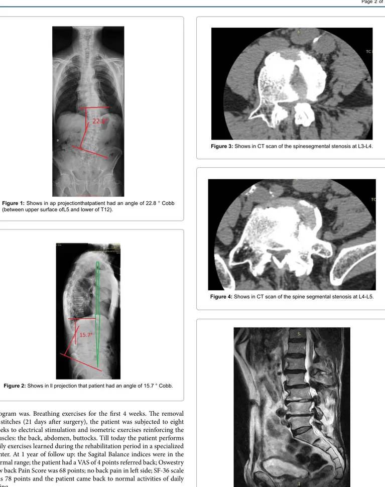
Our patient,a74 year old retired bank worker, came to the center of our Spine Surgery AOUS University Policlinic “Santa Maria alle Scotte” of Siena, reporting an anamnestic history of backache for 6 months, and was negative for idiopathic scoliosis. He reported an ingravescent limitation in the performance of daily activities. Pain measured by VAS scale was an 8, [6] while the Oswestry Low Back Pain Score [7] was 36 points and 46 in SF-36 scale [8].Upon objective examination the patient had an unnatural upright posture while squatting, and there was a scoliotic deflection along the thoracic, thoraco-lumbar, lumbar, and lower back section, with the presence of a rib prominence. The patient also had left radicular syndrome. He had an intermittent claudicatio with pain after 25 meters of walk. The vertebrae with a limiting curve were T12 andL5, while theapical vertebra was L2.X-ray measurement in AP projection (Figure 1) (antero-posterior) revealed that the patient had an angle of 22.8 ° Cobb (between upper surface of T12 and lower of L5) while the LL (latero-lateral) projection (Figure 2) was 15.7 ° Cobb, with the loss of the physiological lumbar lordosis. There was also an excessive pelvic nutation and sagittal line dropped from the center of the soma of C7, which did not pass through the center ofL3’s vertebral body and fell forward at the femoral heads. Delmas index was 97.3, which indicated a rigid column that could be more exposed to vertebral
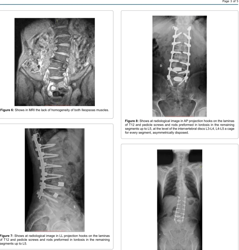
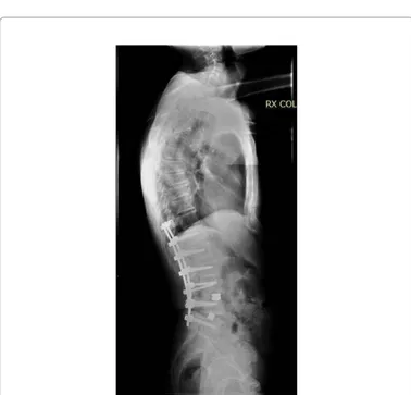
fractures. We sent the patient to have a CT scan of the spine and we found a segmental stenosis at L3-L4 (Figure 3) and L4-L5 lumbar level (Figure 4). The patient also had an RM of the lumbar section, where we found two massive lumbar disc hernias at level L3-L4 and L4-L5 (Figure 5). Although we could appreciate lack of homogeneity of both Ileopsoas muscles at coronal cut (Figure 6), we did give the diagnosis of De Novo Scoliosis. Given the clinical and radiological disease, we decided together with the patient to proceed directly to surgery without conservative treatment except for pain with pain killers. According to our Osteoporosis‘Center: the osteoporosis treatment was carried out by 1 tablet per week of 70 mg alendronic acid + 5,600 I.U. of cholecalciferol, it is still being treated with this dose according our Osteoporosis‘Center. Upon completion of routine pre-operative exams, we decided to perform a surgical correction and stabilization of scoliosis. We administered normal antibiotic and heparin prophylaxis for these interventions in accordance with the Italian guidelines. The duration of surgery was 456 minutes. We decided to correct only the thoracolumbarsection with a hybrid system (hooks on the laminas of T12 and pedicle screws and rods preformed in lordosis in the remaining segments up to L5), at the level of the intervertebral discs L3-L4, L4-L5 (Figures 7 and 8). We performed Transforaminal Lumbar Interbody Fusion (TLIF) Back Surgery with Smith-Petersen’s ostheotomies (Figures 7 and 8), in which we affixed a cage for every segment that was a symmetrically disposed. For the correction of the scoliotic curve in the frontal direction, the cages were placed in front to increase lordotic curve (Figures 7 and 8). We chose to stabilize only the thoraco-lumbar section in order to ensure that the thoracic portion had a development of a kyphosis compensation. One year after surgery (Figures 9 and 10) we restored the physiological curves of the lumbar and thoracic sections. We used like brace the dynamic corset, Spinomed® (Medi GmbH & Co. KG, Bayreuth, Germany) for 3 months from the surgery. The rehabilitation
Figure 1: Shows in ap projectionthatpatient had an angle of 22.8 ° Cobb (between upper surface ofL5 and lower of T12).
Figure 2: Shows in ll projection that patient had an angle of 15.7 ° Cobb.
program was. Breathing exercises for the first 4 weeks. The removal of stitches (21 days after surgery), the patient was subjected to eight weeks to electrical stimulation and isometric exercises reinforcing the muscles: the back, abdomen, buttocks. Till today the patient performs daily exercises learned during the rehabilitation period in a specialized center. At 1 year of follow up: the Sagital Balance indices were in the normal range; the patient had a VAS of 4 points referred back; Oswestry low back Pain Score was 68 points; no back pain in left side; SF-36 scale was 78 points and the patient came back to normal activities of daily living.
Discussion
Figure 3: Shows in CT scan of the spinesegmental stenosis at L3-L4. Figure 4: Shows in CT scan of the spine segmental stenosis at L4-L5. Figure 5: Shows in MRItwo massive lumbar disc’s hernias at level L3-L4 and L4-L5.Figure 6: Shows in MRI the lack of homogeneity of both Ileopsoas muscles.
Figure 7: Shows at radiological image in LL projection hooks on the laminas of T12 and pedicle screws and rods preformed in lordosis in the remaining segments up to L5.
The great functional limitation caused by this pathology due to its deformity, demonstrates that the use of surgical methods is the last treatment approach after failure of more conservative methods [9]. We were looking for a surgical technique that did not involve excessive blood loss or lengthy operating times, and could be compatible with a poor quality of bone due to the advanced age of the patient. Surgery of scoliosis de novo is comparable to surgery of vertebral fractures with posterior long segment fixation because both cases have the objective of surgical restoration of biomechanical stability and the reduction of neurological damage, which is usually imminent or already present [10]. Finally we use the TLIF technique [11]. The results of 24 patients
Figure 8: Shows at radiological image in AP projection hooks on the laminas of T12 and pedicle screws and rods preformed in lordosis in the remaining segments up to L5, at the level of the intervertebral discs L3-L4, L4-L5 a cage for every segment, asymmetrically disposed. Figure 9: Shows in radiological exam in approjectionone year after surgery we restored the physiological curves of the lumbar and thoracic sections.
with treatment in this surgical technique show that TLIF surgery for interbody support was reliable and safe, and that it can be performed with excellent clinical outcomes. We used anterior cage for hypercorrection of lordosis [12]. The experience of Villavicencio et al. [13] demonstrates that AP lumbar interbody fusion surgery is associated with a complication rate more than two times higher, significantly increased blood loss (550 ml vs 231 ml average value), and longer operative (455 minutes vs 255 minutes average value) and hospitalization times (7.2
8.1 day (staged, 16.5%), 3.8 days overall [22]. Five patients (4.7%) received a transfusion, 3 (2.8%) required intensive care unit admission, and 1 (0.9%) required rehabilitation services [22]. Major complications occurred in 13 patients (12.1%): 2 (1.9%) medical, 12 (11.2%) surgical [22]. Of procedures that involved only less invasive techniques (XLIF stand-alone or with percutaneous instrumentation), 9.0% had one or more major complications. In those with supplemental open posterior instrumentation, 20.7% had one or more major complication [22]. Early reoperations (3) (all for deep wound infections) were associated with open posterior instrumentation procedures [22].Their conclusion was the morbidity in adult scoliosis surgery is minimized with less invasive techniques. Di Martino et al. [5] analyzed 63 patients with degenerative scoliosis and operated on by an asymmetric positioning of the cages on the concave side of degenerative scoliosis patients to correct the coronal deformity. They evaluted the radiographic results of the correction by the measurement of the Cobb angles [5]. The clinical results were evaluteted by the Roland -Morris Disability Questionnaire (RMDQ) and by the analysis of complications [5]. Their results were [5]: The RMDQ has improved from a mean preoperative value of 16 points, to an average of 4 points at the last follow-up; The Cobb angle passed from an average of 24° preoperatively to 8° postoperatively. Eight patients sustained intraoperative or early postoperative complication. The used of Spinomed® is based on the principle of bio-feedback activation of the dorsal-lumbar musculature, it responds to the biomechanical principle of the three-point support, while giving a lesser degree of immobilization. The main advantages over the conventional 3-point orthoses are the preservation of dorsal-lumbar muscles function, the absence of pressure on ventral supports and of respiratory restriction, and a larger back support, which can be modeled on individual patient’s spine shape [18]. Postoperative rehabilitation is rarely necessary. On the one hand direct postoperative mobility and activities of the patients’ should be limited for a year in order to ensure a safe healing and stabilization of the bone structure [19]. On the other hand, mobility of the spine as a whole and on the segmental level as well is largely reduced after operation. Problems within the fusion area cannot be addressed by physical means, manipulation of fused segments is obsolete and pain related to costotranversal joint problems can hardly be mobilized without stress to the fusion area. Nevertheless, there is little evidence that operated patients with chronic back pain can benefit from inpatient rehabilitation [19]. The junctional zones (fused area/unfused area) of the spine can be stabilized and obviously pain can be reduced [19]. For the rehabilitation of operated patients with spinal deformities only specialized centres are recommended to assure maximum patient safety. High quality rehabilitation with the help of exercises, re-education and high quality bracing may reduce the costs the community has to bear when surgical intervention can be avoided [19]. Finally it’s the surgical intervention causing the highest costs after low quality conservative management has failed [19].
Conclusion
The aim of this article is not to show or to give guidelines, but to demonstrate a valid and rational alternative to the treatment of this condition, which is increasing due to the aging of the population.
References 1. Trobisch P, Suess O, Schwab F (2010) Idiopathic scoliosis. Dtsch Arztebl Int 107: 875-883. 2. Monticone M, Negrini S (2006) Scoliosi dell’adulto, osteoporosi e back pain. Ital J Rehab medicine 20: 185-187. 3. Vanderpool DW, James JI, Wynne-Davies R (1969) Scoliosis in the elderly. J Bone Joint Surg Am 51: 446-455.
days vs 4.1 days average value) than TLIF techniques for lumbar disc degeneration and instability. Our surgery choice was based on previous studies of three-dimensional spinal deformations, studies on the rigidity of the deformity, and ultimately on the presence of osteoporosis, which influences the stability of the installations. Indeed, the presence of rotatory subluxation in the deformity of the adult implies the need for a study of the three-dimensional column. The correlation of all dimensional parameters is possible with the use of a pelvic incidence parameter, which plays a key role in the regulation of positional pelvic and spinal parameters [14-16]. A low value of pelvic incidence (44° or less) decreases sacral slope and leads to flattened lordosis. A high value of pelvic incidence (62° or more) increases sacral slope and leads to a more pronounced lordosis. For the adult population the standard values of pelvic incidence were 53 ± 9° [17-20] (which reflects a normal distribution of anthropometric data). Our surgery increased lumbar lordosis and the angle between L3-L4, and the pelvic incidence was corrected after one year. Delmas index was 97.3 due to the osteoporotic, rigid column. After the surgery we reduced the rigidity (Delmas index <95) and prevented osteoporotic collapse. Normaly, the surgical treatment of adult scoliosis presents a treatment challenge [21]. Neural decompression with combined anterior/posterior instrumented fusion is often performed [21]. These procedures have been reported to carry a high risk of complication, particularly in the elderly patient population. Over the past decade, less invasive surgical approaches to neural decompression and fusion have been popularized and have recently been applied in the treatment of degenerative scoliosis [21]. In their prospectively studied, Isaacs et al. [22] reported 107 underwent the XLIF procedure with or without supplemental posterior fusion for the treatment of degenerative scoliosis. Their results were: In all, 107 patients (mean age, 68 years; range, 45-87) were treated with XLIF; 28% had at least 1 comorbidity. A mean of 4.4 levels (range, 1-9) were treated per patient. Supplemental pedicle screw fixation was used in 75.7% of patients, 5.6% had lateral fixation, and 18.7% had stand-alone XLIF. Mean operative time and blood loss were 178 minutes (58 minutes/ level) and 50 to 100 mL. Mean hospital stay was 2.9 days (unstaged),
Figure 10: Shows in radiological exam in ll projection one year after surgery we restored the physiological curves of the lumbar and thoracic sections.
14. Duval-Beaupère G, Schmidt C, Cosson P (1992) A Barycentremetric study of the sagittal shape of spine and pelvis: the conditions required for an economic standing position. Ann Biomed Eng 20: 451-462. 15. Legaye J, Duval-Beaupère G, Hecquet J, Marty C (1998) Pelvic incidence: a fundamental pelvic parameter for three-dimensional regulation of spinal sagittal curves. Eur Spine J 7: 99-103. 16. Vaz G, Roussouly P, Berthonnaud E, Dimnet J (2002) Sagittal morphology and equilibrium of pelvis and spine. Eur Spine J 11: 80-87.
17. Boulay C, Tardieu C, Hecquet J, Benaim C, Mouilleseaux B, et al. (2006) Sagittal alignment of spine and pelvis regulated by pelvic incidence: standard values and prediction of lordosis. Eur Spine J 15: 415-422.
18. Pfeifer M, Kohlwey L, Begerow B, Minne HW (2011) Effects of two newly developed spinal orthoses on trunk muscle strength, posture, and quality-of-life in women with postmenopausal osteoporosis: a randomized trial. Am J Phys Med Rehabil 90: 805-815.
19. Weiss HR (2010) Spinal deformities rehabilitation - state of the art review. Scoliosis 5: 28.
20. Negrini S, Aulisa AG, Aulisa L, Circo AB, de Mauroy JC, et al. (2012) 2011 SOSORT guidelines: Orthopaedic and Rehabilitation treatment of idiopathic scoliosis during growth. Scoliosis 7: 3.
21. Schwab FJ, Lafage V, Farcy JP, Bridwell KH, Glassman S, et al. (2008) Predicting outcome and complications in the surgical treatment of adult scoliosis. Spine (Phila Pa 1976) 33: 2243-2247.
22. Isaacs RE, Hyde J, Goodrich JA, Rodgers WB, Phillips FM (2010) A prospective, nonrandomized, multicenter evaluation of extreme lateral interbody fusion for the treatment of adult degenerative scoliosis: perioperative outcomes and complications. Spine (Phila Pa 1976) 35: 322-330.
4. Harms JG, Jeszensky D (1998) The unilateral, transforaminal approach for posterior lumbar interbody fusion. Orthop Traumatol 394: 64-72. 5. Di Martino A, Faldini C, Giannini S, Denaro V (2013) Unilateral cage placement on the concave side to address the deformity in degenerative lumbar scoliosis. GIOT 39: 260-265. 6. Price DD, McGrath PA, Rafii A, Buckingham B (1983) The validation of visual analogue scales as ratio scale measures for chronic and experimental pain. Pain 17: 45-56. 7. Beurskens AJ, de Vet HC, Köke AJ (1996) Responsiveness of functional status in low back pain: a comparison of different instruments. Pain 65: 71-76. 8. Brazier JE, Harper R, Jones NM, O’Cathain A, Thomas KJ, et al. (1992)
Validating the SF-36 health survey questionnaire: new outcome measure for primary care. BMJ 305: 160-164. 9. Wang G, Hu J, Liu X, Cao Y (2015) Surgical treatments for degenerative lumbar scoliosis: a meta analysis. Eur Spine J 24: 1792-1799. 10. Verlaan JJ, Diekerhof CH, Buskens E, van der Tweel I, Verbout AJ, et al. (2004) Surgical treatment of traumatic fractures of the thoracic and lumbar spine: a systematic review of the literature on techniques, complications, and outcome. Spine (Phila Pa 1976) 29: 803-814. 11. Salehi SA, Tawk R, Ganju A, LaMarca F, Liu JC, et al. (2004) Transforaminal lumbar interbody fusion: surgical technique and results in 24 patients. Neurosurgery 54: 368-374.
12. Gödde S, Fritsch E, Dienst M, Kohn D (2003) Influence of cage geometry on sagittal alignment in instrumented posterior lumbar interbody fusion. Spine (Phila Pa 1976) 28: 1693-1699.
13. Villavicencio A, Burneikiene S, Bulsara KR, Thramann JJ (2006) Perioperative Complications in Transforaminal Lumbar Interbody Fusion Versus Anterior– Posterior Reconstruction for Lumbar Disc Degeneration and Instability. Journal of Spinal Disorders & Techniques 29: 92-97.
Submit your next manuscript and get advantages of OMICS Group submissions Unique features: • User friendly/feasible website-translation of your paper to 50 world’s leading languages • Audio Version of published paper • Digital articles to share and explore Special features: • 400 Open Access Journals • 30,000 editorial team • 21 days rapid review process • Quality and quick editorial, review and publication processing • Indexing at PubMed (partial), Scopus, EBSCO, Index Copernicus and Google Scholar etc • Sharing Option: Social Networking Enabled • Authors, Reviewers and Editors rewarded with online Scientific Credits • Better discount for your subsequent articles Submit your manuscript at: http://www.omicsonline.org/submission/ Citation: Muzii VF, Meccariello L, Mazzei G, Vespi M,Carta S, et al. (2015) Is the Short Posterior Stabilization by TLIF and Cages A Good Way for A Correct Spinal Alignment in the De Novo Scoliosis? A Case Report. Orthop Muscular Syst 4: 197. doi:10.4172/2161-0533.1000197