Università degli Studi di Ferrara
DOTTORATO DI RICERCA IN
Farmacologia e Oncologia Molecolare
CICLO XXICOORDINATORE Prof. Pier Andrea Borea
The Role of Opioids on Motor Activity in Physio-pathological Conditions
Settore Scientifico Disciplinare BIO/14
Dottorando Tutore
Dott. Omar Sharif Mabrouk Prof. Michele Morari
1. Introduction
………..11.1 Opioids and their receptors……….2
1.2 Basal Ganglia………..4
1.21 Direct Pathway……….4
1.22 Indirect Pathway………...5
1.23 Summation of pathways………...5
1.3 Opioids in the Basal Ganglia………...6
1.4 Parkinson’s Disease………...7
1.5 Parkinson’s Disease Treatment Options………...10
1.6 Opioids and Parkinson’s Disease………...11
1.61 MOP Receptors and Parkinson’s Disease………...11
1.62 KOP Receptors and Parkinson’s Disease………....12
1.63 DOP Receptors and Parkinson’s Disease………....13
1.64 NOP Receptors and Parkinson’s Disease………....14
2. Aims of Studies
………...163. Materials and Methods
………..193.1 Animal Subjects……….…20 3.2 6-OHDA lesioning……….20 3.3 Behavioral experiments……….………...20 3.31 Bar Test………...21 3.32 Drag test………...21 3.33 Rotarod test………..21
3.34 Inclined grid test………..22
3.4 Microinjection technique………...23
3.5 Microdialysis experiments……….23
3.51 Probe construction………...23
3.52 Surgery and microdialysis procedure………..24
3.6 Microdialysis + Bar test coupling………..25
3.7 Endogenous glutamate and GABA analysis………..26
3.8 Endogenous DA analysis………...27
3.9 Data presentation and statistical analysis………...27
3.10 Materials………...27
4. Results
………....29Section I 4.1.1 Effect of i.c.v. injection of EM-1 and CTOP on motor behavior………31
4.1.2 Effect of intranigral injection of EM-1 on motor behavior………...32
4.1.3 Effect of perfusion with EM-1 in SNr on local GABA and glutamate extracellular levels…………..………...33
4.1.4 Effect of intranigral perfusion of EM-1 on grid test and spontaneous motor activity ………...35
4.1.41 Effect of EM-1 on neck, trunk and forepaw reaction time………...36
4.1.42 Effect of EM-1 on forepaw speed and gait control………...37
4.1.5 Effect of EM-1 on neurotransmitter release in rats undergoing grid test………38
4.1.6 Effect of perfusion with EM-1 in SNr on striatal DA and thalamic GABA release………....39
4.1.7 TTX sensitivity to modulation of EM-1 effects on nigral GABA………..40
4.1.8 Modulation of EM-1 effects on nigral GABA by DArgic and GABAergic antagonists………41
Section II
4.2.1 Effect SNC-80 on motor behavior………... 44
4.2.2 Effects of the DOP receptor antagonist NTD on motor behavior………...46
4.2.3 Effect of systemic administration of DOP receptor ligands on GABA and GLU release in GP and SNr………..48
4.2.4 Effect of regional NTD perfusion on the SNC-80 induced antiakinetic effect…...49
4.2.5 Effect of regional NTD perfusion on SNC-80 induced neurotransmitter release...51
4.2.6 Effect of SNC-80 microinjections into SNr, GP and DLS on motor behavior……….53
4.2.7 Effect of bicuculline microinjections in GP on motor behavior……….55
Section III 4.3.1 Effect of UFP-512 on motor behavior……….58
4.3.2 Effect of UFP-512 on GABA and GLU release in GP………....59
4.3.3 Effect of UFP-512 on GABA and GLU release in SNr………..59
4.3.4 Effect of UFP-512 on GABA and GLU release in VMTh………..60
4.3.5 Effect of UFP-512 on akinesia (bar test) while performing microdialysis……….61
Section IV 4.4.1 The effect of SNC-80 and J113397 on motor activity………...64
4.4.2 The effect of SNC-80 and J113397 on GABA and GLU release in GP……….67
4.4.3 The effect of SNC-80 and J113397 on GABA and GLU release in SNr………67
4.4.4 The effect of SNC-80 and J113397 on GABA and GLU release in VMTh …………..68
4.4.5 Effect of systemic SNC-80 and J113397 on akinesia (bar) test coupled to microdialysis………69
4.4.6 Effect of nigral perfusion of SNC-80 and J113397 on local GABA and GLU release……….71
4.4.7 Effect nigral perfusion of SNC-80 and J113397 on thalamic GABA and GLU release………...72
4.4.8 Effect of nigral perfusion of SNC-80 and J113397 on akinesia (bar) test coupled to microdialysis………....73
5. Discussion
………..75I. The endogenous MOP receptor agonist EM-1 dually affects motor activity and differentially modulates nigro-striatal and nigro-thalamic pathways……….76
II. Stimulation of delta opioid receptors located in substantia nigra reticulata but not globus pallidus or striatum restores motor activity in 6-hydroxydopamine lesioned rats………...79
III.The novel delta opioid receptor agonist UFP-512 dually modulates motor activity in experimental parkinsonism via control of the nigro-thalamic pathway………....82
IV.Combined DOP receptor stimulation and NOP receptor blockade synergistically improves motor performance and impairs nigro-thalamic output in experimental parkinsonism………..85
6. Concluding Remarks
..……….…917. References
………...……..94Abbreviations
6-OHDA 6-hydroxydopamine
ACh acetylcholine
ANOVA analysis of variance
BBB blood brain barrier
CNS central nervous system
CTOP D-Pen-Cys-Tyr-D-Trp-Orn-Thr-Pen-Thr-NH 2
DA dopamine
DAMGO [D-Ala2, N-MePhe4, Gly-ol]-enkephalin
DBS deep-brain stimulation
DLS dorsolateral striatum
DMSO dimethyl sulfoxide
DOP δ-opioid peptide
DPDPE [D-Pen2,D-Pen5]-enkephalin
EM-1 endomorphin-1
EN entopeduncular nucleus
ENK enkephalins
GABA γ-amino butyric acid
GLU glutamate
GP globus pallidus
GPe globus pallidus external
GPi globus pallidus internal
HPLC high pressure liquid chromatography
i.c.v. intracerebroventricular
i.p. intraperitoneal
J-113397 1-[(3R,4R)-1-cyclooctylmethyl-3-hydroxymethyl-4-piperidyl]-3-ethyl-1,3-dihydro-2H-benzimidazol-2-one
KOP κ-opioid peptide
L-DOPA 3,4-dihydroxyphenylalanine
MOP µ-opioid peptide
MPTP 1-methyl-4-phenyl-1,2,3,6-tetrahydropyridine
N/OFQ nociceptin/orphanin FQ
NOP nociceptin/orphanin FQ opioid peptide
NTD naltrindole PKC protein kinase C PPE-A preproenkephalin-A PPE-B preproenkephalin-B RM repeated measures SN substantia nigra
SNc substantia nigra pars compacta
SNr substantia nigra pars reticulata
SNC-80 (+)-4-[(αR)-α-(2S,5R)-allyl-2,5-dimethyl-1-piperazinyl)-3-methoxy-benzyl]-N-N-diethylbenzamide
STN subthalamic nucleus
TTX tetrodotoxin
UFP-512 H-Dmt-Tic-NH-CH(CH2-COOH)-Bid
VMTh ventromedial thalamus
Summary
To date, four principal classes of opioid peptide receptors, μ (MOP), κ (KOP), δ (DOP) and ORL-1 (NOP) have been identified and cloned. The experimental and clinical importance of this receptor family has been amply demonstrated in many diseases, though historically they have mainly been associated with antinociception (pain relief). The current work described herein discusses the impact of opioid systems on movement disorders, namely Parkinson’s disease (PD). PD is a relatively common neurological disease in which the motor symptoms result from the death of a particular dopamine (DA) containing cell population in the substantia nigra pars compacta (SNc). This inevitably results in a dysregulation of the activity of basal ganglia (BG); a set of midbrain structures responsible for normal movement programming and execution. The BG structures highly express opioid receptors and their endogenous peptides and, throughout the course of PD, opioidergic transmission appears dysregulated. The view that opioids are involved in compensatory or pathogenic mechanisms following loss of DA neurons lends support to the notion that they may represent a suitable target for the treatment of PD.
The work contained herein describes the benefits of opioid receptor ligand administration under physiological or pathological conditions (e.g. in experimental models of PD). For instance, we discovered that the endogenous MOP receptor peptide endomorphin-1 (EM-1), when injected or perfused into the substantia nigra pars reticulata (SNr) of naïve rats facilitates and inhibits spontaneous locomotion depending on dose.
Additionally, we have tested DOP receptor agonists in an experimental model of PD (i.e. 6-hydroxydopamine hemilesioned rats). These studies demonstrated the ability of the selective non-peptide DOP receptor agonist SNC-80 to alleviate symptoms in experimental PD, such as akinesia and poor coordinated movement. We have also showed that when administered systemically, the novel peptide agonist UFP-512 causes both facilitation and inhibition of movement depending on the dose used. Lastly, we demonstrated a functional relationship between NOP and DOP receptors in SNr. We showed that in SNr, blockade of NOP receptors with the selective antagonist J-113397 and activation of DOP receptors with SNC-80 synergistically enhances motor performance in experimental PD. In addition to demonstrating the behavioral outcome of opioid receptor stimulation or blockade on a variety of movement-related parameters, we have correlated these behaviors with novel neurochemical findings in each of the presented studies. The overall findings
of the work presented shed additional light on the subject of opioidergic systems and movement disorders and may contribute to future therapeutics for diseases such as PD.
1.1 Opioids and their receptors
The word opium is derived from the Greek name for ‘juice’; the drug being obtained from the secretions of the poppy plant, Papaver somniferum. Cultivation of opium poppies for food, anaesthesia, and ritual purposes dates back to at least the Neolithic Age. The Sumerian, Assyrian, Egyptian, Greek, Roman, Persian and Arab Empires each made widespread use of opium, which was the most potent form of pain relief then available, allowing ancient surgeons to perform prolonged surgical procedures. Opium has been mentioned in the most important medical texts of the ancient world. By the middle of the 16th century, opium’s uses were fairly well understood and in the 18th century opium smoking became popular while its overuse by some individuals became more evident.
Opium contains two main groups of alkaloids. Phenanthrenes, including morphine, codeine, and thebaine, are the main narcotic constituents of the plant. Isoquinolines such as papaverine on the other hand have no significant effect on the central nervous system (CNS). In 1805, the German pharmacist Friedrich Wilhem Adam Sertürner reported the isolation of a pure substance in opium that he named morphium, after Morpheus, the god of dreams. Incidentally, this was the first ever extraction of a pure medicinal substance from plant material. Morphine is by far the most prevalent and important alkaloid in opium, constituting 10-16% of the total alkaloid amount, and is mainly responsible for its effects such as pain relief, euphoria, addiction, lung edema and respiratory difficulties.
The existence of receptors for opiate drugs was proposed by Beckett and Casy in 1954 based on their studies on structure-activity relationships for antinociceptive (e.g. the ability to relieve pain) activity in a series of synthetic opiates (Beckett and Casy, 1954). These receptors were called ‘opioids’ since it was understood that their endogenous ligands were peptides with effects resembling those of opiate drugs. As early as 1965, based on structure-activity analysis studies, Portoghese proposed the existence of more than one opioid receptor type or the possibility of multiple modes of interaction between ligands and opioid receptors (Portoghese, 1965). The first clear evidence that opioid receptors were in fact a diverse population was presented by Martin and colleagues in 1976 (Martin et al., 1976). Extensive pharmacological studies since that time have led to the identification of four opioid receptor types based on their diverse pharmacological actions. The earliest proposed receptor types were named after drugs used during the identification process such as the µ (mu for morphine) and κ (kappa for
ketocyclazocine) receptors (Martin et al., 1976). Those findings clearly linked both analgesia and addictive properties with functions of the µ receptor. Pharmacological analysis of opioid peptide effects in guinea pig ileum and mouse vas deferens led to the proposal of a third opioid receptor named the δ (delta for vas deferens) receptor (Lord et al., 1977). Since their discovery, the nomenclature used to refer to these receptors has changed multiple times. According to the International Union of Basic and Clinical Pharmacology (IUPHAR), µ is currently referred to as mu opioid peptide (MOP) receptor, κ is kappa opioid peptide (KOP) receptor and δ is referred to as delta opioid peptide (DOP) receptor. The most recent addition to the opioid peptide family is the nociceptin/orphanin FQ peptide (NOP) receptor which has high sequence homology compared to classical opioid receptors, particularly the KOP receptor (Bunzow et al., 1994). The endogenous ligand of this receptor was first described by Meunier at the International Narcotics Research Conference in 1995 who subsequently reported the ability of this peptide, which he named nociceptin, to decrease forskolin-stimulated cAMP production, in vitro (Meunier et al., 1995). A peptide of identical sequence isolated in pig brain was reported simultaneously by Reinscheid and colleagues (Reinscheid et al., 1995), who named it orphanin F/Q (OFQ). For the purposes of this text, the nomenclature used to describe the peptide and its receptor will be
nocicpetin/orphanin FQ (N/OFQ) and the nociceptin/orphanin FQ peptide (NOP) receptor,
respectively.
Opioid receptors are part of the G protein-coupled receptor superfamily and are composed of 7 hydrophobic transmembrane domains which couple to specific GTP binding proteins (G proteins). These G proteins are heterodimeric proteins consisting of three distinct subunits, α, β and γ. Opioid receptors are known to couple to Gi (Hawes et al., 2000). Through their
intracellular actions, opioids are also known to inhibit the formation of cAMP, close voltage sensitive Ca2+ channels and enhance outward K+ conductance (Hawes, et al., 2000). The sum of these actions and their ability to reduce neurotransmitter release and neuronal excitability make the opioids primarily inhibitory molecules. Since their discovery, opioid peptide systems have been implicated in variety of diseases and treatments ranging from pain and addiction to mood and movement disorders based on their locations and actions in the CNS.
1.2 Basal Ganglia
The basal ganglia (BG) are a group of subcortical brain structures which control the planning and execution of motor programs. The BG include the striatum, globus pallidus (GP), subthalamic nucleus (STN) and substantia nigra (SN). The dorsolateral part of the striatum (DLS) is positioned along motor circuits where it integrates inputs arriving from thalamus and somatomotor areas of the cortex. The DLS then, in turn, modulates the output structures of BG. In primates (human and non human), the GP is divided into external (GPe) and internal (GPi) segments. In rats, these structures are represented by GP and entopeduncular nucleus (EN), respectively. For the purposes of this text, GPe will be referred to as GP. The SN is also organized into two closely linked segments, the substantia nigra pars compacta (SNc) and the substantia nigra pars reticulata (SNr). The disruption of coherent information flow between these structures results in altered output and disturbed motor activity. The primary neurotransmitters of the BG are γ-amino butyric acid (GABA), glutamate (GLU) and dopamine (DA). GABA being the main inhibitory amino acid neurotransmitter in the CNS, GLU being the main excitatory one and DA, depending on which receptor type it activates, may cause inhibitory (D2-like) or
excitatory responses (D1-like). Two primary pathways (direct and indirect) are described in the
current simplified model of BG function (Albin et al., 1989, Delong, 1990) and these two pathways oppositely modulate the activity of BG output structure (GPi/SNr). These output structures send inhibitory GABAergic projections to the thalamus in effect controlling inhibition and disinhibition of thalamo-cortical projections; the end result being the coordinated execution of motor programs. The striatum is the major input target of projections arriving to the BG. For instance, extensive GLUergic inputs arrive from the cortex as well as the thalamus (Smeal et al., 2008, for review see Smith et al., 2004) while DA arrives via SNc DAergic projections. About 95% of striatal neurons are GABAergic medium-sized spiny neurons (Chang et al., 1982). The remaining neurons found in the striatum are interneurons containing either GABA or acetylcholine (ACh), which act to modulate the activity of the striatofugal pathways (direct and indirect pathways; for review see Pisani et al., 2007).
1.21 Direct Pathway
The direct pathway originates from one population of medium-sized spiny striatal GABAergic neurons which project directly to the GPi/SNr. This cell population mainly express DA D1
release of GABA and its co-transmitters, dynorphin and substance P, in SNr/GPi (for review see Albin et al., 1989). GPi/SNr then sends GABAergic projections to the ventromedial and ventrolateral thalamus (Kha et al., 2001) and excitatory (GLUergic) projections from the thalamus arrive in the cortex. The cortex excites the striatum which then inhibits the GPi/SNr through this pathway. The GPi/SNr is normally tonically active and inhibitory to the thalamus. When the GPi/SNr is inhibited, the thalamus is relieved from inhibition (disinhibition) and excites the cortex, thereby reinforcing the desired movement (Deniau and Chevalier, 1985).
1.22 Indirect Pathway
In the indirect pathway, a separate group of medium-sized spiny striatal GABAergic neurons project to GP. These striato-pallidal projections use ENK as a co-transmitter. DA D2 receptors
are mainly expressed on this population and DA stimulation is responsible for suppressing their activity (Albin et al., 1989). GP then sends GABAergic projections to STN, while the STN projects to GPi/SNr using the excitatory neurotransmitter, GLU. The GPi/SNr projects to thalamus using GABA and the thalamus projects back to the cortex also using GLU. Therefore, the cortex excites the striatum which then inhibits GP. Since GP is inhibitory to the STN, the STN then becomes more active and excites the GPi/SNr. The GPi/SNr being more active then inhibits the thalamus thereby dampening excitation to cortex. In this way, activation of the indirect pathway through striatum causes a relative inhibition of movement.
1.23 Summation of pathways
The opponent parallel pathway hypothesis emphasizes two major paths of information flow from the striatum to the basal ganglia output nuclei: GPi/SNr (in primates) or EN/SNr (in rodents). One is an inhibitory direct pathway from the striatum to the GPi/SNr. The other is an effectively excitatory multisynaptic pathway from the striatum to GPi/SNr. This hypothesis suggests that the two pathways are in balance such that increased activity in the direct pathway causes decreased GPi/SNr output, and increased activity in the indirect pathway causes increased GPi/SNr output. These two pathways converge on the same cells in the GPi/SNr which ultimately allows for the precise control over BG output needed under normal conditions. By adjusting the balance, the cortical targets of the basal ganglia can be facilitated or inhibited. The hypothesis predicts that abnormally decreased output results in excessive movement (chorea) and abnormally increased
(adapted from Carlson 1998, pg 244) Fig. 1 The anatomical organization of the basal ganglia in the human brain (left). Shows BG circuitry in the human brain where the red lines indicate inhibitory input and the blue lines indicate excitatory input (right). The yellow accompanying lines refer to the indirect pathway (striato-pallidal) and the green one refers to the direct pathway (striato-nigral).
1.3 Opioids in the Basal Ganglia
Opioid ligands regulate the release of various neurotransmitters in the CNS. The predominant inhibitory effects of opioids on neurotransmitter release was discovered in the 1950’s by studies demonstrating that opiates inhibit ACh release from electrically stimulated guinea pig ileum (Paton, 1957, Schaumann, 1957), followed by brain in vivo (Beleslin and Polak, 1965) and then brain slices (Sharkawi and Schulman, 1969). Since that time, opioids have also been shown to inhibit the release of other neurotransmitters such as noradrenaline (Montel et al., 1974), DA (Loh et al., 1976) and GABA (Kondo and Iwatsubo, 1978). The BG have one of the highest levels of endogenous opioids and opioid receptors in the brain (Peckys and Landwehrmeyer, 1999). All opioids have been shown to regulate DA functions in different ways and their net effect on transmitter stimulation or inhibition depends on the anatomical localization of their
receptors. An additional layer of complexity to their modulatory effects is based on the fact that opioid receptors may be located presynaptically, which is the typical scenario, or postsynaptically. In the BG, opioid ligands are found in cells containing other classical neurotransmitters such as GABA where they act to modulate transmitter release through their inhibitory actions. Striatal GABAergic neurons comprising the indirect pathway (projecting to GP) express preproenkaphalin-A (PPE-A) whereas striatal neurons of the direct pathway (projecting to SNr) express preproenkephalin-B (PPE-B). Each PPE-A molecule gives rise to 6 copies of methionine-enkephalin (met-ENK) and one copy of leucine-enkephalin (leu-ENK). These ENK are considered the endogenous ligands for the DOP receptor. For the purposes of this text, both leu-ENK and met-ENK will be referred to collectively as ENK. In the BG, ENK are almost exclusively found in striato-pallidal projections (indirect pathway) along with GABA. The direct pathway, on the other hand, is known to express and release substance P and dynorphin, the endogenous ligand of the KOP receptor.
Opioid Receptor Type Localization
MOP Striatum, GPe, GPi, STN, SNr and SNc
DOP Striatum, GPe, GPi, STN and SNr
KOP Striatum, GPi, SNr and SNc
NOP GPe, STN, SNc and SNr
Fig 2. Localization of opioid receptors in BG based on evidence from binding and mRNA expression studies (Mansour et al., 1995, Neal et al., 1999).
1.4 Parkinson’s Disease
In 1817, James Parkinson described in his monograph ‘An essay on the shaking palsy’ a severe movement disorder which he called paralysis agitans, later to be named Parkinson’s disease (PD). PD is also called "primary parkinsonism" or "idiopathic PD" (classically meaning having no known cause). PD is a relatively common disease affecting 0.1% of the world’s population with a prevalence of 1% of the population above 60 years old. Its prevalence increases exponentially from 1% to 5% of the population between 65 and 90 years of age. The cause of the disease is largely unknown and it appears to be without genetic linkage in 90-95% of cases. In those cases, it may be caused in part by the combination of genetic susceptibilities and environmental factors (Lim et al., 2002). While most forms of parkinsonism are idiopathic, secondary cases may result from toxicity most notably from drugs, head trauma, or other medical
disorders. Carlsson and colleagues (1957) experimentally showed that DA loss was a crucial factor underlying the pathological behaviors of PD by depleting catecholamines in mice using reserpine. Following this milestone discovery, DA deficiency was identified in parkinsonian patients (Hornykiewicz, 1966). It is now understood that PD is a progressive neurodegenerative disease which occurs from the loss of DA and non DA neurons in the brain. Nevertheless, PD motor symptoms are caused by the death of SNc DA neurons resulting in a severe reduction in DA at nerve terminals in the caudate nucleus and putamen (e.g. striatum level) as well as in other areas such as GP and STN. However, motor symptoms appear only when the level of degeneration exceeds the critical threshold of 70-80% of nerve terminals and around 50% of midbrain DA containing neurons. This disturbance in BG function results in the dysregulation of movement control leading to tremors, postural instability, rigidity and slowness to initiate (akinesia) and execute (bradykinesia) movements.
Typically, a patient is diagnosed based on medical history and neurological examination conducted by interviewing and observing the patient and applying the Unified Parkinson's Disease Rating Scale which assesses a patient’s behaviors, mood, activity and motor behaviors. Early signs and symptoms of PD may sometimes be overlooked as the effects of normal aging. Due to symptom overlap with other diseases, only 75% of clinical diagnoses of PD are confirmed to be idiopathic PD at autopsy (Gelb et al., 1999). The physician may need to observe the person for some time until it is apparent that the symptoms are consistently present. Usually doctors look for shuffling of feet and lack of swing in the arms since these are hallmark symptoms of the disease.
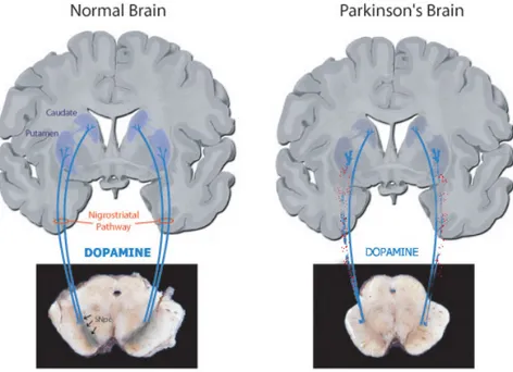
(adapted from Brain-Mind Institute LEN) Fig 3. A normal (left) and a DA denervated (right) nigro-striatal tract as seen in PD. Note the pigmentation due to a build up of melanin in SNc is markedly darker in the normal brain compared to the parkinsonian brain. The loss of DA neurons in SNc results in altered BG signaling and motor complications.
Fig 4. Normal BG inputs and outputs (left) and dyregulated BG inputs and outputs as a result of DA denervation in SNc (right). Lack of DA stimulation in striatal areas causes an increase in GABA release (via lack of inhibitory D2 tone) to GPe and a decrease (via lack of excitatory D1 tone) in striato-nigral GABA release. Ultimately the disinhibition of nigro-thalamic output is followed by reduced GLUergic
1.5 Parkinson’s Disease Treatment Options
Currently, there is no cure for PD, though there are several treatment options that can alleviate symptoms of the disease and improve quality of life of patients. The ‘gold standard’ symptomatic treatment is 3,4-dihydroxyphenylalanine (L-DOPA) which was discovered in the late 1960’s (Cotzias et al., 1967). L-DOPA crosses the blood brain barrier (BBB) when administered orally then is converted to DA by the enzyme L-aromatic amino acid decarboxylase. In order to avoid peripheral decarboxylation, L-DOPA is combined with a decarboxylase inhibitor (Carbidopa or Benserazide) which cannot cross the BBB, limiting its effects to the brain. The net effect of L-DOPA is the replacement of the absent DA in the nigral projections. L-DOPA therapy revolutionized the treatment of PD, providing dramatic benefits to nearly every patient who received it. This treatment is still currently the most widely used and provides satisfactory relief of parkinsonian symptoms for 5-10 years. However, within 5 years, a series of complications begin to develop in more than 40% of PD patients who receive L-DOPA (Ahlskog and Muenter, 2001). These side effects are the “on-off” phenomenon and abnormal involuntary movements (known collectively as dyskinesias). The “on-off” phenomenon refers to the response to L-DOPA either being good (on) or poor (off) and the overall reduction in the duration L-DOPA efficacy. The prevalence and severity of dyskinesias increases with the duration and severity of the disease (Colosimo and De Michele, 1999, Grandas et al., 1999). After 10 years of L-DOPA treatment, dyskinesias have developed in 70-80% of patients and in nearly 100% of patients whose disease onset was below the age of 45-50 years old (Quinn et al., 1987). L-DOPA therapy can also cause non-motor complications such as hallucinations and psychosis (Nausieda et al., 1984, Kuzuhara, 2001) as well as loss of impulse control (Stamey and Jankovic, 2008).
In addition to L-DOPA, DA D2/D3 receptor agonists (typically ropinirole or pramipexole), which
stimulate DA receptors directly without the need for metabolic conversion, are also regularly used to treat PD, either alone or in conjunction with L-DOPA therapy. These agonists are not as effective as L-DOPA in relieving PD motor symptoms, however, their addition to L-DOPA therapy can significantly reduce dyskinesias (Calne, 1993). Alternative treatments have been used with varying degrees of success such as GLU antagonists (Kornhuber et al., 1991), monoamine oxidase inhibitors (Birkmayer et al., 1975), catechol-O-methytransferase inhibitors (Ruottinen and Rinne, 1998) and cholinergic antagonists (Duvoisin, 1967). Beside
pharmacological treatment, some surgical procedures have been performed on PD patients. For example ablative surgeries (i.e. pallidotomy or thalamotomy) or deep brain stimulation (DBS) of the thalamus, GPi or STN. The aim of pallidotomy and DBS is to reduce the excessive inhibitory output from the GPi and SNr (Hill et al., 2000). Taken together, it is clear that the currently available treatments for PD are lacking in some key areas such as therapeutic longevity and side effect profiles. For these reasons, investigators continue to search for alternative therapies with an emphasis on neuroprotective agents to slow or halt the progression of the disease.
1.6 Opioids and Parkinson’s Disease
PD is classically associated with degeneration of nigro-striatal DA cells although the involvement of other factors such as opioid systems have received more attention in the past two decades in an attempt to elucidate the pathophysiology of PD and L-DOPA induced dyskinesias. Interestingly, opioid receptors and their endogenous ligands are present in high concentration in the BG (Gramsch et al., 1979, Emson et al., 1980, Zamir et al., 1984, Mansour et al., 1995) and alterations in endogenous opioids have been reported in postmortem brain from PD patients (Taquet et al., 1983, 1985, Sivam, 1991). Opioid receptor agonists and antagonists have also been shown to affect the behavior of parkinsonian patients (Trabucchi et al., 1982, Sandyk and Snider, 1986, Groppetti et al., 1990). Most of these investigations have emphasized striatal opioid pathways in the pathological process of PD although several more recent observations suggest the likely importance of opioids in other areas of BG such as SNr, SNc and STN (Shen and Johnson, 2002, Marti et al., 2004, Aubert et al., 2007, Mabrouk et al., 2008). Antagonizing excitatory amino acid neurotransmission in the output structures of the BG can alleviate symptoms of experimental parkinsonism (Klogether and Turski, 1990, Brotchie et al., 1991). Since opioids are generally inhibitory in nature and localized in output structures of the BG, this has led investigators to consider the endogenous opioid system a viable target for the treatment of PD.
1.61 MOP Receptors and Parkinson’s Disease
Preclinical and clinical research into the relationship between MOP receptors and PD has been scarce, probably due to the evident unwanted side effects associated with MOP receptor stimulation (e.g. addiction, euphoria and respiratory impairments). Despite this, it is well known
that MOP receptors are highly expressed in several BG structures and the nigro-striatal DA system (Mansour et al., 1995). In non parkinsonian animals, MOP receptor agonists are known to stimulate locomotion when given at lower doses while higher ones cause sedation (Iwamoto, 1981). In fact selective MOP receptor stimulation in SNr (Bontempi and Sharp, 1997) facilitates spontaneous locomotion and turning behavior in rats. This effect on locomotion has been correlated with the effect of MOP receptor stimulation on DA release in striatal areas (Di Chiara and Imperato, 1988) and control of GABAergic nigro-thalamic output neurons (Waszczak et al., 1984). However, studies using the non selective MOP receptor agonist morphine in experimental PD has been shown to be ineffective in enhancing the antiparkinsonian effects of DA agonists (Samadi et al., 2004). Interestingly though, morphine has been shown to reduce the dyskinetic response elicited by L-DOPA in animal models of PD (Samadi et al., 2004) as well as in PD patients (Berg et al., 1999).
1.62 KOP Receptors and Parkinson’s Disease
The endogenous opioid peptide ligand for the KOP receptor is dynorphin. Studies examining the result of DA depletion on dynorphin precursor PPE-B mRNA expression have shown that it is descreased (Gerfen et al., 1990) in striato-nigral neurons. Dynorphin serves as a co-transmitter in DA D1 expressing striatal GABAergic projection neurons extending to the SNr (direct pathway).
Additionally, the KOP receptor is found in abundance in both GP and SNr (Haber and Watson, 1983, Mansour et al., 1993). Indeed, prodynorphin peptide immunoreactivity is dense in the SNr but is virtually absent in the SNc (Vincet et al., 1982). Likewise, KOP receptors are localized in the rat SNr but are not detectable in SNc (Mansour et al., 1987), placing this peptide receptor system in a strategic location to modulate the output of the BG and motor function. Despite this, KOP receptor agonists have been shown to produce contrasting effects compared to MOP and DOP receptor agonists in a number of behavioral paradigms. For instance, KOP receptor stimulation was shown to reduce locomotor activity in naïve mice while DOP (Ito et al., 2008) and MOP (Iwamoto, 1981) stimulation enhance it. Additionally, KOP receptor agonists have been shown to produce dysphoria in contrast to the euphoria brought about by MOP agonists (Mucha and Herz, 1985). Additionally, in experimental PD (6-OHDA hemilesioned rats), nigro-striatal denervation is associated with a decrease in PPE-B mRNA expression, the precursor of dynorphin, in striato-nigral neurons (Gerfen et al., 1990). Since the precursor of dynorphin may
be decreased in PD, investigators sought to restore dynorphin signaling using selective agonists with the hopes of reversing movement abnormalities caused by DA depletion. However, results thus far have been conflicting in terms of the potential benefit of KOP receptor agonists in the treatment of PD. Hughes and colleagues (1998) reported that KOP receptor agonists enadoline and U69,593 increased locomotion in monoamine-depleted rats. This group also demonstrated a synergistic effect between enadoline and L-DOPA and has suggested that KOP receptor agonists may have the potential as an adjunct to L-DOPA therapy in PD. On the other hand, clinical studies using spiradoline in PD patients by Giuffra and colleagues (1993) concluded that selective KOP receptor stimulation would not prove useful alone or in combination with L-DOPA in the treatment of PD. Taken together, role of KOP receptors in PD is not clearly understood, yet further studies are warranted due to its localization profile and its ability to elicit behavioral responses in parkinsonian mice.
1.63 DOP Receptors and Parkinson’s Disease
The DOP receptor and its endogenous ligands, ENK are widely expressed in the basal ganglia, particularly in STN, striatum and, to a lesser extent, GP and SNr (Abou-Khalil et al., 1984, Mansour et al., 1993, Aubert et al. 2007, Hallett and Brotchie, 2007). ENK act as co-transmitters in striatal GABAergic neurons projecting to GP (Haber and Elde, 1981, Gerfen and Young, 1988) where they inhibit GABA release from striato-pallidal terminals (Maneuf et al., 1994) thereby opposing the inhibitory postsynaptic influence produced by striato-pallidal neurons (Stanford and Cooper, 1999). This modulation may be relevant during PD. Indeed, striato-pallidal neurons become pathogenically overactive following DA depletion, leading to overinhibition of pallido-subthalamic GABAergic and disinhibition of subthalamo-nigral GLUergic projections (DeLong, 1990). Consequently, PPE-A expression in striato-pallidal neurons increases (Young et al., 1986, Gerfen et al., 1990) possibly to attenuate exaggerated GABA release and compensate for motor deficits (Maneuf et al., 1994). Consistently, the DOP receptor agonist SNC-80 (Bilsky et al., 1995) promoted spontaneous or L-DOPA induced contralateral turning in 6-OHDA hemilesioned rats (Pinna and Di Chiara, 1998, Hudzik et al., 2000) and reversed akinesia in reserpinized or haloperidol-treated rats as well as MPTP-treated marmosets (Maneuf et al., 1994, Hill et al., 2000, Hille et al., 2001) Based on the evidence that endogenous ENK inhibited K+-evoked GABA release in pallidal slices (Maneuf et al., 1994), it
was proposed that DOP receptor agonists attenuate parkinsonism by inhibiting GABA release from striato-pallidal GABAergic terminals. Recently we showed that SNC-80 attenuated akinesia/bradykinesia and improved overall gait ability in 6-OHDA hemilesioned rats while the selective DOP receptor antagonist naltrindole (NTD; Portoghese et al., 1988) exerted opposite effects (Mabrouk et al., 2008). SNC-80 decreased pallidal GABA as well as nigral GLU and GABA release, suggesting an action at the striatal, pallidal or nigral level. Moreover, only NTD perfusion in SNr (and not GP or DLS) prevented the antiakinetic effect of SNC-80 and its neurochemical correlates. Consistently, microinjections of SNC-80 in SNr (but not GP or DLS) replicated the antiparkinsonian effects of systemic SNC-80. These findings confirm that DOP receptor stimulation, either by synthetic agonists or endogenous ENK, promotes motor function under parkinsonian conditions but challenged the common view that DOP receptor agonists attenuate parkinsonism via intrapallidal mechanisms, suggesting, instead, that nigral DOP receptors are involved.
1.64 NOP Receptors and Parkinson ’s Disease
N/OFQ is a heptadecapeptide that structurally resembles dynorphin A. Its receptors are expressed in both cortical and subcortical motor areas, particularly in the DA containing neurons of SNc. Since the SNc degenerates during the course of PD, investigators sought to explore the relationship between the NOP system and motor functions. Since its discovery, the N/OFQ-NOP receptor system has been termed ‘anti-opioid’ since it has been shown to inhibit the effects of classical opioids. For instance, morphine mediated analgesia in the mouse tail flick test was attenuated by NOP receptor stimulation (Mogil et al., 1996, Calò et al., 1998). Additionally, N/OFQ was shown to suppress striatal DA release from ventral tegmental area (VTA; Murphy and Maidment, 1999) and SNc (Marti et al., 2004) as well as locomotor activity (Meunier et al., 1995, Reinscheid et al., 1995) whereas MOP receptor agonists stimulate DA release by inhibiting intranigral (Lacey et al., 1989) or intra-VTA (Johnson and North, 1992) GABAergic interneurons. A link between N/OFQ and PD has been uncovered by our group. Thus N/OFQergic transmission was found to be upregulated in the DA-depleted SNr of 6-OHDA lesioned rats (Marti et al., 2005). Moreover, systemic or intranigral injections of NOP receptor antagonists such as J-113397 can attenuate experimental parkinsonism in 6-OHDA and haloperidol treated rats (Marti et al., 2004b, 2005) as well as MPTP-treated mice and nonhuman
primates (Viaro et al., 2008). Recent studies have also demonstrated that the deletion of the NOP precursor (preproN/OFQ) or NOP receptor genes gives mice partial resistance to MPTP toxicity and haloperidol-induced catalepsy, respectively (Marti et al., 2005). These findings have prompted investigators to identify novel selective NOP receptor antagonists for PD therapy. Indeed, a novel NOP receptor antagonist TRAP-101 was recently shown to reverse experimental PD in accordance with earlier findings using J-113397 (Marti et al., 2008).
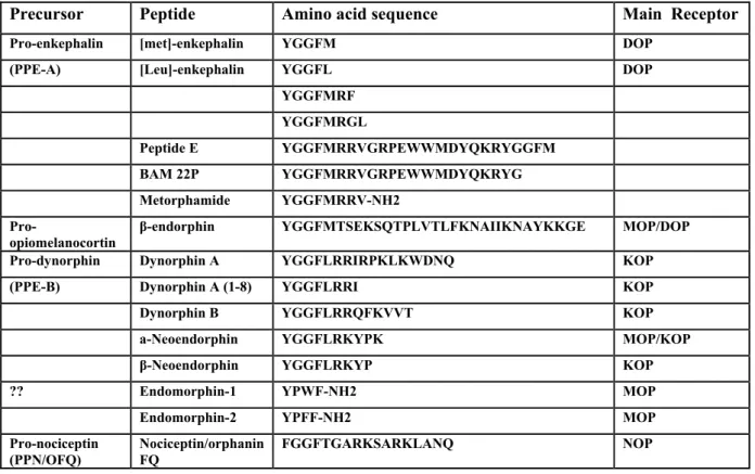
Precursor Peptide Amino acid sequence Main Receptor
Pro-enkephalin [met]-enkephalin YGGFM DOP
(PPE-A) [Leu]-enkephalin YGGFL DOP
YGGFMRF YGGFMRGL Peptide E YGGFMRRVGRPEWWMDYQKRYGGFM BAM 22P YGGFMRRVGRPEWWMDYQKRYG Metorphamide YGGFMRRV-NH2
Pro-opiomelanocortin β-endorphin YGGFMTSEKSQTPLVTLFKNAIIKNAYKKGE MOP/DOP
Pro-dynorphin Dynorphin A YGGFLRRIRPKLKWDNQ KOP
(PPE-B) Dynorphin A (1-8) YGGFLRRI KOP
Dynorphin B YGGFLRRQFKVVT KOP
a-Neoendorphin YGGFLRKYPK MOP/KOP
β-Neoendorphin YGGFLRKYP KOP
?? Endomorphin-1 YPWF-NH2 MOP
Endomorphin-2 YPFF-NH2 MOP
Pro-nociceptin
(PPN/OFQ) Nociceptin/orphanin FQ FGGFTGARKSARKLANQ NOP
The main aim of the work presented in this thesis was to continue the investigations into the involvement of opioid receptors in physiological and pathological conditions affecting the BG, particularly as it relates to PD. These studies were performed in both naïve rats (Section I) as well as in 6-OHDA hemilesioned rats (Section II, III, IV), which is widely regarded as a reliable model of PD. We analyzed motor activity of animals which received various opioid ligands either systemically (i.p.) or directly into brain areas of interest (via perfusion or microinjections). In addition we correlated locomotor activity with neurochemical changes using in vivo
microdialysis to further characterize the mechanism of action of opioid ligands on locomotor behavior.
Section I. The main aim of this study was to investigate whether EM-1, the endogenous ligand of
the MOP administered exogenously into the brain of naïve rats could control locomotor activity and through which mechanism this effect occurred. We then implemented the novel grid test to measure subtle animal behaviors in response to drug treatment such as reaction time and speed of movements. These findings were collected in parallel with in vivo microdialysis to collect neurochemical evidence which could demonstrate the brain area and neurotransmitters involved in the motor actions of EM-1.
Section II. The main aim of this study was to investigate the role of DOP receptors in
parkinsonism and, in particular, to investigate the circuitry underlying the antiparkinsonain actions of the DOP receptor agonist SNC-80. First, we wanted to demonstrate that this drug improved locomotor activity in the 6-OHDA hemilesioned rat model of PD. Following behavioral studies, we sought to determine the precise mechanism of action of DOP receptor agonist induced stimulation of locomotor behavior. We also wanted to understand how
endogenous enkephalinergic tone might influence rat behavior. To this end we used the selective DOP receptor antagonist naltrindole systemically and injected into particular brain areas
including dorsolateral striaum, globus pallidus (GP) and SNr.
Section III. The main aim of this study was characterize the ability of the novel DOP receptor
agonist H-Dmt-Tic-NH-CH(CH2-COOH)-Bid (UFP-512) to attenuate motor deficits in 6-OHDA
hemilesioned rats. Since UFP-512 caused different behavioral effects compared to typical nonpeptide agonists such as SNC-80, we set out to determine the circuitry involved in motor actions of UFP-512 by using microdialysis coupled to behavioral testing. By coupling in vivo microdialysis with the akinesia test, we correlated motor activity with changes in GABA and
GLU release in GP and SNr. Moreover, to test the hypothesis that changes in locomotor behavior were associated with changes in nigro-thalamic transmission, amino acid release in ventromedial thalamus (a target of nigro-thalamic GABAergic projections) was also measured.
Section IV. The main aim of this study was to describe potential interactions between the DOP
and NOP receptor using behavioral techniques in 6-OHDA hemilesioned rats. Since we recently demonstrated that DOP receptor agonists and NOP receptor antagonists improved locomotor activity in experimental models of PD via nigral mechanisms, the behavioral and neurochemical profile of this combination was investigated following both systemic and intranigral administration. In order to characterize whether the observed effects were synergistic or additive, we tested these compounds at subthreshold and submaximal doses or concentrations.
3.1 Animal Subjects
Male Sprague-Dawley rats (150 g; Harlan Italy; S. Pietro al Natisone, Italy) were kept under regular lighting conditions (12 hr light/dark cycle) and given food and water ad libitum. The experimental protocols performed in the present study were approved by Ethical Committee of the University of Ferrara and adequate measures were taken to minimize animal pain and discomfort.
3.2 6-OHDA lesioning
Unilateral lesion of DAergic neurons was induced in isoflurane-anesthetized male rats as previously described (Marti et al., 2005). Eight micrograms of 6-OHDA (dissolved in 4 µl of saline containing 0.02% ascorbic acid) were stereotaxically injected according to the following coordinates from bregma: AP= -4.4 mm, ML= -1.2 mm, DV= -7.8 mm below dura (Paxinos and Watson, 1982). In order to select the rats which had been successfully lesioned the rotational model was employed (Ungerstedt and Arbuthnott, 1970). Two weeks after 6-OHDA injection, denervation was evaluated with a test dose of amphetamine (5 mg/kg i.p., dissolved in saline just before use). Rats showing a turning behavior >7 turns/min in the direction ipsilateral to the lesion were enrolled in the study. This behavior has been associated with >95% loss of striatal DA terminals (Marti et al., 2007) and extracellular DA levels (Marti et al., 2002). Experiments were performed approximately 6-8 weeks after lesion.
3.3 Behavioral experiments
Three behavioral tests were used to evaluate different motor functions as previously described (Marti et al., 2005): the bar test (Kuschinski and Hornykiewicz, 1972; for a review see Sanberg et al., 1988) measures the rat ability to respond to an imposed static posture; 2) the drag test (modification of the “wheelbarrow test”; Schallert et al., 1979) measures rat ability to balance body posture using the forelimbs in response to an externally imposed dynamic stimulus (i.e. backward dragging); 3) the fixed-speed rotarod test (Rozas et al., 1997) measures overall motor performance as an integration of coordination, gait, balance, muscle tone and motivation to run. The three tests were repeated in a fixed sequence (bar, drag and rotarod) before (control)then 20 and 70 min after drug injection. Motor activity was then expressed as percent of performance in
the control session. Rats were trained for approximately 10 days to the specific motor tasks until their motor performance became reproducible.
3.31 Bar Test. Rats were placed on a table and each forepaw was placed alternatively on blocks
of increasing heights (3, 6 and 9 cm). Total time (in seconds) spent by each paw on the blocks was recorded (cut-off time 20 sec).
3.32 Drag test. Rats were lifted from the tail (allowing the forepaws to rest on the table) and
dragged backwards at a constant speed (~20 cm/sec) for a fixed distance (100 cm). The number of steps made by each forepaw was counted by two separate observers.
3.33 Rotarod test. The fixed-speed rotarod test was employed using an established protocol
(Marti et al., 2004). Briefly, rats were trained for 10 days to a complete motor task on the rotarod (i.e. from 5 to 55 rpm; 180 sec each) until their motor performance became reproducible in three consecutive sessions. Rats were then tested at 4 increasing speeds (usually 10, 15, 20, 25 rpm), causing a progressive decrement of performance to about 40% of the maximal response (i.e. the experimental cut-off time).
Fig 6. The bar test. This test measures akinesia or time to initiate movement after an imposed static posture. Animal is placed on 3 separate block heights, 3, 6 and 9 cm and timed.
Fig 7. The drag test. This test measures rat ability to balance body posture using the forelimbs in response to an externally imposed dynamic stimulus (i.e. backward dragging).
Fig 8. The rotarod test. This test measures overall motor performance as an integration of coordination, gait, balance, muscle tone and motivation to run.
3.34 Inclined grid test. A modified version of the inclined grid test originally developed to
evaluate catalepsy in rodents (Ahlenius and Hillegaart, 1986) was employed. A metallic grid (30 x 45 cm) was positioned vertically in the microdialysis cage with a of 60° inclination (Fig 9). Rats undergoing microdialysis were placed in the upper half of the grid and left free to move back to the cage. Each trial started when the rat firmly gripped the grid and terminated when the rat touched the bottom of the cage with both forepaws and hindpaws. Rat motor behavior was videotaped and analyzed posthoc by an experimentally blind investigator using a VCR with slow motion and frame by frame (25 Hz) capabilities. Several motor parameters were recorded: i) latencies of head and forepaw movement (in sec) as a measure of akinesia (Reaction Time; RT);
ii) speed of forepaw movement (in cm/sec) and step length (cm), as a measure of forepaw bradykinesia and gait abnormalities. Each experimental session consisted of 8-10 trials (usually 90-120 sec).
Fig 9. The inclined grid test coupled to microdialysis. This test measures subtle changes in locomotor behavior including reaction time, speed of movements and length of forepaw steps.
3.4 Microinjection technique
Microinjections were performed as previously described (Marti et al., 2004). Microinjection guide cannulas (outer diameter 0.55 mm) were stereotaxically implanted under isoflurane anaesthesia 0.50 mm above the lesioned SNr (AP -5.5, ML -2.2, DV -7.8 from bregma, Paxinos and Watson 1982), GP (AP -1.3, ML -3.3, DV -6.0) and DLS (AP +1.0, ML -3.5, DV -5.5), and secured to the skull by acrylic dental cement and metallic screws. Seven days after surgery, saline or SNC-80 (0.5 µl volume) were injected through a stainless-steel injector (outer diameter 0.30 mm) protruding 1 mm beyond the cannula tip. In a separate set of experiments, bicuculline (1.5 nmol) was injected in GP. At the end of each experiment the placement of the guide cannula was verified by microscopic examination.
3.5 Microdialysis experiments 3.51 Probe construction
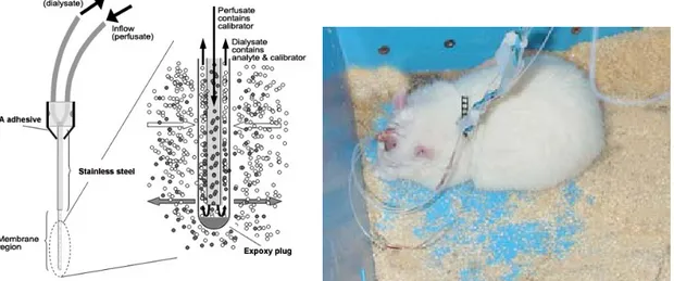
Probes were produced using a concentric design with silica tubing (Polymicro technologies), polyethylene tubing (Smiths Company), stainless steel tubing (outer diameter 0.55 mm) and
dialysis membrane (Hospal Bologna). Inlet portion of probes consisted of 8 cm polyethylene tubing combined with a 3 cm silica tube glued in place with cyanoacrylate (CA) adhesive (Loctite, Attack). The outlet portion of the probe consisted of a 10 cm section of polyethylene tube combined with a 1.5 cm section of silica tubing also glued in place. Silica tubing extended 5 mm into polyethylene tubing when was secured with CA adhesive.
Inlet and outlet silica/polyethylene tubing structures were left to dry and then gently inserted into stainless steel tubing. For SNr, GP and VMTh, the stainless steel used was 20 mm while for studies employee DLS probes, stainless steel was 15 mm. CA adhesive was then used to secure the 3 pieces together and left to dry overnight. Larger tubing was then placed around the point where the 3 pieces converged to further strengthen the structure and more adhesive is added at this site and left to dry overnight.
The inlet portion of the probe with silica extending beyond the stainless steel was then cut to the desired length. For SNr, GP, and VMTh, 1 mm was used while for DLS, 3 mm was used. Membrane is cut and gently inserted over the silica and then cut just beyond where silica ends. Using epoxy adhesive, the membrane is secured to the rest of the structure and the tip is capped leaving a very small space between the end of the silica tubing and the tip of the membrane. The structure is then left to dry for 3 days before experiment. Probes were tested the day of use by pushing purified water through them while checking for any leakage. In vitro recovery for probes were tested periodically and consistenly offered ~10-15% recovery.
3.52 Surgery and microdialysis procedure
Microdialysis probes were stereotaxically implanted, under isoflurane anaesthesia, in the DLS (section I), SNr (section I, II, III, IV), GP (Section II, III, IV), and VMTh (Section III and IV) according to the following coordinates from bregma: DLS; AP +1.0, ML -3.5, DV -6.0, SNr; AP -5.5, ML -2.2, DV -8.3, GP; AP -1.3, ML -3.3, DV -6.5, VMTh; AP -2.3, ML -1.4, DV -7.4. Probes were secured to the skull by acrylic dental cement and metallic screws. After surgery, rats were allowed to recover and experiments were run 24 hr after probe implantation. Microdialysis probes were perfused at a flow rate of 3.0 µl/min with a modified Ringer solution (composition in mM: CaCl2 1.2; KCl 2.7, NaCl 148 and MgCl2 0.85). Samples were collected every 15 min,
starting 6 hr after the onset of probe perfusion. At least 4 stable values were collected before beginning of experiment.
Fig 10. A close up schematic of a microdialysis probe during perfusion (left) and a typical dual probe microdialysis experiment with a rat in home cage (right).
Fig. 11. Example of histological verification of probe placements in SNr and GP. Black points refer to probe tip locations after reviewing brain slices in cryostat.
3.6 Microdialysis + Bar test coupling
The bar test was coupled to microdialysis in order to more accurately characterize the time course of drug effect with respect to neurochemical changes. Bar test, as described above, was simply performed inside microdialysis cages. Animals were handled in their microdialysis cages for 1 hr prior to starting the experiments. Amount of time spent on the bar was measured every 15 minutes (duration of one sample collection) for 45 minutes prior to drug and 90-150 minutes after drug administration. Total time (in sec) spent by each paw on the blocks was recorded (cut-off time 20 sec for each block height).
3.7 Endogenous GLU and GABA analysis
GLU and GABA were measured by HPLC coupled with fluorometric detection as previously described (Marti et al., 2007). Thirty microliters of o-phthaldialdehyde/mercaptoethanol reagent was added to 40 µl aliquots of sample, and 60 µl of the mixture was automatically injected (Triathlon autosampler; Spark Holland, Emmen, Netherlands) onto a 5-C18 Chromsep analytical column (3 mm inner diameter, 10 cm length; Chrompack, Middelburg, Netherlands) run at a flow rate of 0.48 ml/min (Beckman 125 pump; Beckman Instruments, Fullerton, CA, USA) with a mobile phase containing 0.1 M sodium acetate, 10% methanol and 2.2% tetrahydrofuran (pH 6.5). GLU and GABA were detected by means of a fluorescence spectrophotometer FP-2020 Plus (Jasco, Tokyo Japan) with the excitation and the emission wavelengths set at 370 and 450 nm respectively. The limits of detection for GLU and GABA were about 150 fmol/sample. Retention times for GLU and GABA were 3.5 ± 0.2 min and 18.0 ± 0.5 min respectively.
Fig 12. (Left) HPLC coupled to spectrofluorescence detector configured specifically for the detection of GABA and GLU. A) Mobile phase B) Methanol C) Pumps 1 and 2 (Beckman 125) D) Autosampler (Triathalon, Spark Holland) E) 5C-18 chromsep column F) HPLC system interface G) Spectrofluorescence detector (Jasco 2020 plus).
(Right) A typical chromatogram showing the retention times for GLU and GABA. Concentration of amino acids were calculated automatically by peak height analysis.
3.8 Endogenous DA analysis
DA was measured by means of reversed phase HPLC coupled with electrochemical detection. Briefly, 27 µl samples were injected onto a 5-C18 Chromsep analytical column run at a flow rate of 0.4 ml/min (Beckman 118 pump, Beckman Instruments, Fullerton, CA, USA) with a mobile phase containing 75 mM NaH2PO4, 20 µM EDTA, 0.01% triethylamine, 1.5 mM sodium
dodecyl sulphate, 10% methanol and 16% acetonitrile adjusted to pH 5.6 with NaOH. DA was separated under isocratic conditions (~6.5 min retention time) and detected by means of an electrochemical detector (Coulochem II Model 5200; ESA, Inc, Chelmsford, MA, USA) set at +175 mV. The limit of detection for DA was 10 fmol/sample.
3.9 Data presentation and statistical analysis
Motor performance has been calculated as time on bar or on rod (in sec) and number of steps (drag test) and expressed as percent of the control session. In microdialysis studies, GLU and GABA release has been expressed as percentage ± SEM of basal values (calculated as mean of the two samples before the treatment). In text and Figure legends, amino acid dialysate levels were also given in absolute values (in nM).
Motor activity in the inclined grid test has been calculated as time of latency to initiate movements of the head and left and right forepaw (in sec), velocity (in cm/sec) and step length (cm) of left and right forepaw and expressed as percent of the control session. Statistical analysis has been performed on percent data by one-way repeated measure (RM) analysis of variance (ANOVA). In case ANOVA yielded a significant F score, post-hoc analysis was performed by contrast analysis to determine group differences. In case a significant time X treatment interaction was found, the sequentially rejective Bonferroni test was used (implemented on excel spreadsheet) to determine specific differences (i.e. at the single time-point level) between groups. P values <0.05 were considered to be statistically significant.
3.10 Materials
EM-1, UFP-512 and J-113397 were synthesized in the laboratories of the Department of Pharmaceutical Sciences (University of Ferrara). CTOP, bicuculline, raclopride SCH-23390, NTD and SNC-80 were purchased from Tocris Neuramin (Bristol, UK). 6-OHDA hydrobromide and d-amphetamine sulphate were purchased from Sigma (St. Louis, MO, USA). All drugs were
freshly dissolved in Ringer or isosmotic saline just before use with the exception of SNC-80. SNC-80 (10 mg/kg) was dissolved in saline containing 5% DMSO while other doses given systemically (0.1-3 mg/kg) or microinjected (0.1 and 1 nmol) only needed 0.5% DMSO. In both cases, vehicle alone did not affect behavioral or neurochemical responses. SNC-80 was vortexed and sonicated for 15 min or until fully dissolved in solution.
Section I
The endogenous MOP receptor agonist EM-1 dually affects motor activity and
Behavioral Studies I
4.1.1 Effect of i.c.v. injection of EM-1 and CTOP on motor behavior
The effect intracerebroventricular (i.c.v.) administration of EM-1 on physiologically-stimulated motor activity was studied by means of the rotarod test.
In this test, RM ANOVA showed a significant effect of treatment (F4,16=342.7, p<0.0001), but
not time (F1,20=4.3, p=0.051) and a significant time X treatment interaction (F4,20=10.5,
p<0.0001). EM-1 (0.01-10 nmol) biphasically regulated the rotarod performance (Fig. 13A), causing motor facilitation at 0.1 and 1 nmol (∼35% and ∼55%, respectively) and motor impairment at 10 nmol (∼90%). At this dose, EM-1 increased muscle rigidity which probably impaired the execution of the test.
To unravel tonic regulation by endogenous EM-1 and also investigate the selectivity of exogenous EM-1, the selective MOP receptor antagonist CTOP was employed. RM ANOVA on the effect of CTOP alone showed a significant effect of treatment (F2,8=14.9, p=0.0019) but not
time (F1,12=0.4, p=0.5504) and a non significant time X treatment interaction (F2,12=0.4,
p=0.6722). CTOP was ineffective at 1 nmol and produced a long-lasting reduction in rotarod performance at 10 nmol (∼30%) which persisted up to 60 min (Fig. 13B). An ineffective dose of CTOP (1 nmol) prevented the facilitation caused by EM-1 (1 nmol; Fig. 13 C).
0 20 40 60 80 100 120 140 160 180 saline EM-1 0.01 nmol EM-1 0.1 nmol EM-1 1 nmol EM-1 10 nmol
**
10 min 60 min**
**
**
**
A**
ti m e o n r o d (% of c ontr o l) 0 20 40 60 80 100 120 140 160 180 saline CTOP 1 nmol EM-1 1 nmol CTOP + EM-1 10 min 60 min**
**
C ## ## ## ## ti m e on rod (% of c o nt ro l) 0 20 40 60 80 100 120 140 160 saline CTOP 1 nmol CTOP 10 nmol 10 min 60 min**
**
B ti m e on rod (% of c o nt ro l)Fig 13. Effect of i.c.v. injection of EM-1 (0.01-10 nmol; A), CTOP (1 and 10 nmol; B) and their co-administration (EM-1 1 nmol and CTOP 1 nmol; C) on rat motor performance on the rotarod. Each experiment consisted of three different sessions: a control session followed (40 min) by other two sessions performed 10 and 60 min after saline, or drug injection (see Methods). Data are expressed as percentages of basal motor activity in the control session and are means ± SEM of 6-8 determinations per group. Basal motor activity in the rotarod performance was 1044 ± 40 sec (0-55 rpm range).
**p<0.01 significantly different from saline,
4.1.2 Effect of intranigral injection of EM-1 on motor behavior
To investigate the involvement of SNr in central motor effects of EM-1, intranigral microinjections of EM-1 and CTOP were performed and rotarod activity evaluated.
EM-1 (0.01-10 nmol) biphasically regulated rotarod performance (Fig. 14A). RM ANOVA showed a significant effect of treatment (F4,24=402.6, p<0.0001), but not time (F1,26=0.1, p=0.71)
and a non significant time X treatment interaction (F4,26=1.4, p=0.26). Significant increases were
observed at 0.1 and 1 nmol EM-1 (∼30% and ∼40%, respectively) while marked impairment was observed at 10 nmol (∼80%) at 10 min post-injection time and persisted up to 60 min (Fig. 14A).
When administered in SNr, EM-1 evoked spontaneous contralateral turning (Fig. 14B). RM ANOVA showed a significant effect of treatment (F2,12=35.81, p<0.0001), time (F20,360=18.06,
p<0.0001) and a significant time X treatment interaction (F40,360=16.98, p<0.0001). EM-1
induced turning at 10 nmol, but not at 1 nmol (Fig. 14B).
The MOP receptor antagonist CTOP modulated rotarod performance (Fig. 14C). RM ANOVA showed a significant effect of treatment (F2,8=14.9, p=0.0019) but not time (F1,12=0.024, p=0.87)
and a non significant time X treatment interaction (F2,12=0.026, p=0.97). CTOP was ineffective
at 1 nmol yet caused a prolonged reduction of rotarod performance at 10 nmol (∼30%; Fig. 14C). An ineffective dose of CTOP (1 nmol) prevented the facilitation of rotarod performance caused by EM-1 (1 nmol; Fig. 14D).
0 2 0 4 0 6 0 8 0 1 0 0 1 2 0 1 4 0 1 6 0 s a lin e E M - 1 0 .0 1 n m o l E M - 1 0 .1 n m o l E M - 1 1 n m o l E M - 1 1 0 n m o l
**
1 0 m in 6 0 m in**
**
**
**
A*
ti me o n r o d (% of c o n tr o l) -1 5 0 1 5 3 0 4 5 6 0 7 5 9 0 -10 -5 0 5 10 15 20 25 30 35 40 salin e E M-1 1 n m o l E M-1 1 0 n m o l E M -1 (S Nr ) B*
*
*
* *
**
tim e (m in ) s p o n ta neo u s co nt alat er al t u rn s 0 2 0 4 0 6 0 8 0 1 0 0 1 2 0 1 4 0 1 6 0 s a lin e C T O P 1 n m o l C T O P 1 0 n m o l 1 0 m in 6 0 m in**
**
C ti m e o n r o d ( % of co nt ro l) 0 2 0 4 0 6 0 8 0 1 0 0 1 2 0 1 4 0 1 6 0 s a lin e C T O P 1 n m o l E M - 1 1 n m o l 1 0 m in 6 0 m in**
**
D # # # # # # # # ti m e on r o d (% of c o ntr o l) C T O P + E M - 1Fig 14. Effect of intranigral injection of EM-1 (0.01-10 nmol; A-B), CTOP (1 and 10 nmol; C) and their co-administration (EM-1 1 nmol and CTOP 1 nmol; D) on rat motor behavior. EM-1 was injected i.c.v. and motor activity evaluated in the rotarod (A) and spontaneous rotational (B) tests. Each experiment consisted of three different sessions: a control session followed (40 min) by other two sessions performed 10 and 60 min after saline, EM-1, CTOP or their co-injection (see Methods). Data are expressed as percentages of basal motor activity in the control session and are means ± SEM of 6-8 determinations per group. Basal motor activity in the rotarod performance was 1020 ± 29 sec (0-55 rpm range). In spontaneous rotational test data are expressed as absolute values (number of turning executed in 5 min). **p<0.01 significantly different from saline,
##p<0.01 significantly different from EM-1 (RM ANOVA followed by contrast analysis and the sequentially rejective Bonferroni’s test).
Neurochemical studies
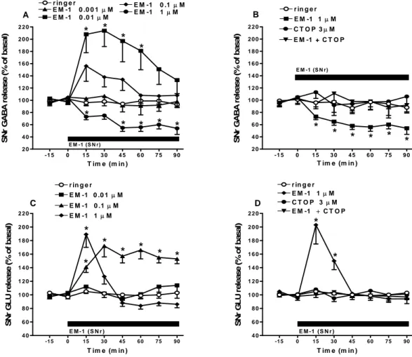
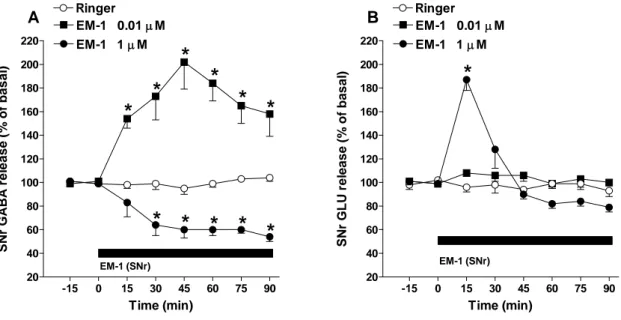
4.1.3 Effect of perfusion with EM-1 in SNr on local GABA and GLU extracellular levels
Since the behavioral data pointed to the involvement of nigral MOP receptors in the effects of EM-1, we investigated how local EM-1 perfusion in the SNr affected local GABA and GLU release. RM ANOVA on GABA levels revealed a significant effect of treatment (F4,20=9.09,
p<0.001), time (F7,167=2.33, p<0.05), and a significant time X treatment interaction (F28,167=2.11,
p=0.026). Post hoc analysis showed that EM-1, ineffective at 0.001 µM, produced a prompt and transient increase at 0.01 µM (~200% at peak) and a prompt and long lasting decrease at 1 µM
(~45% at the end of perfusion) while the stimulation induced by an intermediate concentration (i.e. 0.1 µM) was not significant (Fig. 15A).
To test the specificity of EM-1, high EM-1 concentrations (i.e 1 µM) were challenged with CTOP (3 µM; Fig. 15B). For these data, RM ANOVA on nigral GABA levels revealed a significant effect of treatment (F3,53=9.66, p=0.0002), time (F7,23=21.05, p<0.0001) and time X
treatment interaction (F21,429 = 12.22, p <0.0001). Post hoc analysis showed that intranigral
perfusion with CTOP alone did not modify local GABA levels yet prevented the inhibition induced by EM-1 (Fig. 15B).
Intranigral perfusion with EM-1 (0.01-1 µM; 90 min) was also associated with changes in nigral GLU levels (Fig. 15C). RM ANOVA on GLU levels revealed a significant effect of treatment (F3,17=21.42, p<0.001), time (F7,468=13.45, p<0.0001), and a significant time X treatment
interaction (F21,468=10.68, p<0.0001). Post hoc analysis showed that EM-1, ineffective at 0.01
µM, evoked a prompt and long lasting facilitation (~153% by end of perfusion) at 0.1 µM and a prompt but short-lived increase (~189%) at 1 µM (Fig. 15C).
Again, the specificity of EM-1 was investigated by using CTOP (Fig. 15D). RM ANOVA on nigral GLU levels revealed a non significant effect of treatment (F3,17=1.55, p=0.2130) but a
significant effect of time (F7,436=14.05, p<0.0001) and time X treatment interaction (F21,436=9.82,
p<0.0001). Intranigral perfusion with CTOP (3 µM), ineffective alone, prevented the increase in GLU induced by EM-1 (1 µM; Fig. 15D).