Università degli Studi di Ferrara
DOTTORATO DI RICERCA IN
"Farmacologia e Oncologia Molecolare"
CICLO XXI
COORDINATORE Prof. Pier Andrea Borea
Genetic alterations in epilepsy: the cases of
Unverricht-Lundborg disease, Fragile X and B1 null mutation
Settore Scientifico Disciplinare BIO/14
Dottorando Tutore
Dott. Buzzi Andrea Prof. Simonato Michele
CONTENTS
Abstract………... 1 Riassunto………. 3 Chapter I:EPILEPSY………
5 Generalities: ……… 7Epilepsy and its definition……….7
Epidemiology………...8
Treatment………. 9
Classification……… 14
Models of epilepsy………18
Induced models of epilepsy……… 19
Genetic models of epilepsy……….20
Genes and epilepsy………22
References………..30
GENERAL AIMS OF THE STUDY
AND THESIS STRUCTURE………
33Chapter II:
UNVERRICHT-LUNDBORG DISEASE………
35Introduction……….. 37
Myoclonus………. 37
Myoclonus and epilepsy……… 38
Clinical features of Unverricht-Lundborg disease………. 40
Molecular basis of Unverricht-Lundborg disease……….. 43
Cystatin B (CSTB)………... 43
Molecular alterations of CSTB leading to Unverrich-Lundborg phenotype……… 44
CSTB Knock-out mice: an animal model of Unverricht-Lundborg disease……… 47
Aim of this study………... 49
Paper Franceschetti et al, 2007 (file in attachment for the electronic version of the thesis)……….. insert References………..51
Chapter III:
FRAGILE X………...
55Introduction……….. 57
Fragile X……… 57
Fragile X and Epilepsy……….. 62
Pilocarpine………. 64
Aim of the study……… 67
Materials and Methods……… 68
Animals………. 68
Pilocarpine treatment……… 68
Preparation of tissue for in situ hybridization and immunostaining………. 68
Immunohistochemistry………... 69
Preparation of cRNA probes……….. 70
Optical Density Measurement……… 70
Results……… 72
Pilocarpine seizures cause dendritic targeting of Fmr1 mRNA and FMRP in hippocampus……… 72
Discussion………...75
References………..77
Chapter IV:
BRADYKININS AND SUSCEPTIBILITY TO SEIZURES………..
83Introduction……….. 85
Bradykinins……… 85
Kinins receptors………. 87
Bradykinins and epilepsy………... 91
B1 KO mice……… 93
Kindling……….. 94
Kainate………....95
Aim of the study……….. 97
Materials and methods………... 98
B1 null mice………... 98
Kindling………. 98
Kainate………... 98
Results……… 100
Kindling development……….. 100
Susceptibility to kainate seizures………. 102
Discussion……….. 104
References……….. 105
ABSTRACT
Genetic alterations in epilepsy: the cases of Unverrich-Lundborg disease,
Fragile-X and B1 null mutation
Genetic mechanisms are estimated to underlie about 40% of epilepsies and may present with different manifestations and syndromes. In this thesis, three different cases of genetic implications in epilepsy have been studied: Unverricht-Lundborg disease (ULD), a monogenic Progressive Myoclonic Epilepsy in which disruption of the cystatin B gene results in the manifestation of the pathology; Fragile X, the most frequent genetic mental retardation disease which associates to epileptic seizures in about 25% of the cases; and involvement of the bradykinin system in epilepsy, a possible cause of latent hyper-excitability.
The study on ULD revealed increased brain excitability in mice deficient for cystatin B, the genetic defect associated with the pathology. This hyperexcitability seems to be related to a decreased inhibitory cell number and/or to the activation of microglia. Our findings on Fragile-X reveal that, after a chemically induced epileptic event (pilocarpine status epilepticus), active synapses accumulate Fmr1 mRNA and its protein, FMRP. This phenomenon may account for an involvement in activity-dependent synaptic plasticity and epileptogenesis. Finally, we studied bradykinin involvement in brain excitability, and found that electrical (kindling) and chemical (kainate) epileptic insults exacerbate a latent tissue hyper-excitability in B1 null mice.
These findings are examples of how genetic alterations may alter brain function, directly or indirectly leading to epilepsy manifestations. In depth, investigation of the different aspects of the genetic mechanisms of epilepsy is very important to identify key factors that, in the end, may allow discovering new therapeutic targets and new treatments. Moreover, study of the basic mechanisms underpinning pathological events may also shed light on the normal brain function.
RIASSUNTO
Genetic alterations in epilepsy: the cases of Unverrich-Lundborg disease,
Fragile-X and B1 null mutation
Si stima che intorno al 40% dei casi di epilessia abbiano un’eziologia genetica. Le epilessie genetiche possono manifestarsi con una elevata varietà di sintomi e differenti forme di crisi. In questa tesi abbiamo studiato tre aspetti di questo fenomeno: la patologia di Unverricht-Lundborg (ULD), un esempio di epilessia monogenetica dovuta a mutazioni del gene per la cistatina B; la sindrome dell’X Fragile (FX), la più diffusa forma di ritardo mentale dovuta a cause genetiche (la mutazione del gene Fmr1), che si associa a fenomeni epilettici nel 25% dei casi; ed infine il coinvolgimento delle bradichinine nell’epilessia, una possibile causa di iper-eccitabilità latente e quindi una possibile classe di geni predisponesti alla malattia.
Gli studi sull’ULD hanno dimostrato una maggiore eccitabilità tissutale dei topi KO per la cistatina B, probabilmente riconducibile ad una perdita di neuroni inibitori e/o all’attivazione della microglia. Nello studio sull’FX, l’analisi molecolare dell’insulto epilettico da pilocarpina ha dimostrato che le sinapsi attive accumulano l’mRNA di fmr1 e il suo prodotto proteico, FMRP, implicando un loro ruolo nella plasticità sinaptica attività-dipendente e nell’epilettogenesi. Infine, abbiamo studiato il coinvolgimento della bradichinina nell’eccitabilità cerebrale, e abbiamo scoperto che la delezione del gene codificante il recettore B1 per le bradichinine comporta una ridotta resistenza alle crisi indotte da kindling e da kainato, espressione di una latente iper-eccitabilità.
I dati raccolti in questa tesi rappresentano esempi di come le alterazioni genetiche possano, direttamente o indirettamente, alterare la funzione del cervello e causare manifestazioni di tipo epilettico. Lo studio approfondito delle alterazioni genetiche in epilessia è importante ai fini di identificare bersagli molecolari idonei per lo sviluppo di nuovi farmaci o terapie adeguate. Non solo: lo studio dei meccanismi che sottendono eventi patologici può anche aiutarci a spiegare il normale funzionamento del cervello.
Chapter I
GENERALITIES
Epilepsy and its definition
Through centuries, epilepsy has been described and defined in different ways based on knowledge and superstition of the different ages. Several times epilepsy was associated to divinity, in a good or bad fashion, charging this disease of a magical aura that sometimes survive even to date. Hippocrates, around the 4th century B.C., was among the first describing epilepsy as an ordinary, and not having magical properties, pathology, which origins from the brain. Since then, several other definitions and approaches alternated in time to describe and treat this pathology, resulting in the most peculiar and sometimes ridiculous ways to face the problem.
First clinically defined by John Hughlings Jackson as “an occasional sudden, excessive, rapid and
local discharge of grey matter”, epilepsy, mainly due to its semiology, is still difficult to properly
define.
The International League Against Epilepsy and the International Bureau for Epilepsy define epilepsy as “a disorder of the brain characterized by an enduring predisposition to generate epileptic seizures and by the neurobiologic, cognitive, psychological, and social consequences of this condition. The definition of epilepsy requires the occurrence of at least one epileptic seizure” (Fisher et al, 2005).
Therefore, the term “epilepsy” indicates a brain disorder characterized predominantly by recurrent and unpredictable interruptions of normal brain function, called seizures. Epilepsy is not a singular disease entity but a variety of disorders reflecting underlying brain dysfunction that may result from many different causes. This pathology is not one condition but is a diverse family of disorders, having in common an abnormally increased predisposition to seizures, that are defined as “transient occurrence of signs and/or symptoms due to abnormal excessive or synchronous neuronal activity in the brain.” (Fisher et al, 2005). Discussion about a few points of this new definition of epilepsy is raging, mainly related to: 1) abolishment of the concept of “unprovoked seizure”; 2) absence of the “two seizures” criteria; 3) lack of clear clinical definition of the “enduring alteration in the brain that increases the likelihood of future seizures” (Ahmed et al, 2005; Beghi et al, 2005; Gomez-Alonso et al, 2005).
Therefore, debate on a proper statement which will exhaustively define epilepsy is still a central point in the scientific community, because of its importance in terms of medical, legislative and education communication (Fisher et al, 2005).
Epidemiology
About 1% of world population is estimated to be affected by epilepsy (prevalence), which makes this disease one of the most common neurological disorders. Age specific incidence of epilepsy is higher in children (0-14 years) and in elderly (>60 years) than in adult individuals (15-59 years) (respectively mean incidence rates of epilepsy are: 82,2/100000 per year – 39,7/100000 per year – 34,7/100000 per year; total incidence rate is 47,4/100000 per year). Incidence rate for age-specific epilepsy distribution is significantly different. A non significant trend to higher incidence of epilepsy in males compared to females has been reported showing respectively mean incidence rates of 50,7/100000 per year and 46,2/100000 per year (Kotsopoulos et al, 2002; McHugh and Delanty, 2008). Incidence seems to be higher in males due to the greater exposure to risk factors that cause symptomatic epilepsies. Generally, the median seizure-specific incidence rates are higher for partial (34,4/100000 per year) than for generalized seizures (19,6/100000 per year), but this difference is not significant. The incidence of epilepsy appears to differ from country to country and to change over time. Indeed the incidence rate results to be different in industrialized countries (40-70/100000) compared to the resource-poor countries (100-190/100000) where incidence tends to double. Moreover, in this case, higher rates are found in children and adults than in the elderly. Incidence can change also over time as demonstrated by two studies by Hauser and Kurland, 1975 and Houser et al, 1993 and by Forsgren, 1990 and Forsgren et al, 1996. In these reports, incidence for epilepsy changes with age, doubling for elderly individuals over the young in Hauser studies, and showing a higher incidence for unprovoked seizures in adults in Forsgren studies. Several factors may be involved in this pattern, such as improved antenatal and prenatal care for children, and an increase in life expectancy in the elderly, associated with an increased risk for causes of epilepsy common in old age (for a review see Kotsopoulos et al, 2002; McHugh and Delanty, 2008; Sander, 2003)
Epidemiology terms:
Prevalence: In epidemiology, the prevalence of a disease in a statistical population is defined as the total number of cases of the disease in the
population at a given time, or the total number of cases in the population, divided by the number of individuals in the population. It is used as an estimate of how common a condition is within a population over a certain period of time. It helps physicians or other health professionals understand the probability of certain diagnoses and is routinely used by epidemiologists, health care providers, government agencies, and insurance companies. Mathematically prevalence can be defined as follows: the ratio between the number of individuals in the population with the disease at a given time (a) divided by the sum of individuals in the population with the disease at a given time (a) and individuals in the population without the disease at a given (b).
Prevalence= a/(a+b)
Incidence: Incidence is a measure of the risk of developing some new condition within a specified period of time. Although sometimes loosely
Incidence proportion (also known as cumulative incidence) is the number of new cases within a specified time period divided by the size of the
population initially at risk. For example, if a population initially contains 1,000 non-diseased persons and 28 develop a condition over two years of observation, the incidence proportion is 28 cases per 1,000 persons, i.e. 2.8%.
The incidence rate is the number of new cases per unit of person-time at risk. In the same example as above, the incidence rate is 14 cases per 1000 person-years, because the incidence proportion (28 per 1,000) is divided by the number of years (two). Using person-time rather than just time handles situations where the amount of observation time differs between people, or when the population at risk varies with time. Use of this measure implicitly implies the assumption that the incidence rate is constant over different periods of time, such that for an incidence rate of 14 per 1000 persons-years, 14 cases would be expected for 1000 persons observed for 1 year or 50 persons observed for 20 years.
Incidence should not be confused with prevalence, which is a measure of the total number of cases of disease in a population, rather than the rate of
occurrence of new cases. Thus, incidence conveys information about the risk of contracting the disease, whereas prevalence indicates how widespread the disease is.
Treatment
Epilepsy treatment is symptomatic, mainly aiming to control seizure recurrence, and only surgery can be sometimes curative. No currently available drug is capable of preventing epileptogenesis. Anti-epileptic drugs (AEDs), better if called anticonvulsants, target different biological substrates such as voltage-gated Na-channel (e.g. Phenytoin and its derivatives), GABA system (e.g Barbiturate or Benzodiazepine) and voltage-gated Ca-channels (ethosuximide) (Tab 1).
Table 1: Anti-Epileptic Drugs
YEAR NAME
MECHANISM
OF ACTION TREATMENT ADMINISTRATION SIDE EFFECTS
1857 Bromide ( P otassium bromide) Frequently used as sedatives in the 19th and early 20th century , bromide ion is antiepileptic, and bromide salts are still used as such, particularly in veterinary
medicine
Loss of appetite, nausea/emesis, lethargy, propensity to sleep during the daytime, depression, loss of concentration and memory, confusion, headache, and Bromism Acne-form dermatitis and other forms of skin disease may also be seen, as well as mucous hyper secretion in the lungs. Asthma and rhinitis may worsen. Rarely, tongue disorder, aphten, bad breath, and constipation occur. 1912 Phenobarbital Regulate Chloride permeability reducing excitatory effects of neurotransmitters Still used as AED. It is used to treat generalized convulsive seizures, myoclonus and partial seizures. It is also used in childhood febrile seizures
50-300mg once a day Sedation and hypnosis are the principal side effects of phenobarbital. Central nervous system effects like dizziness, nystagmus and ataxia are also common. In elderly patients, it may cause excitement and confusion while in children, it may result in paradoxical hyperactivity.
1938 Ph en ytoin Regulate sodium channel permeability Effective in treating partial and generalized convulsive seizures, particularly partial convulsive seizures. 100-600mg per day, per os. Intravenous administration is used when patient is on status epilepticus.
At therapeutic doses, phenytoin produces nystagmus, At toxic doses, patients experience sedation, cerebellar ataxia, and ophthalmoparesis, as well as paradoxical seizures. Idiosyncratic side effects are rash and severe allergic reactions. Phenytoin may cause atrophy of the cerebellum when administered at chronically high levels. Phenytoin predispose patients to megaloblastic anemia and gingival hyperplasia. Phenytoin is a known teratogen and suggested to be carcinogenic. Phenytoin has been known to cause Drug-induced Lupus and other dangerous or even fatal skin reactions. (such as Stevens-Johnson syndrome and toxic epidermal necrolysis)
1952 Primidone Methabolized by the liver in Phenobarbital has its same mechanism of action See Phenobarbital 250-1000mg, two or three times per day.
See Phenobarbital 1955 Ethosuximide Primary thalamic regulation of Calcium channel permeability
Used for absence seizures.
500-2000mg CNS common side effects are drowsiness, mental
confusion, insomnia, nervousness, headache, euphoria, ataxia, hiccups, impaired, concentration, irritability, hyperactivity, loss of taste, night terrors. Gastrointestinal side effects are dyspepsia, vomiting, nausea, cramps, constipation, diarrhea, stomach pain, loss of appetite, weight loss, gingival hyperplasia, swelling of tongue. Other side effects involves Genitourinary Hematopoietic Integumentary Ocular apparatus.
1963 Barbexaclone Regulate excitability by modulation of chloride channels. When associated to phenobarbital has awakening properties exerted by the propylexidine component. Treatment of generalized convulsive seizures and partial seizures.
25-200mg See Phenobarbital. Better tolerated than Phenobarbital.
1966 Carbamazepine Regulate sodium channel permeability Drug of choice for partial seizures. Used also in partial and generalized convulsive seizures.
400-2400mg Its common side effects include drowsiness, headaches
and migraines, motor coordination impairment and/or upset stomach. Carbamazepine decrease tolerance to alcohol.
Less common side effects include cardiac arrhythmias, blurry or double vision and/or the temporary loss of blood cells or platelets and in rare cases can cause aplastic anemia. Additionally, carbamazepine may exacerbate pre-existing cases of hypothyroidism, there are also reports of auditory side effects.
1972 Valproate Potenziate GABA inhibitory activity and modulate sodium channel permeability.
Effective for all kind of seizures, it represent the drug of choice for primary
generalized seizures and for benign epilepsy with partial seizures
600-3000mg Common side effects are dyspepsia and/or weight gain.
Less common are fatigue, peripheral edema, dizziness, drowsiness, hair loss, headaches, nausea, sedation and tremors. Valproic acid also causes hyperammonemia, which can lead to brain damage. Rarely, valproic acid can cause blood dyscrasia, impaired liver function, jaundice, thrombocytopenia, and prolonged coagulation times. Valproic acid may also cause acute haematological toxicities cognitive dysfunction and reversible pseudo-atrophic brain changes. It is known as a teratogenic agent.
1974 Clonazepam Increases GABA action Treatment for partial seizures. 0.5-8mg used intravenously in status epilepticus with dosage evaluated patient by patient.
Common side effects are drowsiness, impairment of cognition and judgment, irritability and aggression, psychomotor agitation, lack of motivation, impaired motor function, cognitive impairments. Occasional and rare side effects are serious dysphoria, Thrombocytopenia, serious psychological and psychiatric side effects, induction of seizures or increased frequency of seizures, personality changes, psychosis, incontinence,
1988 Acetazolamide Inhibits CO2 catabolism increasing its concentration in the CNS. This modify the membrane excitability and the modulation of buffer function of glial cells.
Useful for all kind of seizures, but more often used for absence seizures.
250-750mg in association with other AEDs.
Common side effects include numbness and tingling in the fingers and toes, taste alterations (parageusia), frequent urination, and blurred vision, but this usually disappears shortly after stopping the medication. Acetazolamide also increases the risk of developing calcium oxalate and calcium phosphate kidney stones.
1989
Felbamate
Reduces Glycine action
Used for the treatment of partial seizures and secondarily generalized
1200-3600mg in association with other AEDs.
Side effects include decreased appetite, vomiting, insomnia, nausea, dizziness, somnolence, and headache. Many patients report increased alertness with the drug. Two rare but very serious effects include aplastic anemia and hepatic (liver) failure.
1989 Vigabatrin Potenziate GABA action blocking its catabolic enzyme.
Used for the treatment of partial seizures and secondarily generalized
1000-3000mg, in association with other AEDs.
CNS side effects are somnolence, headache, dizziness, nervousness, depression, memory disturbances, diplopia, aggression, ataxia, vertigo, hyperactivity, vision abnormalities, confusion, insomnia, impaired concentration, personality disorder. 11% of children become hyperactive. Some patients develop psychosis during the course of vigabatrin therapy. Other rare CNS side effects include anxiety, emotional lability, irritability, tremor, abnormal gait, and speech disorder. Gastrointestinal side effects are abdominal pain, constipation, vomiting, and nausea.
1991 Lamotrigine Regulate sodium channels reducing the effects of excitatory neurotransmitters
Used for the treatment of partial seizures and secondarily generalized it is also effective for treatment of many kinds of primary or idiopathic generalized seizures 50-600mg, in association with other AEDs.
Common side effects include headaches, dizziness and insomnia. Other side effects may include acne and skin irritation, vivid dreams or nightmares, night sweats, body aches and cramps, muscle aches, dry mouth, fatigue, memory and cognitive problems, irritability, weight changes, hair loss, changes in libido, frequent urination, nausea, and other side effects. In very rare cases, Lamotrigine has been known to cause the development of a dangerous rash called Stevens-Johnson syndrome (or SJS). 1993 Gabapentin Increases inhibitory activity of GABA
Used for the treatment of partial seizures and secondarily generalized
900-2400mg, in association with other AEDs.
Most common side effects include dizziness, drowsiness, and peripheral edema (swelling of extremities). Children 3–12 years of age were observed to be susceptible to mild-to-moderate mood swings, hostility, concentration problems, and hyperactivity. Rare side effects are represented by cases of hepatotoxicity. Gabapentin should be used carefully in patients with renal impairment due to possible accumulation and toxicity. It has a carcinogenic potential.
1995 Topiramate Modulate voltage dependent sodium channels. Potentiate inhibitory activity of GABA and reduces excitatory activity of AMPA-Kainate neurotransmitters
Used for the treatment of partial seizures and secondarily generalized it is also effective for treatment of many kinds of primary or idiopathic generalized seizures 200-600mg, in association with other AEDs.
The most often reported side effects are tiredness, pins and needles in the fingers and toes, dizziness, lowered sense of feeling in the skin, difficulty with language, nausea, diarrhoea, indigestion, dry mouth, weight loss, decrease in appetite, drowsiness, forgetfulness, difficulty with concentration or attention, difficulty in sleeping (insomnia), anxiety, mood swings, depression, changes in taste and vision disorders. Rare side effects are blood clots, blurring of vision and eye pain, reduced sweating and metabolic acidosis
1998 Tiagabine Increases GABA action maintaining GABA at synaptic level for longer time.
Used for the treatment of partial seizures and secondarily generalized
30-50mg, in association with other
AEDs.
Its side effects include confusion, difficulty speaking clearly/stuttering, mild sedation, and, in doses over 8 mg, paresthesia in the body's extremities, particularly the hands and fingers.
2000 Oxcar baz epi ne See
Carbamazepine See Carbamazepine 600-3000mg Oxcarbazepine causes dizziness, drowsiness, blurred or double vision, fatigue and may cause headaches, nausea,
vomiting and stomach pain, diarrhoea, constipation and dry mouth. It can also cause hyponatremia. Concentration loss is also reported to be a frequent side effect. Skin sensitivity to sunlight also may increase.
2001 Levetiracetam Binds synaptic sites of CNS and modulates neuronal burst inhibiting epileptic discharges.
Used for the treatment of partial seizures and secondarily or idiopathic generalized epilepsies 500-1000mg, two or
three times per day Side effects include: hair loss; pins and needles sensation in the extremities; anxiety and psychiatric symptoms
ranging from irritability to depression; and other common side effects like headache and nausea. It is generally well tolerated[6] but may cause sleepiness, weakness, dizziness, and infection. In children, the most common side effects are sleepiness, accidental injury, hostility, irritability, and weakness.
2001 Clobazam Increases GABA action Effective on partial seizures 10-40mg, in association with other
AEDs.
Common side effects are ataxia, somnolence, diplopia, dysarthria and rarely gelastic seizures and urticaria rashes
The choice among the different AEDs depends on the semiology of the disease and the management of the treatment is correlated to the containment of seizures: some patients reach the goal by a monotherapy, some others, unfortunately, require polytherapy with the use of two or more AEDs. Balance between control of seizures and side effects of AEDs is another important point that physicians must take into consideration when starting a treatment for epilepsy. Medication administration is sometimes life-long lasting, some other times it could be slowly withdrawn, depending on recurrence of seizures when drugs are stopped. In spite of the numerous AEDs available, several patients are refractory to pharmacological treatments and continue to experience seizures. Indeed, of those who develop epileptic seizures 47% will be controlled with the first AED prescribed, 32% with the second AED, and 9% with the third. Fourth and subsequent AEDs have at most a 5% chance of bringing remission (Duncan, 2007). This leaves around 7% of individuals that have no chances to control their seizures by pharmacological approaches. Therefore it is of central importance to develop new drugs able to contain seizures or even to prevent epileptogenesis. This latter goal could be reached, not only by empirical experimentation of new chemical compounds, but also by increasing our knowledge of the complex basis of the pathology. When drug treatments result ineffective and patients experience refractory seizures, other medical approaches may be applied. These alternative treatments include surgery, vagus nerve stimulation, ketogenic diet, avoidance therapy.
Epileptic surgery is a delicate tool that does not apply to any kind of epileptic patient. Best candidate for surgery are those individuals that continue to experience seizures despite the fact they are under pharmacological treatment. Focal and very localized epileptic lesions are the target for surgery and only epilepsy with these anatomical characteristics must be taken into consideration when surgery is to apply. Surgery follows after a full assessment of the epileptic conditions.
Video-electroencephalogram monitoring, neuropsychological examination, structural magnetic resonance imaging, functional imaging tests and intracranial electrode recordings are tools used to identify the type of epilepsy, determine the severity of the seizures, and locate the epileptogenic zone (Villanueva et al, 2007). The ictogenic area will be the one dissected from the remaining presumed healthy brain tissue. Mapping the epileptic area must be done with care in order to verify which will be the important areas that might be involved in resection and, if no impairment is forecasted in the dissectible loci, the patient will undergo surgery. Surgery usually reaches 90% of success in healing focal epilepsy. Best surgical results are achieved when small epileptic lesions are dissected, but improvement in the quality of life in partially successful surgery should also be considered a good achievement.
Intractable epilepsy may also be treated by Vagus Nerve Stimulation (VNS). VNS is an alternative treatment for patients refractory to drug therapy and/or in which surgery could not be applied or resulted ineffective. Chronic refractory partial and secondarily generalized seizures are examples of epileptic syndromes that can be treated by VNS. VNS consists of a stimulating device implanted under the skin, which sends electrical impulses to the left side of the vagus nerve in the neck. Its mechanism of action is still on debate. Vagus, the tenth cranial nerve, arises from the medulla and carries both afferent and efferent fibres. The afferent vagal fibres connect to the nucleus of the solitary tract which in turn projects connections to other locations in the central nervous system. Little is understood about exactly how vagal nerve stimulation modulates mood and seizure control but proposed mechanisms seem related to an increase of thalamic synaptic activity. This effect seems to be brought about by release of epinephrine through the nucleus coeruleus and/or involvement of nucleus tractus solitarius, the structure that receives the C fibres of the Vagus nerve. The nucleus tractus solitarius is related to several areas involved in seizures onset (Milby et al, 2008; Ghanem and Early, 2006). Pilot study results demonstrated significant reduction in the frequency, intensity, and duration of seizures with chronic, intermittent VNS (Utman et al, 1993; Penry and Dean, 1990; Uthman et al, 1990; Wilder et al, 1991; Murphy et al, 1995; Labar et al, 1998). Follow up studies report a mean of 50-60% success for VNS when associated to normal pharmacological treatment (Ardesch et al, 2007; Abubakr and Wambacq, 2008; Milby et al, 2008). Indeed, patients experience a reduction of seizure frequency and duration. VNS seems also to progressively increase its effectiveness in seizures containment in time (Ardesch et al, 2007; Milby et al, 2008). Very few are the side effects of this practice, comprising hoarseness of voice, cough and sometimes weight loss; most of them are very mild and bearable for patients.
Ketogenic diet represent another side treatment for intractable refractory epilepsy and for epilepsy at large. The efficacy of the diet is independent of the type of seizure and is effective for both
generalized and partial seizures at varied ages. This therapy is characterized by a diet high in fat and low in carbohydrates and protein. In these conditions, energy is retrieved from fatty acid oxidation and high concentration of ketone bodies (such as β-hydroxybutyrate, acetoacetate, and acetone) are generated. Ketones are then used as nutrients for the body and even for the brain. The use of ketones as source of energy in the brain seems to directly or indirectly affect neurotransmitter biology leading to a possible, even if yet unknown, mechanism of action. High percentage of good outcome is reported for this treatment, also reviewing patients in seizure free conditions of after one year of treatment. Recent findings attributed to ketogenic diet a potential effectiveness in preventing epileptogenesis (for review see: Bough and Rho, 2007; Freeman et al, 2007; Hartman et al, 2007). Avoidance therapy is a therapy applicable only to reflex seizures (see below “classification”). It consists in putting patients in the condition to avoid the precipitating phenomenon of their seizures. Main precipitating phenomena are related to sensory organs and comprise: music and noise, light and dark, touching feelings, tastes and sometimes emotional involvement. This very simple approach is usually successful, leading patients to experience very few seizures.
Classification
Epilepsy is classified by 5 main criteria: o Aetiology (cause of the first seizures) o Semiology (manifestation of the seizures) o Triggering event(s)
o Syndromic symptoms
o Brain anatomic focus-foci (focal/partial or generalized seizures)
Considering aetiology, epilepsy is classified as idiopathic or secondary and probably symptomatic (definition preferred to criptogenic in the last revision of the terms). Each of these comprehends generalized or partial seizure types (Tab. 2).
The term idiopathic describes a syndrome that is only epilepsy, with no underlying structural brain lesion or other neurological signs or symptoms. These are presumed to be genetic and are usually age-dependent (Engel, 2006). Attention must be posed on the correct definition of "idiopathic," which means a disorder onto itself, sui generis, and not aetiology unknown.
Epilepsies described as secondary are related to a known or a suspected disorder of the CNS which is the trigger of the symptoms. Known causes of secondary epilepsies are: head traumas, tumors,
viral and bacterial encephalopathies, anoxia, etc. The probably symptomatic (formerly cryptogenic) epilepsies are referred to epilepsies of unknown nature which are usually suspected to be related to a specific disorder of the brain (Engel, 2006).
The definition of partial (or focal) seizures refers to a specific brain-localized-origin of the seizures. Generalized seizures, instead, refers to a general involvement of both brain hemispheres in the origin of the seizures.
Table 2: Classification by aetiology of the disease.
Idiopathic
Partial seizures type Benign Familiar Epilepsy in Infancy
Childhood benign epilepsy with central or temporal burst
Childhood benign epilepsy with occipital burst
Other form of idiopathic epilepsy with partial seizures
Generalized seizures type Benign myoclonic epilepsy in infancy
Childhood absence epilepsy (Pyknolepsy) Juvenile myoclonic epilepsy
Juvenile absence epilepsy
Generalized epilepsy with tonic-clonic seizures
Secondary and Probably symptomatic epilepsy syndrome
Generalized seizures type Early suppression-burst encephalitis in
infancy West syndrome
Lennox-Gastaut syndrome
Progressive epileptogenic encephalitis
Partial seizures type Severe childhood myoclonic epilepsies
Mesial temporal epilepsy
Other epilepsy defined by their focus of origin
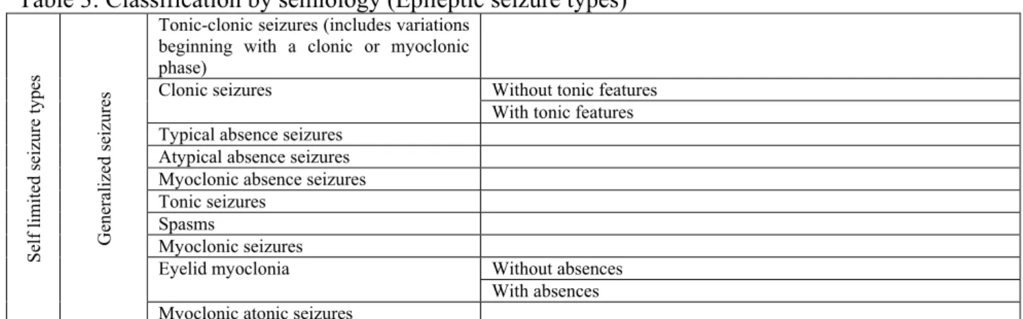
By semiology, epilepsy is classified in self limited seizure types and continuous seizure types and each of these are subdivided in generalized and focal seizures (Tab. 3) (Engel, 2006). Self limited seizures usually end in a reasonable limit of time; on the contrary, continuous seizures are characterized by repetition of the ictal phenomenon for a long period of time with no remission between seizures (i.e. status epilepticus).
Table 3: Classification by semiology (Epileptic seizure types)
Self l imi ted s eiz ure typ es Generalized seizures
Tonic-clonic seizures (includes variations beginning with a clonic or myoclonic phase)
Clonic seizures Without tonic features
With tonic features Typical absence seizures
Atypical absence seizures Myoclonic absence seizures Tonic seizures
Spasms Myoclonic seizures
Eyelid myoclonia Without absences
Negative myoclonus Atonic seizures Fo cal s eizur es
Focal sensory seizures With elementary sensory symptoms (e.g., occipital and parietal lobe
seizures)
With experiential sensory symptoms (e.g., temporo parieto occipital junction seizures)
Focal motor seizures With elementary clonic motor signs
With asymmetrical tonic motor seizures (e.g., supplementary motor seizures)
With typical (temporal lobe) automatisms (e.g., mesial temporal lobe seizures)
With hyperkinetic automatisms With focal negative myoclonus With inhibitory motor seizures Gelastic seizures
Hemiclonic seizures
Secondarily generalized seizures
Continuous seizure typ
es
Generalized
status ep
ilep
ticu
s Generalized tonic-clonic status
epilepticus
Clonic status epilepticus Absence status epilepticus
Tonic status epilepticus Myoclonic status epilepticus
Fo cal s tatus epil epti cus
Epilepsia partialis continua of Kojevnikov
Aura continua
Limbic status epilepticus (psychomotor status)
Hemiconvulsive status
Adapted from Engel (2001) with permission.
By definition, a reflex epilepsy syndrome is a syndrome in which all epileptic seizures are precipitated by sensory stimuli. Reflex seizures that occur in focal and generalized epilepsy syndromes that are also associated with spontaneous seizures are listed on the basis of seizure types. Isolated reflex seizures can also occur in situations that do not necessarily require a diagnosis of epilepsy. Seizures precipitated by other special circumstances, such as fever or alcohol withdrawal, are not reflex seizures (Engel, 2006). Principal stimuli considered as precipitating circumstances of reflex epilepsy syndromes are reported in Tab. 4.
Table 4: Precipitating stimuli for reflex seizures
Precip itatin g st imu li fo r reflex seizures
Visual stimuli Flickering light: color to be specified
when possible Patterns
Other visual stimuli
Thinking Music Eating Praxis Somatosensory Proprioceptive Reading Hot water Startle
An epilepsy syndrome is a complex of signs and symptoms that define a unique epilepsy condition with different aetiologies (Engel, 2006). This must involve more characteristics than just the seizure type such as: age of onset, progressive nature, interictal EEG, associated interictal signs and symptoms, pathophysiologic mechanisms, anatomic substrates, etiological categories and genetic basis (Engel, 2006). The different syndromes are summarized in Tab. 5. In this table are also reported syndrome conditions which, by their nature, must not be considered as epilepsy but as convulsive episodes.
Classification of epileptic syndromes is a difficult matter, considering that a full description of each syndrome is sometimes far from being complete and, for some of them, only a few characteristics have been reported. Including an epilepsy syndrome in an official classification gives it the status of a recognized syndrome. On the other hand, excluding poorly described syndromes from an accepted classification could lead to a reduced investigation in that direction, failing to uncover important features of scarcely reported syndromes (Engel, 2001). For these reasons the reported table has signalled few syndromes as in definition development.
Another important case is raised by syndromes of Idiopathic Generalized Epilepsy (IGE), only eight of which are recognized by ILAE (Nordli, 2005), even if other are described in the literature (Panayiotopoulos, 2005). The difficulties stem from the practical view that IGE are clinically considered as one disease, leading to an easy clinical approach, but in reality IGE comprise several syndromes. This would imply to consider IGE as a complex ensemble of different syndromes which would be more demanding diagnostically. Therefore, in view of a better differential diagnosis, a well organized classification of epilepsy syndromes would be a useful tool for the physicians. Table 5: Epilepsy syndromes and related conditions
Benign familial neonatal seizures Early myoclonic encephalopathy Ohtahara syndrome
aMigrating partial seizures of infancy
West syndrome
Benign myoclonic epilepsy in infancy Benign familial infantile seizures Benign infantile seizures (non-familial) Dravet’s syndrome
HHE syndrome
aMyoclonic status in non-progressive
encephalopathies
Benign childhood epilepsy with centrotemporal spikes
Early onset benign childhood occipital epilepsy (Panayiotopoulos type)
Late onset childhood occipital epilepsy (Gastaut type)
Epilepsy with myoclonic absences Lennox–Gastaut syndrome Landau–Kleffner syndrome
Progressive myoclonus epilepsies
Idiopathic generalized epilepsies with variable phenotypes
Juvenile absence epilepsy Juvenile myoclonic epilepsy
Epilepsy with generalized tonic-clonic seizures only
Reflex epilepsies Idiopathic photosensitive occipital lobe
epilepsy
Other visual sensitive epilepsies Primary reading epilepsy Startle epilepsy
Autosomal dominant nocturnal frontal lobe epilepsy
Familial temporal lobe epilepsies
aGeneralized epilepsies with febrile seizures plus
aFamilial focal epilepsy with variable foci
Symptomatic (or probably symptomatic) focal
epilepsies Limbic epilepsies Mesial temporal lobe epilepsy with hippocampal
sclerosis
Mesial temporal lobe epilepsy defined by specific etiologies
Other types defined by location and etiology
Neocortical epilepsies Rasmussen syndrome
Other types defined by location and etiology Conditions with epileptic seizures that do not
require a diagnosis of epilepsy
Benign neonatal seizures Febrile seizures Reflex seizures
Alcohol-withdrawal seizures
Drug or other chemically induced seizures Immediate and early post cerebral insult seizures
Single seizures or isolated clusters of seizures Rarely repeated seizures (oligoepilepsy)
Adapted from Engel (2001) with permission. (a Syndromes in development)
Models of epilepsy
(For en extensive review of epilepsy modelling see “Models of seizures and epilepsy” Pitkanen A., Schwartzkroin P.A. and Moshé S.L. (2006) Elsevier Academic Press)
The study of epilepsy can not be performed only in humans because of several different reasons, from ethical issues to practical inapplicability, from unavailability of controls to high costs of human research. For all these reasons, modelling epilepsy is of a central importance. Epilepsy models for studying epilepsy are principally used for three reasons:
1. to understand basic mechanisms underlying the pathology; 2. to devise new approaches for diagnosis;
3. to test new drugs or new therapies.
Basic mechanisms of epilepsy are still under strong investigation and epileptic models are a very precious tool to deeply understand the molecular and physiological causes of this complex pathology. Of course, uncovering the mechanisms underpinning the disease will help develop new diagnostic, therapeutic and preventive approaches. Models of epilepsy are the best instruments on
which innovative experimental approaches for diagnosis and therapies can be tested. This will speed up the application of new diagnostic and therapeutic tools, allowing rapid intervention on the disease. In each of these cases, human studies can give a big support, but preliminary data are normally retrieved from in vitro and in vivo experiments.
Epilepsy models should be created or prepared in order to faithfully reproduce the human epileptic condition. Therefore several different models have been developed trying to model all the complex aspects of this pathology. On the other hand, some experimental approaches model only some of the manifestation of epilepsy (epilepsy equivalent) allowing only the investigation on that symptom and not on the whole complex picture of a complete model. In any case, this gives the chance to study an aspect of the disease.
Therefore, the already reported epidemiology that describes 1% of the population as affected from some kind of epilepsy, which in turn explains the economic, social and personal cost of the disease, makes it clear how important is to study epilepsy and how important are the tools that model it.
Induced models of epilepsy
Induced models of epilepsy are developed by application of chemical, electrical or damaging insults on a healthy brain, in order to transform that brain in an ill one, capable to show features of the chosen kind of epilepsy in study.
Table 6: Induced model of epilepsy (classification based on the tools used to develop the models).
In Vitro
Cell culture Models Acute and organotypic slices
Single nerve cells acutely dissociated from animal and
human brain In vitro isolated guinea pig
brain
In Vivo
chemical model of epilepsy
GABA
Pentylenetetrazole Bicuculline Picrotoxin
Glutamic Acid Decarboxylase (GAD) inhibitors
Beta carbolines and convulsant benzodiazepine Ro 5-3663 GHB (gamma-hydroxy-butyrate)
Excitatory Amino-Acid
Kainic acid
Quisqualic acid/alfa-amino-3-Hydroxy-5-Methyl-4-Isoxasole Propionic acid N-Methyl-D-Aspartic acid (NMDA) Homocysteine, homocysteic acid
Acetylcholine related substances Pilocarpine, and litium-pilocarpine Organophosphorus compounds
Other drugs
Strycnine Aminophylline
Insulin induced hypoglycemia Ay-9944
THIP (4,5,6,7 tetrahydroxyisoxazolo (4,5,c) pyridine 3-ol)
Inhalants Fluorothyl Topical application
Metals (cobalt, zinc, antimony, alumina cream, iron
Antibiotic (penicillins and cephalosporins) Tetanus toxin
electrical model of epilepsy
electroshock seizures local electrical stimulation electrical kindling
self sustaining status epilepticus by Perforant Path stimulation self sustaining status epilepticus by Amygdala stimulation
focal status epilepticus by perforant path stimulation in anesthetized rats continuous hippocampal stimulation
lesion model of epilepsy
cortical freeze lesion model
antiproliferative agents (5-azacytidine, methyl-mercury, nitrosureas and carmustine)
Methylazoxymethanol acetate (MAM) model
In-Utero irradiation as a model of cortical dysplasia
Hypoxia-induced seizures and hypoxic encephalopathy in neonatal period Lateral fluid percussion brain injury Chronic Partial Cortical Isolation model
Head Trauma: haemorrhage-Iron Deposit
Stroke
Others
Complex febrile seizures – experimental model in immature rodents
Infection induced seizures Model of neurocysticercosis
Model of herpes virus infection Rasmussen’s encephalitis model
Genetic models of epilepsy
The above reported epilepsy models are related to chemically, electrically or mechanically induced alterations in an otherwise healthy brain. Genetic models of epilepsy, instead, refer to a molecularly and/or anatomically altered brain due to genetic alterations. These models can be naturally selected in animals spontaneously showing epilepsy phenomena or can be created by introducing gene mutations. Either ways, animals show and model a particular kind of epilepsy. On one hand, a naturally occurring seizure behaviour in a spontaneously epileptic animal (i.e. GAERS and WAG/Rij for rats and tottering, lethargic, ducky, stargazer etc for mice); on the other hand, specific seizures and syndromes induced by targeted alteration of genes already known to cause or to be involved in a particular type of epilepsy. Table 7 and 8 reports spontaneous and induced genetic models.
Table 7: Spontaneous models of epilepsy
rats Absence model GAERS rats WAG/Rij rats
mice Spike wave Tottering lethargic ducky stargazer SWE Mocha2j coloboma convulsion Dilute lethal jimpy jittery megencephaly quaking staggerer torpid veritint waddler wabbler-lethal weaver writher
convulsions evoked by sensory stimuli frings
lurcher
Table 8: Induced model of seizures
Gene involved Protein modified
NPY Neuropeptide Y
Syn1 Synapsin 1
Syn2 Synapsin 2
Slc1a (Glt-1) Glutamate transporter
Akp2 Tissue nonspecific alcaline phosphatase (GABA syntesis)
Gad2 Glutamic acid decarboxylase (GABA synthesis)
Gabrβ3 GABAA receptor β3 subunit
Gabrδ GABAA receptor δ subunit
Gria2 Glutamate receptor subunit 2
Htr2c Hydroxytryptamine receptor 2c
Kcna1 Voltage-gated K+ channel
Scn2a Sodium channel type 2a (brain specific)
Kcnj6 (Girk2) Inwardly rectifying K+ channel
Camka Calcium calmodulin kinase α subunit
Itpr1 Inositol 1,4,5 trisphosphate receptor
Gap43 Growth-associated protein
Cdk5r Neuronal-specific activator of cyclin kinase
Otx1 Orthodenticle homolog (homeobox family)
Jrk Jerky (DNA-binding protein)
Pcmt1 Protein L-isoaspartate (D-aspartate) O.methyltransferase (protein repair enzyme)
Hex-a α subunit of β-hexosaminidase (lysosomal enzyme)
Hex-b β subunit of β-hexosaminidase (lysosomal enzyme)
Stfb (Cstb) Cystatin-B (cysteine protease inhibitor)
Psap Sphingolipid activator protein
Il6 Cytokine, interleukin 6
App Amyloid precursor protein
Hdh Huntingtin’s amino-terminal polyglutamine sequence
Polyglutamine repeat 146 unit CAG repeat inserted in hprt gene
Bdnf (homozygous) BDNF
Fyn Tyrosine kinase receptor
Plat Tissue plasminogen activator
Fos Protooncogene Mt-3 Metallothionein-3 … …
Others Many others are already available and several others will be modelled as soon as new
genes will be found responsible for an epileptic syndrome.
GENES AND EPILEPSY
Of the about 1% of the population affected by epilepsy, 30-40% are estimated to have a genetic contribution to the aetiology (Gardiner, 2000; Berkovic et al, 2006). Indeed, among all the classifications for epilepsy, one consistent group is represented by the idiopathic epilepsies which are usually described as epilepsies with no known causes and, therefore, probably genetic. Epidemiologic studies describe also the risk of hereditary transmission of epilepsy from parents experiencing epilepsy or having familiarity for this pathology (Winawer and Shinnar, 2005). Beside a general risk of 5‰ of children born with epilepsy in the general population, there is an increased risk of seizures in siblings and offspring of pro-bands with the epileptic pathology. This is mainly related to parent gender with a maternal higher effect (Ottman et al, 1988), early age at onset (Anderson and Hauser, 1997; Ottman et al, 1996a), number and closeness of affected relatives (Winawer and Shinnar, 2005) and EEG abnormalities including generalized spike waves and photoparoxysmal response (Anderson and Hauser, 1997). Parents affected by idiopathic epilepsies have higher risk to transmit epilepsy than parents affected by symptomatic ones. Hereditary risks depend also from the different seizure types affecting the pro-bands: parents with myoclonic and absence seizures as well as with generalized seizures have higher risk to transmit epilepsy to offspring than people experiencing partial seizures (Eisner et al, 1959;Tsuboi and Christian, 1973; Tsuboi, 1980;Annegers et al, 1982; Beck-Mannagetta and Janz, 1991; Ottman et al, 1998; Ottman et al, 1989). In any event, the data are reassuring; more than 90% of individuals with epilepsy have no affected relatives, and most parents with epilepsy will have no children with epilepsy. In conclusion, the general genetic risk of epilepsy transmission is about 5% for parents having a direct or indirect history of epilepsy (Winawer and Shinnar, 2005).
Genetic epilepsies can be classified based on the mechanism of inheritance in (Gardiner, 2000): o Mendelian disorders;
o Non-Mendelian disorders or complex epilepsies; o Chromosomal disorders.
Mendelian disorders are those in which one single major locus represents the main genetic
modification which is responsible for the epileptic phenotype. Modes of inheritance are the common autosomal dominant, autosomal recessive, X-linked dominant and X-linked recessive.
However, they form an important group because recognition of the characteristic features and presence of a family history enables the correct diagnosis to be made. In these disorders single gene mutation leads to a deleterious change in gene sequence, presumably bringing to either loss of the encoded protein function or gain of function mutation. Either way, a disruption of the pathway including the modified protein is affected, exacerbating the epileptic phenotype. Examples of these diseases are: Benign Familial Neonatal Convulsion (EBN1, EBN2); Autosomal Dominant Nocturnal Frontal Lobe Epilepsy (ADNFLE); Generalized Epilepsy with Febrile Seizures Plus (GEFS+). Several times, epilepsy is one of the major symptoms of Mendelian transmitted disorders such as Unverricht-Lundborg disease, Lafora Disease or Fragile X syndrome.
Non Mendelian disorders (complex epilepsies) are characterized by a complex inheritance and
represent about 70% of the idiopathic epilepsies (Anneger, 1996). They are usually polygenic, as suggested by rapidly diminishing risks beyond first-degree relatives (Ottman et al, 1996b) and high concordance between monozygotic twins (Berkovic et al, 1998). Mechanisms of inheritance for these diseases showed a complex pattern and not only one gene is presumed to be involved. Complex epilepsy arises when, by chance, meiotic reshuffling creates a combination of susceptibility alleles with sufficient effect in the same individual to push neuronal hyperexcitability over the seizure threshold. Each susceptibility allele alone is insufficient to cause seizures, but requires the additive or epistatic interaction of other susceptibility alleles. Complex epilepsy is most often expressed sporadically in an affected individual, and the phenotype does not follow Mendelian inheritance. Nor do the close relatives, who may or may not be affected, necessarily share the same set of epilepsy susceptibility alleles. During intergeneration transmission, each susceptibility allele is segregated away from other unlinked susceptibility alleles (Mulley et al, 2005). This group of genetic epilepsies include Juvenile Myoclonic Epilepsy (JME), Childhood Absence Epilepsy (CAE) and Benign Childhood Epilepsy with Centrotemporal Spikes (BCECTS) (Mulley et al, 2005).
Chromosomal disorders associate to epilepsy many times, as happens in Down Syndrome (trisomy
21p), in trisomy 12p, in trysomy 1q42.3-qter (Tuschl K et al, 2007), monosomy 21q22.3-qter (Tuschl K et al, 2007), etc. They are usually related to a gross cytogenetic abnormality which ordinarily determine a syndrome in which epilepsy may be an important component.
Genetic epilepsy can be also classified based on functional classification as follows (Noebels, 2003): o those linked to primary defects of membrane and synaptic signalling,
o those linked to neuronal plasticity and metabolism, o those linked to network development.
Epilepsies of the first group are characterized by genetic alterations that modify general neurotransmission leading to an imbalance between excitatory and inhibitory stimuli that exacerbate epileptic phenomena. Among those classified in this group, a large number is represented by mutations in neuronal ion channels, the so called channelopathies. Channelopathies have been among the first disorders associated to genetic modifications in epilepsy and gave a boost for a deeper investigation of the genetics of epilepsy.
Impaired neuronal ion channel function has been associated to several pathologies like neuromuscular disorders (Bernard and shevell, 2008), chronic pain (Catterall et al, 2008), migraine (Catterall et al, 2008) and epilepsy (Noebels, 2003; Heron et al, 2007; Bernard and Shevell, 2008,
Catterell et al, 2008). Symptoms of a channelopathy may present either as an abnormal gain of function or as a loss of function of the channel affected by the mutation. Channelopathies usually demonstrate both phenotypic and genetic heterogeneity. Phenotypic heterogeneity means that different mutations in the same gene can cause different diseases. Genetic heterogeneity means that mutations in different genes can result in the same apparent disease phenotype (Bernard and Shevell, 2008).
Numerous are the genetic epilepsies caused by channelopathies, including Autosomal Dominant Nocturnal Frontal Lobe Epilepsy (ADNFLE), caused by mutation in the acetylcholine receptor subunit alfa-4 or subunit alfa-2, Generalized Epilepsy with Febrile Seizures plus (GEFS+) caused by mutation on sodium channel alfa-1 and beta-1 subunit or on GABA-A receptor gamma-2 subunit, Benign Familial Neonatal Seizures (BFNS) due to mutations on potassium channel subunits. Epileptic channelopaties present normally as monogenic inherited alterations (Heron et al, 2007) even though they may associate to complex patterns of inheritance involving more than one mutation to exacerbate the epileptic phenotype (Mulley et al, 2005; Heron et al, 2007).
Synaptic signalling may be disrupted and cause epilepsy also by mutations involving the large protein families that mediate vesicle trafficking and exocytosis (Bock et al, 2001). Seizures appear in mice deleted for the vesicle-anchoring phosphoproteins synapsins 1 and 2; loss of these proteins diminishes the size of the presynaptic vesicle pool and disrupts synaptic depression (Rosahl et al. 1995).
Another synaptic vesicle protein, Sv2A, also regulates synaptic strength by altering the mobility of the releasable pool. Sv2A-deficient mice, alone or combined with Sv2B nulls, show a severe seizure phenotype (Crowder et al. 1999, Janz et al. 1999). In contrast to synapsin mutants, neurons showed sustained release of transmitter in response to brief activation.
At an earlier stage in vesicle biogenesis, incomplete vesicle assembly can also lead to an epileptic phenotype. The mocha mouse encodes the AP3-delta subunit of an adaptor complex that facilitates
incorporation of the Znt3 zinc transporter into synaptic vesicles. Lack of the delta subunit leads to loss of vesicular zinc sequestration, severe EEG hypersychronization, and seizures (Kantheti et al. 1998).
In conclusion, all the mutations affecting neurotransmission may lead to an imbalance of excitatory/inhibitory signalling leading to hyperexcitability, which in turn may manifest as epileptic seizures.
Epilepsy can be observed during the course of many inborn errors of metabolism (IEMs), usually as part of a large clinical spectrum, and several IEMs may manifest with inaugural epileptic seizures (Sedel et al, 2007). These diseases may be subdivided according to the type of clinical presentations into progressive myoclonic epilepsies, epileptic seizures without mental retardation, epilepsies with mental retardation. In the first group are worth to be mentioned ceroid lipofuscinosis, sialidosis, and lafora disease. The second group includes Wilson disease and acute neuropsychiatric porphyries. The last group comprises De Vivo disease, creatine synthesis or transport defects and succinic semialdehyde dehydrogenase. All these disorders are characterized by altered metabolic pathways in which a known protein has been found as the putative responsible for the disease. Unfortunately, the molecular functions of the different proteins remain to be elucidated in most of the diseases and, in many cases, studies are required to better understand the link between protein mutation and epileptic manifestations.
The third group of the functional classification reported above is represented by epilepsies in which the genetic mutation leads to altered cyto-architectonic formation of brain structures. Cortical dysplasias resulting from aberrant patterns of brain development are a frequent substrate for inherited seizure syndromes. The malformations may be microscopic or visible by magnetic resonance imaging and originate from diverse signalling defects affecting migration, proliferation, differentiation, and segmentation. For example, defective proliferation and cell dimension have been associated to deletion of NeuroD/Beta2, a neuronal transcription factor that selectively blocks postnatal proliferation of granule cells in the dentate gyrus. These mice show a hippocampal formation entirely devoid of a granule cell layer, but with a preserved pyramidal cell layer and theta rhythm generation, and exhibit frequent partial motor seizures in adulthood (Liu et al. 2000). The same hippocampal phenotype is associated to a deletion of citron-kinase (citron-K), a Rho effector regulating cytokinesis (Di Cunto et al. 2000). Citron-K-deficient mice lack granule cells and develop fatal seizures before adulthood. Disturbance of cell migration has been linked to mutation of DCX (Kizhatil et al, 2002), the gene encoding for doublecortin, which leads to subcortical band heterotopia syndrome (Feng and Walsh, 2001) comprehensive of seizure phenomena. Periventricular nodular heterotopia has been suggested as consequence of a mutation in the filamin
gene (FLN1) that interacts with F-actin during motility-related cytoskeletal reorganization, and mutation results in a subset of neurons clustered at the ventricular zone. Analysis of mutant human FLN1 neurons and chemically induced heterotopias confirms that key intrinsic excitability changes occur when cells develop outside of their natural lamination pattern; such molecular rearrangements may provoke specific patterns of aberrant receptor expression and loss of repolarizing K+ currents that favor epilepsy (Castro et al. 2001, Battaglia et al. 2002). Mutations in the human reelin gene (RELN) are linked to a lissencephalic cortical neuronal migration defect with seizures (Hong et al. 2000). Finally, genetic disruption of homeobox genes related to specification, regionalization, and terminal differentiation of the neocortex results in epileptic phenotypes. Example for this are deletion for OTX-1 and mutation of ARX.
Transcription factors and protein silencers act together to control the activation and repression of the genetic differentiation program (Robertson and Wolffe 2000). Mutation of genes acting in this pathway are now linked to epilepsy. In this group, two mutations are worth to be mentioned: mutation of the MECP2 gene associated to Rett Syndrome, an X-linked disorder including mental retardation and seizures (Shabazaian et al, 2002; Johnston et al, 2001; Tudor et al, 2002); and deletion of jerky gene, which leads to a limbic seizure phenotype in mice, through a putative defect in translational processing (Liu et al. 2002). A mutation in the human homologue, JH8, has been identified in a case of childhood absence epilepsy (Moore et al. 2001).
Epilepsy is therefore well known as a complex neurological disorder and it is now quite clear that a number of human epilepsy syndromes can result from gene mutations (Crino, 2007). Particular gene mutations may account also for many epilepsy phenotypes that show little if any evidence for inheritance patterns. This may be due to a de-novo mutation. Indeed, one fascinating possibility is that sporadic epilepsies result from somatic mutations occurring in brain progenitor cells, which are not present in the germline DNA (Crino, 2005;Weiss, 2005). Thus, while sporadic epilepsies might not follow Mendelian inheritance patterns, they are nonetheless the result of a genetic or mutational event. In this scenario, a deleterious gene mutation might occur during the many rounds of cellular mitosis occurring throughout the course of brain development. Moreover, a corollary hypothesis is that an accumulation of mutations in a variety of distinct genes, insufficient alone to cause seizures, could lead to neuronal hyperexcitability. Thus, somatic mutations in multiple genes during brain development might lead to seizure onset.
This mechanism may provide a further explanation for complex epilepsies (Crino, 2007). Considering these possibilities, the genomics approach is important for the possible explanation of idiopathic epilepsies. In this respect, single nucleotide polymorphism (SNPs) may play an important
role in the onset of several epileptic manifestations. SNPs represent single basepair changes that are present in more than 1% of the general population and may be synonymous, with no changes in amino-acid in the primary structure of the protein or non-synonymous with amino-acid substitution. In non-synonymous SNPs an altered protein function may be expected and may have functional consequences in terms of excitability or seizure susceptibility. Following this reasoning, clearly results that SNPs relevant to epilepsy occur throughout the genome and thus that subtle alterations in basepair sequence in a set number of candidate genes can culminate in an epilepsy phenotype. This so-called “common variant-common allele” phenomenon suggests that certain types of epilepsy result from an inherent predisposition or predilection based on the unique or additive effects of SNPs within a single or multiple genes (Ottman, 2001; Ottman, 2005). Thus, epilepsy may reflect a “SNP dose effect” in which seizure susceptibility is directly related to the number and functional significance of relevant SNPs throughout the individual genome. Genomic influences may account for the majority of patients with no obvious structural or inherited etiology for their seizures, so-called idiopathic epilepsy.
SNP variations may also account for some forms of symptomatic epilepsy. This notion has practical importance since it may account for the incidence of seizures in some, but not all, patients with tumors, stroke, trauma and so on. For example, even in the setting of brain injury, cortical malformation, or tumor, only a proportion of patients will develop epilepsy. It is conceivable that these individuals develop seizures not just because of injury to the cortex but rather because of combinatorial effects of a brain lesion plus a particular set of predispositional SNPs (Crino, 2007). This point of view opens a discussion on how the genetic background may influence epilepsy onset. Therefore, the so called modifier genes may account for synergistic or antagonistic interactions with known epilepsy susceptibility loci (Durner et al., 2001; for review, see Mulley et al., 2003). These sites in the genome, while not directly responsible for seizures, can affect the impact of gene mutations or sequence alterations that culminate in seizures. Modifier genes may affect the epilepsy phenotype in idiopathic epilepsies and this is well documented by the fact that, in mice, strain backgrounds can determine seizure onset in the setting of an engineered gene defect. Moreover, in humans, variable expressivity among family members is a common feature of inherited epilepsy syndrome of most types, suggesting that genetic modifiers may influence the clinical manifestation of epilepsy.
In the same frame, the genetic background may be responsible for epilepsy onset in symptomatic epilepsies. The idea that genes and acquired factors (traumas, injuries, etc.) interact in human epilepsies is an old one, dating back to Lennox, in the mid-20th century (Lennox, 1951), and is generally accepted for any complex disorder. In this respect, what is thought plausible is that after a
brain damaging event, reparation of the injury may activate mechanisms that enhance hyper-excitability of the damaged areas, which in turn may allow exacerbation of seizures. This relates to the influence of the background by the fact that, in the presence of the same damaging event, not all patients develops seizures over time. This points out how susceptibility loci created by the injury associate to the genetic predisposition to seizures in triggering epileptic phenomena.
Genes and their protein products are also related to epilepsy as a consequence of the seizures. Gene expression is perhaps one of the first studied aspects of gene involvement in epilepsy. Indeed several studies report altered gene expression in the brain experiencing seizures (Aronica and Gorter, 2007; Elliot and Lowenstein, 2004; Likasiuk and Pitkanen, 2004; Lukasiuk et al, 2006; Majores et al, 2004;Morgan et al., 1987;Gall et al., 1991; Newton et al., 2003). The modified gene expression correlates to pathophysiology in all the possible ways, either in a protective attempt to prevent seizures to happen again or, in an aberrant acting fashion, leading to a worsening of the disease. From a mechanistic perspective, it is tacitly assumed that altered mRNA levels predict similar changes in functional protein expression that disrupt normal cell function, although this relationship is not universal. Seizure-induced changes in mRNA expression are usually associated with long-lasting changes in protein expression that have functional relevance in terms of cellular architecture, network reorganization, cell proliferation, cell death, and perhaps most interesting, fostering recurrent seizures. Under a certain point of view, the “holy grail” of gene expression analysis studies in epilepsy has been to define either a single gene or a panel of genes that are the first steps in the epileptogenesis cascade. This mainly in relation to the fact that symptomatic epilepsies are usually a consequence of a plastic modification occurring after a damaging event that may lead to the manifestation of recurrent seizures. Deep investigation in gene expression put in motion during epileptic events may shed light on the putative starters of the neuronal plasticity which at a certain point may exacerbate in epileptic phenomena. The therapeutic implications of these essential genes are numerous.
Finally, it is important to stress how difficult is to associate genetic defects to an epileptic syndrome. Very few genetic associations for idiopathic epilepsy have been replicated and this has tempered enthusiasm for the results of genetic studies in epilepsy. Failure to replicate is most often attributed to multiple testing, type I error considerations (i.e., the belief that the original finding was a false positive) (Tan et al., 2004), population stratification, sample size (Tan et al., 2004; Durner et al., 2006), and genetic heterogeneity (Pal et al, 2008). The main concepts to keep in mind while trying to evaluate a putative gene modification attributable to a idiopathic epilepsy must be independent
replication of the gene association and coherence of evidence. Indeed, independent replication of association is an important criterion in judging evidence, yet the absence of replication may not necessarily invalidate an original finding. Therefore, it has been suggested to adopt a perspective integrating results from different experimental methods, rather than place one over another in importance or insisting only on replication. Coherence of experimental methods is a more informative approach than simple replication, because it forces one to evaluate different kinds of experimental data in the context of findings from other approaches.
Concluding, the last few decades have seen an increasing number of reports on the genetic basis of idiopathic epilepsies and on the involvement of genetics and gene expression in epileptic phenomena, confirming that genes and their protein products play a pivotal role in defining the outcome of the disease and the subtle differences in epileptic manifestations. Therefore, comprehension of the complex genetic mechanisms underlying epileptogenesis may allow a better understanding of the pathology and may help define new therapeutic targets.