Molecular strategies aimed to boost Natural Killer cell-
mediated immunotherapy of Neuroblastoma
PhD school in:
Immunological, Hematological and Rheumatological Sciences (SIER) XXXI cycle
Curriculum: Immunology
PhD school coordinator Prof. Angela Santoni
Supervisor PhD student
Dott. Loredana Cifaldi Irene Veneziani
Sommario
Neuroblastoma - 4 -
MYCN and p53 - 6 -
Neuroblastoma Immune Escape - 9 -
Neuroblastoma Treatment - 10 -
Natural Killer Cells - 14 -
NK cell education and function - 16 -
Inhibitory receptors and their ligands. - 16 -
Activator receptors and their ligands. - 17 -
Nutlin - 20 -
Potential therapeutic use of Nutlin-3a in tumours - 22 -
Aim of the project - 25 -
Materials and methods - 27 -
Results - 33 -
Discussion - 62 -
References - 66 -
- 4 -
Neuroblastoma
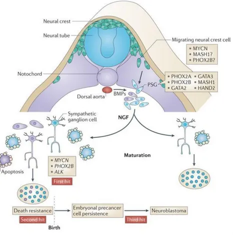
Neuroblastoma (NB) is the most common extra-cranial solid tumour in childhood [1] and it may be considered a malignant manifestation of aberrant sympathetic nervous system development (Fig 1).
Fig 1. Development of the sympathoadrenal lineage of the neural crest (Cheung and Dyer. Nat Rev Cancer 2015)
NB is the primary cause of death from paediatric cancer for children between the age of 1 and 5 years; roughly, it represents 6% of all paediatric tumours and accounts for approximately 15% of all paediatric cancer mortality. The median age at the time of diagnosis is between 18 and 22 mouths and 50% of the patients present a high-risk of metastatic tumour at the time of diagnosis [2].
There is a familial and a sporadic form of NB and both are defined by specific genetic mutations. The first is very rare (<2% of all NBs) and the major predisposition mutation identified in familial NB was in PHOX2B, a gene playing a critical role in the development of neural crest derived autonomic neurons [3]. Another common lesion associated with familial NB is in the ALK receptor tyrosine kinase gene [4]. ALK is expressed in the developing sympathoadrenal lineage of the neural crest, and it may regulate the balance between proliferation and differentiation [5]. While mutations of PHOX2B is relatively rare in sporadic NB, approximately 6–10% of NBs
carry somatic ALK activating mutations, and an additional 3–4% carry high level ALK gene amplifications [6]. However, the most common focal genetic lesion in sporadic NB is the amplification of MYCN, which occurs in approximately 22% of tumours and it is associated with poor outcome [7]. MYCN amplification or over-expression has been described in several other cancers that originate from tissues where MYCN is normally expressed and include retinoblastoma, Wilm’s tumor, rhabdomyosarcoma, medulloblastoma, glioblastoma, and small cell lung cancer [8] (Fig 1).
Others important markers used to diagnose high-risk NB include ATRX mutations, but ATRX alterations alone are not sufficient to promote tumorigenesis, ploidy, chromosomes abnormalities and aberrant expression of tyrosine kinases receptors (Table 1) [2].
High-risk NB
GENE FUNCTION
ALK
ALK cooperates with MYCN to drive malignancy, as activation of ALK results in increased expression of MYCN by elevating activity of the MYCN promoter.
ATRX
ATRX encodes a SWI/SNF chromatin-remodelling ATP-dependent helicase, suggesting ATRX involvement in various developmental processes. However, ATRX mutations alone are not sufficient to promote tumorigenesis.
1p36.3 Deletion of 1p correlates with metastatic diseases.
17q Gain of distal 17q is recurring feature of NB and primary tumours. 17q
gain is itself a powerful independent predictor of poor survival
TrkB
Coexpressed at high levels with its ligand, BDNF, in unfavorable tumours, especially those with MYCN amplification. Activation of the TrkB–BDNF autocrine pathway can lead to invasion, metastasis,
angiogenesis and drug resistance
MYCN Mycn amplification occurs in 25%-33% of the cases and correlates with a
greatly increased risk of fatal outcome. Tab.1 Established characteristics for high-risk NB patients.
International Neuroblastoma Staging System (INSS), first published in 1988, used the extent of the initial surgical procedure to define the stage of the patient. Small localized tumours that were completely resected were considered Stage I lesions. Stage II tumours were small, may or may not have had lymphonode involvement, but could not be completely resected. Stage III lesions
- 6 - were large tumours that crossed the anatomical midline of the patient and could not be completely resected. Lastly, Stage IV and IVS metastatic tumours were differentiated by the fact that IVS patients were <1 year of age with metastatic disease located in liver, skin and less than 10% of the bone marrow [9]. Most infants, with IVS metastatic disease, can be cured with moderate- intensity chemotherapy, and some patients with a special pattern of metastasis have a high likelihood of undergoing spontaneous regression without chemotherapy. Several possible mechanisms are proposed to support spontaneous regression in NBs: (i) neurotrophin deprivation, (ii) loss of telomerase activity, (iii) humoral or cellular immunity and (iv) alterations in epigenetic regulation and possibly other mechanisms, but the exact mechanisms responsible for spontaneous regression (and differentiation) are uncertain [10] [11].
Although the International NB Staging System (INSS) is currently used, a new International Neuroblastoma Risk Group (INRG) Staging System was recently proposed classifying NB patients into 16 risk groups on the assessment of 13 potential biological prognostic factors [12]. According to INRG, risk in NB is classified as low, intermediate, or high. Although low- and intermediate-risk patients generally have a favourable outcome (80%–95% event-free survival rate), high-risk patients have, 50% event-free survival rate, and there is also a subset of “ultra- high” risk patients who do not respond to therapy [13]. The biological hallmark of NB is the complexity of the genetic abnormalities acquired by the tumour cells, and some of these abnormalities are powerful prognostic high-risk NB markers.
MYCN and p53
MYCN belongs to a family that includes MYCL (L-Myc) and MYCN (N-Myc) [14] [15] [16]. While the role of L-Myc is less well understood, N-Myc expression is tissue-restricted, and N- Myc could substitute for c-Myc in murine development [17] [18]. It is crucial to understand that concentration of both c-MYC and MYCN proteins is finely adjusted allowing them to perform their normal functions [19]. While c-Myc is expressed in all proliferating tissues in the adult, MYCN expression in humans and mice is restricted to certain tissues in the developing embryo and is very low or absent in adult tissues [20] [21].
Structurally, c-Myc and MYCN proteins are very similar and both heterodimerize with MAX protein to mediates many of their functions. Myc–Max complexes bind gene promoters by recognizing a DNA sequence, called E-box; MYCN preferentially binds E-box CATGTG as well as the classic CACGTG. Moreover, under N-Myc–amplified conditions such as in NB, MYCN becomes less specific and can bind additional E-box motifs including CATTTG, CATCTG, and
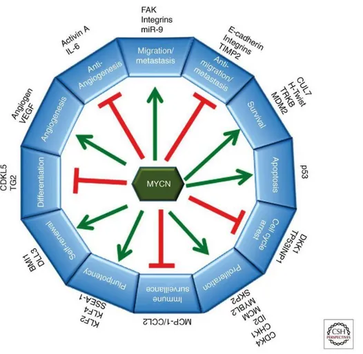
CAACTG [22]. This in turn activates the transcription of downstream genes involved in diverse cellular functions, specifically in NB it can activate transcription of genes involved in metastasis, survival, proliferation, pluripotency, self-renewal, and angiogenesis and can suppress expression of genes that promote differentiation, cell cycle arrest, immune surveillance, and genes that antagonize metastasis and angiogenesis [23] (Fig. 2).
Fig 2. MYCN plays multiple roles in malignancy and maintenance of stem-like state (Huang M. et al. Cold Spring Harb
Perspect Med 2013).
Early studies found that several c-Myc target genes were expressed in some NB cell lines with MYCN amplification: significant overlap between c-Myc and MYCN-regulated gene sets had been reported [24] [25]. By contrast, MYCN expression inversely correlates with that of c-Myc indeed, in MYCN amplified NB cell lines, c-Myc mRNA transcription is repressed by the high levels of MYCN protein [24].
p53 is a tumour suppressor gene that is induced and activated by a variety of potentially tumorigenic stresses, including inappropriate oncogene signalling and DNA damage. p53 is a direct target gene of c-MYC because its promoter contains a non-canonical E-box (CATGTG)
- 8 - located upstream of the transcription initiation site and it is recognized by MYC-MAX heterodimers [26]. Recently, it was demonstrated that p53 is also a direct transcriptional target of MYCN and that p53 accumulation in NB correlates with MYCN amplification [27]. p53, often referred to as “guardian of the genome”, is mutated in up to 60% of many human malignancies, but in NB it is mutated only in 2% of the cases at the time of diagnosis [28]. Normally, p53 function is controlled by a complex network of regulators including p14ARF, mouse double minute
(MDM)2, and MDM4 [29]. In particular, MDM2 binds p53 and blocks its transcription and nuclear export, and mediates p53 degradation because MDM2 has an E3 ubiquitin ligase activity responsible for p53 ubiquitination [30].
Fig.3 – Autoregulatory feedback loop of inhibition of p53 by MDM2. (Annu Rev Pharmacol Toxicol, 2009)
The activation of mammalian ATM promotes p53 responses by stimulating the rapid degradation of both MDM2 and MDM4 through C-terminal phosphorylation of both proteins [31]. The restoration of p53 function induces the expression of target genes such as p21 (CDKN1A), which leads to G1 cell-cycle arrest [32]. Moreover, DNA single strand breaks induce ATR activation which phosphorylates Chk1, leading to G2 cycle arrest [33]. It was demonstrated that both cell cycle arrest and senescence contribute to blocking tumorigenesis [34] and promote the expression of ligands for NK cell-activating receptors [35] [36].
The human MDM2 protein, the main negative controller of p53 function (Fig. 3), is another oncogene that is amplified in a variety of human cancers, including NB. Interestingly, both MDM2 and p53 are regulated by MYCN. Moreover, MDM2 also plays p53-independent roles in oncogenesis regulating MYCN mRNA stabilization [37] [38].
Neuroblastoma Immune Escape
Tumour growth is facilitated by cancer cell-driven immunosuppressive mechanisms. In the last 10 years many studies have contributed to elucidate the immune evasion mechanisms adopted by NB cells to escape the control of the host immune system. These studies have highlighted: • the release of soluble molecules suppressing anti-tumour immune reactivity, such as the
stress inducible NKG2D ligand MICA and the HLA class Ib molecule HLA-G [39];
• the decreased or absent expression of surface ligands for various natural killer (NK) cell- activating receptors, including NCRs, NKG2D and DNAM-1. In particular, defects in the expression of few components of the HLA class I related antigen processing machinery (APM) [40];
• the production of suppressive factors e.g. TGF-beta and IL-10, which may prevent expansion and activation of tumour-infiltrating lymphocytes reducing the expression of NKp30 and NKG2D receptor [41];
• the decreased expression of costimulatory molecules such as CD40, CD80, CD86, OX40L and 4-1BB-L, but not PD-L1and B7H2 in primary NB tumours [42];
• 4Ig-B7-H3 molecules expressed at the NB cell surface can exert a protective role from NK- mediated lysis by interacting with a still undefined inhibitory receptor expressed on NK cells [43].
The main effector cells of the immune system involved in tumour cell recognition are: (i) CD8+ T cells, which specifically recognize through their T cell-receptor (TCR) peptides presented by antigen presenting cells (APC) in the context of MHC class I molecules and mediate cytotoxic functions; (ii) NK cells, innate effectors, which exert their cytotoxic activity through a diverse repertoire of activating and inhibiting receptors that recognize specific ligands on the surface of the target cells; (iii) CD4+ T lymphocytes and macrophages are other important players of anti- tumour immune responses, mainly through the production of cytokines and inflammatory mediators. Defects in HLA class I and APM component expression may render NB cells an excellent target for NK cell-mediated cytotoxicity [44].
- 10 - Neuroblastoma Treatment
A localized NB lesion is generally curable. However, long-term survival for children with advanced disease older than 18 months of age remains poor despite aggressive multimodal therapy. The therapies for these risk categories are very different.
• Low-risk disease can frequently be cured with surgery alone. • Intermediate-risk disease is treated with surgery and chemotherapy.
• High-risk NB is treated with intensive chemotherapy, surgery, radiation therapy, bone marrow / hematopoietic stem cell transplantation [45], biological-based therapy with 13-cis- retinoic acid (isotretinoin or Accutane) [46] and immunotherapy (for example anti GD2- monoclonal antibody usually administered with the cytokines GM-CSF and IL-2) [47]. With the current treatments, patients with low and intermediate risk disease have an excellent prognosis with cure rates above 90% for low risk and 70–90% for intermediate risk. In contrast, therapy for high-risk NB in the last 20 years resulted to be effective only for about 30% of patients.
Chemotherapy agents used in combination have been found to be effective against NB. Agents commonly used for stem cell transplant conditioning are platinum compounds (cisplatin, carboplatin), alkylating agents (cyclophosphamide, ifosfamide, melphalan), topoisomerase II inhibitor (etoposide), anthracycline antibiotics (doxorubicin) and vinca alkaloids (vincristine) and some newer regimens include topoisomerase I inhibitors (topotecan and irinotecan) that have been found to be effective against recurrent disease. Many of this drugs cause a genotoxic stress that lead to DNA damage response (DDR) pathway activation with a consequent induction of ligands for NK-cell activating receptors [48] [49] [50].
Among the new strategies proposed to treat high-risk NB patients, the use of small molecules inhibitors is very promising. There are different compounds targeting proteins associated to NB. The most used inhibitors directly or indirectly target MYCN protein but can also affect downstream targets.
BET (bromodomain and extra-terminal domain) are a family of proteins that regulate the transcription of many genes, including MYCN. These proteins contain 2 bromodomain motifs in the amino terminus and an extraterminal protein–protein interaction domain in the carboxy terminus, constituting an additional class of proteins that regulate chromatin structure [51] [52]. These “chromatin readers” bind to acetylated lysines in chromatin, recruiting additional chromatin, modifying proteins and leading to cell context–dependent gene activation or repression [53]. The BET family consists of BRD2, BRD3, BRD4, and BRDT.
Fig.4. JQ1 mechanism of action (Schnepp and Maris. Cancer Discov. 2013)
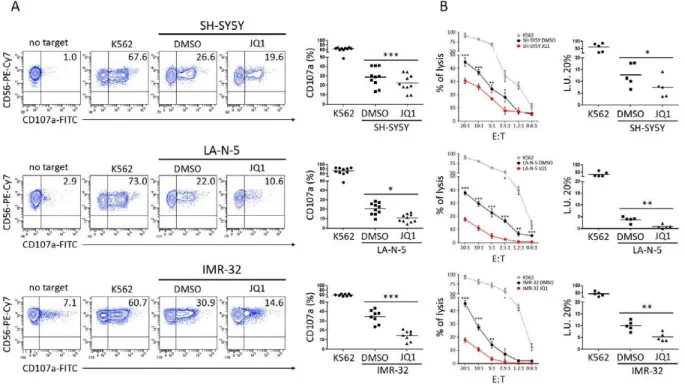
JQ1 is one of the BET bromodomain inhibitor and it can represent a promising strategy to block the growth of MYCN-dependent NB cells [54] [55]. Indeed, it competitively binds to bromodomain, displacing BRD4 fusion oncoprotein from MYCN promoter (Fig. 4).
Among MYCN downstream targets, p53 represents a good candidate because NB is rarely associated with p53 mutations [27]. However, the p53 pathway is often impaired in childhood cancers because of upstream p53/MDM2/p14ARF network aberrations [56]. One of the strategies to affect this pathway is by perturbing the p53/MDM2 interaction. Small molecules as Nutlin-3, a cis-imidazoline compound, can interact with MDM2 by mimicking the p53 N-terminal region where MDM2 binds and can induce p53 activation allowing its post-transcriptional acetylation [57]. Together with wt p53, expression level of MYCN and p14ARF may determine the outcome of the response to MDM2 inhibition [58].
The main prerequisite for p53 activation after Nutlin-3 treatment, however, is an intact p53 protein, as p53 loss-of-function mutations inevitably prevent MDM2 inhibitors from reinstating p53 function [59] (Fig. 5).
- 12 - Fig. 5. Nutlin-3a mechanism of action
NK cells have an important anti-tumor activity because they can limit tumour growth. Moreover, especially NK cells are much less suppressed by tumor microenvironment then T cells and unlike B and T cells of adaptive immunity, they are capable of spontaneously destroying transformed cells without prior sensitization in an MHC-restricted manner [60]. The pre-clinical studies in mice in which NK cells are infused after allogeneic haematopoietic stem cell transplantation (HSCT), are very promising because suppress graft-versus-host-diseases (GVHD) and enhance graft-versus-tumor (GVT) against solid tumors and leukemias [61] [62]. The concurrent administration of NK cell stimulating cytokines, such as IL-2 or IL-15, can promote this effect [63].
Tumor-specific mAbs that recognize tumour antigens expressed on the surface of tumor cells are being used as cancer therapy. GD2 is a promising target for antibody therapy because it is abundant in most NB cells and its expression is restricted to nervous system cells among normal cells [64]. For this reason, an immunotherapy based on the use of anti-GD2 antibody for NB was investigated [47]. Anti-GD2 antibody induces ADCC via NK cells. NK cell-mediated surveillance mechanisms [65] and NK cell stimulation with interleukin IL-2 can also contribute to the antitumor effect [66]. Two intravenous (i.v.) injection of anti-GD2 IgG antibodies, chimeric 14.18 (ch14.18) and mouse 3F8 combined with i.v. injection of interleukin-2 (IL-2) and granulocyte-macrophage colony-stimulating factor (GM-CSF), have been tested in the clinic [47]. Despite the recent success of immunotherapy with the anti-GD2 antibody ch14.18 and cytokines, treatment of high-risk NB remains challenging.
Another strategy to enhance an NK cell-mediated immunotherapy is based on genetic modification of cells. Such as T cells, also NK cells may be genetically engineered to display a
CAR enables the specific targeting or augmented cytotoxicity of NK cells. It was demonstrated that NK-92-scFv(ch14.18)-zeta, a GD2-specific chimeric antigen receptor expressing cell line, successfully lyses partially or multidrug-resistant GD2+ NB cells lines [67]. The reported side
effects of a CAR-T cell based immunotherapy include neurologic toxicity, cytokine release syndrome, tumor lysis syndrome, immunogenicity [68]. For this reason, the long-lasting and production of pro-inflammatory cytokines from CAR-T cells in vivo is the cause of severe side effects [69]. NK cells have been considered better candidates for CARs because their short lifespans last for nearly 2 week in vivo and they mainly produce interferon IFN-γ. NK cells have been used for treating both hematopoietic malignancies (e.g., leukemia, lymphoma, and multiple myeloma) and solid tumors (e.g., melanoma, ovarian cancer, lung cancer, colorectal cancer, and glioblastoma) [70] [71]. NK cells have innate advantages in the treatment of certain solid tumors: IFN- γ has been shown to induce the permanent arrest of the growth of melanoma cells [72]; others solid tumors, such as the ovarian cancer, have high expression of MICA/B and ULBPs, which can promote NK cells function through the activating receptor NKG2D [73]. Moreover, recent studies have shown that IFN-γ can drive T-regulatory cells to promote antitumor immunity, suggesting that NK cells may contribute for the immunotherapy of solid tumors [74]. However, NK cell-immunotherapies for solid tumors, can control disease progression but scarcely abate disease [75] [76]. The possible reasons include (i) low infiltration rate of NK cells to the solid tumor; (ii) inhibitory factors in the tumor microenvironment; and (iii) inhibition from tumor cells, such as residual HLA expression. These crucial issues may be solved enhancing the migration of NK cells to tumor sites, altering the tumor microenvironment and combining with the targeting of checkpoint inhibitors aimed to activate NK cells [77].
- 14 - Fig. 6 Immunotherapy of Neuroblastoma (Nature Reviews, 2013)
Natural Killer Cells
NK cells (around 5-20% of all circulating lymphocytes) are large granular lymphocytes that belong to the innate immune system. Unlike T or B cells of the adaptive immune system, NK cells do not rearrange T cell-receptor or immunoglobulin genes from their germline configuration [78]. The traditional cell surface phenotype defining human NK cells within the lymphocyte gate on the flow cytometric analyser shows an absence of CD3 and expression of CD56, the 140-kDa isoform of neural cell adhesion molecule (NCAM) found on NK cells and a minority of T cells [79]. Murine NK cells do not express CD56 therefore NKp46, a member of the highly conserved natural cytotoxicity receptor (NCR) family of NK cell-activating receptors, is used to define NK cells across species [80] [81].
NK cells are part of hematopoietic system and are derived from CD34+ hematopoietic progenitor cells (HPCs) (Fig.7) [82], but they don’t wholly develop in the bone marrow. As a matter of fact, a subset of NK cells, termed CD56bright for their high-density surface expression of CD56, are relatively dominant in secondary lymphoid tissue (SLT) compared with their more abundant CD56dim NK cell counterpart found in bone marrow, blood, and spleen [83]. CD56bright NK cell population are less mature than the CD56dim subset, that it is characterized by an increased expression of NK cells [84]. In SLT, dendritic cells (DCs) and other antigen presenting cells
(APCs) express membrane bound IL-15, which is required for NK cell maturation, suggesting that SLT may be a site for NK cell development in vivo [85]
Fig.7 Schematic diagram of human NK cell development. (Blood 2001)
Conversely to T cells, NK cells can recognize viral-infected or malignant-transformed cells without a prior exposure and their functions are based on a balance between activator and inhibitor signals [86]. They are a major source of cytokines that influence the host’s immune response. TNF-α initiates cytokines and chemokines release that influence pro-inflammatory cytokine cascades, while interferon gamma (IFN-γ) production by NK cells is known to shape the Th1 immune response, activate APCs to further up-regulate MHC class I expression and improve anti-proliferative effects on viral- and malignant-transformed cells [87].
The majority of circulating NK CD56dim cells share a common killing mechanism with CD8+
cytotoxic T lymphocytes (CTL) display against tumour target cells. NK cell killing of tumour cells proceeds through distinct mechanisms: (i) exocytosis of secretory lysosomes that contain lytic proteins such as perforin, granzymes, and (ii) NK surface expression of ligands that engage death receptors of the tumour necrosis factor (TNF) superfamily on tumour cells (eg, Fas, TRAIL) [88], thereby triggering apoptosis. In addition to antibody–independent natural cytotoxicity, expression of CD16 (FcγRIIIA, the low affinity receptor for IgG) on NK CD56dim cells mediates an antibody-dependent cellular cytotoxicity (ADCC) against IgG–coated cells [89]. Furthermore, NK cells can kill allogeneic cells in hematopoietic transplantation enhancing a graft-versus leukaemia activity [90]. Moreover, elimination of NK cells in mice results in a
- 16 - higher incidence of spontaneous tumours, impaired clearance of inoculated tumour cells, and an increased rate of tumour metastasis [91].
NK cell education and function
NK cell education plays an important role in steady state conditions, where potentially auto- reactive NK cells must maintain tolerance for self, yet recognize infected or transformed cells after proper stimuli [92]. NK cell education is based on the signaling via the inhibitory receptors that recognize self-MHC class I ligand. The activation of NK cells is regulated by the integration of signals deriving from activating and inhibitory receptors expressed on their surface.
Inhibitory receptors and their ligands.
In MHC class I-sufficient humans, the NK cell’s subset lacking inhibitory receptors for self- MHC class I is hypo-responsive to in vitro stimulation through several activating receptors and fail to reject MHC class I-deficient bone marrow in vivo [93]. Thus, expression of self-MHC class I-reactive inhibitory receptors improve the responsive potential of NK cells [94]. Inhibitory NK cell receptors recognizing self-MHC class I are considered the predominant mechanism responsible for NK cell tolerance to self. In human, MHC class I recognition is performed by a distinct set of receptors called killer cell immunoglobulin-like receptors (KIRs) which are specific for different polymorphic MHC class I molecules. Different KIR genes have varied effects on NK cell activity: those KIR genes with immune-receptor tyrosine-based inhibitory motif in the cytoplasmic tails (indicated by L=long in the nomenclature), transmit inhibitory signals after binding their cognate HLA ligand; those with short cytoplasmic tails (S) including immune-receptor tyrosine-based activator motif transmit activating signals [95]. There are two main KIR haplotypes, A and B, which depend upon the genes expressed. The A haplotype contains up to 7 expressed genes (KIR3DL3, KIR2DL3, KIR2DL1, KIR2DL4, KIR3DL1, +/- KIR2DS4, KIR3DL2). KIR B haplotypes contain up to 14 genes, (KIR2DS2, KIR2DL2, KIR2DL5B, KIR2DS3, KIR3DS1, KIR2DL5A, KIR2DS5, KIR2DS1) and the majority of them is stimulatory [96]. Other inhibitory NK cell receptors for human leukocyte antigens (HLA, or human MHC class I) with cytoplasmic ITIMs, such as NKG2A (CD159a) and LIR-1 (ILT2, CD85j), also display variegated expression patterns. The ligand of NKG2A is the non-classical MHC class I molecule HLA-E (Fig. 8 and Fig. 9).
Fig.8 NK-cell receptors and their cognate ligands (CJ Chan et al. Cell Death and Differentiation 2014)
Activator receptors and their ligands.
In contrast to inhibitory receptors, most activating receptors are expressed by all NK cells. Furthermore, activating receptors use diverse signaling cascades, whereas inhibitory receptors appear to use a common mechanism for inhibition [97] [98]. Activating receptors associated with ITAM, can be subdivided into two groups, the first includes rapidly evolving receptors expressed on NK cells, such as KIR2DS, KIR3DS, and NKG2C (CD159c); some activating KIRs bind classical MHC class I [99], whereas NKG2C binds HLA-E [100]. Generally, the binding of activating receptors to MHC class I is less strong than that of their related inhibitory receptor counterparts (Fig. 9).
The second group of ITAM-associated receptors, including CD16, NKp30 (CD337), and NKp46 (CD335), are expressed on most resting NK cells. Natural cytotoxicity receptors (NCRs) NKp30, NKp44, and NKp46 were identified for their role in natural cytotoxicity towards tumour cells [101]. Recently, it was discovered that B7H6 is a functional ligand for the NK cell-activating receptor NKp30 [102]. Information about others NCR receptor’s ligands are not well defined. NK receptor for the Fc fragment of IgG (FcγR) targets immune complexes to effector cells. Multipe FcγRs exist, which differ in ligand affinity, cellular distribution, and effector function.
- 18 - In human, NK cells, macrophages, and polymorphonucler cells (PMN) express FcγRIII (CD16) [103]. Moreover, CD16 represents the only FcγR receptor on NK cells and especially it is expressed on CD56dim NK cells subset [78]. Similar to both NK cell cytokine release and NK
cell-mediated killing, CD16-mediated ADCC is tempered by inhibitory Fc receptors expressed on APCs, as the absence of these inhibitory Fc receptors improves the efficacy of ADCC in vivo [104]. In general, FcγR receptors are protein that contains two canonical Ig-like extracellular domains, a weakly hydrophobic transmembrane domain and a short cytoplasmic domain, (four amino acid) [105]. CD16 often associates with FcϵRI-γ chains or CD3-ζ chains within the cell membrane: both chains have an ITAM in their cytoplasmic tails [106]. Upon FcγR binding, these ITAMs are phosphorylated, and through the activation of PI3K, NF-κb and ERK pathways, NK cell degranulation, cytokine secretion, and finally tumour cell lysis occurs [107]. ADCC stimulates IFNγ release by NK cells activating nearby immune cells to promote antigen presentation and adaptive immune responses.
CD16 is probably the best example of an NK cell receptor coupling the innate and the adaptive immune responses. In fact, the existence of this receptor on NK cells underlines their ability not only to recognize early infected and transformed target cells but also to bind the antibodies that are generated late during the immune response, when the adaptive immunity is activated [108].
NKG2D and DNAM-1 receptors and their ligands
A second category of activating receptors do not contain ITAMs or associate with ITAM– carrying adaptors. They include NKG2D (CD314) and DNAM-1 (CD226) (Fig 9).
NKG2D is a lectin-like type 2 transmembrane receptor expressed as a homodimer in humans by all NK cells [109]. It associates with the adaptor protein DAP10, which carries a phosphatidylinositol-3 kinase (PI3K) binding motif; the phosphorylated form of this tyrosine motif can bind the p85 subunit of PI3K and Grb2 [110]. In tumour microenvironment (TME), many cytokines regulate NKG2D expression on NK cells and its signalling. IL-15 enhances the surface expression of this receptor and increases also the expression of DAP10; on the other hands, cytokines such as IL-4 and TGFβ down regulates NKG2D and other NK cell markers expression in vitro and in vivo as a result to decrease NKG2D dependent cell killing [111] [112].
Fig.9 NK-cell reactivity is determined by the balance between activating and inhibitory signals.
(Nina Shah et al. Blood 2009)
The ligands for NKG2D are self-proteins related to MHC class I molecules. In humans, these ligands consist of the MHC class I chain-related protein (MIC) family (e.g., MICA and MICB) and the UL16-binding protein (ULBP1-6) family [113].
Generally, NKG2D ligands are absent on the cell surface of healthy cells but are frequently upregulated upon cellular stress associated with viral infection and malignant transformation [114] and NKG2D-dependent elimination of tumour cells expressing NKG2D ligands has been well documented in vitro and in tumour transplant experiments [115]. In humans, NKG2D ligands have been described on different primary tumours. MIC and ULBP families of ligands are found prevalently expressed by carcinomas of the breast, lung, colon, ovary, kidney, and prostate, melanomas, gliomas, and leukemia. Regulation of NKG2D ligands exerts at different levels including transcription, mRNA stability, translation, protein stabilization, and excretion/shedding of ligands from cells [116] [117] [118]. DNA damage response mediated by
- 20 - ATM (ataxia telangiectasia mutated) and ATR (ATM and Rad3-related), induces the transcription of an array of NKG2D ligands, such as ULBP1 and ULBP2 and MIC family molecules [50]. In particular, ATM is recruited and activated by DNA double-strand breaks and is involved in several pathways, including the activation of p53 [119] which also induces expression of ligands for NK cell activating receptors such as ULBP1 and ULBP2 [120]. At the post-translational level, NKG2D ligands such MICA, MICB, and ULBPs were found to be cleaved from the cell surface by proteases ADAM10/17 and MMP14 and in some cases by tumour derived exosomes [121] [122].
Another NK activating receptor is DNAM-1 (CD266) that is a 65-kDa surface glycoprotein expressed by NK cells. DNAM-1 triggers NK cell–mediated cytolysis by promoting actin and granule polarization by using immunoreceptor tyrosine tail (ITT)-like motif, Grb2, and other downstream molecules such as Vav-1, PI3K, and phospholipase C-γ1 to activate ERK and Akt [123]. Moreover, in the last few years, it was underlying the recruitment and phosphorylation of Tyr322 and Ser329 in the DNAM-1 cytoplasmic tail for DNAM-1 adhesion and signalling [124]. DNAM-1 has been found to interact with the poliovirus receptor PVR (CD155) and with Nectin- 2 (also known as CD112 or PVRL2), CD96, and TIGIT inhibitory immune receptors [125]. The genes for the DNAM-1 ligands, CD155 and CD112, belong to the nectins, a large family of Ig- like molecules involved in cell-cell adhesion [126] [127]. Additionally, the interaction of NK cell receptors with members of the nectin-like molecules underlines the importance of adhesion in the triggering of NK cell functions; it also shows that β2-integrins, LFA-1, may participate in triggering NK cell-mediated cytotoxicity by interacting with DNAM-1 [128] [129]. Generally, ligands for DNAM-1 activating receptor are not expressed on cell surface but recent studies have been shown that also CD155 and CD112, such as ligands for NKG2D receptor, may be up modulated by activating ATM/ATR pathway on tumour cells of epithelial or neuronal origin. On the other hand, the hypermethylation of PVR promoter-associated CpG islands induces PVR down-modulation in many tumours [130].
Nutlin
Several classes of low molecular weight inhibitors of the p53-MDM2 interaction have been reported to be able to disrupt the binding between these proteins, then restoring p53 functions. Nutlins are the first selective class of cis-imidazoline small-molecules antagonist of the p53- MDM2 interaction. Through extensive chemical modifications of the lead compound, three inhibitors were ultimately obtained: Nutlin-1 and Nutlin-2 are racemic mixtures and Nutlin-3a is
an active enantiomer isolated from racemic Nutlin-3. Nutlin-1, Nutlin-2, and Nutlin-3 disrupted the MDM2-p53 interaction with IC50 values of 260 nM, 140 nM, and 90 nM, respectively, with approximately 150- to 200-fold difference in affinity between enantiomers [57]. Among nutlins compounds, Nutlin-3a has been largely studied for its therapeutic potential and mechanism of action in human cancer. Three amino acid residues from the p53 peptide (Phe19, Trp23, and Leu26) are essential for the p53 binding to the MDM2 hydrophobic pocket [131]. Nutlins bind, in an enantiomer specific manner, to the p53 pocket on the surface of MDM2 by mimicking the interaction of the three critical p53 amino acid residues with the hydrophobic cavity of MDM2.
In vitro Nutlin-3a show excellent cell permeability to cell membranes, stabilize and accumulate
p53 in cell nuclei leading to activation of p53 pathway and the transcription of p53 target genes containing the p53 recognition sequence in their promoter regions (e.g., p21Waf1, MDM2). Although many of the p53 signal transduction components have been identified, the mechanisms that control the choice of p53 response and its execution are still poorly understood. Activation of the p53 pathway by nutlins may induces p21-dependent cell cycle arrest and/or apoptosis, but in normal cells, p53 activation by MDM2 inhibitors leads to cell cycle arrest and not cell death [132] [133] (Fig. 10). Induction of cell cycle arrest by p53 is characterized by depletion of S- phase cells and accumulation at G1/S (74) and/or G2/M (75) [134]. Moreover, several studies have shown that normal cells treated with Nutlin-3a are more resistant to apoptosis then transformed cells because Nutlin-3a is less toxic to normal cells than to neoplastic cells [135] [136]. These results suggest that p53 activation by Nutlin-3a is nontoxic to normal cells and are thus encouraging from a therapeutic perspective.
- 22 - Another modality of action of Nutlin-3a is represented by the possibility to induce a cellular senescence, a characteristic irreversible cell cycle arrest. The first evidence that Nutlin-3a may induce cellular senescence was obtained in a model of oncogenically transformed fibroblasts [136]. Interestingly, a cellular senescence-like phenotype was achieved in neoplastic cells faster than in their corresponding primary cells, suggesting that Nutlin-3a and oncogenic signalling cooperate in mediating cellular senescence. A number of subsequent studies have underlined that senescence-like status induced by Nutlin-3a was strictly dependent on the presence of functional p53 [137] [138].
Generally, in response to genotoxic stress, p53 undergoes post-translational modifications (e.g. phosphorylation), but recently, various studies demonstrated that Nutlin-activated p53 is free of phosphorylation on six key serine residues but functions well or better as a transcription factor than p53 induced by two genotoxic drugs such as doxorubicin and etoposide [139]. This study revealed that p53 phosphorylation is not necessary for p53 activation, and that p53 transcriptional activity depends on its nuclear level rather than phosphorylation status. Moreover, overexpression of MDM2 as a result of amplification of the mdm2 gene locus has been shown to correlate well with wild type (wt) p53 status.
Based on this observation, osteosarcoma cancer cell lines with the amplification of mdm2 gene have responded very well to nutlin treatment both in vitro and in vivo [57].
Potential therapeutic use of Nutlin-3a in tumours
During the past two years, more than 160 studies have been published using Nutlin-3a to study potential therapeutic applications of this compound. All these studies performed in vitro and in animal models confirmed the anti-tumoural potential of the p53/MDM2 inhibitory approach. In addition, most studies demonstrated that patients with functional p53 pathway could take advantage of Nutlin-3a treatment.
With respect to hematological malignancies, acute myeloid leukemia (AML), B-chronic lymphocytic leukemia (B-CLL), and multiple myeloma are potentially attractive tumour types for Nutlin-3a-based therapy, because they show a relatively low percentage of p53 deletion and/or mutation at diagnosis (<15%) [140]. Ex vivo experiments using AML [141], B-CLL [142], and multiple myeloma [143] patient specimens have indeed shown that inhibition of MDM2 by Nutlin-3a effectively triggers apoptosis. Moreover, Nutlin-3a synergizes with doxorubicin and cytosine arabinoside in killing myeloblasts in AML and with doxorubicin, chlorambucil, and fludarabine in killing leukemic cells in B-CLL patient specimens [144]. Importantly, the
combination effect of this drugs with Nutlin-3a is specific for cancer versus normal cells, as revealed by the lack of toxicity to peripheral blood mononuclear cells or bone marrow-derived hematopoietic progenitors and bone marrow stromal epithelium cells [142] [143]. In addition, it was demonstrated that Nutlin-3a might retain its activity in cancers missing upstream signals that regulate p53, such as ATM [141].
On the other hand, all paediatric tumours are very attractive therapeutic targets for Nutlin-3a treatment because most of them are p53 wt at diagnosis. A preclinical study in Rhabdomyosarcoma (RMS) showed that exposure to MDM2-p53 interaction inhibitor, decreased the viability of embryonal rhabdomyosarcoma (eRMS) and alveolar rhabdomyosarcoma (aRMS) cells with wt p53 compared to p53 mutated cell lines where there were only minimal changes. In RMS, MDM2 inhibition induced increased p53 levels and apoptosis markers (Bax, cleaved caspase-3 and cleaved PARP) as well as indicators of cell cycle arrest [145]. No changes were recorded in the mutated p53 RMS cell lines. Moreover, Miyachi et al. found Nutlin-3a induced increases of p53 protein through a post-transcriptional mechanism in RMS cell lines with wt p53 as well as a dose dependent increase in the mRNA levels and protein levels of p21 and MDM2; by contrast, p53 mutated RMS cell lines had little effect on the mRNA and protein levels of p53, p21, and MDM2 [146].
A good cytotoxic activity of Nutlin-3a has been demonstrated also in a variety of in vitro studies carried out on both p53wild-type and p53null NB cell lines. Nevertheless, treatment of a subcutaneous neuroblastoma xenograft model with twice-daily oral Nutlin-3a (200 mg/kg) only partially inhibited tumour growth, despite good sensitivity (IC50 of 5.56 µM) of these cells in vitro [147] [148] [149]. NB cell line with wt p53 treated with Nutlin-3a show a rapid accumulation of p53 protein, within 1–2h of treatment, and in a robust induction of p53 target gene expression, regardless of their differentiation status [150]. The expression levels of MYCN and p14ARF may
determine the outcome of the response to MDM2 inhibition: knockdown and overexpression experiments consistently showed in these studies that both MYCN and p14ARF sensitize NB cells p53 wt to Nutlin-3a treatment. Gamble et al. showed that Nutlin-3a is more active in MYCN amplified compared with non-amplified NB cell lines, by an increased p53 transcriptional response, increased apoptosis and enhanced growth inhibition in MYCN amplified NB cell lines [58].
Of note, p53 is a direct transcriptional target of MYCN and its signalling pathway is potentially functional but impaired by MDM2 binding in MYCN amplified NB cells [27]. It was hypothesised that p53 antagonists such as MDM2 should be more active in MYCN amplified NB cells. Several studies have shown, by pre-clinical studies, the activity of the MDM2/p53 antagonist Nutlin-3a
- 24 - in NB cell lines alone and in combination with conventional chemotherapy (such as cisplatin, etoposide and camptothecin) [151]. A synergistic antitumor effect in NB has also been documented when Nutlin-3a is combined with the CDK inhibitor (R)-roscovitine [152] or with the anti-VEGF monoclonal antibody bevacizumab [153]. An interesting observation is that Nutlin-3 does not only induce G1 cell cycle arrest and apoptosis in NB cells p53 wt, but also premature cellular senescence and neuronal differentiation depending on the cellular background [154]. These observations indicate that Nutlin-3 has pleiotropic activities to counteract the malignant phenotype of NB cells and suggest that selective MDM2/p53 inhibitors are well suited for treating tumours that are arrested in their differentiation, such as NB. Nutlin-3a was also able to induce anti-tumour activity against metastatic NB lesions, as MYCN DNA content, MYCN mRNA levels and the decreased amount of metastatic foci in liver and lungs Nutlin-3a treated mice [148].
Aim of the project
During my PhD I studied molecular mechanisms that induce immune-evasion response in NB and I explored new strategies to render NB cells more susceptible to NK cell-mediated recognition and lysis. The results showed here may represent the base for a new NK cell-mediated immunotherapy of NB.
As reported above, the expression of ligands for NK cell-activating receptors is necessary for a proper anti-tumor NK cell activation and function. In particular, MICA, MICB, ULBP1, ULBP2/5/6 and ULBP3 ligands, are specific for NKG2D receptor and PVR and Nectin-2 ligands are specific for DNAM-1 receptor. In the last years, many studies showed ULBP1, ULBP2/5/6 and ULBP3 gene expression is regulated by c-Myc and p53 transcription factors [120]. p53 and MDM2 are two direct target of MYCN that is the well-established marker of poor prognosis in NB cells and it plays a key role in several pathways involved in metastasis (angiogenesis, motility, and maintenance of pluripotency).
First, based on this consideration, we asked if MYCN was involved, directly or not, in the expression of activating ligands. Then we tried to modulate MYCN expression by using the BET- bromodomain inhibitor JQ1 that, presently, is a good candidate for MYCN targeting. We explored also new molecular strategies to subvert MYCN function, because direct MYCN modulation is still very challenging. Of note, several chemotherapeutic agents have been reported to function as potent immune adjuvants and to be able to induce the expression of ligands for NK cell- activating receptors in tumour cells of different origin. Since in NB p53 gene is mutated only in 2% of the cases, especially in relapse, we also investigated whether the activation of DDR pathway caused by chemotherapeutic agents currently used in the treatment of NB, could positively affect the expression of ligands for NK cell-activating receptors leading to a better recognition and lysis by NK cells.
Despite an initial response to intensive chemotherapy, relapse with chemoresistant disease is common and can rarely be solved. Moreover, the overall long-term survival of high-risk patients currently remains less than 50%, with survivors often having long-term toxicities as a consequence of the intensive chemotherapy. Thus, there is a continuing need to identify novel and less toxic therapies to improve survival of this group of patients.
As reported above, Nutlin-3a is a small molecule that enhances p53 activity by disrupting the interaction of p53 with its inhibitor MDM2; moreover, it was studied that Nutlin-3a is less toxic to normal cells then to transformed cells by restoring p53 function. Of note, p53 is a direct transcription factor of ligands for NK cell-activating receptors [120], and promotes cellular
- 26 - senescence, a characteristic irreversible cell cycle arrest. Interestingly, it was demonstrated that a cellular senescence status is necessary to induce the expression of some ligands for DNAM-1 and NKG2D NK cell-activating receptors [49]. Senescence-like status induced by Nutlin-3a was strictly dependent on the presence of functional p53. For all these reasons, by treating NB cell lines and primary cells with Nutlin-3a and by restoring p53 function, we aim to potentiate the expression of activating ligands on cancer cell surface. The effect of the small molecule Nutlin- 3a on NB cells can be promising for a new NK cell-based immunotherapy.
Materials and methods
Cell lines and reagents
Human NB cell lines were obtained as follow: GICAN and ACN from Interlab Cell Line Collection, Banca Biologica and Cell Factory (www.iclc.it), SK-N-AS, SH-SY5Y, SH-EP, SK- NSH, SK-N-BE(2)c, IMR-32 from the American Type Culture Collection (ATCC), LA-N-1 from Creative Bioarray, LA-N-5 from the Leibniz-Institut DMSZ, SMS-KCNR from Children’s Oncology Group Cell Culture, while the Tet-21/N cell line was kindly provided by Dr M. Schwab (University of Heidelberg, Heidelberg, Germany). The human non-small-cell lung cancer cell line A549 was purchased from Sigma-Aldrich. All NB cell lines were characterized by (i) HLA class I typing by PCR-SSP sets (Genovision) according to the manufacturer’s instructions, and (ii) array CGH (see below). The human erythro-leukemia cell line K562 was purchased from ATCC and used as control target for NK cell functional assays. Cells were grown in RPMI 1640 medium supplemented with 10% FBS (Thermo Fisher Scientific), 2 mM glutamine, 100 mg/mL penicillin and 50 mg/mL streptomycin (Euro Clone S.p.a.). Doxycycline (Sigma Aldrich) was used at 10 ng/mL. Lipofectamine 2000 was used, according to manufacturer’s instructions (Invitrogen), to transfect SK-N-SH cells with pIRVneoSV empty vector or pIRVneoSV-MYCN, both kindly provided by G. Giannini (“La Sapienza” University of Rome, Italy). Cell Tracker Deep Red (Thermo Fisher Scientific) was used at the final concentration of 1µM to mark 5x10⁵ LA-N-5 NB cell line. Cisplatin (Accord Healthcare Limited), etoposide (Teva Italia), irinotecan (Campo, Pfizer), and topotecan (GlaxoSmithKline) were kindly provided by the pharmacy of our institution. JQ1 (Selleckchem) was dissolved in DMSO and used at concentrations of 0,5 µM/L for LAN-5 and SH-SY5Y.
Nutlin-3a (Cayman Chemical) was dissolved in DMSO and used at different doses at on both NB cell lines and NB primary cells, or at 20 and 40mg/kg (Nutlin-3a mg/mouse body weight) for in
vivo studies. Animal experiments were conducted under the auspices of protocols approved by the
Animal Care and Ethics Committee of the University of New South Wales (New South Wales, Australia).
Antibodies, western blotting, flow cytometry and ROS Production.
The following antibodies were used: anti-MYCN, anti-p53, anti-Actin (B8.4.B, FL-393 and I- 19, respectively, Santa Cruz Biotechnology), anti-MYC (Y69, OriGene), and anti-MDM2 (2A10, Calbiochem-Millipore) for western blotting; antiCD107a-FITC (H4A3), anti-CD3-Alexa-700 (UCHT), antiCD56-PE-Cy7 (B159), anti-CD45 (HI30), FITC-conjugated rat anti-mouse IgG1
- 28 - (A85–1) and PE-conjugated rat anti-mouse IgM (R6–60.2) purchased from BD Biosciences; anti- ULBP1-PE (170818), anti-ULBP2/5/6-PE (165903), anti-ULBP3-PE (166510), anti-MICA (159227), anti-MICB (236511), antiTRAIL/R2-APC (17908), anti-CD155/PVR-PE (300907), antiNectin-2/CD112-APC (610603) purchased from R&D Systems; W6/32 which recognizes human fully-assembled MHC class I heavy chains and goat F(ab’)2 Fragment anti-mouse IgG FITC (IM1619, Dako) for flow cytometry; anti-MICA, antiNectin-2 (62540 and 154895, respectively, Abcam) and anti-PVR (Novus Biological) for immunohistochemistry assay. Apoptosis of tumour cells was evaluated with APC-conjugated AnnexinV (BD-Pharmingen) and propidium iodide (PI) (Sigma-Aldrich). ROS production was evaluated in drug-treated NB cell lines by using CellROX Deep Red Reagent (C10422, Invitrogen) and measured by flow cytometry. Flow cytometry was performed on FACSCantoII (BD Bioscences) and analyzed by FACSDiva Software (BD Biosciences). Whole-cell extracts were quantified by the bicinchoninic acid assay (Thermo Fisher Scientific), resolved on 8–10% SDS-PAGE and electroblotted. Filters were probed with primary antibodies followed by goat anti-mouse IgG HRP conjugated (Jackson).
Patient samples
Tumour samples from 12 NB patients and sections of normal intestinal mucosa and colon carcinoma, diagnosed at the Bambino Gesu Children’s Hospital, were used. For each patient, written informed parental consent and approval by the Ethical Committee of the Institution were obtained.
Array CGH
DNA from NB cell lines was tested by high-resolution array comparative genomic hybridization (CGH) SNP arrays. The test involved the use of a 180 K platform with a mean resolution of approximately 40 kb (4 x 180 platform, Agilent Technologies). A copy number variant was defined as a displacement of the normal value of at least three consecutive probes, and the mapping positions refer to the Genome Assembly hg19 (build 37). SNP-array and oligoarray data were analysed with Genomic Workbench 7.0.40 software (Agilent). The quality of the test was assessed on the strength of the QCmetrics values. Polymorphisms (http://projects.trag.ca/variation/) were not included because considered normal variants.
Senescence-associated -galactosidase (SA -Gal) activity
Subconfluent Tet-21/N left untreated or treated with doxycycline, cultured in six-well plates, were fixed using 4% formaldehyde for 10 min at room temperature, washed twice with PBS and then stained for -gal activity at pH 6.0, according to manufacturer’s instructions (Promega). Images were acquired on an Olympus IX51 inverted microscope, and SA -Gal positive (blue) cells were counted for five fields of view (x 20 magnification) per well.
ChIP analysis
Cells were treated with formaldehyde (1% final concentration), added directly to the culture dishes, to cross-link protein complexes to the DNA. The reaction was stopped by adding glycine to a final concentration of 0.125M for 5 min at RT. Cells were washed with cold phosphate buffered saline, scraped and lysed in L1 buffer (2mM EDTA, 50mM Tris-HCl [pH8.1], 0.1% NP40, 10% Glicerol and protease and phosphatase inhibitors) for 20 min at 4°C in rotation. The lysates were homogenized by 15 dounce strokes and then centrifuged at 5000 rpm for 5 min at 4°C. Nuclear pellets were resuspended in L2 buffer (5mM EDTA, 50mM Tris-HCl (pH8.0), SDS 1% and protease and phosphatase inhibitors) and kept on ice for 10 min. Nuclear lysates were sonicated to obtain chromatin fragments of an average length of 200 to 500 bp and centrifuged at 10000 rpm for 10 min at 4°C. After determining the DNA concentrations, each chromatin sample was divided into aliquots of 150 µg. The sonicated supernatant fractions were diluted 10 fold with dilution buffer (5mM EDTA, 50mM Tris-HCl (pH8.0), NP40 0.5%, NaCl 200 mM and protease and phosphatase inhibitors). For each condition one aliquot for the specific antibody (anti p53) and one aliquot for the IgG control were incubated with Protein A Sepharose, saturated with BSA and salmon sperm, for 3 hours at 4°C on a rotating platform. The pre-cleared chromatin samples were centrifuged at 14000 rpm for 5 min and incubated with the respective antibody or IgG overnight with gently rotation at 4°C. Immuno-precipitated samples were recovered by incubation with saturated protein A sepharose on a rotating platform for 3 hours at 4°C. Before washing, the supernatant of the IgG control was taken as input sample. After extensive washing (5 min at 4°C in rotation and subsequent centrifugation at 3000rpm for 2 min) with wash buffers (2 washes with 0.1% SDS, 1% Triton X-100, 2mM EDTA, 20mM Tris-HCl, pH 8.1, 150 mM NaCl; 2 washes with 0.1% SDS, 1% triton X-100, 2mM EDTA, 20mM Tris-HCl, pH 8.1, 500mM NaCl; 1wash with 0.25M LiCl, 1% NP40, 1% deoxycholate, 1mM EDTA, 10mM Tris-HCl, pH 8.1; 1 wash with 10mM HCl, 1mM EDTA, pH 8.0) samples were eluted in elution buffer (100 mM NaHCO3, SDS 1%) at room temperature while vortexing for 30 min. After the elution, samples were centrifuged at 13000 RPM for 5 minutes and the eluate was collected. The samples
- 30 - treated with RNAse A for 10 min at RT were incubated at 67°C over night to reverse the protein- DNA cross-linking. Then in each sample the NaHCO3 was neutralized with 6 µl Tris-HCl 1M
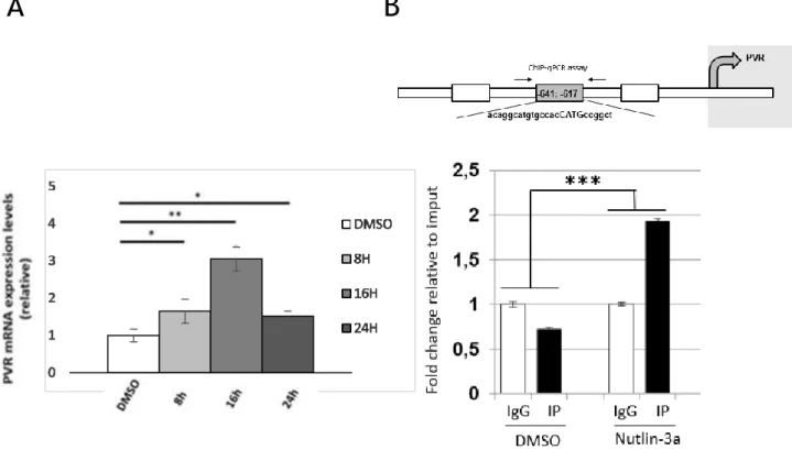
(pH 6-7.5). After treatment with proteinase K, the DNA was extracted with phenol-chloroform, precipitated with ethanol and re-suspended in 50 μl of distilled water. 5 ng of the immunoprecipitated, IgG and input samples were used for PCR with the specific oligonucleotides spanning PVR promoter: PVR P 53BS 3F AGGCTGGTCTTGAACTCCTG and PVR P 53BS 3R CCATTGCGCCACTACACTAC. The reaction was performed in triplicate using 5 ng of DNA, GoTaq qPCR Master Mix (Promega) and the relative qPCR primer pair in the termocycler “CFX Connect Real time PCR detection system” (Bio-Rad). The primer pair efficiency, the relative quantity of each immunoprecipitated and IgG (ΔC(t)) respect to Input sample and the Standard Error of the mean of the relative quantity were determined with CFX ManagerTM
software (Bio-Rad). The percentage of the relative quantity of each immunoprecipitated sample was normalized respect to IgG and expressed as percentage of input chromatin (% input).
Quantitative mRNA expression
Total RNA was extracted using TRIzol Reagent (Thermo Fisher Scientific). First-strand cDNA was synthesized using the SuperScript II First Strand cDNA synthesis kit (Thermo Fisher Scientific). Quantitative real-time PCR (qPCR) reactions were performed using pre-validated TaqMan gene expression assays from Applied Biosystems, Thermo Fisher Scientific (Hs00792195_m1 for MICA, Hs00360941_m1 for ULBP1, Hs00607609_m1 for ULBP2, Hs00225909_m1 for ULBP3, Hs00197846_m1 for PVR, Hs01071562_m1 for Nectin-2). Relative gene expression was determined using the 2-ΔΔCt method and 2-ΔCt considered as
expression level, with GAPDH (Hs02758991_g1) as endogenous control.
NK cell isolation
Human NK cells were isolated from peripheral blood mononuclear cells (PBMCs) of healthy donors with the RosetteSep NK-cell enrichment mixture method (StemCell Technologies) and Ficoll-Paque Plus (Lympholyte Cedarlane) centrifugation. NK cells were routinely checked for the CD3-CD56+ immunophenotype by flow cytometry and those with purity greater than 90%
were cultured with 600 IU/mL of recombinant human IL-2 (PeproTech) at 37°C and used up to 5 d after isolation for in vitro functional assays. NK cells isolated for in mice injections were expanded in NK MACS medium (Miltenyi Biotec).
Cytotoxicity and degranulation assay
NK cell cytotoxic activity was tested by a standard 4-h 51Cr release assay. Degranulation assay
was performed by co-culturing NK cells with target cells at 1:1 ratio for 3 h, in complete medium in presence of anti-CD107a and in the last 2 h of GolgiStop (BD Bioscence). Then, cells were stained with anti-CD56 and anti-CD45 and expression of CD107a was evaluated by flow cytometry in the CD56+CD45+ subset. Specific lysis was converted to lytic units (L.U.) calculated
from the curve of the percentage lysis. One lytic unit is defined as the number of NK cells required to produce 20% lysis of 106 target cells during the 4 h of incubation.
Immunohistochemistry assay
LA-N-5 bearing NSG mice were treated intraperitoneally with DMSO or Nutlin-3a at 20mg/kg (drug dose/mouse body weight) and at 40mg/kg every two days for two weeks. Tumour masses were harvested, formaldehyde-fixed, thus paraffin-embedded blocks were cut into 3-mm sections and baked for 60 min at 56°C in a dehydration oven. Tissues slides from DMSO, Nutlin-3a at 20mg/kg and at 40mg/kg masses were mounted on the same slide and parallel stained for the same antibody. Antigen retrieval and deparaffinization were performed on a PT-Link (Agilent Technologies) using the EnVision FLEX Target Retrieval Solution kits at high pH (Agilent Technologies) for PVR and Nectin-2, as per manufacturer’s instruction. Slides were then blocked for endogenous peroxidase for 10 min with a peroxidase blocking solution (Agilent Technologies), rinsed in the appropriate wash buffer (Agilent Technologies), and incubated for 30 min with 5% PBS/BSA. Slides were then incubated overnight at 4°C with primary antibodies PVR (1:50) and Nectin-2 (1:300). Twenty minutes incubation with secondary antibody coupled with peroxidase (Agilent Technologies) has been subsequently performed. Bound peroxidase was detected with diaminobenzidine (DAB) solution and EnVision FLEX Substrate buffer containing peroxide (Agilent Technologies). Tissue sections were counterstained with EnVision FLEX Haematoxylin (Agilent Technologies). Formaldehyde-fixed paraffin-embedded blocks from NB patients were prepared as described above and the expression of MICA (1:200) and Nectin-2 (1:300) was evaluated. Sections of normal intestinal mucosa, colon carcinoma and kidney were used as positive controls for MICA, Nectin-2 and PVR respectively. Isotype- matched mouse mAbs were used as negative controls.
- 32 -
Xenograft neuroblastoma model and treatment of NSG mice
All in vivo experiments utilized 4- to 6-week-old female NSG (NOD.Cg-Prkdcscid Il2rgtm1Wjl/SzJ) mice (Charles River Laboratories Italia srl), Briefly, 5 x 10⁶ cells of LA-N-5 NB cells resuspended in 100-mL PBS were injected subcutaneously (sc) into flank of the mice [155]. Mice were randomly divided into 4 groups (5 mice for each group), and treated with 1) control solvent, 2) 3a, 3) IL-2 4) NK cells plus IL-2 or 5) NK cells plus IL-2 plus Nutlin-3a. 40mg/kg of Nutlin-3a was intraperitoneally (i.p.) injected every two days for three cycles. NK cells were peritumorally injected 48 hours after the first Nutlin-3a injection, every 5 days, for two cycles. IL-2 (10x103 U/mouse) was injected i.p. every day, starting from the first NK cell injection and for the entire NK cell treatment. Tumour size was assessed every day by caliper measurement, until 45 days after LA-N-5 injection. All animal experiments were performed in accordance with a protocol approved by the Italian Ministry of Health and Usage Committee of Children's Hospital Bambino Gesù.
Statistical analysis
Digital images of western blotting were analysed by Image J (http:// rsbweb.nih.gov/ij/index.html) and statistical significance of densitometric values was evaluated by the two-tailed unpaired Student’s t-test. Normalized values were analysed for correlation by the regression analysis using GraphPad software. p values lower than 0.05 were considered to be statistically significant.
Results
Expression of MYCN inversely correlates with that of ligands for NK cell-activating receptors in NB cell lines
NK-cell cytotoxicity is triggered by the interaction of NK-cell activating receptors with their specific ligands. In particular, NKG2D receptor recognizes MICA, MICB, ULBP1, ULBP2/5/ 6 and ULBP3 ligands [156], while DNAM1 receptor interacts with PVR and Nectin-2 ligands [125]. We asked whether the expression levels of the MYCN oncogene correlated with a mechanism of immune escape involving down-regulation of NK-cell-activating receptor ligands on NB cells. To investigate this possible correlation, we assessed the protein levels of MYCN by western blotting (Fig.12) and the surface expression of activating ligands by flow cytometry in a panel of 12 NB cell lines (Fig.11), including 6 MYCN non-amplified (non-MNA) and 6 MYCN amplified (MNA).
Regression analysis of normalized densitometric values of western blotting and mean fluorescence intensity values of flow cytometry analysis revealed a significant inverse correlation between the expression of MYCN and that of the activating ligands MICA, MICB, ULBP1, ULBP2/5/6, ULBP3 and PVR. A trend toward an inverse correlation was also found between expression of MYCN and that of MHC class I and of Nectin-2. On the other hand, MYCN expression did not correlate with that of TRAIL-R2 (Fig.11).
- 34 - Fig.11 : Scatter plots showing the correlation between MYCN and the surface expression of activating ligands for NK- cell receptors in twelve NB cell lines. The means of four independent immunoblot analyses of MYCN expression in NB cell
lines are plotted against the means of fifteen independent flow cytometric analyses of surface expression of the indicated activating ligands; Spearman correlation r and p value are shown for each plot.* p<0.05, **p<0.01.
c-MYC and p53 are two transcription factors involved in the induction of ULBP ligands [120]. Of note, p53 gene is a direct transcriptional targets of MYCN in NB [157].
To assess whether p53 and c-MYC could be involved in the expression of activating ligands in NB, the status of MYCN, c-MYC, p53 and MDM2 was evaluated in NB cell lines in terms of amplification, gain, deletion and protein expression by high-resolution array CGH analysis and western blotting (Fig.12), respectively. p53 gene was lost in four NB cell lines tested [SK-N-AS, ACN, SK-N-BE(2)c and LA-N-1 [158]], three of them [SK-N-AS, ACN and SK-N-BE(2)c] also lost MDM2 gene. c-MYC gene gain was detected in two NB cells lines (SK-N-AS and ACN), whereas MYCN gene amplification or gain were found in five [SK-N-BE(2)c, LA-N-1, LA-N-5, SMS-KCNR, IMR-32] and two (GICAN, SK-N-SH) NB cell lines, respectively (Table 2).
All these genetic aberrations were confirmed at the protein level, except for SK-N-BE(2)c, which expressed high levels of mutant p53, due to a previously reported missense mutation. Consistently with previously published studies, MYCN expression was inversely correlated with c-MYC expression. In contrast, MYCN expression directly correlated with both p53 and MDM2 expression, as evaluated in p53 wt NB cell lines (SH-EP, GICAN, SH-SY-5Y, SK-N-SH,
LA-Tab 2. Arr
ay CGH analy
sis about the
status of MYCN, c -MYC, p53 and MDM2
- 36 - N-5, SMS-KCNR, IMR-32 and Tet-21/N). This indicates that p53 is more functional in non- MNA NB cells, even if expressed at low levels, than in MNA NB cells in which it is inhibited by MDM2.
Fig.12 MYCN is inversely correlated with c-MYC and directly correlated with p53 and MDM2 in NB cell lines Immunoblot
analysis of MYCN, c-MYC, p53 and MDM2 in the NB cell lines indicated. An anti-Actin Ab was used for normalization. One representative experiment out of the five performed is shown. Scatter plots showing the correlation between MYCN and c-MYC (B), p53 (C) or MDM2 (D) in twelve NB cell lines. The average of densitometric values of 5 independent experiments for each marker is plotted; Spearman correlation r and p values value are show for each plot.
The modulation of MYCN expression affects that of ligands for NK cell-activating- receptors and the susceptibility of Neuroblastoma cells to NK cell-mediated lysis
To investigate whether modulation of MYCN expression could affect that of activating ligands, we used the conditionally MYCN-expressing Tet-21/N cell line that loses MYCN expression following treatment with doxycycline (Doxy). Doxycycline treatment caused a drastic down- regulation of MYCN expression, early induction of c-MYC expression (at 8 h) and delayed reduction of p53 and MDM2 expression (at 24 h) (Fig.13). Following 16 h treatment with doxycycline, the expression of MICA, ULBP2/5/6, ULBP3 and PVR was significantly higher in
Doxy-treated Tet-21/N than in untreated Tet-21/N cells. Up-regulation of NK cell-activating- receptor ligands was detected up to 24 h after doxycycline treatment, with a peak at 16 h (Fig.14).
Fig 13. Modulation of MYCN affects the expression of ligands for NK-cell activating receptors. Representative exemple of
immunoblot analysis of MYCN, c-Myc, p53 and MDM2 in Tet-21/N either left untreated (0) or treated with doxycycline for the indicated time. An anti-actin Ab was used for normalization. Densitometry analysis of Actin-normalized proteins values of three independent experiments are shown. Mean ± SD; * p<0.05, **p<0.01.
Fig.14 Representative
flow cytometric analysis of surface expression of activating ligands for NK- cell receptors in Tet-21/N either left untreated (medium, gray line) or treated with doxycycline for 16 h (Doxy, red line); dotted lines, isotype- matched negative controls (left panel). Summary of five independentflow cytometric analyses (right panel). p values, compared with untreated and Doxy- treated Tet-21/N cells (two- tailed paired Student's t- test); *p < 0.05, **p < 0.01.
- 38 - In Tet-21/N cell line a state of cell senescence was detected 16 h after doxycycline treatment, as evaluated by senescence associated-β-galactosidase (SA-β-Gal) staining (Fig.15). Three days after doxycycline treatment, the occurrence of cell differentiation, revealed by changes of cell morphology, became evident (Fig.15). This finding is consistent with results reported by other authors [49] on the expression of activating ligands in senescent cells, but not in mature and differentiated cells.
Fig.15 SA-β-Gal staining and morphology of Tet-21/N cells left untreated or treated with doxycycline for 3 or 10 days, bar
50 m. The percentage of SA-β-Gal positive cells, evaluated from 5 different fields for each condition, by counting at least 200 cells per field, is reported, mean ± sd (right panel), (two-tailed paired Student t test) *p<0.05, ***p<0.001.
To further confirm that MYCN expression affects that of ligands for NK cell-activating- receptors, we transiently transfected a non-MNA NB cell line as SK-N-SH with piRV-neoSV vector bearing MYCN cDNA (SK-N-SH-MYCN) or the empty vector (SK-N-SH-ctrl) as control. The overexpression of MYCN, peaking at 16 h and gradually decreasing until 72 h, induced downregulation of c-MYC (from 16 to 72 h) and upregulation of both p53 (from 16 to 48 h) and MDM2 (from 16 to 24 h), as revealed by western blotting (Fig.16). SK-N-SH-MYCN cells showed decreased surface expression of activating ligands as compared with SK-N-SH-ctrl cells, significantly evident 48 h after transfection.
Fig.16 Representative example of immunoblot analysis of SK-N-SH either left transfected with empty vector (ctrl) or with
piRVneoSV-MYCN (MYCN) for the indicated time (left panel). Densitometry analysis of Actin-normalized proteins values of three independent experiments are shown (right panel).
SK-N-SH-MYCN cells showed decreased surface expression of activating ligands as compared with SK-N-SH-ctrl cells, significantly evident 48 h after transfection (Fig.17).
Fig.17. Representative flow cytometric analysis of surface expression of activating ligands for NK- cell receptors in SK-N-SH transfected for 48 h with control vector (ctrl, gray line) or with MYCN cDNA vector (MYCN, blue line); dotted lines, isotype-matched negative controls (left panel).
Summary of five independent flow cytometric analyses (right panel). p values, compared with ctrl and MYCN cDNA vector transfected cells (two-tailed paired Student's t-test); *p < 0.05,**p < 0.01.
40 - - Later, to test whether the increased expression of NK cell-activating receptor ligands induced by
MYCN modulation could affect NK cell-mediated recognition of NB cells, we performed degranulation and cytotoxicity assays using untreated and Doxy-treated Tet-21/N cells, as well as SK-N-SH-ctrl and SK-N-SH-MYCN cell lines as targets. After 16 h treatment with doxycycline, Tet-21/N cells were significantly more susceptible to NK cell-mediated lysis than untreated cells in both degranulation and cytotoxicity assays. Conversely, SK-N-SH-MYCN cell lines were less susceptible to NK cell recognition and lysis, compared with control cells, as evaluated in both degranulation and cytotoxicity assays (Fig. 18).