UNIVERSITA’ DI PISA
Doctorate Course in
Fisiopatologia Medica e Farmacologia
Ph.D Thesis
ANTI-VIRAL CITRULLINATED PEPTIDES ANTIBODIES
IN RHEUMATOID ARTHRITIS
F
EDERICO
P
RATESI
Tutor
CONTENTS
CHAPTER 1
G
ENERAL INTRODUCTION 6EPIDEMIOLOGY OF RHEUMATOID ARTHRITIS 7
RISK FACTORS 9
Genetic factors 9
Infectious agents 10
Age and gender 10
Smoking 10 Socio-economic factors 11 Hormonal factors 11 Dietary factors 11 Ethnicity 12 HISTORY OF RA 12 PATHOLOGY 14 DIAGNOSIS OF RA 15 REFERENCES 17
CHAPTER 2
A
NTI CITRULLINATED PEPTIDES/
PROTEINS ANTIBODIES INRA
AUTOANTIBODIES IN RHEUMATOID ARTHRITIS 21
Rheumatoid factors 21
Type II collagen and other components of articular cartilage 22
Anti-RA33 or anti-hnRNP A2 22
Antibodies to calpastatin 23
Antibodies to stress proteins 23
Anti-Sa antibodies 24
ANTI CITRULLINATED PEPTIDES/PROTEINS ANTIBODIES
HISTORY OF ACPA 25
CITRULLINATION 29
PEPTIDYLARGININE DEIMINASE (PAD) 30
REFERENCES 33
CHAPTER 3
A
NTI VIRAL CITRULLINATED PEPTIDES ANTIBODIES INRA
INTRODUCTION 44
PATIENTS AND METHODS 46
Patients 46
Peptide synthesis 46
Binding of anti-citrullinated peptides/proteins antibodies 46 Purification of anti-deiminated viral peptide antibodies 47
Competition assay 47
ELISA for anti viral deiminated peptide IgG, IgM, IgA antibody detection 47
Statistical analysis 48
RESULTS 49
DISCUSSION 60
REFERENCES 63
CHAPTER 4
G
ENETIC CONTROL OFACPA
PRODUCTIONIN
R
HEUMATOIDA
RTHRITIS 66INTRODUCTION 66
HLA GENETICS OF RHEUMATOID ARTHRITIS 67
ROLE FOR NON-HLA GENES IN THE SUSCEPTIBILITY TO RA 72
TNFR2 at 1p36 72
PTPN22 at 1p13 72
PADI4 at 1p36 73
HLA AND ACPA 74
GENE-ENVIRONMENT INTERACTION IN RA. THE ROLE OF SMOKING,
HLA-SE AND ACPA
75
PATIENTS AND METHODS
Patients 77
HLA-DRB1 genotyping 77
HLA-DRB1 allele classification 78
Binding of anti-viral citrullinated peptides/proteins antibodies 78
Statistical analysis 79
RESULTS
Demographic and immunologic characteristics of RA patients 80
Allele frequency for HLA-DRB1 polymorphisms 80
Relationships between HLA-DRB1 status and anti-viral citrullinated peptides antibodies
80
Binding of citrullinated viral peptides to HLA-SE positive molecules 83
DISCUSSION 84
REFERENCES 87
CHAPTER 5
E
PSTEIN-B
ARRV
IRUS ANDR
HEUMATOIDA
RTHRITIS INTRODUCTIONEpstein Barr Virus (EBV) 93
Epstein Barr Virus and rheumatoid arthritis 94
PATIENTS AND METHODS
Purification of anti-VCP antibodies 97
Peripheral blood cells isolation 97
Viral preparation 97
EBV infection 98
Preparation of cell lysates and immunoblotting 98
RESULTS 100 DISCUSSION 103 REFERENCES 106
CHAPTER 6
CONCLUSIONS
111CHAPTER 7
SUMMARY
117RIASSUNTO
118CHAPTER 8
A
CKNOWLEDGMENTS 122CHAPTER 9
CHAPTER 1
GENERAL INTRODUCTION
Rheumatoid arthritis is a chronic systemic inflammatory autoimmune disease primarily characterized by a bilateral symmetrical polyarticular arthritis, which is often erosive. It is probably the most common autoimmune disease (about 1% of the world population), affects three times as many women as men, and usually appears in middle age [1].
Although the etiology of RA remains unknown, it is widely accepted that multiple accumulative/compounding genetic and environmental ‘hits’ are required between the initiation of self-peptide recognition, subsequent loss of tolerance, and the development of autoimmunity [2]. Disease onset is often insidious, initially affecting the small joints of the hands (proximal interphalangeal joints), feet, and wrists.
Clinical course of the disease varies between individuals but may be mild, relapsing– remitting, or progressive.
The systemic complications and the severity of articular manifestations (with associated functional impairment) may have a debilitating and disabling impact on the patient, causing significant morbidity, reduced quality of life, and even premature mortality. It is therefore important that the disease is diagnosed and treated early to slow/stop joint damage, increase/maintain joint function, establish remission, and maximize quality of life. It has also been widely reported that, although the early stages of the disease are responsive to disease-modifying anti-rheumatic drugs (DMARDS), once the clinical disease is established, this effect is lost [3].
RA is currently diagnosed using the American College of Rheumatology (ACR) 1987 revised criteria [4]that are primarily based on clinical parameters, the only serological criteria being IgM RF. However, the criteria may be insufficient for the diagnosis of early RA, as they are based upon measurements for disease classification predominately featuring manifestations typical of later-stage disease.
Diagnosis may be complicated by the insidious (and sometimes nonspecific) onset of the disease, resulting in delayed diagnosis, often to the detriment of the joint.
Numerous serological markers of RA have been described [5] over the past half century, and namely rheumatoid factor (RF) and antibodies against a range of citrullinated proteins
including anti-perinuclear factor, anti-keratin antibody, anti-filaggrin antibody (AFA), and anti-Sa (vimentin) antibody. However, many others have been discarded in the routine clinical setting due to their lack of sensitivity, specificity, and/or laborious detection methods. An ideal marker would be sensitive, specific, detectable in early disease and prognostic, enabling clarification of disease subsets and the early identification of patients likely to develop severe disease and therefore requiring aggressive early treatment to prevent significant joint damage. Equally, patients with transient/mild arthritis would be spared unnecessary immunosuppressive therapy and its associated side effects, also representing a cost saving. The candidate marker should also be quantitative, to allow monitoring of disease (as anti-dsDNA antibodies in systemic lupus erythematosus, SLE) and measurement of response to treatment.
EPIDEMIOLOGY OF RA
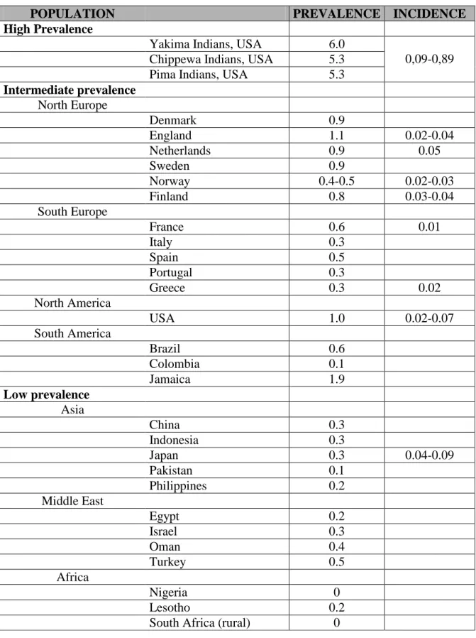
Several prevalence and incidence studies of RA have been reported during the last decades [rev. in 6], suggesting a considerable variation of the disease occurrence among different populations. The majority of prevalence studies carried out in Northern European and North American areas estimate a prevalence of 0.5–1.1%. Studies from Southern European countries report a prevalence of 0.3–0.7%. Studies from developing countries also report a relatively lower prevalence of the disease (between 0.1% and 0.5%). A higher prevalence has been reported in certain Native Americans, and a very low frequency of RA in some areas of rural Africa [7].
Table 1 presents prevalence data for RA in different population groups. These populations have been divided into those with high, intermediate and low prevalence rates of the disease which closely agree with the geographic area in which they are located.
The annual incidence rates of RA vary between 20 and 50 cases per 100,000 inhabitants in North American and North European countries. There are only few studies from Southern European countries indicating a relatively lower occurrence of the disease. There are no studies on RA incidence from developing countries [8-13].
Mortality rates are higher among RA patients than in the general population. The mortality rates reported vary widely among studies. They are higher in hospital-based studies and relatively lower (but still higher than in the general population) in population-based studies. The expected survival of RA patients is likely to decrease 3–10 years according to the severity
between RA patients and the general population they come from. It can be said that the majority of affected individuals die from the same causes as the general population, but at a younger age [6].
POPULATION PREVALENCE INCIDENCE
High Prevalence
Yakima Indians, USA 6.0
Chippewa Indians, USA 5.3
Pima Indians, USA 5.3
0,09-0,89 Intermediate prevalence North Europe Denmark 0.9 England 1.1 0.02-0.04 Netherlands 0.9 0.05 Sweden 0.9 Norway 0.4-0.5 0.02-0.03 Finland 0.8 0.03-0.04 South Europe France 0.6 0.01 Italy 0.3 Spain 0.5 Portugal 0.3 Greece 0.3 0.02 North America USA 1.0 0.02-0.07 South America Brazil 0.6 Colombia 0.1 Jamaica 1.9 Low prevalence Asia China 0.3 Indonesia 0.3 Japan 0.3 0.04-0.09 Pakistan 0.1 Philippines 0.2 Middle East Egypt 0.2 Israel 0.3 Oman 0.4 Turkey 0.5 Africa Nigeria 0 Lesotho 0.2
South Africa (rural) 0
RISK FACTORS
There is a general consensus that RA is a multifactorial disease, resulting from the interaction of both genetic and environmental factors, which contribute to its occurrence and expression [1-3].
Several environmental factors have been suspected and studied as possibly related to an increased risk of RA, as well as to a worse or improved prognosis of the disease. However, the impact of most of these factors on the risk of developing RA and the expression of the disease remains uncertain. On the other hand, there is epidemiologic evidence that genetic factors are related to an increased risk of RA. The nature and the impact of this genetic risk is becoming clearer during the last years. We describe here the most important personal, lifestyle, environmental, and genetic factors that have been proposed and studied as influencing the occurrence of the disease. Some of these factors have also been studied as possibly associated with the course and the severity of RA.
Genetic factors
There is evidence that the occurrence and the severity of RA are determined by genetic factors. Twin and family studies strongly suggested that the risk of the disease among relatives of affected individuals is influenced by shared genetic factors [rev. in 14, 15, 16] Studies carried out in Caucasian patients with established and advanced disease indicated an association of RA with HLA alleles encoding a “shared epitope” (called “rheumatoid epitope”). These studies also suggested a significant association of “shared epitope” with disease severity, autoantibody production and outcome. The role of other genes has also been studied and several associations have been described. Some studies indicate an association between certain tumor necrosis factor alpha alleles and RA occurrence and severity, but others failed to confirm those associations. A number of DR specificities have been reported to be protective factors of RA. Interactions between genes in the etiology of RA have also been proposed.
At the moment, it is not clear whether genetic factors are related to the risk of RA or to the severity of the disease or both of them. Although the available data do not clarify completely the role of genetic factors in disease susceptibility and severity, their impact on the epidemiology of RA is generally accepted. The significant variations observed in the incidence and prevalence of RA among different populations or ethnic groups could partly
been explained by genetic variation in the HLA region, and variation in the prevalence of “shared epitope” in different populations.
Infectious agents
A potential involvement of infectious agents in the occurrence of RA has been suggested and studied for decades [rev in 17]. It is possible that those agents could trigger the development of the disease in a genetically susceptible host. Several infectious agents have been implicated in the etiology of the disease, but there is not epidemiological evidence that those agents could explain a considerable fraction of RA cases.
Several potential associations of RA with infectious agents have been suggested [17]. They include parvovirus, rubella virus, Epstein–Barr virus, borrelia burgdorferi, and others. Increased titers of antibodies among RA patients and clinical syndromes similar to RA induced by some of these agents have been reported. However, the role of infectious agents in the occurrence of the disease remains unclear. Incident cases of RA do not report a history of increased number of infections, or any specific infection. Furthermore, cases of RA do not present any time or space clustering, as would be expected if there was a direct association of RA development with infections.
Age and gender
The incidence of RA is higher in women than in men [18, 19]. The sex ratio varies in most studies from about 2:1 to about 3:1. This difference suggests an influence of reproductive and hormonal factors in the occurrence of the disease. However, the findings of several studies give a conflicting picture on this issue. For the moment, it is not clear how gender influences the occurrence of RA [18, 19].
The age of disease onset presents a peak in the fifth decade of life according to the majority of epidemiological studies. Some more recent studies suggest a later onset of the disease [20, 21].
Smoking
Smoking is likely to influence the risk of developing RA, the production of RA specific autoantibodies and the course of the disease [22]. The increased risk of RA associated with smoking has been suggested in cross-sectional as well as in longitudinal studies. The association appears to be dose-dependent, and is most clear for heavy smokers. The severity and the outcome of RA appears also to be influenced by smoking, although it is not clear
which clinical characteristics of the disease are related to smoking. An increased risk for seropositive disease is also related to smoking habits [23]
.
Socioeconomic factors
Socioeconomic factors appear to influence the course and the outcome of RA rather than the risk of developing RA. Occupation, educational level, marital status, and social group have been studied as possible risk factors for disease susceptibility, or predictors for disease severity and outcome. The results of these studies are conflicting mainly as far as the impact of socioeconomic factors on the risk of developing RA is concerned. However, the available data suggest an association of adverse socioeconomic status with worse prognosis of the disease [24, 25].
Hormonal factors
The higher occurrence of RA in females than in males suggests a possible role of hormonal factors in disease susceptibility [25, 26]. Furthermore, estrogens are known to have a stimulatory effect on the immune system. Epidemiological studies tried to investigate this association by studying the possible protective role of pregnancy, the use of oral contraceptives and the hormone replacement therapy after menopause.
Parity has been associated with reduced risk of developing RA. However, it is difficult to clarify whether this association reflects any protective effect of pregnancy or an increased risk for infertility before the development of RA. There is also evidence that pregnancy is associated with remission of the disease in RA patients [27]. This effect reverts in the post-partum period.
The potential protective effect of the use of oral contraceptives has been studied also. It seems that oral contraceptives have a modulatory effect, protecting mainly against severe disease rather than protecting against RA occurrence. There are only few studies on the effect of hormone replacement therapy, presenting a conflicting picture.
Dietary factors
Several epidemiological studies suggest a potential protective effect of lifelong consumption of fish, olive oil, and cooked vegetables. The protective role of fish consumption has been attributed to the effect of omega-3 long chain polyunsaturated fatty acids against poly-inflammatory disease. Mediterranean diet as a whole has also been reported as a lifestyle
factor reducing the risk of developing RA, and protecting against severe course of the disease. These observations could partly explain the geographical variations of the disease occurrence, and severity.
Ethnicity
Differences in the occurrence and the clinical expression of RA among different populations have been reported. The significant geographic variations of the disease occurrence, and the increased incidence observed in some specific ethnic groups, suggest an association of RA with ethnicity. The differences observed may reside in the different distribution of environmental and genetic factors, as well as in their interaction. Lifestyle factors possibly associated with the risk and the severity of RA (such as dietary factors) may differ significantly among ethnic groups. The prevalence of rheumatoid epitope is likely to present important variations in different racial groups. In addition, the association of rheumatoid epitope with the development of erosive disease seems to present significant differences among ethnic groups. These observations indicate that ethnicity could be considered as an independent risk factor, reflecting interactions between several genetic and environmental factors.
HISTORY OF RA
Did RA exist in the ancient world?
No medical writings has been discovered with descriptions of rheumatoid arthritis until the seventeenth century, when Thomas Sydenham (1624– 1689) gave a fairly reasonable account, publishing the first case report in 1676.
However, if the first description of RA dates from the seventheenth century, paleontological findings suggest that RA may be an ancient disease.
This hypothesis is supported by the discovery of skeletal remains of American Indians that lived 3000-5000 years ago in Alabama. In some of these skeletons symmetrically distributed joint erosions, characteristic of RA, could be observed [27].
In the tenth century the Bizantine historian and philosopher Michael Psellus, in his Chronographia, described the emperor Constantine IX joints, suggesting he suffered from severe RA : “I myself saw his fingers, once so beautiful formed, completely altered from their
natural shape, warped and twisted with hollows here and projection there, so that they were incapable of grasping anything at all. His feet were bent and his knees, crooked like the point of a man’s elbow, were swollen, making it impossible for him to walk steadily or to stand upright for any length of time [28]
However, Psellus also records that the Emperor was ‘naturally inclined to sexual indulgence but could find no satisfaction in cheap harlotry’, This might suggest that the Emperor suffered from a reactive arthritis (Reiter’ s disease) which can lead to severe joint destruction.
An argument in favour of the existence of RA in the Old world comes from paintings by Dutch and Flemish painters. The most convincing was the joint changes in the fingers of the serving maid in the painting, The Printer’s Family, by Jacob Jordaens (1593– 1678). Another painting suggesting the existence of RA is “The Three Graces” (1638), by Paul Rubens (1577-1640), showing a right hand clumsily represented for some but deformed by rheumatoid arthritis for others. Moreover, in the painting “The Miracle of Saint Ignatius Loyola”, 1618 swelling of the metacarpophalangeal and interphalangeal joints can be observed.
Presently, the most prevalent hypothesis is that rheumatoid arthritis originated in the New World, and spread to the Old World after Columbus discovered America in 1492.
Thus, it is likely that rheumatoid arthritis had been present as adisease for a long time, maybe since the time of Sydenham (1624-1689),but the nomenclature had been confused with terms like rheumaticgout, chronic rheumatism, rheumalgia, scorbutic rheumatism,etc.
Actually, the first precise description of RA date from the end of the eighteenth century. In 1782, Jon Petursson described a chronic, symmetric, destructive, inflammatory polyarthritis different from osteoarthritis and sometimes accompanied by systemic manifestations [29].
The first surely unambiguous report of rheumatoid arthritis is considered the description that the young Parisian doctor named Augustine Jacob Landrè-Beauvais made in 1800 writing his MD thesis [30].
After reviewing the main features of ordinary and regular gout, the scientist pointed out that the disease he named “asthenic gout” exhibited several distinctive features, including predominance in women, a chronic course, involvement of main joints at the onset, and decline in general health. He was convinced that he has being describing an yet unreported entity.
Nevertheless, the name “rheumatoid arthritis” was only given in 1859 by the English Physician Alfred Baring Garrod, considered as “the father of rheumatology”, who discussed
the differential diagnosis of rheumatoid arthritis with respect to different other conditions like rheumatic gout, chronic rheumatism, rheumalgia, scorbutic rheumatism. He rejected previous definitions like “chronic rheumatism” and “rheumatic gout” and chose the name “rheumatoid arthritis”. Moreover he divided the disease in acute, chronic and irregular forms of generalized and localized type [31].
The name has remained ever since.
PATHOLOGY
The histologic changes in RA are not disease-specific but largely depend on the organ involved. The primary inflammatory joint lesion involves the synovium [1, 2, 3]. In contrast to hematogenous infective arthritis, synovial changes are a primary event an do not follow changes in the adjacent bon marrow. The earliest changes are injury to the synovial microvasculature with occlusion of the lumen, swelling of endothelial cells and gaps between endothelial cells. This stage is usually associated with mild proliferation of the superficial lining cell layer. Two cell types constitute the synovial lining: a bone marrow derived type A synoviocytes, which has macrophagic features and the mesenchimal type B synoviocytes. Both cell types contribute to the synovial hyperplasia, suggesting a paracrine interaction between these two cell types. This stage of inflammation is associated with congestion, edema and fibrin exudation. Cellular infiltration occurs in early disease and initially is mainly represented by T lymphocytes. The nodular aggregates consist mainly of CD4+ T cells, while CD8+ T cells dominate in the diffuse infiltrate. Close cell-to-cell contact with dendritic cells and macrophages has been described. In later stages true lymphoid follicles with germinal centers may appear, although rheumatoid synovitis presents with a diffuse lymphocytic infiltrate without follicles or pseudofollicles in many patients. Plasma cells are usually found in more advanced stages of inflammation.
As a consequence of inflammation the synovium becomes hypertrophic from the proliferation of blood vessels and synovial fibroblasts and from multiplication and enlargement of the synovial lining layers.
Granulation tissue extends to the cartilage and is known as pannus. The tissue actively invades and destroys the periarticular bone and cartilage at the margin between synovium and bone.
Clinically evident extra-articular manifestations occur in approximately 20% of RA patients, although sub-clinical involvement is likely to be more frequent. Rheumatoid nodules have the histologic appears of a foreign body granuloma with three distinct layers. Multicentric fibrinoid necrosis is sourrounded by palisading elongated cells arranged radially and granulation tissue with inflammatory cells. The composition of necrotic material varies. No conclusions about the necrosis-inducing events can be drawn from histologic examination, but since nodules often occur over pressure points, minor trauma may precipitate the necrosis. The elongated cells are modified macrophages that are aligned parallel to collagen fibers. Tenosynovitis is present in the majority of patients. Involvement of the tendons themselves is common and manifests as a non specific inflammatory infiltrate and less frequently as formation of characteristic nodules with central necrosis.
Vascular involvement is usually confined to small segments of terminal arteries and lacks distinctive histologic characteristics.
In general immune complex deposition may contribute to, but does not explain completely, the spectrum of extra-articular manifestations. A common theme appears to be the mononuclear cells infiltrate with varying degrees of fibrinoid necrosis and nodule formation.
DIAGNOSIS OF RA
Given the seriousness of the disease, an early and accurate diagnosis of RA is mandatory. The 1987 ACR (American College of Rheumatology) criteria for the classification of RA [4] are commonly used for its diagnosis and are listed in the following table.
For classification purposes, a patient is said to have RA if he or she has satisfied at least 4 of the following 7 criteria. Criteria 1 through 4 must have been present for at least 6 weeks. Patients with 2 clinical diagnoses are not excluded.
CRITERION DEFINITION
Morning stiffness Morning stiffness in and around the joints, lasting at least 1 hour before maximal improvement
Arthritis of 3 or more joint areas for at least 6 weeks
At least 3 joint areas simultaneously have had soft tissue swelling or fluid observed by a physician. The 14 possible areas are right or left proximal interphalangeal (PIP),
metacarpophalangeal (MCP), wrist, elbow, knee, ankle, and metatarsophalangela MTP joints
Arthritis of hand joints for at least 6 weeks At least 1 are swollen in a wrist, MCP or PIP joint
Symmetric arthritis Simultaneous involvement of the same joint areas (as defined in criterion 2) on both sides of the body
Rheumatoid nodules Subcutaneous nodules, over bony
prominences, or exterior surfaces, or in
juxtaarticular regions, observed by a physician Serum rheumatoid factor Demonstration of abnormal amounts of serum
rheumatoid factor by any method for which the results has been positive in less than 5% of normal control subjects
Radiographic changes Radiographic changes typical of RA on postero-anterior hand and wrist radiograph, which must include erosion unequivocal bony decalcification localized in or most marked adjacent to the involved joints
REFERENCES
1. Rheumathoid arthritis. R.N. Maini, N. J. Zvaifler, in “Rheumatology”, J.H. Clipper, P.A. Dieppe Eds, Mobsy, London 1994
2. Todesco S, Gambari PF. Malattie reumatiche, 3th edition. 2002; pag. 32-37,141-143. 3. Cotran RS, Kumar V, Robbins SL. The pathologic basis of disease, 5th edition.1997;
1401:1404.
4. Arnett FC, Edworthy SM, Bloch DA, McShane DJ, Fries JF, Cooper NS, Healey LA, Kaplan SR, Liang MH, Luthra HS, et al. The American Rheumatism Association 1987 revised criteria for the classification of rheumatoid arthritis. Arthritis Rheum. 1988;31:315-24
5. Bridges SL. Update on autoantibodies in RA. Curr Rheumatol Rep. 2004;6:343-50 6. Alamanos Y and Drosos A. Epidemiology of adult rheumatoid arthritis. Autoimm Rev.
2005; 4: 130-36.
7. Silman AJ, Ollier W, Holligan S, Birrell F, Adebajo A, Asuzu MC, et al. Absence of rheumatoid arthritis in a rural Nigerian population. J Rheumatol 1993;20:618–22.
8. Riise T, Jacobsen BK, Gran JT. Incidence and prevalence of rheumatoid arthritis in the county of Troms, northern Norway. J Rheumatol 2001;27:1386–9.
9. Gabriel SE, Crowson CS, O’Fallon WM. The epidemiology of rheumatoid arthritis in Rochester, Minnesota, 1955–1985. Arthritis Rheum 1999;42:415–20.
10. Simonsson M, Bergman S, Jacobsson LT, Petersson IF, Svensson B. The prevalence of rheumatoid arthritis in Sweden. Scand J Rheumatol 1999;28:340–3.
11. Symmons D, Turner G, Webb R, Asten P, Barrett E, Lunt M et al. The prevalence of rheumatoid arthritis in the United Kingdom: new estimates for a new century. Rheumatology 2002;41:793–800.
12. Power D, Codd M, Ivers L, Sant S, Barry M. Prevalence of rheumatoid arthritis in Dublin, Ireland: a population based survey. Ir J Med Sci 1999;168:197– 200.
13. Aho K, Kaipiainen-Seppanen O, Heliovaara M, Klaukka T. Epidemiology of rheumatoid arthritis in Finland. Semin Arthritis Rheum 1998;27:325–34
14. Dieude P and Cornelis F. Genetic Basis of rheumatoid arthritis. Joint Bone Spine. 2005; 72: 520-26.
15. Orozco G, Rueda B, Martin J. Genetic basis of rheumatoid arthritis. Biomed & Pharmacother. 2006; 60: 656-662.
16. Bowes J and Barton A. Recent advances in the genetics of RA susceptibility. Rheumatology. 2008; 47: 399-402.
17. Kobayashi S, Momohara S, Kamatani N, Okamoto H. Molecular aspects of rheumatoid arthritis: role of environmental factors. FEBS J. 2008 Sep;275(18):4456-62
18. Riise T, Jacobsen BK, Gran JT. Incidence and prevalence of rheumatoid arthritis in the county of Troms, northern Norway. J Rheumatol 2001;27:1386–9.
19. Gabriel SE, Crowson CS, O’Fallon WM. The epidemiology of rheumatoid arthritis in Rochester, Minnesota, 1955–1985. Arthritis Rheum 1999;42:415–20.
20. Doran MF, Pond GR, Crowson CS, O’Fallon WM, Gabriel SE. Trends in incidence and mortality in rheumatoid arthritis in Rochester, Minnesota, over a forty-year period. Arthritis Rheum 2002;46:625–31.
21. Pfeil A, Hansch A, Lehmann G, Eidner T, Schäfer ML, Oelzner P, Renz DM, Wolf G, Hein G, Kaiser WA, Böttcher J. Impact of sex, age, body mass index and handedness on finger joint space width in patients with prolonged rheumatoid arthritis using computer-aided joint space analysis. Rheumatol Int. 2009; 29: 517-24
22. Klareskog L, Padyukov L, Alfredsson L. Smoking as a trigger for inflammatory rheumatic diseases. Curr Opin Rheumatol. 2007 Jan;19(1):49-54
23. Klareskog L, Stolt P, Lundberg K, Källberg H, Bengtsson C, Grunewald J, Rönnelid J, Harris HE, Ulfgren AK, Rantapää-Dahlqvist S, Eklund A, Padyukov L, Alfredsson L. A new model for an etiology of rheumatoid arthritis: smoking may trigger HLA-DR (shared epitope)-restricted immune reactions to autoantigens modified by citrullination. Arthritis Rheum. 2006 Jan;54(1):38-46.
24. MacGregor AJ, Silman AJ. Rheumatoid arthritis and other synovial disorders: classification and epidemiology. In: Hochberg MC, Silman AJ, Smolen JS, Weinblatt ME, Weisman MH, editors. Rheumatology, 3rd ed. Mosby. p. 757– 63.
25. Silman AJ, Hochberg MC. Epidemiology of the rheumatic diseases. 2nd edition. Oxford University Press.
26. Buyon JP. The effects of pregnancy on autoimmune diseases. J Leukoc Biol 1998;63:281– 7.
27. Rothschild BM, Turner KR, De Luca MA. Symmetrical erosive peripheral polyarthritis in thelate Archaic period of Alabama. Science 1988; 241: 1498-1501
28. Psellus M. Chronographia. Book VI. From: www.fordham.edu/halsall/basis/psellus-chrono06.htlm
29. Jonsson H and Helgason J. Rheumatoid arthritis in an Icelandic textbook from 1782. Scand J Rheumatol 1996; 25:134-37
30. Landre-Beauvais AJ. The first description of rheumatoid arthritis. Unbridged text of the doctoral dissertation presented in 1800. Joint Bone Spine 2001; 68:130-143
CHAPTER 2
Anti-Citrullinated Peptide/proteins Antibodies in RA
AUTOANTIBODIES IN RHEUMATOID ARTHRITIS
A number of autoantibodies have now been described in RA and their clinical associations and role as markers of disease examined [1]. Of these only three (rheumatoid factors (RFs), antibodies to citrullinated antigens such as fillagrin and anti-CCP antibody [2], and antibodies to immunoglobulin binding protein (BiP) [3] have shown sufficient sensitivity and specificity to be considered clinically useful, and only two (RFs and anti-CCP antibody) are used in clinical practice.
Rheumatoid Factor
RFs, the first described human autoantibodies [4] are directed at the Fc region of IgG and are usually of IgM isotype. They are detectable in up to 10% of normal individuals, 70–80% of patients with RA and in other systemic diseases such as Sjogren's syndrome and many systemic infections [5]. RFs and the B cells that synthesise and secrete them are believed to have a physiological role as part of the immune system. However RFs from healthy individuals and patients with RA differ considerably. RFs in healthy individuals are synthesised by CD5 + B cells, exhibit low affinity for IgG, polyreactivity and a low ratio of replacement to silent mutations in their CDRs [6 and 7] suggesting the presence of a mechanism to suppress the affinity maturation of RFs. In rheumatoid synovium, B cells produce high affinity RFs with multiple replacement mutations in their CDRs, indicating a process dependent on T cell help affinity maturation. In addition synoviocytes themselves can support the survival and differentiation of B cells [8]. Hence in RA synovium RF-producing B cells could encounter a milieu that would support their survival and provide the T cell help necessary for the production of high affinity RFs [9]. There is evidence that they could contribute to disease by the formation of immune complexes [10] and complement fixation [11], and also by functioning as highly efficient antigen-presenting cells [12]. The correlation of RF status and progression of joint damage in RA [13] supports a role in pathogenesis
although they are by no means an absolute requirement for severe disease and joint destruction.
Type II collagen and other components of articular cartilage
The idea that immune responses directed against joint-related antigens may play a role in the pathogenesis of RA has been pursued for many years. The possible importance of immune responses to type II collagen (CII) was first suggested nearly 30 years ago. The specific expression of this molecule in articular cartilage as well as its ability to induce a destructive arthritis in experimental animals made it an attractive candidate autoantigen in human RA. Antibodies to native and denatured CII have been demonstrated in serum of patients with RA (10–30%) [14] as well as in synovial fluid, and collagen–anticollagen immune complexes described in synovial tissue [15]. A dominant epitope of anti-CII antibodies has been identified [16]. T cell responses have been found in approximately 50% of antibody positive patients [Snowden N, Reynolds I, Morgan K and Holt L. T cell responses to human type II collagen in patients with rheumatoid arthritis and healthy controls. Arthritis Rheum.1997; 40: 1210–1218]. Antibodies to CII are present early in the disease course, appear to decline with time and may be predictive of a more severe outcome [17]. However, T cell responses to CII are also found in 37% of normal individuals [18], many patients do not demonstrate any form of immune response to CII and no major associations with clinical parameters have been described. Attempts to induce tolerance in patients with RA by the oral administration of collagen have met limited success [19]. Hence the relevance of anti-CII T cell responses and autoantibodies remains unclear.
Anti-RA33 or anti-hnRNP A2
Antibodies to the 33 kDa A2 protein of the heterogeneous nuclear ribonucleoprotein complex (anti-RA33 or anti-hnRNP A2), a component of the spliceosome, were first demonstrated in approximately 35% of patients with RA by Western blotting [20]. They are not disease-specific, however, occurring in 20% of patients with SLE and 40–60% with MCTD, usually concomitantly with antibodies to U1-snRNP or Sm; these latter antibodies do not occur in RA [21]. The major autoantibody binding sites have been shown to be conformation-dependent and found to be different in MCTD patients from RA and SLE [22]. Interestingly anti-RA33 has been shown to occur in two experimental models of arthritis, in TNF-transgenic mice [23]
and in the arthritis prone MRL/lpr mice [24]. The presence of anti-RA33 in SLE appears to define a subset of patients with destructive arthritis emphasising the link with joint pathology [25]. Although shown to be present early in the disease, anti-RA33 has not been shown to have any predictive value for disease severity or progression.
Antibodies to calpastatin
Calpastatin is a natural inhibitor of the calpains, a family of intracellular calcium-dependent cysteine proteases unique because of their cytosolic rather than lysosomal location. Calpains are over-expressed in diseased synovial tissue [26] and secreted calpains can degrade components of articular cartilage [27]. Two groups independently described antibodies to calpastatin in approximately 50% of RA sera and although not specific for RA they occur at a higher frequency than in connective tissue diseases [28 and 29].
It has therefore been postulated that autoantibodies to calpastatin may potentiate joint destruction by interference with the calpain–calpastatin interaction [30]. Some support for this hypothesis comes from the in vitro observation that calpains are released from calpain– calpastatin complexes in the presence of RA sera [31]. Antibodies to calpastatin however are not associated with disease severity, and more recent studies have suggested they may be less frequent than previously believed [32]. The importance therefore of anti-calpastatin antibodies in RA remains unclear.
Antibodies to stress proteins
Heat shock proteins and BiP, a 78 kDa chaperone are members of a highly conserved family of proteins with critical roles in cellular homeostasis in both normal conditions as well as during cellular stress [33]. The close homology between bacterial and human HSPs suggested the possibility that molecular mimicry arising from cross-reaction of anti-bacterial HSP responses with the human counterpart might play a role in disease pathogenesis. Many studies in the early 1990s identified antibodies to the human heat shock proteins HSP-65, -70 and -90 in subjects with RA as well as a number of other diseases and healthy controls, however the inconsistent data obtained and lack of disease specificity did not support the initial enthusiasm for this idea [34]. Indeed T cell responses to heat shock protein derived epitopes were found to be immunoregulatory rather than disease-potentiating in experimental models [35]. In
addition BiP is over-expressed in the rheumatoid joint and is present in both early and pre-disease sera [36]. Finally BiP itself may have immunomodulatory properties quite apart from its role as an antigen [37] and therefore may represent a novel therapy in itself.
Anti-Sa antibodies
An RA-specific autoantibody was discovered in 1994 and named after a patient as anti-Savoie (anti-Sa) [38]. This antibody was present in the sera of 43% of RA patients but not in many other autoimmune diseases and in healthy individuals. In addition, 27% of RF negative RA patients also tested positive for anti-Sa. Using immunoblot technique, anti- Sa reacted with a 50 kDa protein present in the spleen, placenta, as well as in the RA synovium. Numerous studies have been performed since that time, and the overall specificity of anti-Sa was found to be 92–98%, whereas the sensitivity was about 40% [39]. The high specificity is coupled with substantial prognostic value [40 and 41], as anti-Sa positivity has been associated with more active and destructive disease [42]. Thus, anti-Sa has been thought to have important diagnostic and prognostic relevance in RA.
Recently, Sa has been identified as modified form of vimentin, with anti-Sa autoantibodies only found to target modified vimentin and not the native form [43]. Vimentin is an intermediate filament protein, one of the cytoskeletal proteins providing structural support in mesenchymal cells. The protein is normally unmodified but converted during cell death and tissue inflammation. Anti-Sa antibodies were found to be predictive of poor outcome in RA, characterized by more rapid development and more severe joint damage [44].
ANTI CITRULLINATED PROTEINS/PEPTIDES ANTIBODIES (ACPA)
HISTORY OF ACPA
Among the above mentioned autoantibodies, the only serological test routinely used is to determine the presence of rheumatoid factors (RF) in the serum. RF are antibodies directed to the constant region of immunoglobulins of the IgG subclass, and their presence is determined by agglutination assays, nephelometry, or ELISA-based tests. RF can be detected in up to 70– 80% of RA patients, but is also detected in relatively high percentages in other autoimmune and infectious diseases, and in up to 15% of healthy individuals, especially elder people [45]. Antibodies of a more specific nature have also been found in the sera of RA patients.
In 1964 Nienhuis and Mandema discovered that RA patient sera contain highly specific antibodies named anti perinuclear factor antibodies (APF) [46].
These antibodies specifically label the perinuclear factor, a component of a number of so-called keratohyaline granules surrounding the nucleus, in indirect immunofluorescence (IIF) using buccal mucosa (BM) cells. These antiperinuclear factor (APF) antibodies are reported to be present in 49–91% of RA patients with a specificity between 73 and 99% [47]. Not every BM cells donated are suitable as an antigenic substrate. A high percentage of APF-positive BM cells can only be found in 5% of the donors.
This dependence on the availability of suitable donors and the inconvenience of the IIF test format are the main reasons why the APF test is not routinely used. Anyway APF are produced in the initial phases of the disease and could be used for an early diagnosis, being present in 20% of RA patients before clinical manifestations.
In 1979, Young et al. described the presence of anti keratin antibodies (AKA), whose presence can be determined by IIF on cryosections of the stratum corneum of rat esophagus epithelium [48]. The sensitivity of the AKA test varies between 36 and 59%, with a specificity between 88 and 99% [49]. Again, the presence of AKA is not routinely tested because of the inconvenient antigenic substrate and the use of IIF that is regarded as a problem in a routine laboratory setting.
The nature of the antigen recognized by both the AKA and APF antibodies has been unclear until 1993, when Simon et al. showed that the target of AKA is filaggrin, a filament protein.
It was shown that the APF and a monoclonal antibody specific for human profilaggrin produce an identical staining pattern in IIF [50].
A 40-kD protein extracted from human epidermis, specifically labeled by RA sera in Western blots, was identified as a neutral/acidic isoform of filaggrin [51]. Moreover, it was demonstrated that the IgG fraction, affinity-purified from patient sera by using the 40-kD protein, was reactive in both the APF and AKA test, indicating that the AKA and APF antibodies are at least partially overlapped [52].
Filaggrin (filament-aggregating protein) is produced during the late stages of terminal differentiation of epithelial cells in mammals. It is synthesized as a heavily phosphorylated precursor protein (profilaggrin) that consists of 10–12 filaggrin repeats [53]. Profilaggrin is deposited in granules, and filaggrin is released by proteolytic cleavage during differentiation of the cells. During this stage the protein is dephosphorylated, and 20% of the arginine residues are converted into citrulline by the enzyme peptidylarginine deiminase. The large heterogeneity in amino acid sequence of filaggrin (30–40% of the amino acid residues of the filaggrin repeat units are variable) and the partial citrullination of arginine residues result in an extensive charge heterogeneity.
Anti filaggrin antibodies do not bind the precursor or the non deiminated form of the molecule, but react only towards filaggrin deiminated either in vitro or in vivo [54].
Thus, citrullyl residues are an essential constituent of epitopes recognized by these autoantibodies. In 1998, Schellekens et al. showed that AFA bind filaggrin derived synthetic peptides, where arginine has been substituted by citrulline during in vitro synthesis [55]. A high percentage of RA sera reacts with one or more modified peptide and generally the presence of two citrulline increases the antigenicity.
Moreover, further improvement in antigenicity has been reached by inserting two cysteine residues at the tails of the peptide and oxidizing them, thus creating a cyclic citrullinated peptide (CCP), employed in the anti-CCP1 diagnostic assay [56].
The anti-CCP antibody (CCP2) assay was developed using a mixture of synthetically formed cyclic citrullinated peptides, which show no homology to tissue proteins and were generated using RA sera to screen citrullinated peptide libraries (14 million peptides). The cyclic structure (formed with disulfide bonds) provides optimal exposure of the citrulline residues, enhancing recognition of the peptide by the autoantibodies and therefore increasing the sensitivity of the assay.
Anti-CCP antibodies are highly specific for RA and may appear many years before the onset of symptoms.
Typically, 50%–70% of early RA patients are anti-CCP positive [57 and 58], and the phenotype is thereafter very stable, i.e., very few patients shift from being anti-CCP positive to being anti-CCP negative or vice versa, even after treatment with disease-modifying antirheumatic drugs [59 and 60]. This qualitative phenotypic stability is also seen after treatment with TNF-blocking agents, although ACPA concentrations remain stable in some [61 and 62], but not in all [63 and 64] studies. Comparatively few individuals (typically approximately 2%) in a population of healthy controls are positive for anti-CCP antibodies.
Whereas filaggrin is not present in the RA joint, this protein itself is probably not the in vivo target of ACPA in RA, but rather an antigen cross-recognized in vitro. Indeed, epithelial tissues are not particularly affected during RA and filaggrin is not expressed in articular tissues.
By immunochemical analysis of exhaustive sequential extracts of rheumatoid synovial membranes, Masson-Bessiere et al. [65] demonstrated that these tissues contain several citrullinated proteins, among which only two were significantly and specifically targeted by ACPA. On the basis of several biochemical and immunological arguments, these two proteins were identified as citrullinated forms of the a- and b-chains of fibrin.
Subsequent studies demonstrated that citrullinated fibrinogen is abundant in the inflamed synovium [66, 67, 68]. Using in-house assays, several studies confirmed the presence of anti citrullinated fibrinogen (ACF) antibodies in the sera of RA patients [69, 70, 71]. The diagnostic performance of the assays was very similar to that of anti-CCP2 ELISA, with a very high agreement rate [72]. ACF antibodies were good predictors of RA at 1 year in patients with early arthritis, and for radiographic progression after 1 years. An association was observed between HLA-DRB1*0404 alleles and the production of ACF antibodies [73].
In the recent years, Sa, an autoantigen in RA, has been identified as citrullinated vimentin, with anti-Sa autoantibodies only found to target citrullinated vimentin and not the native form [74]
Vimentin is secreted and citrullinated by macrophages in response to apoptosis, or by proinflammatory cytokines, such as tumor necrosis factor-α [75 and 76]. Vimentin contains 43 arginine residues, which can be potentially citrullinated by PAD.
These results suggest that citrullinated vimentin may be a newly identified, promising autoantigen in RA. To detect antibodies to citrullinated vimentin, a new ELISA system has recently been developed [77]. This assay utilizes genetically mutated citrullinated vimentin (MCV) to improve the performance of the test.
When introducing a cut-off level resulting in 95% specificity, it has been observed a 69.7% sensitivity for the anti-MCV assay when compared with 74.8% for anti-CCP2 ELISA.
The agreement rate between anti-MCV and anti-CCP2 tests was 88.2%. A strong correlation between anti-MCV and anti-CCP2 levels in RA sera has also been found, suggesting potential cross reactivity. However, the presence of anti-MCV positive patients in the anti-CCP2 negative group suggests that these antibodies may be directed to citrullinated epitopes present only in the anti-MCV assay.
Among other proteins that can be citrullinated and thus become a target of ACPA, there are alpha enolase, fibronectin, and nuclear proteins.
Alpha-enolase is recognized in its in vitro citrullinated form by RA sera [78]
Epitope mapping identified an immunodominant citrullinated peptide on alpha-enolase (citrullinated alpha-enolase peptide 1) able to detect ACPA [79].
Antibodies to this epitope were observed in 37-62% of sera obtained from patients with RA, 3% of sera obtained from disease control subjects, and 2% of sera obtained from healthy control subjects. Binding was inhibited with homologous peptide but not with the arginine-containing control peptide or with 4 citrullinated peptides from other portions of the molecule, indicating that antibody binding was dependent on both citrulline and flanking amino acids The immunodominant peptide showed 82% homology with enolase from Porphyromonas
gingivalis, and the levels of antibodies to citrullinated alpha-enolase peptide 1 correlated with
the levels of antibodies to the bacterial peptide (r2=0.803, P<0.0001). Affinity-purified antibodies to the human peptide cross-reacted with citrullinated recombinant P. gingivalis enolase.
These data on sequence similarity and cross-reactivity with bacterial enolase may suggest a role for bacterial infection, particularly with P. gingivalis, in priming autoimmunity in a subset of patients with RA, but may also be due to the highly conservation among species of the sequence of enzyme such the alpha enolase.
Moreover, alpha-enolase can be found in the regions of the synovium staned by anticitrulline antibodies [80], but Western blotting of immunoprecipitates from synovial cells failed to confirm in vivo citrullination of alpha enolase .
Fibronectin colocalizes with citrulline reactivity on parallel stainings of consecutive sections and on 1-dimensional Western blotting after immunoprecipitation of fibronectin [81]. However, as fibronectin can crosslink with fibrin, deimination of fibronectin needs to be confirmed by identification of the citrulline containing epitopes.
Finally, immunohistochemical analysis of RA and control synovium also revealed nuclear staining suggesting that deiminated nuclear proteins, such as histones (known to be deiminated during transcriptional control processes) [82, 83, 84] may also be present.
CITRULLINATION
Peptide-bound arginine residues can undergo this modification and the resulting citrulline remains part of the protein as peptidyl citrulline. Citrulline is not a natural amino acid in proteins, therefore may induce immune response. The conversion of arginine (Arg) to citrulline (Cit) changes the charge of the amino acid. Arginine is strongly basic (pI 10.76) due to the presence of a guanidino group that could easily be protonated at physiological pH. The removal of the imino moiety (a transformation referred to as deimination) is an enzymatic reaction catalyzed by peptidylarginine deiminases (PAD). The resulting citrulline lacks the strong basic character; it is a neutral amino acid similar to Asn or Gln.
At the protein level, the reaction leads to a 1 Da mass reduction for each Arg modified. Basic charge(s) are lost that will influence the overall charge, charge distribution, isoelectric point, as well as the ionic and hydrogen bond forming abilities of the protein [85, 86].
The deimination process could change the primary, secondary and tertiary structures of proteins. In vitro analysis revealed that high degree of citrullination could denature proteins [86]. Experiments with trichohyalin (THH) and filaggrin suggest that modification of 5% of the Arg residues starts to destroy the tertiary structure, and modification of more than 10% of the arginines leads to a complete loss of ordered structure, causing denaturation of the protein. This phenomenon could likely be attributed to the loss of basic residues resulting in altered charge distribution, loss of ionic interactions and H-bonds [86]. In vivo, under physiological conditions we cannot assume that Citrullination is a denaturation process. In general, it can be presumed that the modification alters the protein structure and results in a somewhat looser, less organised, more open configuration [86]. The deimination may also influence the interaction of the molecule with other proteins. The wide array of primary effects of citrullination leads to far-reaching physiological and pathological consequences.
PEPTIDYLARGININE DEIMINASE (PAD)
In their pioneering study in 1977, Rogers and Taylor [87] described for the first time a calcium-dependent enzyme involved in the deimination of a hair follicle protein. Further works demonstrated that deimination is catalyzed by a family of enzymes, the peptidylarginine deiminases (PADs) (EC 3.5.3.15).
Five paralogous genes encode the 5 human PAD isoforms, PADI1, 2, 3, 4 and 6. The genes PADI are clustered on chromosome 1p35-36, a region conserved with synteny on the mouse chromosome 4E1. The coding sequences of these 5 genes display 59-71% nucleotide identity and 45-55% aminoacid identity. Available genome data reveal the existence of the five PADs in mammals, of three PADs in birds (Gallus gallus), and one in amphibians (Xenopus laevis) and fish (Danio rerio, Takifugu rubripes and Tetraodon nigroviridis). The gene PADI2 first diverged from the common ancestor, has been submitted to high negative selective pressure, and has probably retained the ancestral biochemical function [88]. The PADI genes may have been fixed through specialization of expression in tissues, or neofunctionalization through different biochemical requirements [89, 90]. Not only the expression of the genes PADI is regulated at the transcriptional level (as that of PADI2 [91, 92], or PADI3 [93], but also at the post-transcriptional (PADI4) [94] and translational (PADI2 [92] levels. PADI2 is expressed in a wide array of tissues; PADI1, mainly in epidermis, prostate, testis, placenta, spleen and thymus; PADI3, in epidermis and hair follicles; and PADI4, in the haematological system [95]. PADI6 is expressed essentially in oocytes/ovary and testis, and during embryonic development [96].
PAD and RA – Expression and functions
A role for PADI4 in RA has been suggested on the basis of different experimental evidences [rev. in Pratesi et al., Autoimm Rev. in press]
PADI4 can be detected in synovial tissue from RA patients, in T cells and B cells (nuclei) surrounding small capillaries, macrophages (cytoplasm and nuclei), which are very abundant in the inflamed RA synovial membrane and in granulocytes (nuclei) which are evenly distributed in the tissue [97]. Because of this abundant inflammatory infiltrate, the PADI4 content is high in RA synovia. Moreover, extensive angiogenesis is a primary feature of RA synovial tissue: the nuclei of endothelial cells surrounding small capillaries express PADI4 [97]. In RA synovial tissue there is a significant amount of fibrin deposition; most of these fibrin deposits form a solid block and some fibrin appear as a mesh or spongy structure with loosely organized cells: the latter expressed large amounts of intracellular and extracellular PADI4 protein [97].
The role of PADI4 in the pathogenesis of RA is still a matter of debate, and different possible roles of the enzyme in this disorder have been suggested. As mentioned above, the RA
enzymes and, under certain conditions, contain citrullinated proteins. The macrophages in the RA synovial tissue (in RA 80–100% of synovial lining cells are macrophage-like cells, compared with 20–30% in normal synovium) show signs of activation; short term activated macrophages are protected from apoptosis, whereas long term (>24 hours) activated macrophages are very prone to cell death [98]. Calcium influx in the dying macrophages triggers activation of PAD and subsequent citrullination of several intracellular proteins. Furthermore, PAD enzymes may leak out of dead cells, enabling them to citrullinate extracellular proteins [97, 98].
Proteins involved in the apoptotic process may be targeted by PADI4. During apoptosis, cytokeratins undergo caspases digestion, and this event seems fundamental for the morphological features of apoptosis such as cell shrinkage, nuclear fragmentation, and apoptotic body formation [99]. Upon citrullination, cytokeratins are resistant to the digestion of caspases; thus, PADI4 may in the end interfere with apoptosis [99, 100], contributing to synovial hyperplasia [101]. Another protein involved in the apoptotic process that can be a substrate for PAD is fibronectin (Fn) [81]: this protein is expressed at high levels in arthritic joints and it mediates various physiological processes through interactions with cell-surface integrin receptors and growth factors. Upon in vitro citrullination, Fn no longer stimulates the apoptosis of monocytes induced from cultured HL-60 cells. Thus, citrullination can modify the affinity of Fibronectin for its receptors and growth factors, contributing to mechanisms of RA pathogenesis such as perturbed angiogenesis and apoptosis [81].
Nonetheless, studies from Hung et al show that PADI4 is able to induce apoptosis through cell cycle arrest in a mythocondria mediated pathway, and that PADI4 SNPs associated with RA have a higher activity in inducing apoptosis of Jurkat cells [25, 37].
Thus, a definitive role for PADI4 in apoptosis remains to be elucidated. However, several data suggest a deregulation of apoptosis in RA pathogenesis, and many cell types expressing PADI4 are involved in the disease. PADI4 may have different effects in different cell types, leading to increased inflammation, tissue formation, neoangiogenesis, and finally to over-production of deiminated proteins.
Citrullinated proteins may lose/change their physiological functions, as in the case of antithrombin: this protein is involved in the control of inflammation, regulating the pleomorfic activities of thrombin, such as fibrin clots formation, angiogenesis induction, proinflammatory mediators elevation. Upon citrullination, antithrombin loses its anti-inflammatory activity possibly contributing to the perpetuation of inflammation at the sites of PADI4 activity [102].
REFERENCES
1. Bridges SL. Update on autoantibodies in RA. Curr Rheumatol Rep. 2004;6:343-50 2. Schellekens GA, Visser H, de Jong BA, van den Hoogen FH, Hazes JM and Breedveld
FC. The diagnostic properties of rheumatoid arthritis antibodies recognizing a cyclic citrullinated peptide, Arthritis Rheum. 2000; 43:155–163
3. Blass S, Union A, Raymackers J, Schumann F, Ungethum U and Muller-Steinbach S. The stress protein BiP is overexpressed and is a major B and T cell target in rheumatoid arthritis, Arthritis Rheum. 2001; 44: 761–771
4. Waaler E, On the occurrence of a factor in human serum activating the specific agglutination of sheep blood corpuscles, Acta Pathol. Microbiol. Scand. 1940; 17: 172 5. Tighe H and Carson DA, Rheumatoid factor. In: S. Ruddy, E.D. Harris and C. Sledge,
Editors, Kelley’s textbook of rheumatology, W.B. Saunders Company, Philadelphia 2001;151–161
6. Borretzen M, Chapman C, Natvig JV and Thompson KM. Differences in mutational patterns between rheumatoid factors in health and disease are related to variable heavy chain family and germ-line gene usage, Eur. J. Immunol.1997; 27:735–741
7. Mantovani L, Wilder RL and Casali P. Human rheumatoid B-1a (CD5+ B) cells make somatically hypermutated high affinity IgM rheumatoid factors. J. Immunol. 1993; 151: 473–488
8. Edwards JC, Leigh RD and Cambridge G. Expression of molecules involved in B lymphocyte survival and differentiation by synovial fibroblasts, Clin. Exp. Immunol.1997; 108: 407–414
9. Edwards JC and Cambridge G. Is rheumatoid arthritis a failure of B cell death in synovium? Ann. Rheum. Dis. 1995; 54: 696–700
10. Edwards JC and Cambridge G. Rheumatoid arthritis: the predictable effect of small immune complexes in which antibody is also antigen, Br. J. Rheumatol. 1998; 37: 126– 130
11. Brown PB, Nardella FA and Mannik M. Human complement activation by self-associated IgG rheumatoid factors, Arthritis Rheum. 1982; 25: 1101–1107
12. H. Tighe, P.P. Chen, R. Tucker, T.J. Kipps, J. Roudier and F.R. Jirik et al., Function of B cells expressing a human immunoglobulin M rheumatoid factor autoantibody in transgenic mice, J. Exp. Med. 1993 177: 109–118
13. Masi AT, Maldonado-Cocco MA, Kaplan SB, Feigenbaum SL and Chandler RW. Prospective study of the early course of rheumatoid arthritis in young adults: comparison of patients with and without rheumatoid factor positivity at entry and identification of variables correlating with outcome. Semin. Arthritis Rheum. 1976; 4: 299–326
14. Clague RB, Morgan K, Reynolds I and Williams HJ. The prevalence of serum IgG antibodies to type II collagen in American patients with rheumatoid arthritis. Br. J. Rheumatol 1994;. 33: 336–338
15. Clague RB and Moore LJ. IgG and IgM antibody to native type II collagen in rheumatoid arthritis serum and synovial fluid. Evidence for the presence of collagen– anticollagen immune complexes in synovial fluid. Arthritis Rheum. 1984; 27: 1370– 1377
16. Kraetsch HG, Unger C, Wernhoff P, Schneider C, Kalden JR and Holmdahl R. Cartilage-specific autoimmunity in rheumatoid arthritis: characterization of a triple helical B cell epitope in the integrin-binding-domain of collagen type II, Eur. J. Immunol. 2001; 31: 1666–1673
17. Cook AD, Rowley MJ, Mackay IR, Gough A and Emery P. Antibodies to type II collagen in early rheumatoid arthritis. Correlation with disease progression. Arthritis Rheum.1996; 39: 1720–1727
18. Snowden N, Reynolds I, Morgan K and Holt L. T cell responses to human type II collagen in patients with rheumatoid arthritis and healthy controls. Arthritis Rheum.1997; 40: 1210–1218
19. Barnett ML, Kremer JM, St Clair EW, Clegg DO, Furst D and Weisman M. Treatment of rheumatoid arthritis with oral type II collagen. Results of a multicenter, double-blind, placebo-controlled trial. Arthritis Rheum. 1998; 41: 290–297
20. Hassfeld W, Steiner G, Hartmuth K, Kolarz G, Scherak O and Graninger W. Demonstration of a new antinuclear antibody (anti-RA33) that is highly specific for rheumatoid arthritis. Arthritis Rheum. 1898; 32: 1515–1520
21. Hassfeld W, Steiner G, Studnicka-Benke A, Skriner K, Graninger W and Fischer I. Autoimmune response to the spliceosome. An immunologic link between rheumatoid arthritis, mixed connective tissue disease, and systemic lupus erythematosus, Arthritis Rheum. 1995; 38: 777–785
22. Skriner K, Sommergruber WH, Tremmel V, Fischer I, Barta A and Smolen JS. Anti-A2/RA33 autoantibodies are directed to the RNA binding region of the A2 protein of
the heterogeneous nuclear ribonucleoprotein complex. Differential epitope recognition in rheumatoid arthritis, systemic lupus erythematosus, and mixed connective tissue disease. J. Clin. Invest. 1997; 100: 127–135
23. Hayer S, Tohidast-Akrad M, Haralambous S, Jahn-Schmid B, Skriner K and Trembleau S. Aberrant expression of the autoantigen heterogeneous nuclear ribonucleoprotein-A2 (RA33) and spontaneous formation of rheumatoid arthritis-associated anti-RA33 autoantibodies in TNF-alpha transgenic mice, J. Immunol. 2005; 175: 8327–8336
24. Steiner G, Skriner K, Plows D, Kollias G, Cohen P and Klareskog L. Anti-A2/RA33 autoantibodies in animal models of rheumatoid arthritis and SLE (abstract), Arthritis Rheum. 1997; 40: S154
25. Richter Cohen M, Steiner G, Smolen JS and Isenberg DA. Erosive arthritis in systemic lupus erythematosus: analysis of a distinct clinical and serological subset, Br. J. Rheumatol. 1998; 37: 421–424
26. Szomor Z, Shimizu K, Fujimori Y, Yamamoto S and Yamamuro T. Appearance of calpain correlates with arthritis and cartilage destruction in collagen induced arthritic knee joints of mice, Ann. Rheum. Dis. 1995; 54: 477–483
27. Suzuki K, Shimizu K, Hamamoto T, Nakagawa Y, Murachi T and Yamamuro T. Characterization of proteoglycan degradation by calpain, Biochem. J. 1992; 285: 857– 862
28. Despres N, Talbot G, Plouffe B, Boire G and Menard HA. Detection and expression of a cDNA clone that encodes a polypeptide containing two inhibitory domains of human calpastatin and its recognition by rheumatoid arthritis sera, J. Clin. Invest. 1995; 95: 1891–1896
29. Mimori T, Suganuma K, Tanami Y, Nojima T, Matsumura M and Fujii T. Autoantibodies to calpastatin (an endogenous inhibitor for calcium-dependent neutral protease, calpain) in systemic rheumatic diseases, Proc. Natl. Acad. Sci. USA 1995; 92: 7267–7271
30. Menard H and el-Amine M. The calpain–calpastatin system in rheumatoid arthritis. Immunol. Today 1996; 17: 545–547
31. Mimori T, Suganuma K, Tanami Y, Nojima T, Matsumura M and Fujii T. Autoantibodies to calpastatin (an endogenous inhibitor for calcium-dependent neutral protease, calpain) in systemic rheumatic diseases, Proc. Natl. Acad. Sci. USA 1995; 92: 7267–7271