University of Pisa
Research Doctorate School in Biological and Molecular
Sciences
Ph.D. Thesis
REGULATORY GENES INVOLVED IN
THE DIFFERENTIATION OF
MESENCHYMAL STEM AND
PROGENITOR CELLS
Candidate: Supervisor: Jacopo Picchi Dr. Maria Cristina Magli
1.2.1 Hematopoietic stem cells (HSCs) 13
1.2.2 Cancer stem cells (CSCs) 17
1.2.3 Plasticity of Adult Stem Cells 21
1.2.4 Stem Cell Niche 27
2. MESENCHYMAL STEM CELLS (MSCs) 47
2.1 Isolation of MSCs 50
2.2 Immunoregolatory and trophic activity of MSCs 55
2.3 Osteogenesis 56
2.4 Adipogenesis 64
3. HOMEOBOX-‐CONTAINING GENES 74
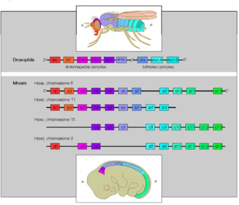
3.1 Hox genes 77
3.1.1 Hox genes in skeletal development 81
3.1.2 Hox genes as positional cell memory 89
3.1.3 Epigenetic control of Hox activity 92
3.1.4 Hox code and tissue regeneration 96
3.2 TALE genes 102
3.3 Otx genes 114
4. THE STEM CELL GENE KIT 125
4.1 Kit/GFP transgenic mouse lines 130
5. AIM OF THE STUDY 133
MATERIALS AND METHODS 137
6. MATERIALS AND METHODS 138
6.1 Mice 138
6.2 Isolation and expansion of murine MSCs 139
6.3 Colony Forming Unit-‐Fibroblast (CFU-‐F) assay 140
6.4 In vitro differentiation of murine MSCs 140
6.5 Cell cultures of murine pre-‐osteoblastic MC3T3-‐E1 cell line 141
6.6 Senescence Associated β-‐Gal Staining 142
6.7 In Situ Immunofluorescence 142
6.9 Flow cytometric analysis 144
6.10 Real-‐Time PCR 144
6.11 Statistical analysis 145
RESULTS AND DISCUSSION 146
7. CHARACTERIZATION OF MSCS DERIVED FROM DIFFERENT
BODY SITES-‐RESULTS 147
7.1 Immunophenotype and in vitro growth and differentiation 147
8.1 Immunophenotype and in vitro growth and differentiation 166
8.2 HOX expression signatures 168
8.3 TALE expression profiles 171
8.4 Conclusion 173
9. ROLE OF OTX1 IN THE CONTROL OF MSCs-‐RESULTS 174
9.1 Otx1 expressions in murine and human cells 174
9.2 Otx1 protein expression in murine cells 181
9.3 Effects of Otx1 inactivation 183
10. ROLE OF OTX1 IN THE CONTROL OF MSCs-‐DISCUSSION
192
10.1 Otx1 activity during in vitro osteogenic differentiation of murine
and human MSCs 193
10.2 Expression and localization of the Otx1 protein during
osteogenic differentiation of murine MSCs 196
10.3 Effects of Otx1 inactivation 201
10.4 Phenotypic alterations 201
10.5 Enhanced survival and proliferation 203
10.6 Impaired osteogenic differentiation 206
10.7 Conclusion 208
11. KIT ACTIVITY DURING IN VITRO ADIPOGENESIS-‐RESULTS
210
11.1 Kit expression in human and murine MSCs 210
11.2 Kit protein expression and localization in murine cells 215
11.3 Analysis of Kit/GFP MSCs 219
12. KIT ACTIVITY DURING IN VITRO ADIPOGENESIS-‐
DISCUSSION 221
12.1 Kit receptor is involved in the adipogenic differentiation 221
12.2 Expression of the transgene Kit/GFP in the mesenchymal
system 226
12.3 Conclusion 227
CONCLUSIONS 228
13. CONCLUSION 229
grow ex vivo and differentiate in vivo and in vitro into multiple mesodermal phenotypes. They can be isolated from most fetal and adult tissues, including bone marrow, fat, teeth. Moreover, they can easily be expanded and differentiated in vitro. In recent years MSCs have raised great interest for their properties and characteristic as potential therapeutical tools for a wide spectrum of human pathologies. However, several aspects of their biology still need to be clarified. Developmental regulators coding for growth factors, receptors and transcription factors are involved in the regulation of stem and progenitor cells of different tissues. In this view, the present study has been mainly focused on homeodomain transcription factors of different families (Hox, Tale, and Otx) and on the receptor of the Stem Cell Factor (Kit), investigating their possible role in the control of mesenchymal stem and progenitor cells. Different experimental strategies have been employed, including both expression and functional studies in mouse models and human cells.
A relevant question is whether MSCs derived from different sources exhibit equivalent biological properties. Here I have compared cellular and molecular characteristics of human MSCs derived from different body locations. Results have highlighted that they exhibit the same immunophenotipic profile, but distinct
in vitro growth and differentiation properties, and are
characterized by specific HOX codes and TALE signatures. Thus, they may not provide equivalent cell sources for regenerative
MSCs. Otx1 encodes a key factor in neural development and plays an important role also in blood cell production. Here I showed that Otx1 expression is modulated during the osteogenic differentiation of both murine and human MSCs and its inactivation is associated with phenotypical alterations and enhanced survival and proliferation of MSCs, as well as an impairment of their osteogenic capacity. These findings indicate that Otx1 is implicated in the control of both proliferation and differentiation of MSCs.
Finally, another important pleiotropic molecule is Kit, coding for the Stem Cell Factor (SCF) tyrosine-kinase receptor. Kit is a pivotal regulator of several types of stem and progenitor cells. However, its role in MSCs is still controversial. My results have shown that Kit is active also in MSCs, providing the first evidence on its involvement in the adipogenic process.
The identification of new molecular cues controlling self-renewal, commitment and differentiation of mesenchymal stem and progenitor cells has both biological and clinical relevance.
1. THE STEM CELLS
During embryogenesis, a single fertilized oocyte gives rise to a multicellular organism whose cells and tissues have adopted differentiated characteristics or fates to perform the specified functions of each organ of the body. As embryos develop, cells that have acquired their particular fate proliferate, enabling tissues and organs to grown. Even after an animal is fully grow, however, many tissues and organs maintain a process known as homeostasis, where as cells die, either by natural death or by injury, they are replenished. This remarkable feature has ancient origins, dating back to the most primitive animals. Some amphibians, for istance, can regenerate a limb or tail when severed, and the neurons of bird brains can readily regenerate. While mammals seem to have lost at least some of this wonderful plasticity, their liver can partially regenerate, provided that the injury is not too severe, and their epidermis and hair of their skin can readily repair when wounded or cut. Additionaly, the epidermis, hair, small intestine, and hematopoietic system are all examples of adult tissues that are naturally in a dinamic state: even in the absence of injury, these structures continually divide, terminally differentiate and die.
The outstanding ability of an embryo to diversify and of certain adult tissues to regenerate throughout life is a direct result of stem cells. Stem cells have both the capacity to self-renew, that is, to divide and create additional stem cells, and also to differentiate along a specified molecular pathway. Embryonic stem cells are very nearly totipotent, as they have the ability of choosing all of the differentiation pathways that specify the animal. In contrast,
adult stem cells that reside within an adult organ or tissue have more restricted ability to differentiate, as they are able to select a differentiation program from only few possible pathways. In the last years, some spectacular findings have exploded many long-standing dogmas in the stem cell world, giving adult stem cells a new lease on life (Fuchs and Segre, 2000). Adult stem cells have raised great interest for their properties and characteristics as potential therapeutical tools for a wide spectrum of pathologies. There is, therefore, considerable interest in understanding the regulatory transcriptional and signaling networks which underlie stem cell properties, such as pluri-multipotency and self-renewal, as well as specificity of commitment and differentiation.
1.1 Embryonic Stem Cells
Mammalian development commences when an oocyte is fertilized by a sperm forming a single celled embryo, the zygote. Consistent with the definition, the zygote is totipotent, meaning that this single cell has the potential to develop into an embryo with all the specialized cells that make up a living being, as well as into the placenta support structure necessary for fetal development. Thus, each totipotent cell is a self-contained entity that can give rise to the whole organism. This is said to be true for the zygote and for early embryonic blastomeres up to at least the 4-cell stage embryo. Experimentally, totipotency can be demonstrated by the isolation of a single blastomere from a preimplantiation embryo and subsequently monitoring its ability to support a term birth following transfer into a suitable recipient.
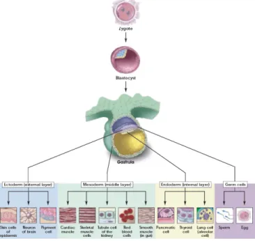
several mammalian species including nonhuman primates. As embryo development progresses to the 8-cell stage and beyond depending on the species, the individual blastomeres that comprise the embryo gradually lose their totipotency. It is generally believed that this restriction in developmental potential indicates irreversible differentiation and specialization of early embryonic cells into the first two lineages, the inner cell mass (ICM) that includes cells that will give rise to the fetus and the trophoectoderm (TE), and an outer layer of cells that is destined to an extraembryonic fate. At least some of the embryo’s ICM cells are pluripotent, meaning that they have the potential to differentiate into any of the three germ layers (endoderm, mesoderm or ectoderm) and they can form virtually every somatic and germ cell type in the body (Fig. 1).
These ICM cells are self sustained and their pluripotency is maintained by endogenously expressed factors, as the transcription factors Oct4, Nanog and Sox2. In vivo, pluripotent cells within the ICM exist transiently; as the developmental program unfolds they differentiate into cells of the next embryonic or fetal stage. However, they can be isolated, adapted and propagated in vitro in an undifferentiated state as embryonic stem (ES) cells (Fig. 2) (Evans and Kaufman, 1981; Martin, 1981; Brook and Gardner, 1997).
Figure 2: ES in vitro.
Murine ES cell remain undifferentiated when grown in the presence of cytokine leukemia inhibitory factor (LIF) and cultured on murine embryonic fibroblasts (MEFs) (Smith et al., 1988; Williams et al., 1988). The effect of LIF is to inhibit
differentiation and support proliferation of undifferentiated stem cells. When LIF is withdrawn, most types of ES cells differentiated spontaneously to form aggregates known, in view of their similarity to post-implantation embryonic tissues, as embryoid bodies (Desbaillets et al., 2000; Itskovitz-Eldor et al., 2000). These spherical structures are comprised of derivatives of all three germ layers and continued in vitro culture of embryoid bodies leads to the formation of a range of differentiated cell types including cardiomyocytes, hematopoietic cells, endothelial cells, neural cells, skeletal muscle, chondrocytes, adipocytes and pancreatic islets (Wiles M.V. and Keller, 1991; Schmitt et al., 1991; Maltsev et al., 1993; Wang et al., 1994; Bain et al., 1995; Fraichard et al., 1995; Wobus et al., 1997; Dani et al., 1997; Kramer et al., 2000; Hamazaki et al., 2001; Lumelsky et al., 2001). In each case, despite the use of growth factors favouring the differentiation of a particular cell type, the resulting cultures are heterogeneous.
With similar techniques to those developed for mouse embryos, stem cell lines have been derived from human blastocysts (Thomson et al., 1998). Human ES cells derived from the inner cell mass removed from embryos grown into blastocysts. The embryo from which the initial human ES cells were derived were produced by in vitro fertilization and donated with informed consent of the parents. Another approach to deriving these cells is somatic nuclear transfer or cloning. This procedure uses the transfer of a somatic cell nucleus from an individual into an enucleated oocyte (McGrath and Solter, 1983; Campbell et al., 1996; Wilmut et al., 1997). The cells are then allowed to undergo
embryonic development to the blastocyst stage prior the isolation of ES cells from the inner cell mass. These cells show several morphological and behavioural differences from murine ES cells: they grow slowlier and tend to form flat rather than spherical colonies (Amit et al., 2000; Odorico et al., 2001). LIF does not have the same effect on human ES cells and, in order to maintain them in an undifferentiated state, the presence of both MEFs feeder layers and basic fibroblast growth factor (bFGF) is required (Martin, 1981; Evans and Kaufman, 1981). As with murine ES cells, modification of culture medium in which human ES cells are grown can promote the differentiation of certain lineages (Schuldiner et al., 2000), displaying cells with the distinct morphology of neurons, epithelium, cardiomyocytes (Reubinoff et al., 2000; Odorico et al., 2001; Kehat et al., 2001). In addition, spontaneous in vitro formation of functionally active pancreatic β cells has also been observed (Assady et al., 2001). While no one doubts that ES cells are likely the most flexible of all stem cells, their use is the subject of intense debate particularly because of the ethical issues. Moreover, experimental results indicate that these cells can origin teratocarcinoma when injected subcutaneously in athymic mice (Martin, 1981). Therefore, the search is prompted for alternative stem cells sources. A major breakthrough was achieved in 2006, when it was shown that pluripotent stem cells could be obtained by transducing mouse embryonic and adult fibroblasts with a limited set of genes encoding four transcriptional factors (Oct4, Sox2, c-Myc and Klf4). These reprogrammed cells, named induced pluripotent stem (iPS) cells, resembled ES cells in many of their
characteristic (Takahashi and Yamanaka, 2006). However, these iPS cells had a different global gene expression pattern from ES cells and failed to produce adult chimeric mice. In 2007, germline transmission was achieved with mouse iPS cells (Meissner et al., 2007; Okita et al., 2007; Wernig et al., 2007), and iPS cells were generated from human fibroblasts (Takahashi et al., 2007; Yu et al., 2007; Park et al., 2008b). Given the potential to generate patient-specific cell populations without the need for human embryonic cells, iPS cell technology has been received with great excitement by research and medical communities. However, many questions regarding the actual molecular process of induced reprogramming remain unanswered and need to be addressed before iPS cells can go to the clinic.
1.2 Adult Stem Cells
In the adult organism many tissues and/or organs, as a consequence physiologic cell turn over or tissue injury, are capable of maintaining, generating and replacing terminally differentiated cells: this process of homeostasis is guaranteed by adult stem cells. Tissue-specific stem cells share common properties: self-renewal and multipotency. First, they can divide indefinitely, producing a population of identical progeny. Second, adult stem cells can undergo an asymmetric division to produce two dissimilar daughter cells. One is identical to the parent and contributes to maintain the stem cell population. The other is characterized by a reduced proliferative capacity and more restricted developmental potential than its parent, becoming a
“progenitor” or “precursor” cell, committed to terminal differentiation in the specialized cell types of the tissue where it is located (Fig. 3). Adult stem cells are often quiescent or relatively slow-cycling cells able to respond to specific environmental signals. When a stem cell undergoes a commitment to differentiate, it often first enters a transient state of rapid proliferation. Upon exhaustion of its proliferative potential, the transiently amplifying cell withdraws from its cycle and executes its terminal differentiation (Potten et al., 1979).
Figure 3: The possible choices for a daughter cell of a stem cell division. It
can either self-renew (that is, remain a stem cell) or commit to a pathway leading to differentiation. In many cases where it commits to differentiation, it first becomes a precursor cell, which proliferates before differentiating.
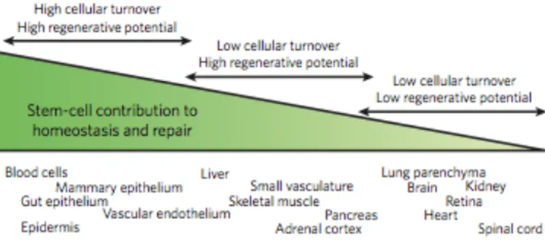
Adult stem cells reside in most mammalian tissues, but the extent to which they contribute to normal homeostasis and repair varies widely. Along this spectrum, tissues generally fall into one of three categories. First, tissues with high turnover (such as blood, skin and gut) have a prominent stem-cell compartment and, by definition, have high regenerative capacity. Second, tissues with
strategies to ensure effective repair in the setting of acute injury. In skeletal muscle, for example, differentiated myofibres are unable to proliferate to generate new tissue, so muscle must rely on resident stem cells for all turnover and repair. For the liver, it seems that differentiated hepatocytes can proliferate sufficiently to mediate effective tissue remodelling, repair and replacement normally, whereas stem cells might be recruited in the setting of severe injury. Third, tissues with low turnover and low regenerative potential might have stem cells that mediate only limited tissue repair. Although there is much interest in harnessing the potential of stem cells in the brain and heart for therapeutic purposes, for example, there is limited endogenous repair capacity of these tissues following acute injuries (Fig. 4) (Rando, 2006).
Figure 4: Tissue heterogeneity and stem-cell functionality for homeostasis and repair.
As a result, stem cells must be identified prospectively and purified carefully in order to study their properties. The dream to “rebuilt” human organs is turning slowly into reality. Many
researchers believe that stem cells therapy has the potential to dramatically change the treatment of human diseases and adult stem cells have captured the popular imagination with the promise of enhanced tissue repair, the treatment of degenerative diseases and even the amelioration of dysfunction associated with normal ageing. However, several stem cells therapies already exist. An example is provided by bone marrow stromal stem cells taken from the patient, expanded in vitro and then transplanted into the injured tendons or ligaments of the same patient.
1.2.1 Hematopoietic stem cells (HSCs)
Although it seems reasonable to propose that each tissue arises from a tissue-specific stem cell, the rigorous identification and isolation of the somatic stem cells has been accomplished only in a few instances. The best-characterized stem cells are those responsible for blood cell production, the hematopoiesis. All experimental strategies and conceptual paradigms applicable to stem cells in general were first defined in hematopietic system. The hallmark properties of the Hematopoietic Stem Cells (HSCs) were defined in 1963 by Till and Mc-Culloch (Siminovitch et al., 1963). They defined HSCs as cells that are individually capable of both self-renewal and multilineage differentiation, yelding greatly expanded numbers of lymphocytes, myeloid cells and erythrocytes. In fact, around 2x10¹¹ erythrocytes and 1010 white blood cells must be replaced each day to maintain adult human hematopoiesis. Terminally differentiated cells are by themselves incapable of further growth and must, therefore, be replaced
through proliferation and development of HSCs (Goldman, 1982). The process, by which stem cells give rise to terminally differentiated cells, occurs through a hierarchy of progenitor cells, and during commitment, cells can undergo extensive proliferation and sequential differentiation, accompanied by a decrease in self-renewal capacity (Dexter et al., 1984; Golde, 1991). The primary function of this transit population is to increase the number of mature cells produced by each stem cell division. The immediate progeny of HSCs in adult bone marrow are the common myeloid precursor (CMP) and the common lymphoid precursor (CLP) (Akashi et al., 2000; Hao et al., 2001; Manz et al., 2002). CLP is thought to give rise to all lymphoid lineage (B, T and NK), but little is known about its development (Spangrude and Cooper, 2000). CMP can be associated with two cell types that can form in vivo (CFU-S) and in vitro (CFU-Mix) colonies containing multiple cell types. CFU-S (Colony Forming Unit-Spleen) can produce colonies on the spleen of irradiated mice injected with bone marrow cells (Till and Mc-Culloch, 1961). The other pluripotent progenitor is the CFU-Mix (Colony Forming Unit-Mix) that generate mixed colonies in cultures containing an appropriate cocktail of cytokines. CMP can give rise to progenitors committed to the generation of erythrocytes and megakaryocytes (MEP) or granulocytes and macrophages (CFU-GM). These cells, capable of developing along more than one lineage, undergo commitment and become progressively restricted to a specific differentiative fate, such as Colony Forming Granulocytes (CFU-G) and Colony Formin Unit-Macrophages (CFU-M), the direct precursors of granulocytes and
monocytes, respectively. Similarly, two stages of different maturation have been identified within the erythroid lineage: an early cell (Burst Forming Unit-Erythroid or BFU-E) and a late precursor (Colony Forming Unit-Erythroid or CFU-E) capable of forming large and small erythroid colonies, respectively.
Murine HSCs were initially identified on the basis of their ability to form colonies in the spleens of lethally irradiated mice following bone-marrow transfer. However, subsequent studies highlighted that spleen colonies were not always formed by multipotent cells (Magli et al., 1982; Metcalf, 1999)and that HSCs were defined by their ability to sustain long term hematopoiesis in vivo. Subsequently, a number of assays have been developed to monitor HSC activity in vitro and in vivo. In
vitro clonogenic assays are a powerful means to quantitatively
and qualitatively study a broad spectrum of progenitors (Metcalf, 1999), but they do not sustain HSCs growth. The most widely accepted assay is the capacity of HSCs to provide lifelong reconstitution of all blood-cell lineages after transplantation into lethally irradiated recipients. The strictest version of this long-term repopulating (LTR) assay, known as serial transplantation, requires that HSC-containing donor bone marrow can be re-transplanted into secondary, and even tertiary, recipients while retaining both self-renewal and multi-lineage differentiation capacity (Okada et al., 1993; Harrison et al., 1978; Kim et al., 2000). Multipotent long-term HSCs (LT-HSCs) reside in the bone marrow and through a process of asymmetric cell division, can self-renew to sustain the stem cell pool or differentiate into short-term HSCs (ST-HSCs) or lineage-restricted progenitors that
undergo extensive proliferation and differentiation to produce terminally differentiated, functional hematopoietic cells. ST-HSCs or multipotent progenitors (MPPs) are only able to sustain hematopoiesis in the short term, whereas the LT-HSCs must persist for the lifespan of the organism to perpetually replenish the hematopoietic system. Despite many exhaustive studies, researchers have yet to find a single molecular marker that is expressed exclusively by HSCs. However, they can be isolated from bone marrow or peripheral blood using enrichment and/or single-cell sorting (fluorescence-activated cell sorting-FACS) based on cell surface markers and/or vital dye staining. The identification and purification of HSCs relies on the unique cell surface molecule expression found on these cells compared with the remainder of bone marrow cells including closely related hematopoietic progenitor cell counterparts. Almost all HSCs purification strategies revolve around the cell surface phenotype of positive selection for the markers c-Kit and Sca-1 and negative selection for markers of mature hematopoietic cell lineages (RBCs, T-cells, B-cells, NK-cells, monocytes and granulocytes). Although this c-Kit+Lin-Sca-1+ (KLS) phenotype greatly enriches for hematopoietic reconstituting activity, this bone marrow compartment contains progenitor cells in addition to long-term HSCs. In fact only 10% of KLS cells are bona fide long-term HSCs, and as such the KLS compartment should be regarded as merely enriched for HSCs. A variety of strategies have been used to further enrich bone marrow for HSCs, with or without the KLS as a foundation. These strategies include identification of HSCs as KLS-CD34-Flk-2-, KLS-CD150+CD48- cells, the
Hoechst-effluxing side population (SP), and associated variations on that theme. The Hoechst dye is retained at low levels in HSCs because of their ability to efflux the dye via membrane transport pumps, which are highly active in these cells compared with other bone marrow cell types. The distinctive staining pattern of HSCs, easily observed when Hoechst fluorescence is displayed at two different wavelengths, results in their presence at the side of the Hoechst fluorescence profile, hence the ‘‘SP.’’ Several studies have shown that even without other markers such as KLS, the SP is remarkably enriched for HSC activity, and most of the SP cells bear other surface markers of HSC, such as KLS. In addition, virtually all of the long-term HSC activity is contained within this SP fraction (reviewed in Challen et al., 2009). Alternative methods use a signature of SLAM family of cell surface molecules. SLAM (Signaling lymphocyte activation molecule) family receptors is a group of molecules, all belonging to a subset of immunoglobulin gene superfamily, and originally thought to be involved in T-cell stimulation. A combination of SLAM molecules (CD150+CD244-CD48-) has been recently shown to mark HSCs from fetal liver stage through adult hematopoiesis, including mobilized and aging HSCs (Kiel et al., 2005; Kim et al., 2006; Yilmaz et al., 2006). Thus, SLAM markers introduce a code that may be more generally applicable for identifying HSCs.
1.2.2 Cancer stem cells (CSCs)
During the last years, a growing body of evidence indicate that also neoplastic tissues can harbour stem cells. Based on
functional and immunophenotypic analysis of cell subpopulation with modern technologies, cancer has started to be increasingly consider as a stem-cell disorder, in which the continuous growth and propagation of the whole tumor depends on a small population of self-renewing tumor stem cells. The prevailing theory of tumor initiation is that cells accumulate genetic and epigenetic alterations that ultimately confer a growth and/or survival advantage to the nascent cancer. It has been well-accepted that these events occur in terminally differentiated cell types and that the progression of cancer is directly correlated to the ability of those cells to divide indefinitely. Attempts to target and destroy this transformed population of cells have been modestly effective, although it is becoming increasingly evident that targeted therapies alone cannot completely eradicate an established tumor. This observation suggests that there may be a separate and distinct subpopulation of cells that is resistant to the aforementioned therapy and can also repopulate the tumor with malignant cells, as needed. This minor subset of cells is more commonly referred to as Cancer Stem Cells (CSCs). Because of varying experimental strategies that isolate and characterize cancer stem cells, clearly defining the traits that identify cancer stem cells has become troublesome. The current definition includes two stipulations that help delineate these cells: (1) must possess the ability to self-renew and (2) must be able to resupply the tumor with the various lineages of cells of which it is comprised (Clarke et al., 2006). The existing definition has thereby yielded an alternative expression for CSCs that describes
their functionality: thus, ‘‘tumor-initiating cell’’ has now become synonymous with cancer stem cell.
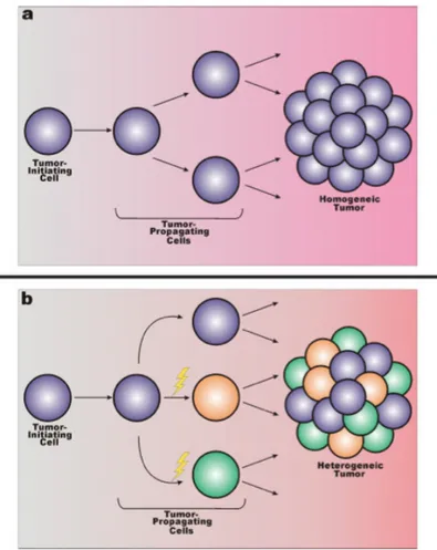
Similarly to normal adult stem cells, they can committed and generate transient-amplifying cells. These progenitor cells are thought to be the cell type that most actively support tumor growth (Fig. 5).
Figure 5: Overview of tumor stem cells in cancer. Cancer stem cells
(initiating cells) divide asymmetrically, resulting in self-renewal of the tumor-initiating cell and production of a daughter cell known as a transient-amplifying cell (progenitor cell). The transient-transient-amplifying cell is not thought to possess self-renewing capabilities, but instead divides indefinitely to contribute to cancer progression.
A relevant question asks if the tumor-initiating cell and the tumor-propagating cell are the same entity. Logic dictates that any cell that can initiate tumor formation should also possess the ability to continue supporting tumor growth. This simple premise, however, does not explain the heterogeneic cellular distribution within solid tumors; if the initiating cell and the tumor-propagating cell are identical throughout development, once
could argue that the cellular constituency of the tumor would be fairly homogenous. Because this is certainly not the case in solid tumors, one possible explanation is that the tumor-initiating cell continues to gradually acquire additional mutations that contribute to cellular heterogeneity; this also accounts for how the tumor-initiating cell and tumor-propagating cell may become distinct components over time (Fig. 6).
Figure 6: Is the tumor-initiating cell also the tumor-propagating cell. Two primary models that speculate the individuality of the tumor-initiating cell and the propagating cell. a: If the initiating cell and the
tumor-propagating cell are the same entity, the cellular population of the solid tumor should be relatively genetically similar (indicated by violet spheres). b: Possible secondary model where the tumor-initiating cell gives rise to the tumor-propagating cell, which can acquire genetic mutations (yellow lightning
bolt) that further potentiate tumor growth, as well as lead to cellular heterogeneity (indicated by multiple sphere colors).
However, this explanation does not discount the significance of the tumor-initiating cell to the cancer because it likely retains the ability to supply the growing tumor with malignant progenitors that are likely to subsequently become tumor-propagating cells (Lee and Herlyn, 2007).
Cancer stem cells were first identified in acute myeloid leukemia when surface markers were used to distinguish the stem cell population from the remaining cells with limited proliferative potential (Dick, 2005). In solid tumors, cancer stem cells have been identified in breast cancer (Shackleton et al., 2006; Ponti et al., 2005), glioblastomas (Singh et al., 2003), lung cancer (Kim et al., 2005), ovarian cancer (Bapat et al., 2005), prostate cancer (Collins et al., 2005), and epithelial gastric cancer (Houghton et al., 2004). Isolating and defining tumor-initiating stem cells should lead to the development of highly effective, less toxic therapies for cancer patients. The relative efficacy of traditional chemotherapeutics is highly dependent on both the stage and the type of disease; future drug formulations may be able to bypass the shortcomings of those compounds by targeting only the tumor-initiating cells.
1.2.3 Plasticity of Adult Stem Cells
In the adult soma, stem cells generally have been thought of as tissue-specific, able to give rise only to progeny cells
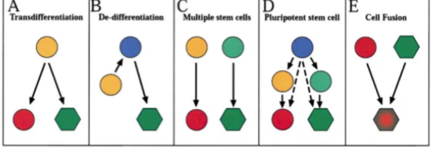
corresponding to their tissue of origin. However, recent experiments have challenged this notion and called into question the lineage commitment of various adult stem cell populations by suggesting that under certain circumstances these cells may “transdifferentiate” to contribute to a much wider spectrum of differentiated progeny than previously anticipated. Transdifferentiation describes the conversion of a cell of one tissue lineage into a cell of an entirely distinct lineage, with concomitant loss of the tissue-specific markers and function of the original cell type, and acquisition of markers and function of the transdifferentiated cell type. The suggestion that adult stem cells may transdifferentiate has in turn given rise to the concept of stem cell plasticity, which holds that the lineage determination of a differentiating stem cell may not be rigidly defined, but is instead flexible, allowing these cells to respond to a variety of microenvironmental regenerative cues (Blau et al., 2001). Suggestions of stem cell plasticity in the adult could indicate a need to reexamine precursor-progeny relationships during the early developmental steps when germ layer specification occurs, especially if rare cells with high proliferative potential from one tissue (e.g., mesoderm) may be found within, and perhaps nurtured by, another tissue (e.g., endoderm). A telling precedent for rare cells within an organ giving rise to most mature cells within that organ derives from early studies on the development of thymic lymphocytes. In these experiments, transfilter incubation of thymic epithelium and mesenchyme appeared to implicate endoderm, not mesoderm, cells as thymocyte precursors (Auerbach, 1961); however, subsequent examination
showed that these cells, called hematocytoblasts, were in fact rare mesoderm-derived precursors nested within the thymic endoderm that gave rise to most, if not all, thymic lymphocytes (Moore and Owen, 1967). Moreover, the concept that adult HSCs function solely to maintain hematopoietic cell lineages was challenged by a series of papers suggesting that unfractionated bone marrow cells, or bone marrow cells enriched by various methods for hematopoietic stem cell activity, could be seen to contribute at low levels to multiple nonhematopoietic tissues following transfer into lethally irradiated, and often injured, recipient mice or humans (reviewed in Herzog et al., 2003 and Goodell, 2003). Such studies have reported the expression of donor-derived genetic markers in non- hematopoietic cells within the skin (Krause et al., 2001), lung epithelium (Krause et al., 2001; Theise et al., 2002), intestinal epithelium (Krause et al., 2001), kidney epithelium (Kale et al., 2003), liver parenchyma (Krause et al., 2001; Lagasse et al., 2000; Petersen et al., 1999; Theise et al., 2000; Vassilopoulos et al., 2003; Wang et al., 2003b), pancreas (Ianus et al., 2003), skeletal muscle (Brazelton et al., 2003; Camargo et al., 2003; Corbel et al., 2003; Ferrari et al., 1998; Fukada et al., 2002; Gussoni et al., 1999; LaBarge and Blau, 2002), endothelium (Jackson et al., 2001), myocardium (Jackson et al., 2001; Barile et al., 2009), and CNS neurons in the cortex and cerebellum (Brazelton et al., 2000; Mezey et al., 2000; Priller et al., 2001; Weimann et al., 2003a, 2003b). Such findings were extended by some to a general hypothesis of adult stem cell plasticity, wherein adult stem cells from one tissue were considered to be roughly equivalent in developmental potential to
adult stem cells in another tissue, with the outcome of stem cell differentiation largely determined by different microenvironments encountered following differential trafficking from the bloodstream (Blau et al., 2001). Studies of bone marrow or stem cell transdifferentiation have been highly controversial (Anderson et al., 2001; Goodell, 2003; Raff, 2003). Clearly the mechanism invoked may depend on the cell populations and tissues analyzed and on the method of injury, if any, employed. In only a few cases has the actual mechanism, by which such unexpected contributions of cells across tissue lineage boundaries occur been studied, much less elucidated. Cell transdifferentiation is one mechanism by which stem cells potentially could contribute to cell types of different lineages. This lineage conversion was proposed to occur directly, by activation of an otherwise dormant differentiation program to alter the lineage specificity of the cell (Figure 7A). Lineage conversion also could theoretically occur via dedifferentiation of a tissue-specific cell to a more primitive, multipotent cell and subsequent redifferentiation along a new lineage pathway (Figure 7B). Extensive evidence suggests that urodele amphibians regenerate amputated limb, tail, eye, jaw, and heart structures through a process that appears to involve the dedifferentiation of mature cells near the edge of the wound, forming a cluster of “naive” progenitor cells referred to as the blastema, and subsequent activation of a regenerative process reminiscent of the developmental program that originally functioned to specify limb formation (Brockes and Kumar, 2002). It should be noted, however, that a possible role for preexisting multipotent adult
stem cells in amphibian regeneration can not be excluded entirely. Nonetheless, in these systems, blastema cells do not appear to become totally uncommitted pluripotent stem cells; instead, they retain memory of their earlier identity, as indicated by the fact that iris epithelial cells from the eye give rise to lens even if transplanted into an amputated limb (Reyer et al., 1973; Ito et al., 1999), and limb blastema transplanted into the eye still generates limb structures (Kim and Stocum, 1986). Dedifferentiation of cells in adult mammals has not been clearly and unequivocally documented, and at present, no evidence directly supports transdifferentiation or dedifferentiation events as an explanation for stem cell or BM cell plasticity in vivo (Tanaka, 2003).
A third explanation for observations of adult stem cell plasticity relates to the purity or homogeneity of the test population. In order to demonstrate definitively transdifferentiation of a particular lineage-specific stem cell, it is essential to exclude the possibility that multiple, distinct stem cells could be contributing to the observed outcome, ideally by evaluating the potential of single, prospectively isolated, stem cells (Fig. 7C).
Contributions across multiple tissue types also could arise through the action of a single, rare pluripotent stem cell present in bone marrow, and/or other tissues, which possibly copurifies in protocols designed to enrich for tissue-specific stem cells such as bone marrow HSCs (Figure 7D). The possible existence of pluripotent stem cells within the bone marrow of mice and humans has previously been proposed and this idea has gained support from the isolation through culture of multipotent (actually
pluripotent) adult progenitor cells (MAPC), which give rise to tissues of multiple germ layers following intravenous transplant, and contribute substantially to most, if not all, tissues following injection into blastocysts (Jiang et al., 2002a; Serafini et al., 2007). However, whether cells with such extensive developmental potential exist normally in adult animals or are endowed with such properties via the culture conditions employed is currently unknown, and no direct evidence implicating these cells in bone marrow plasticity studies has been obtained.
The final mechanism to explain observations of stem cell plasticity is cell-cell fusion (Figure 7E). Fusion between the egg and sperm is the initiating event in vertebrate development, and in adult animals, cell-cell fusion occurs naturally in the generation of multinucleated skeletal myofibers from myoblasts and of osteoclasts from monocyte/macrophage lineage cells (Anderson, 2000; Vignery, 2000). Cell-cell fusion has been implicated in contributions of transplanted bone marrow cells to liver hepatocytes, cardiac myocytes, and Purkinje neurons (Alvarez- Dolado et al., 2003; Vassilopoulos et al., 2003; Wang et al., 2003b; Weimann et al., 2003b). Several authors have proposed that heterotypic cell fusions represent a physiological process designed to rejuvenate and/or repair a wide variety of tissues (Alvarez-Dolado et al., 2003; Blau, 2002). However, the low frequency with which such events have been observed (typically less than 1% of cardiomyocytes and 0.1% of hepatocytes or Purkinje cells in nonselective transplant model;
Alvarez-Dolado et al., 2003) implies that this rare phenomenon is unlikely to contribute significantly to normal tissue regeneration.
Figure 7: Schematic diagram depicting potential mechanisms and explanations for observations of adult stem cell plasticity.
Given the current data available from both mouse and human studies, recruitment of cells from heterologous sources is unlikely to be a normal, robust, or highly utilized mechanism for adult tissue regeneration. Nonetheless, it is possible that by a concerted effort aimed at dissecting this rare phenomenon, rigorously identifying, purifying and potentially expanding the appropriate cell populations responsible for “plasticity,” characterizing the tissue-specific and injury-related signals that recruit, stimulate, or regulate plasticity, and determining the mechanism(s) underlying fusion or plasticity, it may eventually enhance tissue regeneration via this mechanism to clinically useful levels.
1.2.4 Stem Cell Niche
A stem cell niche can be defined as a spatial structure in which stem cells are housed and maintained by allowing self-renewal. Structurally, the niche is formed by supporting cells that provide
a microenvironment for stem cells as well as the signals emanating from the supporting cells. Although the concept of the stem-cell niche was initially proposed in vertebrates, the D.
melanogaster ovarian and testicular niches controlling germline
stem-cell maintenance and differentiation were the first to be characterized. In higher organisms, the analysis of stem-cell-niche interactions has been hampered by their unknown location. However, during the past few years, substantial progress has been made in localizing adult stem cells in situ. Many studies have indicated that most adult tissue stem cells (such as HSCs, or epidermal stem cells (ESCs) in the skin) divide infrequently and can be quiescent for weeks or even months. In support of this notion, the adult stem cell pool is largely resistant to classical chemotherapeutic agents that target cycling cells. In addition, HSCs that efficiently engraft after transplantation are mainly quiescent and considered to be metabolically inactive. Moreover, when the DNA of adult stem cells is labelled during cellular proliferation by nucleotide analogues (such as 3H-thymidine or bromodeoxyuridine (BrdU labelling)), or by the histone H2B-enhanced-green-fluorescent-protein fusion protein (H2B-EGFP), the DNA label can be retained for months and has consequently been used to locate quiescent stem cells in situ.
The vast majority of cell divisions are symmetrical, producing identical daughter cells and leading (in the absence of apoptosis) to increased numbers of cells. This process is readily observed for cells in culture and also occurs during organogenesis, where substantial cellular expansion (including stem cells) occurs during embryogenesis. By contrast, under homeostatic conditions
in the adult, the number of tissue stem cells in a particular organ remains relatively constant, despite the fact that they proliferate, because they not only self-renew but also produce differentiated progeny.
The ability of adult stem cells to both self-renew and differentiate is critical for tissue homeostasis. The stem cell population would become depleted if cell differentiation overwhelmed self-renewal. Similarly, unchecked stem cell self-renewal would expand the stem cell population excessively, risking tumorigenesis (Jones and Fuller, 2004). An important function of the stem cell niche, therefore, is to regulate the balance between cellular self-renewal and differentiation. One mechanism that ensures this balance is the control of asymmetric/symmetric stem cell division. This balance could be achieved if the number of stem cells dividing symmetrically to generate two identical daughter cells with stem-cell function was equivalent to the number of stem stem-cells giving rise to two differentiated daughter cells. However, because this mechanism does not function at the single-cell level, and would require close coordination of two separate stem-cell populations, it is commonly assumed that an individual stem cell can give rise to two non-identical daughter cells, one maintaining stem-cell identity and the other becoming a differentiated cell. There are two mechanisms by which this asymmetry can be achieved, depending on whether it occurs pre- (divisional asymmetry), or post- (environmental asymmetry) cell division (Fig. 8).
Figure 8: A model of asymmetric cell division. a) During divisional
asymmetry, cell-fate determinants are asymmetrically localized to only one of the two daughter cells, which retains stem-cell fate, while the second daughter cell differentiates. b) During environmental asymmetry, after division, one of two identical daughter cells remains in the self-renewing niche microenvironment while the other relocates outside the niche to a different, differentiation-promoting microenvironment.
In divisional asymmetry, specific cell-fate determinants in the cytoplasm (mRNA and/or proteins) redistribute unequally before the onset of cell division. During mitosis, the cleavage plane is oriented such that only one daughter cell receives the determinants. Therefore, two non-identical daughter cells are produced, one retaining the stem-cell fate while the other initiates differentiation (Fig. 8a). An alternative way to achieve asymmetry is by exposure of the two daughter stem cells to different extrinsic signals provided by distinct local microenvironments. Therefore, a stem cell would first undergo a
symmetric self-renewing division, producing two identical daughter cells. While one daughter cell would remain in the niche microenvironment, conserving its stem-cell fate, the other would contact (passively or actively) a different microenvironment that would no longer preserve its stem-cell phenotype but would instead produce signals initiating differentiation (Spradling et al., 2001; Ohlstein et al., 2004). Therefore, as with divisional asymmetry, the final product would be two non-identical daughter cells but achieved post-cell-division and not pre-cell-division (Fig. 8b).
It is specific cues from specific sites that allow stem cells to persist, and to change in number and fate. Importantly, it is also the niche that provides the modulation in stem-cell function needed under conditions of physiologic challenge. It is this dynamic capability that makes the ‘niche’ concept particularly important and central to the realization of regenerative medicine. It is the ability of the niche to impose functions on stem cells that makes the concept important in disease.
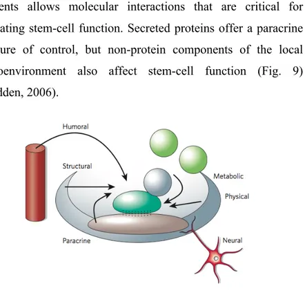
Cells and extracellular-matrix components in the stem-cell niche are relatively predictable, although the complexity and integration of these elements is far from being understood. Early studies provided evidence that heterologous cell types created a three-dimensional structure in which stem cells reside. Recent data raises the possibility that a regulatory microenvironment might include stem cells simply resident on the basement membrane with homologous cell-cell interactions. Cells, matrix glycoproteins and the three-dimensional spaces they form provide ultrastructure for a stem-cell niche. The contact between these
elements allows molecular interactions that are critical for regulating stem-cell function. Secreted proteins offer a paracrine measure of control, but non-protein components of the local microenvironment also affect stem-cell function (Fig. 9) (Scadden, 2006).
Figure 9: Inputs feeding back on stem-cell function in the niche.
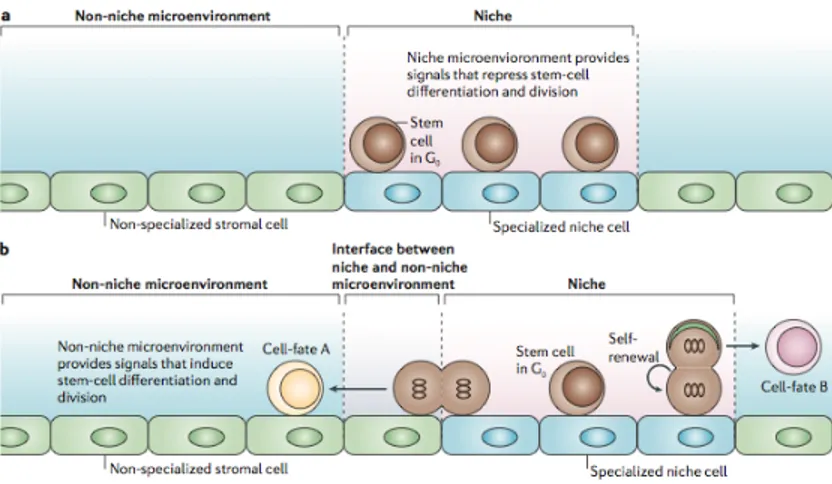
It is currently unclear whether all postulated stem-cell-niche functions (storage of quiescent stem cells, self-renewal and inhibition of differentiation) can be provided by a single niche, or whether different types of niches coexist (Fig. 10). Resting (G0)
stem cells are stored in quiescent niches. Specialized niche cells generate a differentiation- and/or division-repressive environment. Under conditions of stress these might be mobilized to generate mature cells as required, and might then return to empty niches for storage or self-renewal (Fig. 10a). The main function of a self-renewing niche would be to guarantee that (by environmental and/or divisional asymmetry) one of the two
daughters of a dividing stem cell maintains the stem-cell fate while the other produces differentiating progenitors (Murphy et al., 2005). Such a self-renewing stem-cell niche would be more complex than a quiescent-storage niche but would be the essential unit that maintains normal tissue homeostasis. In this type of niche, one can propose that quiescent stem cells would be anchored in the center of the niche, whereas self-renewing stem cells would be located close to the border separating the niche from the non-niche microenvironment, which could provide signals that would induce differentiation and/or cell division (Fig. 10b).
Figure 10: Different types of niches. a) Quiescent-storage niche. b)
Self-renewing niche.
The best-characterized stem-cell niche is the bone HSCs niche. The term ‘niche’ for the specific HSC bone-marrow microenvironment was first coined by Schofield, who proposed that HSCs are in intimate contact with bone, and that cell-cell contact was responsible for the apparently unlimited proliferative
More recently, several mutant mice, in which hematopoiesis is defective as a consequence of primary defects in bone development or remodeling, have implicated osteoblasts and/or osteoclasts in the formation and function of the bone-marrow HSC environment or niche. For example, mice lacking core binding factor α1 (CBFα1; also known as Runx2), which is one of the earliest osteoblast-specific transcription factors, have defective bone-marrow hematopoiesis and extensive extramedullary hematopoiesis, owing to defects in osteoblast differentiation and the consequent failure to form bone (Deguchi et al., 1999; Ducy et al., 2000). A direct role for the involvement of osteoblasts in HSC regulation and/or maintenance in vivo has been obtained from two studies in which osteoblast numbers were experimentally increased or decreased. In the first study osteoblast-specific expression of a constitutively active form of parathyroid hormone (PTH) or the PTH/PTH-related protein receptor (PPR), which is an important regulator of calcium homeostasis, and therefore bone formation and resorption, was achieved using the type 1 collagen α1 (Col1a1) promoter. This resulted in a simultaneous increase in the number of both osteoblasts and HSCs in the bone marrow. Moreover, the maintenance of HSCs in vitro was more efficient when supported by stromal cells that were isolated from these transgenic mice, presumably because of an increase in the proportion of osteoblasts in the stromal-cell population compared to stromal cells from wild-type mice (Calvi et al., 2003).
In a second study mice lacking bone morphogenetic protein (BMP) receptor 1A (BMPR1A, which is normally expressed on
osteoblasts lining the endosteum) in the bone-marrow stroma showed a simultaneous increase in the number of both osteoblasts and repopulating HSCs, although the number of more differentiated cells remained unchanged (Zhang et al., 2003). Moreover, Lin– LRCs and osteoblasts were shown to be in direct contact through homotypic N-cadherin interactions. Therefore, specialized spindle-shaped N-cadherin-expressing osteoblasts (SNOs) located in the endosteum were postulated to be essential components of the HSC bone-marrow niche. Both studies show that an increase in the number of osteoblasts directly correlates with the number of functional LTR HSCs, indicating that osteoblasts (or a subset of these cells) are an essential part of the niche and are limiting for niche size and/or activity. Although N-cadherin-expressing PPR+BMPR1A+ osteoblasts seem to be necessary and rate-limiting for niche function, it is probable that other cell types, such as osteoclasts, stromal fibroblasts and endothelial cells, also contribute to niche formation, activity or architecture. In fact, activating osteoclasts through either RANKL (necessary for osteoclastogenesis) or stress results in mobilization of the HSCs into the circulation (Fig. 11) (Kollet et al., 2006; Purton and Scadden, 2006).
Figure 11: Osteoblasts and osteoclasts seem to participate as cellular components of the endosteal hematopoietic stem cell niche. Activation of
the respective cellular elements are associated with different phenomena. Increasing osteoblast activity through stimulation of the parathyroid hormone receptor or inactivation of the bone morphogenic protein receptor-1a results in an increase in the number of hematopoietic stem cells (HSC). In contrast, activating osteoclasts through either RANKL or stress results in mobilization of the HSC into the circulation.
The presence of a second specialized HSC microenvironment in the bone marrow has recently been postulated, as a large proportion of CD150+ HSCs were observed to be attached to the fenestrated endothelium of bone-marrow sinusoids (Kiel et al., 2005). A close interaction between HSCs and endothelial cells is not unexpected because both lineages arise from a common embryonic precursor, the hemangioblast (Huber et al., 2004). Bone-marrow sinusoidal endothelial cells constitutively express cytokines such as CXC-chemokine ligand 12 (CXCL12) and adhesion molecules such as endothelial-cell (E)-selectin and vascular cell-adhesion molecule 1 (VCAM1) that are important for HSC mobilization, homing and engraftment (Fig. 13) (Rafii et al., 1997; Avecilla et al., 2004; Sipkins et al., 2005). A vascular bone-marrow HSC niche has previously been predicted to form during HSC mobilization after myeloablation. Quiescent HSCs detach from the endosteal niche and migrate towards the center of the bone marrow to the vascular zone from where they re-establish hematopoiesis (Heissig et al., 2002; Kopp et al., 2005). The recent finding that CD150+ HSCs are attached to the sinusoidal endothelium now raises the possibility that a vascular bone-marrow HSC niche might also exist during homeostasis (Kiel et al., 2005). Why have two apparently distinct HSC niches
in the bone marrow? Putative HSCs that have been identified by LRC assays are almost exclusively located in the endosteal niche, indicating that this niche might contain the most dormant HSCs and therefore serve as a quiescent-storage niche, or a self-renewing niche comprising both quiescent and self-self-renewing HSCs. In contrast to label-retaining HSCs that have not divided for many weeks, the CD150+ HSC population comprises both long-term quiescent and self-renewing HSCs, because 3.8% of the cells are proliferating at any given time. Because many of the proliferating cells are in contact with bone-marrow sinusoidal endothelial cells, it is probable that the vascular bone-marrow HSC niche contains self-renewing, rather than long-term dormant, HSCs. The location of CD150+ HSCs, in close proximity to sinusoids, would enable them to constantly monitor the concentration of blood-borne factors that reflect the status of the hematopoietic system. Under hematological stress, a rapid and robust response could be mounted, and if necessary more HSCs could be recruited from endosteal niches (Fig. 12). It is probable that the pool of HSCs located in the vascular and self-renewing endosteal niches are freely exchanged to maintain homeostasis in a constantly changing hematopoietic environment. In addition, HSCs that are located in the self-renewing endosteal niche produce multipotential progenitors (MPPs) by divisional and/or environmental asymmetry (Fig. 12). Because deletion of osteoblasts results in extramedullary hematopoiesis (Visnjic et al., 2004), the vascular bone-marrow HSC niche alone might not be sufficient to maintain long-term hematopoiesis. This indicates that in the bone marrow the vascular niche might be a secondary
niche, requiring an influx of HSCs from the primary endosteal niches (Fig. 12). Collectively, the vascular and endosteal niches strongly cooperate to control HSC quiescence and self-renewing activity (and therefore HSC number), as well as the production of early progenitors to maintain homeostasis or re-establish it after injury.
Figure 12: Model of bone-marrow HSCs niches.
Although the vast majority of HSCs in the adult mouse are located in the bone marrow, HSCs show remarkable motility. In response to specific signals they can exit and re-enter the endosteal bone-marrow HSC niche, processes known as mobilization and homing, respectively (Fig. 13). These opposing biological processes are controlled by overlapping but distinct
molecular mechanisms (Papayannopoulou, 2003; Lapidot et al., 2005; Cancelas, 2005). HSCs bound to the bone-marrow niche are mobilized in response to granulocyte colony-stimulating factor (G-CSF) or cyclophosphamide, or after peripheral myeloablation following treatment with 5-fluorouracil (5-FU). After extravasation from the bone-marrow cords into the microvasculature, HSCs enter the circulation and are distributed to peripheral tissues such as the spleen or liver. HSCs locate close to endothelial cells in the splenic red pulp. They home to the bone-marrow cords through the circulation, a process that is controlled by a number of adhesion molecules such as very late antigen 4 (VLA4), VLA5, lymphocyte function-associated antigen 1 (LFA1) or selectins. After entering the bone marrow, HSCs specifically lodge in the niche, a process requiring membrane-bound stem-cell factor (SCF), CXC-chemokine ligand 12 (CXCL12), osteopontin (OPN), hyaluronic acid, and their corresponding receptors (Fig. 13) (reviewed in Wilson and Trumpp, 2006).
Although little is known about the signals that are exchanged between HSCs and osteoblasts in situ, several receptors, membrane-anchored proteins and secreted factors are expressed by both cell types (Taichman, 2005). Comparative gene-expression profiling has recently been performed on HSC-supporting and non-HSC-supporting stromal cell lines, identifying a number of new molecules that might regulate endosteal bone-marrow HSC-niche activity (Fig. 14).
Several Notch receptors and Notch-receptor ligands are expressed in the bone marrow (Radtke et al., 2004), leading to the suggestion that Notch signaling has a role in HSC self-renewal and/or clonal expansion. Support for this hypothesis has been provided by in vitro culture of purified HSCs on various stromal cell lines (Varnum-Finney et al., 2000; Duncan et al., 2005). Moreover, as expression of the Notch ligand Jagged-1 is upregulated on osteoblasts that are exposed to PTH, the concomitant increase in HSCs has been postulated to be caused by increased Notch signaling (Calvi et al., 2003).
One mechanism by which osteoblasts might regulate the number of HSCs in the bone marrow is through secretion of osteopontin (OPN), an acidic glycoprotein, into the bone matrix (Denhardt and Guo, 1993). OPN-deficient mice have a two-fold increase in HSCs and, because the same effect was observed by transplanting wild-type HSCs into lethally irradiated OPN mutant recipients, OPN production by osteoblasts has a negative effect on HSC number (Stier et al., 2005). In addition, OPN has been postulated to act as a negative regulator of HSCs by actively maintaining their quiescence (Nilsson et al., 2005).
The steel (Sl) locus encodes both membrane-bound SCF and secreted SCF. The latter is produced by alternative splicing followed by proteolytic cleavage of membrane-bound SCF (Flanagan et al., 1991). SCF binds and activates KIT, which is expressed by LTR HSCs as well as other stem cells. Mutations at either of these loci affect migration and differentiation of primordial germ cells, neural-crest-derived melanoblasts, and hematopoietic cells (Lyman et al., 1998). Analysis of the different SCF and KIT mutant mice showed that although not essential for the generation and initial clonal expansion of HSCs in the embryo and fetal liver, they are crucial for long-term maintenance and self-renewal of adult HSCs, raising the possibility that the SCF-KIT pathway mediates endosteal bone-marrow HSC niche activity (Fig. 14). Importantly, membrane-bound SCF is expressed by osteoblasts and has a higher and more sustained capacity to activate KIT on the cell surface of HSCs than secreted SCF (Miyazawa et al., 1995). In addition, membrane-bound SCF is a potent stimulator of adhesion of HSCs or hematopoietic progenitor cells to stromal cells (Kinashi and Springer, 1994) because it can activate VLA4 and VLA5, indicating that membrane-bound SCF can affect the adhesive properties of the endosteal niche by modifying the functional state of specific integrins (Kovach et al., 1995).
N-cadherin is expressed by both SNOs and a subset of LSK HSCs (Wilson et al., 2007). In addition, N-cadherin expression by HSCs localizes asymmetrically to the side of their attachment to SNOs. Therefore, homotypic N-cadherin interactions have been postulated to be an important component of the anchor that
links HSCs to SNOs in the endosteal niche. Indirect support for the importance of N-cadherin has been obtained from studies showing that MYC and tyrosine kinase receptor 2 (TIE2) control N-cadherin expression by HSCs in an antagonistic manner. The effects of MYC and TIE2 on HSCs and on N-cadherin expression correlate with a key function for N-cadherin in the retention of HSCs in the endosteal niche (Arai et al., 2004; Suda et al., 2005; Murphy et al., 2005).
In adult bone marrow, TIE2 (which is expressed specifically by LT-HSCs) is activated by angiopoietin-1 (ANG1), which is secreted by osteoblasts, leading to upregulation of N-cadherin expression by HSCs, providing the first example of a secreted factor promoting HSC-osteoblast adhesion. Interestingly, the ANG1-TIE2 signaling pathway prevents HSC division and maintains HSC quiescence, both in vitro and in vivo (Fleming et al., 1993; Cheng et al., 2000). Collectively, these data strongly support the hypothesis that N-cadherin-expressing ANG1+ osteoblasts form a niche that maintains quiescence and prevents self-renewal or differentiation through TIE2 signaling (Fig. 14). TIE2-mediated quiescence is potentially caused by positively regulating the cyclin-dependent-kinase inhibitor p21 (also known as CIP1 and WAF1). HSCs express high levels of p21, and mice lacking p21 show increased HSC proliferation at the expense of long-term self-renewal, indicating that p21 is essential for maintenance of quiescence in HSCs. In contrast to TIE2, transcription of the gene encoding p21 is negatively regulated by MYC, which is expressed at low levels by HSCs but increases
during initiation of HSC differentiation in a converse expression pattern to that of p21 (Cheng et al., 2001; Wu et al., 2003).
It has recently been shown that the transmembrane metallo- proteinase ADAM10 (a disintegrin and metalloproteinase-10) is able to cleave N-cadherin that is expressed at the cell surface of fibroblasts and neuronal cells. This leads to the redistribution of β-catenin (which is associated with the intracellular portion of N-cadherin) from the cell surface to the cytoplasmic β-catenin pool, thereby decreasing the signaling threshold required for the expression of target genes of the canonical WNT signaling pathway (which is mediated through β-catenin signal transduction cascades), such as the genes encoding cyclin D1 and MYC (Reiss et al., 2005). A similar re-distribution of β-catenin has also been reported after E-cadherin cleavage (Ito et al., 1999), indicating that high levels of expression of cadherins, as observed for HSCs, might decrease cytoplasmic β-catenin levels and therefore negatively regulate expression of β-catenin target genes. It is intriguing that the N-cadherin, TIE2, MYC, p21 and β-catenin pathways are apparently interconnected, leading to the suggestion that they might cooperatively control quiescence, self-renewal and initiation of HSC differentiation through interaction with the niche (Murphy et al., 2005).
Figure 14: A model of the endosteal niche-stem-cell synapse.
The picture emerging from accumulating genetic and functional data indicates that molecular crosstalk between HSCs and niche cells (particularly osteoblasts) involves a large number of molecules (cadherins, integrins, chemokines, cytokines, signaling molecules and receptors) that mediate at least two types of interaction. First, adhesive cell-extracellular-matrix (ECM) interactions such as CD44 binding to OPN or hyaluronic acid, and cell-cell interactions, such as those mediated by heterotypic VLA4-VCAM1 interactions and homotypic N-cadherin interactions. The main function of these interactions would be to maintain HSCs in close proximity to cells in the endosteal bone-marrow niche. In addition, most adhesion receptors are also linked to intracellular signaling cascades and actively participate in the signaling network controlling HSC maintenance. Second, ligand-receptor interactions, through which intracellular signaling