See discussions, stats, and author profiles for this publication at: https://www.researchgate.net/publication/305778956
In the Treatment of Subacromial Impingement Syndrome Is Extra Corporeal
Shock Wave Therapy Combined With Isokinetic Exercise for Rotator Cuff
More Effective Than Extra Corporeal Sh...
Article · July 2016 DOI: 10.2519/jospt.2016.4629 CITATIONS 4 READS 296 12 authors, including:
Some of the authors of this publication are also working on these related projects: GreatAge Study: Aging in PopulationView project
The Italian Longitudinal Study on AgingView project Andrea Santamato
Università degli studi di Foggia
116PUBLICATIONS 2,211CITATIONS
SEE PROFILE
Francesco Panza
Università degli Studi di Bari Aldo Moro
426PUBLICATIONS 16,266CITATIONS
SEE PROFILE
Angela Notarnicola
Università degli Studi di Bari Aldo Moro
118PUBLICATIONS 953CITATIONS
SEE PROFILE
Francesca Fortunato
Università degli studi di Foggia
12PUBLICATIONS 140CITATIONS
[
research
report
]
R
otator cuff tendinopathy
includes external or inter-
nal impingement,
ten-dinitis, tendinosis with
degeneration, and partial-thickness ten-don tears.22 The mechanisms of rotator cuff tendinopathy have been classified as extrinsic, intrinsic, or a combination of both.62 Extrinsic mechanisms are those causing compression of the rotator cuff tendons, and intrinsic mechanisms are those associated with degeneration of the rotator cuff tendon.72 Neer60 proposed an extrinsic mechanism for the etiology of rotator cuff tendinopathy, with compres-sion of the rotator cuff tendons and asso-ciated tissues between the anteroinferior corner of the acromion and the greater tuberosity of the humerus,19 and coined the term subacromial impingementsyn-drome (SAIS).61 The diagnosis of SAIS inherently implies an extrinsic compres-sion mechanism due to narrowing of the T
TSTUDY DESIGN: Single-blind randomized trial.
T
TBACKGROUND: Extracorporeal shockwave therapy (ESWT) has been shown to produce good results in the treatment of subacromial impinge-ment syndrome (SAIS). The efficacy of a combined administration of ESWT and isokinetic exercise (IE) has not yet been studied.
T
TOBJECTIVES: To evaluate the efficacy of focused ESWT combined with IE for the rotator cuff versus focused ESWT alone in the treatment of SAIS. The secondary objective was to assess the isokinetic torque recovery (external rotation at 210°/s, 180°/s, and 120°/s).
T
TMETHODS: Thirty participants with SAIS were randomly assigned to a focused-ESWT group or focused ESWT–plus-IE group. Subjects of both groups received 3 treatment sessions of focused ESWT over a period of 10 days. Participants in the second group also received IE for 10 therapy ses-sions. Outcome measures were the Constant-Mur-ley score (CMS), the visual analog scale (VAS), and isokinetic parameters (peak torque and total work calculated from 5 repetitions) measured with the isokinetic test. Subjects were assessed at baseline, 10 days after the last treatment session with
focused ESWT, and after 2 months of follow-up.
T
TRESULTS: At 2 months posttreatment, partici-pants in the focused ESWT–plus-IE group showed significantly less pain (focused-ESWT VAS, 3.4 ± 0.8 versus focused ESWT–plus-IE VAS, 1.5 ± 0.5;
P<.001) and greater improvement in
function-ality (focused-ESWT CMS, 75.9 ± 6.7 versus focused ESWT–plus-IE CMS, 92.1 ± 6.3; P<.001) and muscle endurance than the subjects in the focused-ESWT group.
T
TCONCLUSION: In subjects with SAIS, combined administration of focused ESWT and IE for the rotator cuff resulted in greater reduction of pain, as well as superior functional recovery and muscle endurance in the short to medium term, compared with ESWT alone.
T
TLEVEL OF EVIDENCE: Therapy, 2b. Trial reg-istration: unregistered 2011 trial. J Orthop Sports
Phys Ther 2016;46(9):714-725. Epub 5 Aug 2016. doi:10.2519/jospt.2016.4629
T
TKEY WORDS: Constant-Murley score, isokinetic
parameters, muscle torque, physical therapy, rotator cuff tendinopathy, shoulder pain, visual analog scale
1Department of Physical Medicine and Rehabilitation, OORR Hospital, University of Foggia, Foggia, Italy. 2Neurodegenerative Disease Unit, Department of Basic Medicine, Neuroscience,
and Sense Organs, University of Bari Aldo Moro, Bari, Italy. 3Department of Clinical Research in Neurology, University of Bari Aldo Moro, Tricase, Italy. 4Gerontology-Geriatrics Research
Laboratory, IRCCS Casa Sollievo della Sofferenza, San Giovanni Rotondo, Foggia, Italy. 5Orthopaedics and Traumatology Sections, Department of Basic Medicine, Neuroscience, and Sense
Organs, University of Bari Aldo Moro, Bari, Italy. 6Section of Hygiene, Department of Medical and Surgical Sciences, University of Foggia, Foggia, Italy. 7Dementia Research Group, School
of Clinical Sciences, University of Bristol, Frenchay Hospital, Bristol, UK. 8Physical Medicine and Rehabilitation Section, Department of Basic Medicine, Neuroscience, and Sense Organs,
University of Bari Aldo Moro, Bari, Italy. The Institutional Review Board of the University of Foggia (Foggia, Italy) approved this study. This research was supported by Programmi di Ricerca Scientifica di Rilevante Interesse Nazionale 2009 (grant 2009E4RM4Z). The authors certify that they have no affiliations with or financial involvement in any organization or entity with a direct financial interest in the subject matter or materials discussed in the article. Address correspondence to Dr Francesco Panza, Department of Clinical Research in Neurology, University of Bari Aldo Moro, Pia Fondazione Cardinale G. Panico, Tricase, Lecce, Italy. E-mail: [email protected] T Copyright ©2016 Journal of Orthopaedic & Sports Physical Therapy®
ANDREA SANTAMATO, MD1 • FRANCESCO PANZA, MD, PhD2-4 • ANGELA NOTARNICOLA, MD5 • GENNARO CASSATELLA, MD1
FRANCESCA FORTUNATO, MD, PhD6 • JULA LAURA DE SANCTIS, MD1 • GIOVANNI VALENO, MD1 • PATRICK G. KEHOE, PhD7
DAVIDE SERIPA, PhD4 • GIANCARLO LOGROSCINO, MD, PhD2,3 • PIETRO FIORE, MD8 • MAURIZIO RANIERI, MD, PhD1
Is Extracorporeal Shockwave Therapy
Combined With Isokinetic Exercise
More Effective Than Extracorporeal
Shockwave Therapy Alone for
Subacromial Impingement Syndrome?
A Randomized Clinical Trial
subacromial space, which may not accu-rately represent all rotator cuff tendon pathology.72 A unique extrinsic mecha-nism, internal impingement, described particularly in overhead athletes,6,48 is at-tributed to compression of the posterior articular surface of the tendons between the humeral head and glenoid when the arm is in full external rotation, abduc-tion, and extension,15 and is not related to subacromial-space narrowing. In con-trast to extrinsic mechanisms of rotator cuff tendinopathy, Codman and Akerson9 postulated an intrinsic mechanism due to degeneration within the tendon with tensile/shear overload, including altera-tions in biology, mechanical properties, morphology, and vascularity, confounded by aging.75,78 Subacromial impingement syndrome is characterized by pain on the anterior/posterior and lateral shoulder, extended to the deltoid and biceps mus-cles at rest and during abduction; forced internal rotation; and resisted motions. Several causative mechanisms have been proposed, such as continuous lesions during the glenohumeral joint movement caused by subacromial contact, subcora-coid space, coracoacromial ligament, and coracoacromial articulation; alteration of acromial morphology3; alteration of arterial vascularization of the humeral head24,70; overuse syndrome; or altera-tions of the tensile properties of the su-praspinatus tendon.39 Moreover, scapular dyskinesia can be considered another im-portant mechanism of SAIS.29,56
Management of SAIS includes nu-merous interventions, depending on the severity of pain and the stage of the ten-don lesion as described by Neer61: stage 1, the presence of edema and hemorrhage; stage 2, deterioration of the tendon and bursa; or stage 3, bone spurs and par-tial- or full-thickness tendon rupture. Analgesic and nonsteroidal anti-inflam-matory drugs,2 steroid injections,5 and physical therapy treatment techniques have often been reported, with mixed results.74,80 Systematic reviews have sug-gested that physical therapy has not provided unequivocal results because of
the notable variability of the underlying lesions.26,47,79 However, according to the recommendations from the Philadelphia Panel, an expert panel on selected reha-bilitation interventions for shoulder pain, therapeutic exercise was an acceptable intervention for SAIS.45,64 The large, un-restricted range of motion (ROM) of the glenohumeral joint and limited inherent bony stability necessitate dynamic mus-cular stabilization to ensure normal joint arthrokinematics. Although pain can still reduce their efficacy, rehabilitative exer-cise approaches for the treatment of SAIS include stretching, isometric and isotonic exercises, Codman exercises, and active and passive ROM exercises.13,56 Isomet-ric and isotonic exercises are designed to strengthen the weakened rotator cuff musculature, thus restoring its ability to counteract the action of the deltoid mus-cle.52,58 Scapular-stability exercises are included in the rehabilitation of people with SAIS because electromyographical studies have highlighted increased activ-ity in the upper trapezius, with decreased activity in the serratus anterior and the middle and lower fibers of the trape-zius, and asynchronous timing deficits in subjects with SAIS.52,57 Application of isokinetic exercise (IE) and testing for the upper extremity may be very useful, due to the demanding muscular work re-quired in daily activities.53,55
Extracorporeal shockwave therapy (ESWT) has been suggested as a benefi-cial treatment for SAIS that may decrease the need for surgery.30,49,81 Shockwaves are defined as transient pressure oscil-lations that propagate in 3 dimensions and typically bring about a clear increase in pressure within a few nanoseconds.38 The process involves very rapidly rising positive pressure impulses from 5 to 120 MPa in around 5 nanoseconds, followed by a decrease to negative pressure values of –20 MPa.77 The theoretical benefits are reduction of calcification, promotion of soft tissue healing, and inhibition of pain receptors to achieve relief from pain.31,51 There are 2 types of ESWT, focused and radial, that differ in how they penetrate or
dissipate at the skin. Focused waves tar-get tissues with higher penetration power and impact than those of radial waves, which are directed radially at the skin.20 To date, many studies on employment of ESWT in treating tendinopathies have been published.8,12,25,28,30,31,33,36,49,51,63,67,73,76,81
Different studies on rehabilitation af-ter ESWT have verified the efficacy of ec-centric exercises; however, this is limited to the treatment of Achilles tendinopathy and jumper’s knee.43,68,69 The objective of the present study was to test ESWT for SAIS associated with isokinetic rehabilita-tion to correct related muscle dysfuncrehabilita-tion. Some studies have suggested that active and passive exercise may be useful in the management of SAIS.13,47 Furthermore, scapular and humeral kinematic abnor-malities can cause dynamic narrowing of the subacromial space, leading to rotator cuff tendon compression secondary to superior translation of the humeral head or aberrant scapular motion that causes the acromion to move inferiorly.29,52 The muscle-strengthening exercise is usu-ally integrated in the treatment program to maintain the regained ROM, prevent disuse atrophy, and restore muscle func-tion. Malliou and colleagues53 compared different training methods for improving muscular performance in the shoulder rotator cuff. They determined that iso-kinetic strengthening is the most effective method of altering strength ratios. Beneka and colleagues1 found that the isokinetic method was more effective at increasing rotator strength. The restoration of imbal-ances may be due to strengthening of the internal and external rotators. To the au-thors’ knowledge, no studies to date have tested the possible benefits of ESWT and IE for SAIS. The aim of this study was to compare the effectiveness of 2 protocols for the treatment of SAIS (focused ESWT alone and focused ESWT combined with IE) in a single-blind randomized trial. The 2 approaches were compared us-ing traditional assessment scores (visual analog scale [VAS] and Constant-Murley score [CMS]).11,65 The secondary objective was to assess the isokinetic torque recov-Journal of Orthopaedic & Sports Physical Therapy® Downloaded from www.jospt.org at on September 5, 2016. For personal use only. No other uses without permission. Copyright © 2016 Journal of Orthopaedic & Sports Physical Therapy®. All rights reserved.
[
research
report
]
ery (external rotation at 210°/s, 180°/s, and 120°/s).
METHODS
Setting and Participants
T
he present study was asingle-blind randomized trial conducted according to the Declaration of Helsinki, the guidelines for good clini-cal practice, and the Consolidated Stan-dards of Reporting Trials (CONSORT) statement guidelines.71 The Institutional Review Board of the University of Foggia (Foggia, Italy) approved the study proto-col. Consecutive outpatients older than 18 years of age and with pain in the shoulder for at least 4 weeks prior to the study were potentially eligible to participate in this study. Individuals who had clinical signs of unilateral SAIS, attending the Depart-ment of Physical Medicine and Rehabili-tation at OORR Hospital (University of Foggia) from January 2011 to July 2011, were invited to participate in the study and were screened for study eligibility.
Diagnostic criteria for SAIS were the presence of shoulder pain, pain on ab-duction of the shoulder with painful arc, a positive impingement sign (Hawkins sign),32 and a positive impingement test (relief of pain within 15 minutes after in-jection of local anesthetic [bupivacaine 5 mL] into the subacromial space). All pa-tients were also evaluated by radiography, ultrasonography, or magnetic resonance imaging (MRI) of the shoulder (for MRI, extremely low acromiohumeral, coraco-humeral, and coracoclavicular intervals were quantitative measures for SAIS di-agnosis)35,42 to confirm the diagnosis of stage 1 (presence of edema and hemor-rhage) or 2 (deterioration of the tendon and bursa) SAIS, according to Neer’s classification.61 We used the diagnostic criteria for ultrasonography described by Naredo and colleagues.59 This technique included a dynamic examination of the supraspinatus tendon obtained by mov-ing the patient’s arm from a neutral posi-tion to 90° of abducposi-tion in order to detect encroachment of the acromion into the
rotator cuff. Subjects were excluded from the study if they met any of the follow-ing criteria: shoulder affected bilaterally, receiving anesthetic or corticosteroid in-jections within 4 weeks of study enroll-ment, surgery or previous fractures of the ipsilateral humeral head of the affected shoulder, a history of acute trauma, known osteoarthritis in the acromiocla-vicular or glenohumeral joints (identified by conventional radiography to assess ac-romion shape), calcifications exceeding 2 cm in the rotator cuff tendons or signs of a rupture of the cuff, cervical myofascial pain syndrome, radicular pain, diabetes mellitus type 1 or 2, thyroid dysfunctions,
pacemaker, neurological pathologies, and anxious depressive syndromes.
Concealed allocation was performed with random numbers generated be-fore the beginning of the study. Bebe-fore the study commenced, a random-inte-ger generator generated 100 random integers, and individual, sequentially numbered index cards with the random assignments were folded and placed in sealed, opaque envelopes. A physician member of the research team (A.N.), who was blinded to the baseline examination findings, opened the envelopes to allo-cate the interventions according to group assignments.
Consecutive subjects with SAIS screened for eligibility, n = 45
Consecutive subjects with SAIS screened for eligibility, n = 45
Eligible, n = 32
Eligible, n = 32
Agreed to participate and signed informed-consent statement, n = 30
Agreed to participate and signed informed-consent statement, n = 30
Allocated to focused ESWT alone, n = 15
Allocated to focused ESWT
alone, n = 15 Allocated to focused ESWT Allocated to focused ESWT plus IE, n = 15plus IE, n = 15
Received allocated intervention, n = 15
Received allocated intervention, n = 15
Not eligible, n = 13 • Previous fractures of the
humeral head of the affected shoulder, n = 2 • Rupture of the cuff, n = 3 • Neoplastic pathology,
n = 2
• Diabetes mellitus type 1 or 2, n = 4 • Patients with a
pacemaker, defibrillator, or neurostimulator, n = 2
Not eligible, n = 13 • Previous fractures of the
humeral head of the affected shoulder, n = 2 • Rupture of the cuff, n = 3 • Neoplastic pathology,
n = 2
• Diabetes mellitus type 1 or 2, n = 4 • Patients with a pacemaker, defibrillator, or neurostimulator, n = 2 Random assignment Random assignment Declined to participate, n = 2 Declined to participate, n = 2 Analyzed, n = 15 Analyzed, n = 15 Received allocated intervention, n = 15 Received allocated intervention, n = 15 Analyzed, n = 15 Analyzed, n = 15
FIGURE. Flow diagram of subject retention and recruitment for focused ESWT alone and ESWT plus IE for SAIS.
Abbreviations: ESWT, extracorporeal shockwave therapy; IE, isokinetic exercise; SAIS, subacromial impingement syndrome.
Outcome Measures
All participants underwent clinical ex-amination that was performed by 1 physician, who was blinded to group assignment, using the VAS65 and CMS as primary outcome measures11 and the isokinetic test as the secondary outcome measure.40
The VAS, a 10-cm line with a left end point of “no shoulder pain” and a right end point of “worst pain ever,” was used to measure pain65 during external rotation and abduction movements of the affected shoulder. In the acute pain setting, the VAS has been shown to have very good test-retest reliability (intraclass correla-tion coefficient [ICC] = 0.99).4 The VAS is generally accepted as a valid measure of acute pain, with good construct va-lidity.41,84 At a consensus meeting of the Initiative on Methods, Measurement, and Pain Assessment in Clinical Tri-als (IMMPACT), the results of several studies suggested that a score change of approximately 1 point or 15% to 20% rep-resents the minimal clinically important difference (MCID) for the VAS and for similar numeric rating scales (0-10) for pain intensity.14
The CMS is a 100-point scoring sys-tem of which 35 points are from patient self-report of pain and function and the remaining 65 points are allocated to ob-jective assessment of ROM and strength.
The self-report assessment includes a single item for pain (15 points) and 4 items for activities of daily living (work, 4 points; recreation, 4 points; sleep, 2 points; and ability to work at various levels, 10 points). The objective assess-ment includes ROM (forward elevation, 10 points; external rotation, 10 points; internal rotation, 10 points; abduction, 10 points) and power (scoring based on the number of pounds of pull the patient can resist in abduction, to a maximum of 25 points).11 The CMS has good psy-chometric properties, reflecting shoulder function with accuracy, test-retest reli-ability (ICC = 0.80),10 and reproducibil-ity.23 Unfortunately, to date, there are no studies to provide data on the MCID for the CMS, despite the fact that the error estimates (95% confidence interval of the standard error of measurement [SEM], ±17.7)10 and responsiveness (standardized response mean, 0.59),44 that is, the ability of a measure to detect change over time, have been reported.
Isokinetic testing has been advocated as a reliable method for quantifying many parameters of shoulder muscle func-tion.27,40 Testing should be comfortable and safe, and result in reproducible and accurate measurements of specific func-tions, such as muscle torque of the rotator cuff. We evaluated the peak torque and total work calculated from 5 repetitions
of external rotation with the arm in the scapular plane (45° of abduction, 30° of flexion, and the elbow in 90° of flexion), to compare the results of the isokinetic test (concentric testing) after 5 sessions at each of 3 different speeds (120°/s, 180°/s, and 210°/s). The position cho-sen for isokinetic assessment of shoulder rotators allowed the best reproducibility and reliability for internal and external rotators.21 These speeds were chosen to avoid further tendon impairments with higher resistance. All subjects performed the isokinetic test using the HUMAC NORM for CYBEX dynamometer (Com-puter Sports Medicine Inc, Stoughton, MA). Patient stabilization, in a seated po-sition, was achieved with 4 Velcro straps: 1 across the hips, 2 across the chest, and 1 around the forearm.17 The unaffected side was tested first, followed by the affected side. Peak torque and total work calcu-lated from 5 repetitions were used to as-sess muscular impairment. All isokinetic testing was conducted by 1 investigator (G.V.), who was blinded to group allo-cation. The results are presented as the difference between mean values of the affected and the unaffected side.
Interventions
After baseline evaluation, both treatment groups were treated with 3 sessions over 10 days (ie, each session was followed by a 3-day interval) of medium-energy (0.12 mJ/mm2) focused ESWT, administered with an electrohydraulic lithotripter (EvoTron; High Medical Technologies AG, Lengwil, Switzerland). We chose 3 ESWT sessions after considering previ-ous studies describing a variable num-ber of treatments, ranging from 3 to 5, for shoulder pain in rotator cuff tendi-nopathy.37 Each participant received 700 pulses of shockwaves per session, focused on the localized area of the supraspinatus tendon insertion. All therapy was carried out without anesthesia. Subjects in the first group received focused ESWT alone. Ten days after the last focused ESWT, subjects in the second group received 10 additional sessions of IE, 3 times per
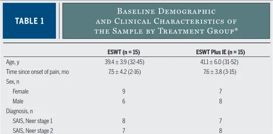
TABLE 1
and Clinical Characteristics of
Baseline Demographic
the Sample by Treatment Group*
Abbreviations: ESWT, extracorporeal shockwave therapy; IE, isokinetic exercise; SAIS, subacromial impingement syndrome.
*Values are mean ± SD (range) unless otherwise indicated.
ESWT (n = 15) ESWT Plus IE (n = 15)
Age, y 39.4 ± 3.9 (32-45) 41.1 ± 6.0 (31-52) Time since onset of pain, mo 7.5 ± 4.2 (2-16) 7.6 ± 3.8 (3-15) Sex, n
Female 9 7
Male 6 8
Diagnosis, n
SAIS, Neer stage 1 8 7 SAIS, Neer stage 2 7 8
[
research
report
]
week. We chose this time interval before starting with IE, given the possible pain increase during ESWT. Each subject was positioned for eccentric/concentric shoulder internal/external rotation in 45° of shoulder abduction, 30° of flexion, and the elbow in 90° of flexion.21 The training protocol consisted of 3 sets of 10 repeti-tions during the first week at the concen-tric and eccenconcen-tric speeds of 240°/s and 180°/s. During the second week, training was progressed to 4 sets of 10 repetitions. In the last week, the concentric speed was decreased to 120°/s, while the eccentric speed was decreased to 90°/s.34 Further-more, in this final week of training, the participants received 5 repetitions of iso-metric training at 30°/s of external rota-tion. We used training speeds reported in previous studies.34 The rationale for
decreasing speeds over time in the train-ing program is that low speeds can stress rotator cuff tendons during the perfor-mances, so the progressive increase of training enhances rotator cuff muscle strength. Subjects were assessed at base-line (before the first treatment session), 10 days after the last treatment session with focused ESWT, and 2 months af-ter the last session with focused ESWT. The ethical committee stipulated that all participants could receive other interven-tions that were not part of the research protocol of the present study in case of failure after 2 months. After 6 months from baseline, a phone interview with all subjects treated was made to assess clinical improvements and, in case of insufficient treatment success, to allow for other physical therapy interventions.
Therefore, our purpose was to examine the effectiveness of isokinetics for im-proving power output with isokinetic shoulder measurements in order to es-tablish shoulder strength values and bal-ance ratios (conventional and functional) that can be employed in preventive, train-ing, and rehabilitation strategies for shoulder injuries. Participants received no other physical therapy intervention for shoulder pain during the study and 4 to 5 weeks prior to the study. The sub-jects were instructed to avoid analgesic/ anti-inflammatory drugs for the dura-tion of the physical therapy period and to abstain from the execution of painful activities of daily living involving the af-fected shoulder.
Statistical Analysis
To assess the distribution of the vari-ables, we used the Bartlett test. The data for explored variables (ie, VAS and CMS scores) and isokinetic data (peak torque, total work) at 210°/s, 180°/s, and 120°/s were analyzed using separate 2-by-3 (group-by-time) mixed-model analyses of variance (P values less than .05 were significant). With regard to the isokinet-ic variables, the variables analyzed were difference between mean values of peak torque and total work between affected and unaffected side. For power analysis and sample-size estimation, G*Power Version 3.1.10 software (Heinrich-Heine Universität, Düsseldorf, Germany) was used. Data analyses were performed us-ing Stata/MP Version 10.1 (StataCorp LP, College Station, TX). Our sample of 15 patients in each group provided a power greater than 80% to detect a difference of 3.5 points on the VAS between the 2 measurements, assessing an SD of 1, cor-relation of 0.7, and alpha of .05.
RESULTS
A
total of 45 consecutiveoutpa-tients (25 women and 20 men) were screened for study eligibility. At the end of the evaluation, 30 individuals with SAIS who fulfilled the selection criteria
TABLE 2
Within- and Between-Group
Differences of All Outcome Measures
of Participants Who Received Focused
ESWT Alone and Focused ESWT Plus IE
Abbreviations: CMS, Constant-Murley score; ESWT, extracorporeal shockwave therapy; IE, isokinetic exercise; VAS, visual analog scale.
*Values are mean ± SD unless otherwise indicated.
†Values are mean difference (95% confidence interval) unless otherwise indicated.
Measure Baseline 10 d Posttreatment 2 mo Posttreatment P Value
Outcomes* VAS ESWT 8.1 ± 0.6 5.1 ± 0.9 3.4 ± 0.8 <.001 ESWT plus IE 8.2 ± 0.8 4.9 ± 1.3 1.5 ± 0.5 <.001 P value >.05 <.001 CMS ESWT 49.7 ± 7.9 65.1 ± 7.7 75.9 ± 6.7 <.001 ESWT plus IE 45.6 ± 9.8 63.6 ± 8.7 92.1 ± 6.3 <.001 P value >.05 <.001 Within-group change score
from baseline† VAS ESWT –3.0 (–3.6, –2.5) –4.7 (–5.3, –4.2) ESWT plus IE –3.3 (–4.0, –2.6) –6.7 (–7.2, –6.2) CMS ESWT 15.4 (12.5, 18.2) 26.2 (23.2, 29.2) ESWT plus IE 18.0 (14.7, 21.3) 46.5 (41.9, 51.2) Between-group difference in change score† VAS –0.2 (–0.8, 0.4) –1.9 (–2.5, –1.4) CMS –1.5 (–5.4, 2.5) 16.2 (12.6, 19.8)
and agreed to participate were enrolled in the study (16 women, 14 men; mean ± SD age, 40.2 ± 5.0 years). Written informed consent was obtained from all subjects. Reasons for exclusion are shown in a flow diagram of subject recruitment and retention (FIGURE). Enrolled participants were randomly assigned to 1 of 2 groups: 15 subjects (9 women, 6 men; mean ± SD age, 39.4 ± 3.9 years) who received focused ESWT alone, and 15 individuals (7 women, 8 men; mean ± SD age, 41.1 ± 6 years) who received focused ESWT plus IE. All 30 participants completed the trial and were included in the analy-sis (TABLE 1). None of the participants reported adverse effects or reported tak-ing analgesic/anti-inflammatory drugs during the period of their participation in the study. There was a statistically sig-nificant group-by-time interaction for the 2-by-3 mixed-model analyses of variance for VAS and CMS scores, and for all iso-kinetic data (peak torque and total work at 210°/s, 180°/s, and 120°/s) (P<.05). In those who received focused ESWT and focused ESWT plus IE, a statistically significant decrease (P<.05) in VAS pain scores and a statistically significant in-crease (P<.05) in CMS scores were found 10 days after the last treatment session with focused ESWT and after 2 months of follow-up (TABLE 2). The VAS score re-duction was statistically greater in the fo-cused ESWT–plus-IE group (mean score, 1.5 ± 0.5) in comparison with the focused-ESWT group (mean score, 3.4 ± 0.8) af-ter 2 months of follow-up. Moreover, the CMS score was statistically greater in the focused ESWT–plus-IE group (mean, 92.1 ± 6.3) than in the focused-ESWT group (mean, 75.9 ± 6.7) after 2 months of follow-up (TABLE 2).
For isokinetic test data, considering the differences between affected and un-affected shoulders evaluated at baseline for patients of both groups, the positive effects of this treatment on rotator cuff tendons were associated with a statisti-cally significant reduction (P<.05) in affected versus unaffected shoulders for peak torque and total work output
at 210°/s, 180°/s, and 120°/s 10 days after the last treatment session with ESWT and after the 2-month follow-up
(TABLE 3). After the treatment, isokinetic test data for subjects receiving ESWT alone for the affected shoulder showed a
TABLE 3
Changes in Peak Torque and
Total Work Difference for External
Rotation Between Affected and Unaffected
Shoulders in Participants With Subacromial
Impingement Syndrome in the Group
With Focused ESWT Alone and in the Group
With Focused ESWT Plus IE
Measure Baseline 10 d Posttreatment 2 mo Posttreatment P Value
Outcomes*
External rotation at 210°/s Peak torque, Nm/kg body weight
ESWT 40.9 ± 20.7 21.8 ± 10.6 17.9 ± 7.9 <.001 ESWT plus IE 38.9 ± 25.9 21.3 ± 13.3 9.7 ± 6.9 <.001 Total work, J/kg body weight
ESWT 56.2 ± 17.5 41.7 ± 13.7 35.5 ± 12.1 <.01 ESWT plus IE 51 ± 19.1 42.8 ± 19.2 21.5 ± 16.1 <.001 External rotation at 180°/s
Peak torque, Nm/kg body weight
ESWT 46.7 ± 12.7 40.1 ± 11.7 31.0 ± 8.4 <.01 ESWT plus IE 43.6 ± 13.4 27.3 ± 14.7 14.6 ± 13.6 <.001 Total work, J/kg body weight
ESWT 47.1 ± 18.5 38.8 ± 19.1 28.0 ± 4.3 <.05 ESWT plus IE 44.7 ± 22.5 36.9 ± 21.1 15.5 ± 11.9 <.001 External rotation at 120°/s
Peak torque, Nm/kg body weight
ESWT 41.3 ± 10.1 28 ± 7.3 23.9 ± 8 <.001 ESWT plus IE 39.2 ± 14 22.5 ± 11.6 9.9 ± 9.2 <.001 Total work, J/kg body weight
ESWT 57.8 ± 10.1 42.3 ± 14.2 34 ± 10.4 <.001 ESWT plus IE 51.5 ± 12.5 44.3 ± 18.1 27.7 ± 12.8 <.001 Within-group change score from baseline†
External rotation at 210°/s Peak torque, Nm/kg body weight
ESWT –19.2 (–27.5, –10.7) –23.0 (–32.0, –13.9) ESWT plus IE –17.6 (–29.6, –5.6) –29.1 (–42.8, –15.4) Total work, J/kg body weight
ESWT –14.5 (–18.0, –10.9) –20.7 (–25.3, –16.1) ESWT plus IE –8.2 (–18.4, 2.1) –29.5 (–44.5, –14.5) External rotation at 180°/s
Peak torque, Nm/kg body weight
ESWT –6.6 (–8.8, –4.4) –15.7 (–18.7, –11.8) ESWT plus IE –16.3 (–23.8, –8.7) –29.0 (–37.4, –20.6) Total work, J/kg body weight
ESWT –8.3 (–12.8, –3.8) –19.1 (–25.2, –13.1) ESWT plus IE –7.8 (–17.1, 1.5) –29.2 (–38.9, –19.5)
Table continues on page 720.
[
research
report
]
statistically significant increase (P<.05) in mean values for external rotation peak torque at 210°/s and total work output at 210°/s, 180°/s, and 120°/s 10 days after the last treatment session with ESWT and after 2 months of follow-up (TABLE 4). Subjects who received both ESWT and IE showed a statistically significant increase (P<.05) in mean external rotation peak torque and total work output at 210°/s, 180°/s, and 120°/s 10 days after the last treatment session with ESWT and after 2 months of follow-up (TABLE 4).
Moreover, the effectiveness of IE for muscle functional recovery can be dem-onstrated considering isokinetic test data differences between both shoulders in both groups. The observed differences between affected and unaffected shoul-ders in peak torque at 180°/s were sig-nificantly reduced 10 days after the last treatment session with ESWT (P<.05) and after 2 months of follow-up (P<.001), while at 120°/s and 210°/s, peak torque was significantly reduced only after 2 months of follow-up (P<.001 and P<.01, respectively) in subjects in the focused ESWT–plus-IE group compared to sub-jects in the focused-ESWT group. In terms of total work output, these differ-ences were statistically reduced only after 2 months of follow-up at 210°/s (P<.05) and 180°/s (P<.05) in subjects in the fo-cused ESWT–plus-IE group compared to subjects in the focused-ESWT group. Fi-nally, after the 2-month follow-up, there was a statistically significant increase in total work output at 210°/s (P<.05), 180°/s (P<.05), and 120°/s (P<.001) in subjects in the focused ESWT–plus-IE group compared to subjects in the fo-cused-ESWT group.
During phone interviews 6 months after baseline, 9 subjects in the focused-ESWT group (60%) and 11 subjects in the focused ESWT–plus-IE group (73.3%) re-ferred to having an improvement of their clinical picture (P<.05). Five participants in the focused-ESWT group (33.3%) and 4 in the focused ESWT–plus-IE group (26.7%) reported worsened pain (P<.05) and underwent another type of physical
therapy. Only 1 subject in the focused-ES-WT group (6.7%) submitted to surgery.
DISCUSSION
I
n the present study, individuals with SAIS who received a combined intervention of focused ESWT plus IE reported reduced pain and showed successful functional recovery in the short to medium term. To date, no stud-ies have tested the possible benefits of ESWT and IE for SAIS. Among physi-cal modalities, ESWT represents an important treatment for a number of musculoskeletal conditions, including tendinopathies and enthesopathies.30,73 On the other hand, several studies have emphasized the positive effect of bothfocused and radial ESWT for the treat-ment of chronic calcific tendinitis of the shoulder.7,12,25,28,30,33,36,49,51,63,66,67,81 There is, however, less evidence to support the effectiveness of ESWT for SAIS without calcification.30,49,76,81 In the present study, following ESWT, participants in both groups reported a statistically signifi-cant improvement in the overall clinical picture as assessed by the VAS and CMS for pain and isokinetic testing. The first important result was the effectiveness of ESWT in the treatment of pain, function-ality, and muscle weakness resulting from SAIS. When the ESWT was combined with IE, the subjects showed a statistical-ly significantstatistical-ly greater reduction in pain and improvements in articular move-ment, functionality, and muscle
endur-TABLE 3
Changes in Peak Torque and
Total Work Difference for External
Rotation Between Affected and Unaffected
Shoulders in Participants With Subacromial
Impingement Syndrome in the Group
With Focused ESWT Alone and in the Group
With Focused ESWT Plus IE (continued)
Abbreviations: ESWT, extracorporeal shockwave therapy; IE, isokinetic exercise.
*Values are mean ± SD unless otherwise indicated.
†Values are mean difference (95% confidence interval) unless otherwise indicated.
Measure Baseline 10 d Posttreatment 2 mo Posttreatment P Value
Within-group change score from baseline†
(continued)
External rotation at 120°/s Peak torque, Nm/kg body weight
ESWT –13.3 (–16.1, –10.6) –17.4 (–29.9, –13.8) ESWT plus IE –16.7 (–20.2, –13.3) –29.3 (–35.5, –23.0) Total work, J/kg body weight
ESWT –15.5 (–19.5, –11.4) –23.8 (–26.8, –20.7) ESWT plus IE –7.2 (–15.8, 1.4) –23.8 (–31.5, –16.3) Between-group difference in change score†
External rotation at 210°/s
Peak torque, Nm/kg body weight –0.5 (–4.2, 3.2) –8.2 (–10.9, –5.4) Total work, J/kg body weight 1.1 (–5.7, 7.8) –14.0 (–23.3, –4.7) External rotation at 180°/s
Peak torque, Nm/kg body weight –12.8 (–22.2, –3.4) –16.4 (–23.6, –10.1) Total work, J/kg body weight –1.9 (–8.9, 5.1) –12.5 (–20.4, –4.7) External rotation at 120°/s
Peak torque, Nm/kg body weight –5.5 (–9.3, –1.7) –14.0 (–18.5, –9.4) Total work, J/kg body weight 2.0 (–3.9, 7.9) –6.3 (–9.7, –2.9)
ance of the affected shoulder compared with those treated only with ESWT. The contrasting findings of ESWT alone for
SAIS could be explained by the multi-factorial pathoetiology of this syndrome, caused not only by the degenerative
process of tendons but also by abnor-mal scapular and humeral kinematics, postural abnormalities, rotator cuff and scapular muscle performance deficits, and decreased extensibility of pectoralis minor or posterior shoulder tissues.29 Treatment of patients with impingement symptoms commonly includes exercises intended to restore “normal” movement patterns.
Based on these concepts, a combined treatment of ESWT and IE for reha-bilitation of SAIS may be an effective treatment protocol. Isokinetic exercise is useful in the treatment of shoulder dysfunction to increase the muscle’s endurance and torque, reducing the in-voluntary hyperactivity of rotator cuff muscles responsible for weakness and pain during shoulder movements.17,34 Therefore, the isokinetic test may play an important part in the measurement of muscular performance before and after a rehabilitative treatment. The objective documentation that the isokinetic test provides allows clinicians and research-ers to report muscle torque, power, work, and endurance as important outcome measures of an evidence-based reha-bilitation program after injury.17,50 The subjects enrolled in the present study received concentric external rotation ex-ercises for the affected shoulder because eccentric contraction induces significant engagement of the supraspinatus muscle tendon, which had just been challenged by ESWT. Furthermore, the supraspi-natus and infraspisupraspi-natus components of the rotator cuff contribute to a variable proportion of the total torque of abduc-tion (25%-50%) and external rotaabduc-tion (50%-75%) throughout the ROM, so the external rotation work helps to train these muscles predominantly.46 In fact, the purpose of IE is to increase muscle endurance. It is known that SAIS is not restricted to overuse activities, and also involves weakness and shoulder muscle impairment. Therefore, any improve-ment in total work, a parameter of muscle endurance, can be considered more use-ful than peak torque in reducing the risk
TABLE 4
Within-Group Differences for the Affected
Shoulder of Patients With Subacromial
Impingement Syndrome in the Group With
Focused ESWT Alone and in the Group With
Focused ESWT Plus IE in Isokinetic Testing
Data for External Rotation at 210°/s,
180°/s, and 120°/s
Measures Baseline 10 d Posttreatment 2 mo Posttreatment P Value
Outcomes*
External rotation at 210°/s Peak torque, Nm/kg body weight
ESWT 13.9 ± 4.7 14.8 ± 4.4 16.9 ± 3.2 <.05 ESWT plus IE 12.8 ± 2.9 12.8 ± 2.9 16.5 ± 3.3 <.05 Total work, J/kg body weight
ESWT 17.7 ± 8.7 23.1 ± 9.7 33.8 ± 9.6 <.001 ESWT plus IE 20.0 ± 16.1 28.1 ± 16.8 53.9 ± 27.3 <.001 External rotation at 180°/s
Peak torque, Nm/kg body weight
ESWT 13.4 ± 2.8 14.3 ± 2.9 16.9 ± 3.9 >.05 ESWT plus IE 12.0 ± 2.8 13.4 ± 2.8 17.5 ± 7.1 <.01 Total work, J/kg body weight
ESWT 18.6 ± 10.8 23.9 ± 11.2 34.7 ± 13.5 <.01 ESWT plus IE 22.9 ± 19.0 31.9 ± 18.1 62.4 ± 30.8 <.001 External rotation at 120°/s
Peak torque, Nm/kg body weight
ESWT 12.6 ± 4.1 14.3 ± 5.1 17 ± 5.9 >.05 ESWT plus IE 11.3 ± 2.5 12.5 ± 3.8 17.9 ± 9.1 <.01 Total work, J/kg body weight
ESWT 18.9 ± 7.3 23.8 ± 8.2 34.3 ± 8.7 <.001 ESWT plus IE 22.8 ± 12.9 26.3 ± 13.1 52.6 ± 16.2 <.001 Within-group change score from baseline†
External rotation at 210°/s Peak torque, Nm/kg body weight
ESWT 0.9 (0.01, 1.8) 3.0 (1.1, 4.9) ESWT plus IE 0.0 (–1.2, 1.2) 3.7 (2.3, 5.0) Total work, J/kg body weight
ESWT 5.4 (3.6, 7.2) 16.1 (13.5, 18.6) ESWT plus IE 8.1 (4.1, 11.9) 33.9 (23.4, 44.3) External rotation at 180°/s
Peak torque, Nm/kg body weight
ESWT 0.9 (0.08, 1.8) 3.5 (1.5, 5.6) ESWT plus IE 1.4 (0.2, 2.7) 5.5 (2.4, 8.6) Total work, J/kg body weight
ESWT 5.3 (3.2, 7.5) 16.1 (11.4, 20.7) ESWT plus IE 9.0 (2.8, 15.2) 39.5 (27.0, 52.1)
Table continues on page 722.
[
research
report
]
of recurrence of painful shoulder tendini-tis. After ESWT, some studies suggested the efficacy of eccentric exercises for the treatment of Achilles tendinopathy and jumper’s knee,43,68,69 confirming the pres-ent findings on SAIS. Moreover, evidence from a recent randomized controlled trial indicated that supervised exercises were more effective than radial ESWT for the treatment of subacromial shoulder pain in the short to medium term.16 No sig-nificant difference was found between supervised exercises and radial ESWT at 1 year of follow-up.18 These findings con-firmed the rationale of the present study of adding IE to ESWT in the treatment of SAIS.
The improvements in the VAS, CMS, and isokinetic data could be explained
by the positive metabolic effect of ESWT on tendons and pain.54,82 Wang and col-leagues83 also stated that ESWT caused tissue healing, whereas other authors have hypothesized that increases in trans-forming growth factor β1 and insulin-like
growth factor 1 expression may mediate mitogenic and anabolic responses of tendon tissue and tenocytes, as well as neovascularization, that contribute to the success of ESWT in resolving tendi-nitis.8,36 Tendon healing and pain reduc-tion induce the recovery of rotator cuff muscle strength. However, participants receiving additional IE showed a statis-tically significant increase in the mean value of external rotation peak torque at 210°/s and total work output of 5 repeti-tions at 210°/s, 180°/s, and120°/s after 2
months of follow-up compared with par-ticipants receiving focused ESWT alone. We also noted a statistically significant improvement of external rotation peak torque only at 210°/s, suggesting that at the end of ESWT, shoulder pain may still be present and thus may interfere with IE at slow velocity (external rotation at 180°/s and 120°/s). Total work of 5 repe-titions at 210°/s, 180°/s, and 120°/s in the focused ESWT–plus-IE group was higher than in the focused-ESWT group because it is related to major endurance after IE. The positive effect of ESWT and ESWT combined with IE was evaluated when comparing differences in isokinetic data between both shoulders of participants in both groups. Peak torque and total work of 5 repetitions at 210°/s, 180°/s, and 120°/s showed a statistically significant improvement 10 days after ESWT and after 2 months of follow-up. This may be related to the positive effect of ESWT on tendons, to restore rotator cuff func-tion and strength. This effect was more evident for the focused ESWT–plus-IE group than for the focused-ESWT group at external rotation at 210°/s and 180°/s, whereas the differences in isokinetic values between unaffected and affected shoulders in both groups were not statis-tically significant at external rotation at 120°/s. This may be due to difficulties for patients at slow velocities.
The present study showed promis-ing treatment effects durpromis-ing rehabilita-tion of SAIS. Further clinical studies are needed to verify the effects over a longer follow-up and in terms of reducing any recurrence of problems. Limitations of this study include the lack of a placebo or sham control group that received no treatment whatsoever, which restricts our ability to claim cause and effect. Results from 1 trial reporting no differ-ence between sham therapy and ESWT in noncalcific tendinitis of the rotator cuff suggest that a sham group should have been included.76 Another limita-tion is the lack of longer follow-up data, which reduces the clinical application of our findings to long-term SAIS
treat-TABLE 4
Within-Group Differences for the Affected
Shoulder of Patients With Subacromial
Impingement Syndrome in the Group With
Focused ESWT Alone and in the Group With
Focused ESWT Plus IE in Isokinetic Testing
Data for External Rotation at 210°/s,
180°/s, and 120°/s (continued)
Abbreviations: ESWT, extracorporeal shockwave therapy; IE, isokinetic exercise.
*Values are mean ± SD unless otherwise indicated.
†Values are mean difference (95% confidence interval) unless otherwise indicated.
Measure Baseline 10 d Posttreatment 2 mo Posttreatment P Value
Within-group change score from baseline†
(continued)
External rotation at 120°/s Peak torque, Nm/kg body weight
ESWT 1.7 (0.5, 2.8) 4.4 (2.7, 6.1) ESWT plus IE 1.2 (0.03, 2.4) 6.6 (2.2, 10.9) Total work, J/kg body weight
ESWT 4.8 (3.2, 6.4) 15.4 (12.0, 18.8) ESWT plus IE 3.5 (0.9, 6.1) 29.8 (21.7, 37.8) Between-group difference in change score†
External rotation at 210°/s
Peak torque, Nm/kg body weight –2.0 (–5.0, 1.0) –0.4 (–2.8, 2.0) Total work, J/kg body weight 5.0 (–2.3, 12.2) 20.1 (7.9, 32.2) External rotation at 180°/s
Peak torque, Nm/kg body weight –0.9 (–2.9, 1.1) –0.6 (–2.1, 3.2) Total work, J/kg body weight 8.0 (0.4, 15.4) 27.7 (13.3, 42.1) External rotation at 120°/s
Peak torque, Nm/kg body weight –1.8 (–3.5, –0.9) 0.9 (–1.9, 3.6) Total work, J/kg body weight 2.5 (–2.3, 7.3) 18.3 (11.4, 25.3)
ment. The sample size was also small, al-though it yielded statistically significant results, which now need to be replicated in larger samples over longer follow-up periods. Furthermore, the absence of radiographic imaging or MRI, which is useful to evaluate anatomical structures of the rotator cuff at follow-up, may be a potential limitation. Notwithstanding the good psychometric properties of the 2 measurement tools used in the pres-ent study, we only have MCID data on the VAS, limiting our ability to attribute clinical significance to the differences between groups observed on the CMS. However, the difference in the change in the VAS scores between groups (1.9 points) surpassed the MCID for this tool.14 On the other hand, the 95% con-fidence interval of the SEM for the CMS was ±17.7 points,10 and the between-group difference did not surpass the SEM (16.2 points). Finally, an important and valuable aspect of this trial is the safety of this treatment. In fact, no patients inter-rupted ESWT for pain relief, and IE was well tolerated and performed.
CONCLUSION
I
n conclusion, in the treatment of SAIS, ESWT combined with IE re-duced pain and improved functional-ity and muscle endurance of the affected shoulder. In the attempt to explain the mechanisms underlying the effective-ness of this combined treatment for SAIS, we speculated that ESWT may induce a metabolic stimulation of tendon tissue, whereas IE may improve scapulo-hu-meral kinematics and reduce rotator cuff and scapular muscle performance deficits resulting from SAIS. We therefore pro-posed the potential value of providing IE after ESWT to improve patient outcomes for treatment of SAIS rather than using ESWT alone.T
KEY POINTS
FINDINGS: In patients with SAIS, com-bined administration of focused ESWT and IE for the rotator cuff resulted in
greater reduction in pain, as well as su-perior functional recovery and muscle endurance of the affected shoulder, in the short to medium term compared to ESWT alone.
IMPLICATIONS: In SAIS, IE after ESWT ap-pears to improve patient outcomes more than ESWT alone.
CAUTION: The lack of a placebo control group, the lack of a longer follow-up period, and the small sample size were limitations of the present study.
REFERENCES
1. Beneka A, Malliou P, Giannakopoulos K,
Kyriala-nis P, Godolias G. Different training modes for the rotator cuff muscle group: a comparative study. Isokinet Exerc Sci. 2002;10:73-79.
2. Bertin P, Behier JM, Noel E, Leroux JL.
Cele-coxib is as efficacious as naproxen in the man-agement of acute shoulder pain. J Int Med Res. 2003;31:102-112.
3. Bigliani L, Morrison D, April E. The morphology
of the acromion and its relationship to rotator cuff tears. Orthop Trans. 1986;10:216.
4. Bijur PE, Latimer CT, Gallagher EJ. Validation of
a verbally administered numerical rating scale of acute pain for use in the emergency depart-ment. Acad Emerg Med. 2003;10:390-392.
5. Blair B, Rokito AS, Cuomo F, Jarolem K,
Zucker-man JD. Efficacy of injections of corticosteroids for subacromial impingement syndrome. J Bone
Joint Surg Am. 1996;78:1685-1689.
6. Burkhart SS, Morgan CD, Kibler WB. The
dis-abled throwing shoulder: spectrum of pathology part III: the SICK scapula, scapular dyskinesis, the kinetic chain, and rehabilitation.
Arthrosco-py. 2003;19:641-661. http://dx.doi.org/10.1016/
S0749-8063(03)00389-X
7. Cacchio A, Paoloni M, Barile A, et al.
Effective-ness of radial shock-wave therapy for calcific tendinitis of the shoulder: single-blind, random-ized clinical study. Phys Ther. 2006;86:672-682.
8. Chen YJ, Wang CJ, Yang KD, et al. Extracorporeal
shock waves promote healing of collagenase-induced Achilles tendinitis and increase TGF-β1 and IGF-I expression. J Orthop Res. 2004;22:854-861. http://dx.doi.org/10.1016/j. orthres.2003.10.013
9. Codman EA, Akerson IB. The pathology
associ-ated with rupture of the supraspinatus tendon.
Ann Surg. 1931;93:348-359.
10. Conboy VB, Morris RW, Kiss J, Carr AJ. An
eval-uation of the Constant-Murley shoulder assess-ment. J Bone Joint Surg Br. 1996;78:229-232.
11. Constant CR, Murley AH. A clinical method of
functional assessment of the shoulder. Clin
Orthop Relat Res. 1987:160-164.
12. Cosentino R, De Stefano R, Selvi E, et al.
Ex-tracorporeal shock wave therapy for chronic calcific tendinitis of the shoulder: single blind study. Ann Rheum Dis. 2003;62:248-250. http://dx.doi.org/10.1136/ard.62.3.248
13. Desmeules F, Côté CH, Frémont P. Therapeutic
exercise and orthopedic manual therapy for im-pingement syndrome: a systematic review. Clin
J Sport Med. 2003;13:176-182.
14. Dworkin RH, Turk DC, Wyrwich KW, et al.
Interpreting the clinical importance of treat-ment outcomes in chronic pain clinical trials: IMMPACT recommendations. J Pain. 2008;9:105-121. http://dx.doi.org/10.1016/j. jpain.2007.09.005
15. Edelson G, Teitz C. Internal impingement
in the shoulder. J Shoulder Elbow Surg. 2000;9:308-315. http://dx.doi.org/10.1067/ mse.2000.105449
16. Ellenbecker TS, Davies GJ. The application
of isokinetics in testing and rehabilita-tion of the shoulder complex. J Athl Train. 2000;35:338-350.
17. Engebretsen K, Grotle M, Bautz-Holter E,
Ekeberg OM, Juel NG, Brox JI. Supervised exercises compared with radial extracorporeal shock-wave therapy for subacromial shoulder pain: 1-year results of a single-blind randomized controlled trial. Phys Ther. 2011;91:37-47. http:// dx.doi.org/10.2522/ptj.20090338
18. Engebretsen K, Grotle M, Bautz-Holter E, et al.
Radial extracorporeal shockwave treatment compared with supervised exercises in patients with subacromial pain syndrome: single blind randomised study. BMJ. 2009;339:b3360. http://dx.doi.org/10.1136/bmj.b3360
19. Flatow EL, Soslowsky LJ, Ticker JB, et al.
Excur-sion of the rotator cuff under the acromion. Pat-terns of subacromial contact. Am J Sports Med. 1994;22:779-788.
20. Foldager CB, Kearney C, Spector M. Clinical
application of extracorporeal shock wave therapy in orthopedics: focused versus un-focused shock waves. Ultrasound Med Biol. 2012;38:1673-1680. http://dx.doi.org/10.1016/j. ultrasmedbio.2012.06.004
21. Forthomme B, Dvir Z, Crielaard JM, Croisier JL.
Isokinetic assessment of the shoulder rotators: a study of optimal test position. Clin Physiol
Funct Imaging. 2011;31:227-232. http://dx.doi.
org/10.1111/j.1475-097X.2010.01005.x
22. Fredberg U, Stengaard-Pedersen K.
Chronic tendinopathy tissue pathology, pain mechanisms, and etiology with a spe-cial focus on inflammation. Scand J Med
Sci Sports. 2008;18:3-15. http://dx.doi.
org/10.1111/j.1600-0838.2007.00746.x
23. Gazielly DF, Gleyze P, Montagnon C. Functional
and anatomical results after rotator cuff repair.
Clin Orthop Relat Res. 1994:43-53.
24. Gerber C, Schneeberger AG, Vinh TS. The
arterial vascularization of the humeral head. An anatomical study. J Bone Joint Surg Am. 1990;72:1486-1494.
25. Gerdesmeyer L, Wagenpfeil S, Haake M, et al.
[
research
report
]
Extracorporeal shock wave therapy for the treatment of chronic calcifying tendonitis of the rotator cuff: a randomized controlled trial.
JAMA. 2003;290:2573-2580. http://dx.doi.
org/10.1001/jama.290.19.2573
26. Grant HJ, Arthur A, Pichora DR. Evaluation of
interventions for rotator cuff pathology: a sys-tematic review. J Hand Ther. 2004;17:274-299. http://dx.doi.org/10.1197/j.jht.2004.02.013
27. Greenfield BH, Donatelli R, Wooden MJ,
Wilkes J. Isokinetic evaluation of shoulder rotational strength between the plane of scap-ula and the frontal plane. Am J Sports Med. 1990;18:124-128.
28. Haake M, Deike B, Thon A, Schmitt J. Exact
fo-cusing of extracorporeal shock wave therapy for calcifying tendinopathy. Clin Orthop Relat Res. 2002:323-331.
29. Hallström E, Kärrholm J. Shoulder kinematics in
25 patients with impingement and 12 controls.
Clin Orthop Relat Res. 2006;448:22-27.
30. Harniman E, Carette S, Kennedy C, Beaton D.
Extracorporeal shock wave therapy for calcific and noncalcific tendonitis of the rotator cuff: a systematic review. J Hand Ther. 2004;17:132-151. http://dx.doi.org/10.1197/j.jht.2004.02.003
31. Haupt G. Use of extracorporeal shock
waves in the treatment of pseudarthrosis, tendinopathy and other orthopedic dis-eases. J Urol. 1997;158:4-11. http://dx.doi. org/10.1097/00005392-199707000-00003
32. Hawkins RJ, Kennedy JC. Impingement
syndrome in athletes. Am J Sports Med. 1980;8:151-158.
33. Hearnden A, Desai A, Karmegam A, Flannery M.
Extracorporeal shock wave therapy in chronic calcific tendonitis of the shoulder – is it effec-tive? Acta Orthop Belg. 2009;75:25-31.
34. Heiderscheit BC, McLean KP, Davies GJ. The
effects of isokinetic vs. plyometric training on the shoulder internal rotators. J Orthop Sports
Phys Ther. 1996;23:125-133. http://dx.doi.
org/10.2519/jospt.1996.23.2.125
35. Hekimoğlu B, Aydin H, Kızılgöz V, Tatar IG, Ersan Ö. Quantitative measurement of humero-acro-mial, humero-coracoid, and coraco-clavicular intervals for the diagnosis of subacromial and subcoracoid impingement of shoulder joint.
Clin Imaging. 2013;37:201-210. http://dx.doi.
org/10.1016/j.clinimag.2012.07.006
36. Hsu RW, Hsu WH, Tai CL, Lee KF. Effect
of shock-wave therapy on patellar tendi-nopathy in a rabbit model. J Orthop Res. 2004;22:221-227. http://dx.doi.org/10.1016/ S0736-0266(03)00138-4
37. Huisstede BM, Gebremariam L, van der Sande
R, Hay EM, Koes BW. Evidence for effective-ness of extracorporal [sic] shock-wave therapy (ESWT) to treat calcific and non-calcific rotator cuff tendinosis – a systematic review. Man Ther. 2011;16:419-433. http://dx.doi.org/10.1016/j. math.2011.02.005
38. Hundt E. Die Physik. Mannheim, West Germany:
Bibliographisches Institut; 1974.
39. Itoi E, Berglund LJ, Grabowski JJ, et al. Tensile
properties of the supraspinatus tendon. J
Orthop Res. 1995;13:578-584. http://dx.doi.
org/10.1002/jor.1100130413
40. Ivey FM, Jr., Calhoun JH, Rusche K, Bierschenk
J. Isokinetic testing of shoulder strength: normal values. Arch Phys Med Rehabil. 1985;66:384-386.
41. Jensen MP, Karoly P, Braver S. The
measure-ment of clinical pain intensity: a comparison of six methods. Pain. 1986;27:117-126. http:// dx.doi.org/10.1016/0304-3959(86)90228-9
42. Kassarjian A, Bencardino JT, Palmer WE.
MR imaging of the rotator cuff. Radiol Clin
North Am. 2006;44:503-523. http://dx.doi.
org/10.1016/j.rcl.2006.04.005
43. Kearney R, Costa ML. Insertional Achilles
ten-dinopathy management: a systematic review.
Foot Ankle Int. 2010;31:689-694. http://dx.doi.
org/10.3113/FAI.2010.0689
44. Kirkley A, Griffin S, McLintock H, Ng L. The
de-velopment and evaluation of a disease-specific quality of life measurement tool for shoulder instability. The Western Ontario Shoulder Instability Index (WOSI). Am J Sports Med. 1998;26:764-772.
45. Kromer TO, de Bie RA, Bastiaenen CH.
Phys-iotherapy in patients with clinical signs of shoulder impingement syndrome: a randomized controlled trial. J Rehabil Med. 2013;45:488-497. http://dx.doi.org/10.2340/16501977-1142
46. Kuhlman JR, Iannotti JP, Kelly MJ, Riegler FX,
Gevaert ML, Ergin TM. Isokinetic and isometric measurement of strength of external rotation and abduction of the shoulder. J Bone Joint
Surg Am. 1992;74:1320-1333.
47. Kuhn JE. Exercise in the treatment of rotator
cuff impingement: a systematic review and a synthesized evidence-based rehabilitation pro-tocol. J Shoulder Elbow Surg. 2009;18:138-160. http://dx.doi.org/10.1016/j.jse.2008.06.004
48. Kvitne RS, Jobe FW. The diagnosis and
treat-ment of anterior instability in the throwing athlete. Clin Orthop Relat Res. 1993:107-123.
49. Lee SY, Cheng B, Grimmer-Somers K. The
mid-term effectiveness of extracorporeal shockwave therapy in the management of chronic calcific shoulder tendinitis. J Shoulder Elbow Surg. 2011;20:845-854. http://dx.doi.org/10.1016/j. jse.2010.10.024
50. Leroux JL, Codine P, Thomas E, Pocholle M,
Mailhe D, Blotman F. Isokinetic evaluation of rotational strength in normal shoulders and shoulders with impingement syndrome. Clin
Orthop Relat Res. 1994:108-115.
51. Loew M, Daecke W, Kusnierczak D,
Rah-manzadeh M, Ewerbeck V. Shock-wave therapy is effective for chronic calcifying tendinitis of the shoulder. J Bone Joint Surg Br. 1999;81:863-867.
52. Ludewig PM, Cook TM. Alterations in shoulder
kinematics and associated muscle activity in people with symptoms of shoulder impinge-ment. Phys Ther. 2000;80:276-291.
53. Malliou PC, Giannakopoulos K, Beneka AG,
Gioftsidou A, Godolias G. Effective ways of restoring muscular imbalances of the rotator cuff muscle group: a comparative study of various training methods. Br J Sports Med. 2004;38:766-772. http://dx.doi.org/10.1136/ bjsm.2003.009548
54. Mariotto S, de Prati AC, Cavalieri E, Amelio
E, Marlinghaus E, Suzuki H. Extracorpo-real shock wave therapy in inflammatory diseases: molecular mechanism that trig-gers anti-inflammatory action. Curr Med
Chem. 2009;16:2366-2372. http://dx.doi.
org/10.2174/092986709788682119
55. Meister K, Andrews JR. Classification and
treatment of rotator cuff injuries in the over-hand athlete. J Orthop Sports Phys Ther. 1993;18:413-421. http://dx.doi.org/10.2519/ jospt.1993.18.2.413
56. Michener LA, Walsworth MK, Burnet EN.
Effec-tiveness of rehabilitation for patients with sub-acromial impingement syndrome: a systematic review. J Hand Ther. 2004;17:152-164. http:// dx.doi.org/10.1197/j.jht.2004.02.004
57. Moraes GF, Faria CD, Teixeira-Salmela LF.
Scapu-lar muscle recruitment patterns and isokinetic strength ratios of the shoulder rotator muscles in individuals with and without impingement syndrome. J Shoulder Elbow Surg. 2008;17:48S-53S. http://dx.doi.org/10.1016/j.jse.2007.08.007
58. Morrison DS, Greenbaum BS, Einhorn A.
Shoulder impingement. Orthop Clin North Am. 2000;31:285-293.
59. Naredo E, Aguado P, De Miguel E, et al. Painful
shoulder: comparison of physical examination and ultrasonographic findings. Ann Rheum Dis. 2002;61:132-136. http://dx.doi.org/10.1136/ ard.61.2.132
60. Neer CS, 2nd. Anterior acromioplasty for the
chronic impingement syndrome in the shoulder: a preliminary report. J Bone Joint Surg Am. 1972;54:41-50.
61. Neer CS, 2nd. Impingement lesions. Clin Orthop
Relat Res. 1983:70-77.
62. Nho SJ, Yadav H, Shindle MK, MacGillivray JD.
Rotator cuff degeneration: etiology and patho-genesis. Am J Sports Med. 2008;36:987-993. http://dx.doi.org/10.1177/0363546508317344
63. Perlick L, Luring C, Bathis H, Perlick C, Kraft
C, Diedrich O. Efficacy of extracorporal [sic] shock-wave treatment for calcific tendinitis of the shoulder: experimental and clinical results.
J Orthop Sci. 2003;8:777-783. http://dx.doi.
org/10.1007/s00776-003-0720-0
64. Philadelphia Panel. Philadelphia Panel
evidence-based clinical practice guidelines on selected rehabilitation interventions for shoul-der pain. Phys Ther. 2001;81:1719-1730.
65. Price DD, Bush FM, Long S, Harkins SW. A
com-parison of pain measurement characteristics of mechanical visual analogue and simple numeri-cal rating snumeri-cales. Pain. 1994;56:217-226. http:// dx.doi.org/10.1016/0304-3959(94)90097-3
66. Rebuzzi E, Coletti N, Schiavetti S, Giusto F.
MORE INFORMATION
WWW.JOSPT.ORG
@
Arthroscopy surgery versus shock wave therapyfor chronic calcifying tendinitis of the shoulder.
J Orthop Traumatol. 2008;9:179-185. http://
dx.doi.org/10.1007/s10195-008-0024-4
67. Rompe JD, Bürger R, Hopf C, Eysel P. Shoulder
function after extracorporal [sic] shock wave therapy for calcific tendinitis. J Shoulder
Elbow Surg. 1998;7:505-509. http://dx.doi.
org/10.1016/S1058-2746(98)90203-8
68. Rompe JD, Furia J, Maffulli N. Eccentric
load-ing compared with shock wave treatment for chronic insertional Achilles tendinopathy. A randomized, controlled trial. J Bone Joint Surg
Am. 2008;90:52-61. http://dx.doi.org/10.2106/
JBJS.F.01494
69. Rompe JD, Furia J, Maffulli N. Eccentric
load-ing versus eccentric loadload-ing plus shock-wave treatment for midportion Achilles tendi-nopathy: a randomized controlled trial. Am J
Sports Med. 2009;37:463-470. http://dx.doi.
org/10.1177/0363546508326983
70. Rothman RH, Parke WW. The vascular anatomy
of the rotator cuff. Clin Orthop Relat Res. 1965;41:176-186.
71. Schulz KF, Altman DG, Moher D. CONSORT
2010 statement: updated guidelines for report-ing parallel group randomised trials. BMJ. 2010;340:c332. http://dx.doi.org/10.1136/bmj. c332
72. Seitz AL, McClure PW, Finucane S,
Board-man ND, 3rd, Michener LA. Mechanisms of rotator cuff tendinopathy: intrinsic, extrin-sic, or both? Clin Biomech (Bristol, Avon). 2011;26:1-12. http://dx.doi.org/10.1016/j.
clinbiomech.2010.08.001
73. Sems A, Dimeff R, Iannotti JP. Extracorporeal
shock wave therapy in the treatment of chronic tendinopathies. J Am Acad Orthop Surg. 2006;14:195-204.
74. Senbursa G, Baltaci G, Atay A. Comparison
of conservative treatment with and without manual physical therapy for patients with shoulder impingement syndrome: a prospec-tive, randomized clinical trial. Knee Surg Sports
Traumatol Arthrosc. 2007;15:915-921. http://
dx.doi.org/10.1007/s00167-007-0288-x
75. Sher JS, Uribe JW, Posada A, Murphy BJ,
Zlatkin MB. Abnormal findings on magnetic resonance images of asymptomatic shoulders.
J Bone Joint Surg Am. 1995;77:10-15.
76. Speed CA, Richards C, Nichols D, et al.
Extra-corporeal shock-wave therapy for tendonitis of the rotator cuff. A double-blind, randomised, controlled trial. J Bone Joint Surg Br. 2002;84:509-512.
77. Staudenraus J. In vivo Strasswllenmessung. In:
Chaussy C, Eisenberger F, Jocham D, Wilbert D, eds. Die Stoßwelle: Forschung und Klinik. Tübin-gen, Germany: Attempto Verlag; 1995:S21-S26.
78. Tempelhof S, Rupp S, Seil R. Age-related
prevalence of rotator cuff tears in asymp-tomatic shoulders. J Shoulder Elbow Surg. 1999;8:296-299. http://dx.doi.org/10.1016/ S1058-2746(99)90148-9
79. van der Heijden GJ. Shoulder disorders: a
state-of-the-art review. Best Pract Res Clin
Rheuma-tol. 1999;13:287-309. http://dx.doi.org/10.1053/
berh.1999.0021
80. van der Heijden GJ, van der Windt DA, de
Win-ter AF. Physiotherapy for patients with soft tis-sue shoulder disorders: a systematic review of randomised clinical trials. BMJ. 1997;315:25-30. http://dx.doi.org/10.1136/bmj.315.7099.25
81. Vavken P, Holinka J, Rompe JD, Dorotka R.
Focused extracorporeal shock wave therapy in calcifying tendinitis of the shoulder: a meta-analysis. Sports Health. 2009;1:137-144. http:// dx.doi.org/10.1177/1941738108331197
82. Vetrano M, d’Alessandro F, Torrisi MR, Ferretti A,
Vulpiani MC, Visco V. Extracorporeal shock wave therapy promotes cell proliferation and collagen synthesis of primary cultured human teno-cytes. Knee Surg Sports Traumatol Arthrosc. 2011;19:2159-2168. http://dx.doi.org/10.1007/ s00167-011-1534-9
83. Wang CJ, Wang FS, Yang KD, Weng LH, Sun YC,
Yang YJ. The effect of shock wave treatment at the tendon-bone interface—an histomorpho-logical and biomechanical study in rabbits. J
Orthop Res. 2005;23:274-280. http://dx.doi.
org/10.1016/j.orthres.2004.07.004
84. Williamson A, Hoggart B. Pain: a review of
three commonly used pain rating scales. J
Clin Nurs. 2005;14:798-804. http://dx.doi.
org/10.1111/j.1365-2702.2005.01121.x
PUBLISH
Your Manuscript in a Journal With International Reach
JOSPT offers authors of accepted papers an international audience. The
Journal is currently distributed to the members of APTA’s Orthopaedic
and Sports Physical Therapy Sections and 33 orthopaedics, manual therapy, and sports groups in 26 countries who provide online access either as a member benefit or at a discount. As a result, the Journal is now distributed monthly to more than 30,000 individuals around the world who specialize in musculoskeletal and sports-related rehabilitation, health, and wellness. In addition, JOSPT reaches students and faculty, physical therapists and physicians at more than 1,500 institutions in 56 countries. Please review our Information for and Instructions to Authors
at www.jospt.org in the Info Center for Authors and submit your manuscript for peer review at http://mc.manuscriptcentral.com/jospt.