O R I G I N A L A R T I C L E
Aberrant disulphide bonding contributes to the ER
retention of alpha1-antitrypsin de
ficiency variants
Riccardo Ronzoni
1
, Romina Berardelli
1
, Daniela Medicina
2
, Roberto Sitia
3
,
Bibek Gooptu
4,5
and Anna Maria Fra
1,
*
1
Department of Molecular and Translational Medicine, University of Brescia, Brescia, Italy,
2Department of
Pathology, Spedali Civili, Brescia, Italy,
3Università Vita-Salute San Raffaele, Milan, Italy,
4Institute of Structural
and Molecular Biology/Crystallography, Birkbeck College, University of London, London, UK and
5Division of
Asthma, Allergy and Lung Biology, King’s College, London, UK
*To whom correspondence should be addressed at: Department of Molecular and Translational Medicine, University of Brescia, Viale Europa, 11, 25123 Brescia, Italy. Tel: +39 0303717468; Fax: +39 0303701157; Email: [email protected]
Abstract
Mutations in alpha1-antitrypsin (AAT) can cause the protein to polymerise and be retained in the endoplasmic reticulum (ER) of hepatocytes. The ensuing systemic AAT deficiency leads to pulmonary emphysema, while intracellular polymers are toxic and cause chronic liver disease. The severity of this process varies considerably between individuals, suggesting the involvement of mechanistic co-factors and potential for therapeutically beneficial interventions. We show in Hepa1.6 cells that the mildly polymerogenic I (Arg39Cys) AAT mutant forms aberrant inter- and intra-molecular disulphide bonds involving the acquired Cys39 and the only cysteine residue in the wild-type (M) sequence (Cys232). Substitution of Cys39 to serine partially restores secretion, showing that disulphide bonding contributes to the intracellular retention of I AAT. Covalent homodimers mediated by inter-Cys232 bonding alone are also observed in cells expressing the common Z and other polymerising AAT variants where conformational behaviour is abnormal, but not in those expressing M AAT. Prevention of such disulphide linkage through the introduction of the Cys232Ser mutation or by treatment of cells with reducing agents increases Z AAT secretion. Our results reveal that disulphide interactions enhance intracellular accumulation of AAT mutants and implicate the oxidative ER state as a pathogenic co-factor. Redox modulation, e.g. by anti-oxidant strategies, may therefore be beneficial in AAT deficiency-associated liver disease.
Introduction
Alpha1-antitrypsin (AAT) is an acute phase glycoprotein pro-duced and secreted by liver cells. It inhibits neutrophil elastase, protecting lung tissue from excessive damage during inflamma-tory responses. Mutations in the SERPINA1 gene (OMIM 107400) encoding AAT cause the hereditary autosomal disorder alpha-1 antitrypsin deficiency (AATD, OMIM 613490) (1). Pathological AAT variants are either‘null’ (no detectable levels of AAT in the plasma, e.g. due to premature stop codons) or‘deficient’, where missense mutations or small deletions result in synthesis of con-formationally altered AAT and retention within the endoplasmic
reticulum (ER). These mutants engage with a number of ER quality-control mechanisms.
The most frequent genotype associated with severe AATD is homozygosity for the Z (Glu342Lys) allele. This is associated with plasma AAT levels that are 10–15% of normal and early-onset emphysema in adults due to protease:antiprotease imbalance in the lung. A subset of ZZ patients also develop liver diseases, e.g. neonatal hepatitis, liver cirrhosis and hepato-cellular carcinoma. Liver disease is associated histopathological-ly with accumulation of Z AAT protein inclusions in the ER of hepatocytes (2). The majority of synthesized Z AAT is degraded
Received: November 11, 2015. Revised and Accepted: December 3, 2015
© The Author 2015. Published by Oxford University Press. All rights reserved. For Permissions, please email: [email protected] doi: 10.1093/hmg/ddv501
Advance Access Publication Date: 8 December 2015 Original Article
642
at Università degli Studi di Brescia on August 22, 2016
http://hmg.oxfordjournals.org/
by ER-associated degradation (ERAD) (3,4). However, a proportion escapes degradation and self-associates to form ordered poly-meric structures that accumulate in dilated cisternae of the ER as inclusion bodies. Liver manifestations vary significantly among ZZ patients, implicating additional genetic and environ-mental factors in the onset of liver pathologies associated with AATD. Hypomorphic variants of ERAD or autophagy pathway components have been proposed as co-factors predisposing to accumulation of Z AAT polymers to cause hepatotoxicity (5).
Besides Z, many other AAT mutants have been identified in AATD patients. The S variant (Glu264Val) is relatively frequent in South-West European countries and is associated with a milder secretory deficiency. Rarer variants are increasingly iden-tified in AATD patients, often compound heterozygotes who also carry the Z allele (1,6). A subset of rare variants form polymeric structures and intracellular inclusions similar to Z AAT and so contribute to both liver disease risk and severe secretory defect. Examples include Mmalton (ΔPhe52), Siiyama (Ser53Pro), King’s (His334Pro) and Pbrescia (Gly225Arg) (1).
A further variant in this class is the I variant (Arg39Cys), that is now well-recognized in Europe (7). It wasfirst reported in a IZ heterozygous patient with emphysema (8). A child with IZ geno-type was then reported with evidence of liver disease. In vitro cell-free assays indicated that purified I AAT was as polymerogenic as the S variant. This similarity may be rationalized by reference to the X-ray crystallographic structure of AAT; Arg39, the site of the I mutation, lies within part of the A helix of AAT that interacts directly with Glu264, the site of the S mutation (9).
The amino acid substitution of the I variant causes AAT to acquire a second cysteine residue in addition to Cys232 that is pre-sent in the wild-type (M) AAT. Cysteines normally form disulphide bonds in the ER environment (10). This process is catalyzed by members of the protein disulphide isomerase (PDI) family in appropriate redox conditions (11,12). Non-native intra- or inter-molecular disulphide bonds may form transiently during folding but can be isomerized by PDI family members to more stable native disulphides. Mutations affecting cysteine residues (e.g. causing their gain or loss) may result in abnormal intra- or inter-chain disulphides with ER-resident or cargo proteins that prevent export of the unfolded/misfolded proteins. We investigated the role of such events in the transport and aggregation of the I variant and other deficiency mutants in Hepa 1.6 cells. Our results show that aberrant intra- and inter-molecular disulphide bonding be-tween disease-variant AAT molecules contributes to ER retention and impedes transport to distal compartments of the secretory pathway. The effects are greatest for the I variant, but this process also affects the more common Z variant.
Results
AAT variants form intra-cellular disulphide-linked complexes
We set out to investigate whether the additional cysteine found in I AAT besides Cys232 in the wild-type (M) sequence could mediate aberrant disulphide bonds and contribute to secretion defects. We used a liver-derived cellular model previously used to characterize other rare deficiency AAT variants (13,14). We compared the I mu-tant with M AAT, the common Z (Glu342Lys) mumu-tant and two fur-ther mild deficiency variants containing an extra cysteine residue: the previously reported F variant (Arg223Cys) (15) and a novel, nat-urally occurring mutant we have named Brixia (Phe35Cys). The Brixia allele was identified in a 40-year-old male heterozygote re-ferred at Spedali Civili (Brescia, Italy) because of mildly reduced
plasma AAT levels (89 mg/dl; 90–220 mg/dl normal range). The new allele shows a T>G transversion in exon2 (c.176T>G) resulting in Phe35Cys substitution.
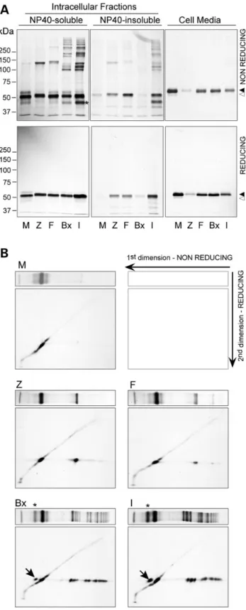
Wefirst analysed the five AAT variants by metabolic labelling of Hepa1.6 transient transfectants. Importantly, post-lysis, artefactual disulphide bonding interactions were prevented by N-ethylmaleimide (NEM) treatment. All fractions were immuno-precipitated by a polyclonal anti-AAT antibody and analysed by sodium dodecyl sulphate-polyAcrilamyde gel electrophoresis (SDS-PAGE) under non-reducing conditions in order to detect the presence of disulphide-linked complexes (Fig.1A, upper panels).
Wild-type M AAT showed the expected pattern: detected mainly as an immature high-mannose monomer in the NP40-soluble cel-lular fraction and efficiently secreted in the cell media as a mature glycosylated form. All mutants were secreted as fully glycosylated monomers but at lower levels compared with M, with I being the most deficient among the rare variants (by densitometric analysis Z, F, Brixia and I were, respectively, 8, 49, 55 and 31% of the M levels). The I mutant forms multiple high molecular weight complexes both in soluble and insoluble cellular fractions. These dissociate upon reduction (Fig.1A, lower panels), confirming that they are sta-bilized by disulphide bonds. The faster migrating complex has an apparent molecular weight of 100 kDa, followed by higher molecu-lar weight complexes. The Brixia mutation occurs close to that of the I variant on the A helix and shows similar disulphide-linked complexes with the 100 kDa form prevalent. A 140 kDa covalent complex was also observed in F, Z and other disease associated AAT mutants (Supplementary Material, Fig. S1), but not in M AAT, suggesting formation of dimers by Cys232 in variants with abnor-mal conformation and ER retention. Similar results were obtained by immunoblot analysis with anti-AAT antibodies (not shown).
To further investigate the nature of the disulphide-linked com-plexes formed by AAT mutants, we performed diagonal gels, con-sisting of non-reducing SDS-PAGE in thefirst dimension followed by a reducing SDS-PAGE in the second dimension (Fig.1B). As ex-pected, the wild-type AAT M (top left panel) shows the monomeric form and runs in the second dimension as a train of spots along the diagonal, indicating that neither intra- nor inter-chain disul-phide bonds are formed as the protein undergoes glycan process-ing. The disulphide complex of the Z mutant with an apparent molecular weight of 140 kDa migrates in the reducing dimension as a high-mannose immature monomer, suggesting that the com-plex is a covalently linked Z AAT homodimer. I and Brixia mutants show similar behaviour, with all major disulphide complexes dissociating into monomeric AAT. No other proteins were found associated with AAT in this analysis, under the experimental con-ditions adopted, even at longer exposure times. This is consistent with the retained higher mass species being composed entirely of AAT subunits with a range of disulphide linkage patterns. We can-not formally exclude interactions of AAT with additional long-lived proteins or methionine/cysteine-poor proteins, due to the nature of the pulse-chase radiolabelling approach. However, our findings suggest that if they occur, heterotypic interactions are a minor contributor to the observed profile of disulphide-adducted AAT species. Importantly, a proportion of I and Brixia AAT runs faster than fully extended monomers in the first dimension (Fig.1A and B, asterisks) and then migrates as a full-length mono-mer in the reducing second dimension (arrows), consistent with the presence of an intra-chain disulphide bond. An intra-chain form was not evident for the F mutant. In this case, the acquired Cys223 lies within the sameβ-strand as Cys232 in the native struc-ture. Formation of an intra-chain bond would likely necessitate major disruption of the protein backbone and stabilising interactions relative to the native structure with kinetically and
at Università degli Studi di Brescia on August 22, 2016
http://hmg.oxfordjournals.org/
energetically unfavourable consequences. We therefore favour this over the alternative interpretation that a disulphide bridge can form but does not result in a significant change in migration on SDS-PAGE. The presence of a second cysteine in the F variant is also associated with less extensive formation of higher molecu-lar weight disulphide species than the other AAT mutants con-taining two cysteines.
Disulphide-linked complexes are distinct from classical polymers
Inclusions formed in the hepatocytes of Z homozygotes are com-posed of polymers that are well-recognized by the conformation-specific monoclonal antibody (mAb) 2C1 (16). To date all AAT polymers that have been assessed in tissue or cell models have also been recognized by the 2C1 mAb. To investigate whether disulphide-linked complexes are associated with 2C1-positive polymeric structures, we performed immunoprecipitation of NP40-soluble fractions obtained as in Figure1with the 2C1 mAb (Fig.2, left). The residual solution was then subjected to immuno-precipitation for total AAT using a polyclonal antibody (Fig.2, right). About 25% of total Z variant migrating in SDS-PAGE as monomer is immunoprecipitated by 2C1 mAb reflecting the frac-tion of intracellular Z in polymeric forms. Instead the disulphide-linked dimers and the other disulphide-disulphide-linked species were less efficiently immunoprecipitated by 2C1 (<10%). These observations suggest that the disulphide-linked species are not incorporated into classical polymers recognized by 2C1 mAb, but may be incor-porated in distinct aggregation states.
Cysteine-mediated interactions contribute to ER retention of AAT mutants
To dissect the individual roles of the two cysteines in determin-ing the intracellular fate of I AAT, we generated two I AAT
Figure 2. 2C1 fails to immunoprecipitate disulphide-linked complexes. NP-40 soluble cell lysates of Hepa1.6 cells expressing M, Z, F, Brixia (Bx) or I, metabolically
labelled for 4 h with35S Met/Cys as in Figure1A, were immunoprecipitated with
the 2C1 mAb (IP1, left side), and the leftover further immunoprecipitated by an anti-total AAT polyclonal antibody (IP2, right side). Immunoprecipitated samples were analysed by non-reducing 8% SDS-PAGE and autoradiography. The results shown are representative of two independent experiments.
Figure 1. AAT mutants form intracellular disulphide-linked complexes. Hepa1.6 cells were transfected with vectors encoding the AAT variants M, Z, F, Brixia (Bx) or I. Twenty-four hours after transfection the cells were metabolically
labelled for 4 h with35S Met/Cys. Cell media were collected, the cells lysed in 1%
NP40 buffer and the NP40-insoluble fractions solubilized in 1% SDS buffer. (A) AAT was immunoprecipitated from the culture media and the intracellular fractions and analysed by 8% SDS-PAGE under non-reducing conditions (upper panels) or reducing conditions (lower panels), followed by autoradiography. White arrowhead: immature AAT. Black arrowhead: mature AAT. (B) NP-40 soluble cell lysates obtained as in (A) were analysed by diagonal gels. The samples were
first resolved by non-reducing 8% SDS-PAGE; for the second dimension, individual lanes were cut, reduced with Laemmli buffer containing 100 mM DTT and further subjected to 8% SDS-PAGE analysis and autoradiography. First dimension lanes are shown at the top of each panel. Asterisks indicate the I and Brixia AAT conformers with an intrachain disulphide bond, which run in the second dimension as reduced monomeric AAT (arrows). The results shown are representative of two independent experiments.
at Università degli Studi di Brescia on August 22, 2016
http://hmg.oxfordjournals.org/
variants (IC39Sand IC232S) with Cys39 or Cys232, respectively,
sub-stituted with serines. Serine residues are chemically similar to cysteines but cannot form disulphide bonds. We expressed these artificial mutants in Hepa1.6 cells and compared their behaviour with that of M, Z and I AAT. Cells were metabolically labelled and analysed by non-reducing SDS-PAGE (Fig.3). IC39S
AAT formed a Cys232-Cys232 disulphide-bonded dimer that mi-grated close to the molecular weight marker of 140 kDa. A dimer with identical behaviour was observed in cells expressing Z AAT. On the other hand, IC232Sexpression formed a Cys39-Cys39
disul-phide-linked dimer that migrated more rapidly (apparent mass
100 kDa) likely due to a compact structure and was also found in the cell media (Fig.3B). A further dimer species was apparent in cells expressing the I variant that migrated intermediate be-tween these points and likely involves both cysteine residues. Formation of higher order complexes was indicated by slower mi-grating species.
The disulphide-linked pattern observed for Brixia was more consistent with that seen in IC232Sthan in I variant containing
Cys232. Most likely, therefore, Brixia dimer formation was mediated by Cys35 rather than Cys232. The variation in disulphide bonding between the I and Brixia variants, which both have two cysteines in near-identical positions, indicates differences in dynamic folding and/or conformational behaviour.
Levels of AAT secreted by cells expressing different AAT var-iants were then analysed by quantitative enzyme-linked immuno-sorbent assay (ELISA) (Fig.3C). F and Brixia mutant secretion was mildly deficient relative to the M AAT control (75 and 80%, respect-ively) while Z and I showed severe deficiency (respectively, 13 and 20%). Notably, secretion of the IC39Sand IC232Smutants was
signifi-cantly higher than that of I, indicating that the formation of het-erogeneous disulphide complexes in the ER contributes to the secretory defect of the I variant. Interestingly, intracellular co-ag-gregation and disulphide bonding of I and Z AAT were demon-strated when they were co-expressed to model hepatocyte behaviour in IZ compound heterozygotes (Supplementary Mater-ial, Fig. S2). Such individuals are more likely to be encountered in clinical practice than I homozygotes. The observation that co-ex-pression of a second, milder deficiency variant can result in com-plex formation with the Z variant in the ER environment validates a previous proposal based upon biophysical studies in cell-free conditions (9).
Effects of disulphide bonding on maturation and secretion of I AAT
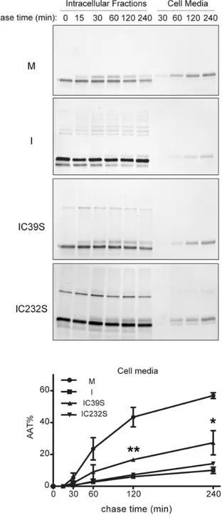
To further investigate the kinetics of formation of the disulphide-linked complexes and their effects on secretion, we performed pulse-chase experiments (Fig.4). The electrophoretic patterns of the I mutant revealed that intra-chain disulphides and Cys39-mediated homodimers form very soon after synthesis. Inter-chain Cys232-Cys232 bonds appear to form more slowly, and the three dimer isoforms are similarly represented from the 30 min time point. Intra-chain bonding is associated with reduced per-sistence of monomeric AAT since the population of this species at-tenuates steadily. This likely reports abnormal conformational behaviour, favouring degradation and/or polymerization. Higher order forms become prevalent at later time points. As expected, IC39Sand IC232SAAT form only homodimers and show increased
secretion with faster intracellular maturation to the 56 kDa glyco-sylated form that acquires Golgi processing. Some IC232S
homodi-mers were secreted, whilst only IC39Smonomers accumulated
extracellularly. The distribution of AAT in the intracellular frac-tions and in the cell media at different time points was quantified by densitometry on reducing SDS-PAGE gels and are shown in Supplementary Material, Figure S3. The effects of aberrant disul-phide bonding upon I AAT secretion were determined by densito-metric analysis of data from two independent pulse-chase experiments (Fig.4A, lower panel). At 240 min of chase, when about 60% of the wild-type M was found in the culture medium, 10, 26 and 13%, respectively, of I, IC39Sand IC232SAAT were secreted.
The significant increase in secretion associated with Cys39Ser substitution further confirms that Cys39-mediated disulphide bonding promotes I AAT retention.
Figure 3. Effects of Cys39 and Cys232 substitution. Hepa1.6 cells were transfected
with vectors encoding the AAT variants M, Z, F, Brixia (Bx) or I, and the artificial
mutants IC39S and IC232S. (A and B) Transfected cells were metabolically
labelled for 4 h and processed as in Figure1. Immunoprecipitated AAT from the
NP-40-soluble cellular fractions ( panel A) and cell media ( panel B) were analysed by non-reducing 8% SDS-PAGE and autoradiography. (C) Transfected Hepa1.6 cells were incubated for 6 h in serum-free culture medium. Secreted
AAT levels in the media were quantified by sandwich ELISA using a standard
curve of purified AAT and expressed as percentages of the wild-type M levels. Results are presented as means and SD of three independent transfection experiments. Statistical significance was determined by one-way ANOVA
followed by the Bonferroni’s post-hoc test using GraphPad Prism version 5
(Graphpad software Inc., San Diego, CA, USA): *P < 0.05; **P < 0.01.
at Università degli Studi di Brescia on August 22, 2016
http://hmg.oxfordjournals.org/
Preventing aberrant disulphide bond formation increases secretion of Z AAT
To specifically address the role of aberrant Cys232 thiol interac-tions in the retention of Z AAT, we performed four independent pulse-chase experiments, comparing Z with a Z mutant in which Cys232 was substituted for a serine (ZC232S). Cys232Ser
sub-stitution significantly increased Golgi maturation and secretion
of Z AAT (Fig.5A), indicating that Cys232-mediated disulphide bonding plays a role in the intracellular retention of Z AAT.
Cell treatment with reducing agents has been used previously to shift the ER to a more reducing state and impair the formation of disulphides on cargo proteins (17,18). Therefore, we next assessed whether such treatment could thereby increase the secretion of Z AAT from Hepa1.6 cells similarly to the effect of the Cys232Ser mutation. A significant increase in secretion of Z AAT was ob-served upon treatment with 0.5 mM dithiothreitol (DTT) (Fig.5B). This was of similar magnitude to the increase in secretion result-ing from the introduction of the Cys232Ser mutation. To assess how far this increase could be attributed to the rescue of Cys232-mediated aggregation, as opposed to the known pleiotropic effects of DTT upon cells, we similarly treated cells expressing ZC232SAAT.
Treatment of these control cells did not increase AAT secretion whatsoever, indicating that this effect of DTT was indeed due to inhibition of Cys232-mediated Z AAT aggregation within cells. Only fully glycosylated AAT was detected in the cell media upon treatment with DTT and we did not observe major effects on the intracellular degradation of the proteins. Consistent results were obtained by measuring secreted AAT by ELISA in the culture media of Hepa1.6 cells expressing Z, treated with either 2ME or DTT. Notably, the change in I AAT secretion was smaller and did not achieve significance, consistent with the more extensive disul-phide bonding patterns observed in this mutant.
Discussion
The oxidizing environment of the ER lumen favours disulphide bonding during the biosynthesis and maturation of secreted pro-teins. The process is assisted by numerous disulphide isomerases and oxidases, working in a tightly controlled redox environment (11). The interaction between free exposed cysteines and disul-phide isomerases also serve to prevent unfolded and unas-sembled proteins from exiting the ER, and so are fundamental in protein quality-control within the early secretory compartment (10). Such thiol-dependent ER retention was originally described for unassembled IgM subunits (19,20) and was then shown for other substrates (21,22). Moreover proteins exposing free cysteine residues may be retrieved from ER-Golgi intermediate compart-ment to the ER by pH-dependent interactions with ERp44 (23,24). The stringency of thiol-mediated retention can be modulated by the local pKaof the thiol group, as demonstrated by the lower
re-activity of the IgA C-terminal cysteine due to the presence of an adjacent negatively charged amino acid (25).
Human AAT appears to represent an exception to this general model of thiol-dependent quality-control: it contains a single cysteine residue (Cys232) at a solvent-exposed site yet is an effi-ciently secreted protein. The reason for this discrepancy is un-clear but does not obviously relate to masking of Cys232 that might render it poorly reactive and/or not accessible in the native structure. Biochemical studies have demonstrated that Cys232 of recombinant AAT has a pKaof 6.86 and can be efficiently oxidized
in vitro (26). Moreover, high resolution crystal structures of AAT indicate that the thiol group of Cys232 is highly exposed to the solvent and electron density maps are consistent with an oxi-dized state (PDB IDs: 2QUG and 3NE4 (27,28). Whether Cys232 in-teracts with protein isomerases in the secretory pathway, however, remains to be elucidated.
Here we report that the Z mutant as well as other transport-incompetent AAT variants form disulphide-linked homodimers in the ER. This suggests that Cys232 participates in disulphide bonding events that report abnormal conformational behaviour of AAT and stabilize aberrant conformations in pathogenic AAT
Figure 4. Effects of disulphide bonding on maturation and secretion of I AAT.
Hepa1.6 cells expressing M, I, IC39S or IC232S were pulsed with35S Met/Cys for
10 min and chased for the indicated times. AAT was immunoprecipitated from the culture media and the intracellular fractions and analysed by non-reducing 8% SDS-PAGE and autoradiography. The kinetics of secretion of the mutants, determined by densitometric analysis as in Supplementary Material, Figure S3, is represented in the graphs as means and SD of two independent experiments.
Statistical significance of IC39S versus I secretion was determined at each chase
time point by t-test analysis: *P < 0.05; **P < 0.01.
at Università degli Studi di Brescia on August 22, 2016
http://hmg.oxfordjournals.org/
Figure 5. Preventing aberrant disulphide bond formation increases secretion of Z AAT. (A) Hepa1.6 cells expressing Z or ZC232Swere pulsed with35S Met/Cys for 10 min and
chased for the indicated times. AAT was immunoprecipitated from the culture media and the intracellular fractions, and analysed by non-reducing 8% SDS-PAGE and autoradiography. The kinetics of secretion of the AAT mutants, determined by densitometric analysis, is represented in the graph as means and SD of four independent experiments. Statistical significance of ZC232S versus Z secretion levels was determined at each chase time point by t-test analysis: *P < 0.05; **P < 0.01; ***P < 0.001. (B) The kinetics of secretion of Z and ZC232S in three independent pulse-chase experiments performed in the absence or presence of 0.5 mM DTT are represented in the graphs as means and SD. Statistical significance of DTT effect on secretion levels was determined at each chase time point by t-test analysis: *P < 0.05; **P < 0.01; ***P < 0.001. Autoradiographies of one of the experiments quantified are shown in the left panels. (C) Hepa1.6 cells expressing M, I, Z or ZC232S were incubated for 6 h in serum-free culture medium in the absence (NT) or presence of 2-ME or DTT at the indicated mM concentrations. AAT levels in the cell media were quantified by sandwich ELISA using a standard curve of purified AAT and expressed for each variant as fold increase relative to NT sample, set as 1. Results from two independent experiments, each one in duplicates, are presented as means and SD. Statistical significance was determined by one-way ANOVA followed by Bonferroni’s post-hoc test using GraphPad Prism version 5 (GraphPad software Inc, San Diego, CA, USA): *P < 0.05; ***P < 0.001.
at Università degli Studi di Brescia on August 22, 2016
http://hmg.oxfordjournals.org/
mutants. These interactions may be favoured by cellular oxida-tive conditions such as those observed in liver tissues of PiZ mice (29). Under our experimental conditions, we did not detect major disulphide interactions of AAT mutants with resident ER isomerases. However this does not preclude their existence, since such interactions are highly dynamic and may form transi-ently during protein folding. In our view such interactions with isomerases and other ER protein folding quality-control proteins are likely to occur. These could be further interrogated in future high sensitivity (e.g. mass spectrometry-based) interactome studies involving immunoprecipitation of intracellular aggre-gates of AAT variants characterized here. Proteins identified as potential interactors could then be validated by reciprocal immu-noprecipitation and functional studies.
The Cys232-mediated dimers we observe for the Z and other polymerogenic variants are reminiscent of those previously reported for the truncated AAT variant NHK (334stop), whose degradation requires EDEM (ER-degradation enhancing alpha-mannosidase-like protein) and ERdJ5 (30,31). Moreover, the intracellular accumulation of such dimers as NP40-insoluble ag-gregates is consistent with their incorporation into high molecular weight polymers. However, disulphide-linked dimers were not im-munoprecipitated effectively by the anti-polymer mAb 2C1 raising the possibility that in vivo these disulphide-linked states are incor-porated in non-classical polymers that may co-exist alongside polymers recognized by the 2C1 mAb. It is now known that AAT and other members of the serpin protein superfamily to which it belongs can assemble into a range of distinct polymeric conforma-tions (1,32). The different polymer architectures share many char-acteristics, but vary in the degree to which each protein subunit is folded relative to the native state. This could account for the differ-ential recognition of AAT polymers by the 2C1 mAb described here, with differently linked polymers having different predispos-ition to abnormal disulphide bond formation. Alternatively, the polymers may all be similar in structure but the observed disul-phide bonding may cause disruption of the 2C1 epitope or render it less accessible due to local effects.
Aberrant disulphide bonding by gain- or loss-of-cysteine mu-tants expressed in the ER has been previously reported for other genetic diseases (33–36). Cysteine-mediated retention of the I AAT mutant is a striking example of the consequences of a non-native cysteine in the protein maturation in the ER. The presence of an additional cysteine residue (Cys39) in the I mutant leads to the formation of an intra-chain and multiple inter-chain disulphide-linkages involving both Cys39 and Cys232. Gain-of-cysteine AAT mutants may therefore be regarded as a subset of AAT deficiency variants, with a distinctive molecular pathogen-esis characterized by increased aberrant disulphide binding events. Notably, our studies have also provided thefirst proof of co-aggregation of different AAT deficiency variants in the ER. Specifically, we observed formation of mixed disulphide com-plexes by the co-expressed I and Z mutants. Such heteropolymer-ization was previously hypothesized on the basis of biophysical data in a cell-free model (9). Since polymer load correlates with disease severity, this has implications for risk stratification of compound heterozygotes such as the IZ cases reported in the literature.
Our mutagenesis data formally demonstrate that aberrant disulphide bonding can contribute to the secretory deficiency of AAT mutants in cells. This effect is particularly evident for I AAT. Here aberrant and complex patterns of intra- and inter-molecu-lar thiol interactions mediated by the two cysteines combine with the inherent polymerogenic tendency of cysteine or serine mutations at position 39. This combination enhances
intracellular aggregation of I AAT in a way that makes in relatively refractory to treatment with reducing agents at concentrations previously used to modulate the ER folding and ER-associated degradation (30,37,38). On the other hand, a simpler pattern of aberrant disulphide bonding contributes to accumulation of the most clinically important variant, Z AAT, within cells. This is more tractable to anti-oxidant treatments to improve secretion. Our data are consistent with the previous understanding that non-covalent polymerization is the primary mechanism of intra-cellular AAT retention, with abnormal disulphide bond forma-tion playing an exacerbating role. Anti-oxidant strategies would therefore be highly complementary to other approaches in development to block or reverse polymerization (39).
Concluding remarks
Previous studies have established that misfolding and aberrant conformational behaviour of AAT mutants determine their intra-cellular retention, degradation and tendency to form ordered polymers. Here we show that cysteine-mediated interactions within the ER contribute to the intracellular retention of the AAT deficiency mutants. The ER redox state may therefore be re-garded as a modifier factor for AATD and represent a useful therapeutic target.
Materials and Methods
Reagents and antibodiesUnless stated otherwise, reagents and culture media were from Sigma-Aldrich. The rabbit polyclonal anti-AAT was from DAKO; the 2C1 mAb (16) was kindly provided by David Lomas (CIMR, Uni-versity of Cambridge, UK). The rabbit polyclonal anti-HA (H6908) was from Sigma-Aldrich.
Expression vectors for the AAT variants
The Brixia allele was identified in a male heterozygous subject by sequencing all coding exons (II–V) of the SERPINA1 gene (RefSeq: NG_008290), as previously described (14). The expression vectors encoding for human M1(Val213) and Z AAT were reported previ-ously (14). The vectors for the tagged AAT variants M-myc, M-HA and Z-HA (40) were kind gifts of Dr Giulia Baldini (University of Arkansas for Medical Sciences, Little Rock, AR, USA). The vectors encoding the Brixia, I and F variants, and the artificial mutants IC39S, IC232S, ZC232S, I-myc and I-HA, were obtained by site-directed
mutagenesis, using the QuikChange II Site-Directed Mutagenesis Kit (Stratagene) and the primers listed in Supplementary Mater-ial, Table S1.
Cell culture and transfections
Hepa1.6 is a mouse hepatoma cell line (ATCC CRL-1830) grown in dulbecco’s modified eagle medium (DMEM) supplemented with 10% fetal bovine serum. Transient transfections were performed with Polyethyleneimine‘MAX’ (PEI) (Polysciences). For a 10 cm2
well, 20 µg PEI and 3 µg plasmid were incubated for 20 min in 400 µl DMEM, diluted to 1.5 ml with normal culture medium and added to the cell layer for 5 h.
Metabolic labelling experiments
Transiently transfected Hepa1.6 cells were pulsed for 10 min with
35SCys/Met (EasyTag™ Express Protein Labelling, Perkin Elmer) in
DMEM without Cys/Met-1% dialysed FCS, and then chased in
at Università degli Studi di Brescia on August 22, 2016
http://hmg.oxfordjournals.org/
normal culture medium for 0, 15, 30, 60, 120 and 240 min. Long metabolic labelling was performed with35S Cys/Met for 5 h.
After labelling, the media were centrifuged at 600g for 5 min and supplemented with 10 mM NEM. Cells were washed with phosphate buffered saline (PBS)/10 mM NEM and lysed in a buffer containing 50 mM Tris–HCl (pH 7.4), 150 mM NaCl, 1% NP40, 10 mM NEM and protease inhibitors. The NP40-insoluble frac-tions were separated by centrifugation at 12 000 g, dissolved in a buffer containing 10 mM Tris, pH 7.4, 150 mM NaCl, 0.2% NP40, 0.1% SDS, 1 mM MgCl2and 20 µg/ml DNAse I, andfinally
di-luted 10-fold with 1% NP40 buffer. The radiolabelled AAT in the cell lysates and media was immunoprecipitated with an anti-AAT polyclonal antibody (DAKO) and analysed by 8% SDS-PAGE under reducing or non-reducing conditions followed by autoradi-ography. Densitometric analysis of AAT bands was performed by Image Quant 5.2 (Molecular Dynamics).
Diagonal gels
The NP40-soluble cell extracts from metabolically labelled Hepa1.6 cells were resolved by SDS-PAGE in non-reducing conditions. All the lanes were cut and reduced by incubation with 2× Laemmli Sample Buffer containing 100 mM DTT for 10 min. For the second dimension, lane slices were placed onto a 8% acrylamide gel and run as a conventional SDS-PAGE followed by autoradiography of the dried gels.
Western blot analysis
For immunoblot analysis, NP40-soluble cell extracts were ob-tained from transfected Hepa1.6 cells as described above for pulse experiments, resolved by SDS-PAGE on pre-casted 7.5% mini PROTEAN TGX gels (Biorad) and transferred to LF-PVDF membranes by the Turbo Blot Transfer System (Biorad). Mem-branes were saturated in low-fat milk, probed with the indicated primary antibodies and horseradish peroxidase (HRP)-conju-gated secondary antibodies (GE Healthcare), revealed by ECL (LiteAblot®plus, Euroclone).
Secretion assays and sandwich ELISA
To quantify secreted AAT, Hepa1.6 transfected cells were washed to remove serum and incubated for 6 h in serum-free culture me-dium, supplemented with 2-mercaptoethanol (2-ME) or DTT when indicated. ELISA plates (half-volume, Costar) were coated overnight at 4°C with 1 µg/ml anti-AAT antibodies (DAKO), washed with PBS/0.1% Tween 20 (PBS-T) and then blocked with 0.25% BSA/PBS-T for 75 min at 37°C. Serial dilutions of cell media were added to the plate and incubated at 37°C for 1 h. For the standard curve, we used serial dilutions of purified AAT (Millipore). Bound AAT was revealed by incubating for 1 h with 1 µg/ml HRP-conjugated goat anti-hAAT (Abcam) followed by TMB-plus substrate (Thermo Scientific). After blocking the reac-tion with 3 M HCl, absorbance was measured at 450 nm using an ELISA plate reader.
Supplementary Material
Supplementary Material is available at HMG online.
Acknowledgements
We thank Elena Miranda Banos (La Sapienza University, Rome, Italy), Ilaria Ferrarotti (San Matteo Hospital, Pavia, Italy), David Lomas (University College London, UK), Stefan Marciniak
(University of Cambridge, UK), Luisa Schiaffonati (University of Brescia, Italy) and Luciano Corda (Spedali Civili, Brescia, Italy) for informative discussions. We are also grateful to David Lomas (University College London, UK) for providing the 2C1 mAb and to Dr Giulia Baldini (University of Arkansas for Medical Sciences, Little Rock, USA) for the expression vectors encoding the tagged AAT variants ZHA and Mmyc.
Conflict of Interest statement. None declared.
Funding
This work was supported by Grifols through the eALTA award to R.R., by Fondazione Cariplo (Grant 2013-0967 to A.M.F.) and by the Italian Patients’ Association Alfa1.
References
1. Gooptu, B., Dickens, J.A. and Lomas, D.A. (2014) The molecu-lar and cellumolecu-lar pathology of alpha(1)-antitrypsin deficiency. Trends Mol. Med., 20, 116–127.
2. Lomas, D.A., Evans, D.L., Finch, J.T. and Carrell, R.W. (1992) The mechanism of Z alpha 1-antitrypsin accumulation in the liver. Nature, 357, 605–607.
3. Kroeger, H., Miranda, E., MacLeod, I., Perez, J., Crowther, D.C., Marciniak, S.J. and Lomas, D.A. (2009) Endoplasmic reticu-lum-associated degradation (ERAD) and autophagy cooper-ate to degrade polymerogenic mutant serpins. J. Biol. Chem., 284, 22793–22802.
4. Teckman, J.H., Burrows, J., Hidvegi, T., Schmidt, B., Hale, P.D. and Perlmutter, D.H. (2001) The proteasome participates in degradation of mutant alpha 1-antitrypsin Z in the endoplas-mic reticulum of hepatoma-derived hepatocytes. J. Biol. Chem., 276, 44865–44872.
5. Perlmutter, D.H. (2011) Alpha-1-antitrypsin deficiency: im-portance of proteasomal and autophagic degradative path-ways in disposal of liver disease-associated protein aggregates. Annu. Rev. Med., 62, 333–345.
6. Piras, B., Ferrarotti, I., Lara, B., Martinez, M.T., Bustamante, A., Ottaviani, S., Pirina, P., Luisetti, M. and Miravitlles, M. (2013) Clinical phenotypes of Italian and Spanish patients with alpha1-antitrypsin deficiency. Eur. Respir. J., 42, 54–64. 7. Rodriguez-Frias, F., Miravitlles, M., Vidal, R., Camos, S. and
Jardi, R. (2012) Rare alpha-1-antitrypsin variants: are they really so rare? Ther. Adv. Respir. Dis., 6, 79–85.
8. Baur, X. and Bencze, K. (1987) Study of familial alpha-1-pro-teinase inhibitor deficiency including a rare proalpha-1-pro-teinase in-hibitor phenotype (IZ). I. Alpha-1-phenotyping and clinical investigations. Respiration, 51, 188–195.
9. Mahadeva, R., Chang, W.S., Dafforn, T.R., Oakley, D.J., Fore-man, R.C., Calvin, J., Wight, D.G. and Lomas, D.A. (1999) Het-eropolymerization of S, I, and Z alpha1-antitrypsin and liver cirrhosis. J. Clin. Invest., 103, 999–1006.
10. Anelli, T. and Sitia, R. (2008) Protein quality control in the early secretory pathway. Embo J., 27, 315–327.
11. Bulleid, N.J. and Ellgaard, L. (2011) Multiple ways to make dis-ulfides. Trends Biochem. Sci., 36, 485–492.
12. Kojer, K. and Riemer, J. (2014) Balancing oxidative protein folding: the influences of reducing pathways on disulfide bond formation. Biochim. Biophys. Acta, 1844, 1383–1390. 13. Fra, A.M., Gooptu, B., Ferrarotti, I., Miranda, E., Scabini, R.,
Ronzoni, R., Benini, F., Corda, L., Medicina, D., Luisetti, M. et al. (2012) Three new alpha1-antitrypsin deficiency variants
at Università degli Studi di Brescia on August 22, 2016
http://hmg.oxfordjournals.org/
help to define a C-terminal region regulating conformational change and polymerization. PLoS One, 7, e38405.
14. Medicina, D., Montani, N., Fra, A.M., Tiberio, L., Corda, L., Miranda, E., Pezzini, A., Bonetti, F., Ingrassia, R., Scabini, R. et al. (2009) Molecular characterization of the new defective P (brescia) alpha1-antitrypsin allele. Hum. Mutat., 30, E771–e38781. 15. Okayama, H., Brantly, M., Holmes, M. and Crystal, R.G. (1991) Characterization of the molecular basis of the alpha 1-anti-trypsin F allele. Am. J. Hum. Genet., 48, 1154–1158.
16. Miranda, E., Perez, J., Ekeowa, U.I., Hadzic, N., Kalsheker, N., Gooptu, B., Portmann, B., Belorgey, D., Hill, M., Chambers, S. et al. (2010) A novel monoclonal antibody to characterize pathogenic polymers in liver disease associated with alpha1-antitrypsin deficiency. Hepatology, 52, 1078–1088. 17. Lodish, H.F. and Kong, N. (1993) The secretory pathway is
nor-mal in dithiothreitol-treated cells, but disulfide-bonded pro-teins are reduced and reversibly retained in the endoplasmic reticulum. J. Biol. Chem., 268, 20598–20605.
18. van Lith, M., Tiwari, S., Pediani, J., Milligan, G. and Bulleid, N.J. (2011) Real-time monitoring of redox changes in the mam-malian endoplasmic reticulum. J. Cell Sci., 124, 2349–2356. 19. Fra, A.M., Fagioli, C., Finazzi, D., Sitia, R. and Alberini, C.M.
(1993) Quality control of ER synthesized proteins: an exposed thiol group as a three-way switch mediating assembly, reten-tion and degradareten-tion. Embo J., 12, 4755–4761.
20. Sitia, R., Neuberger, M., Alberini, C., Bet, P., Fra, A., Valetti, C., Williams, G. and Milstein, C. (1990) Developmental regulation of IgM secretion: the role of the carboxy-terminal cysteine. Cell, 60, 781–790.
21. Fraldi, A., Zito, E., Annunziata, F., Lombardi, A., Cozzolino, M., Monti, M., Spampanato, C., Ballabio, A., Pucci, P., Sitia, R. et al. (2008) Multistep, sequential control of the trafficking and function of the multiple sulfatase deficiency gene product, SUMF1 by PDI, ERGIC-53 and ERp44. Hum. Mol. Genet., 17, 2610–2621.
22. Wang, Z.V., Schraw, T.D., Kim, J.Y., Khan, T., Rajala, M.W., Fol-lenzi, A. and Scherer, P.E. (2007) Secretion of the adipocyte-specific secretory protein adiponectin critically depends on thiol-mediated protein retention. Mol. Cell Biol., 27, 3716–3731. 23. Hampe, L., Radjainia, M., Xu, C., Harris, P.W., Bashiri, G., Gold-stone, D.C., Brimble, M.A., Wang, Y. and Mitra, A.K. (2015) Regulation and quality control of adiponectin assembly by endoplasmic reticulum chaperone ERp44. J. Biol. Chem., 290, 18111–18123.
24. Vavassori, S., Cortini, M., Masui, S., Sannino, S., Anelli, T., Caserta, I.R., Fagioli, C., Mossuto, M.F., Fornili, A., van Anken, E. et al. (2013) A pH-regulated quality control cycle for surveillance of secretory protein assembly. Mol. Cell, 50, 783–792.
25. Guenzi, S., Fra, A.M., Sparvoli, A., Bet, P., Rocco, M. and Sitia, R. (1994) The efficiency of cysteine-mediated intracellular re-tention determines the differential fate of secretory IgA and IgM in B and plasma cells. Eur. J. Immunol., 24, 2477–2482. 26. Griffiths, S.W., King, J. and Cooney, C.L. (2002) The reactivity
and oxidation pathway of cysteine 232 in recombinant human alpha 1-antitrypsin. J. Biol. Chem., 277, 25486–25492. 27. Patschull, A.O., Segu, L., Nyon, M.P., Lomas, D.A., Nobeli, I.,
Barrett, T.E. and Gooptu, B. (2011) Therapeutic target-site
variability in alpha1-antitrypsin characterized at high reso-lution. Acta Crystallogr. Sect. F Struct. Biol. Cryst. Commun., 67, 1492–1497.
28. Pearce, M.C., Morton, C.J., Feil, S.C., Hansen, G., Adams, J.J., Parker, M.W. and Bottomley, S.P. (2008) Preventing serpin ag-gregation: the molecular mechanism of citrate action upon antitrypsin unfolding. Protein Sci., 17, 2127–2133.
29. Marcus, N.Y., Blomenkamp, K., Ahmad, M. and Teckman, J.H. (2012) Oxidative stress contributes to liver damage in a mur-ine model of alpha-1-antitrypsin deficiency. Exp. Biol. Med. (Maywood), 237, 1163–1172.
30. Hosokawa, N., Wada, I., Natsuka, Y. and Nagata, K. (2006) EDEM accelerates ERAD by preventing aberrant dimer formation of misfolded alpha1-antitrypsin. Genes Cells, 11, 465–476.
31. Ushioda, R., Hoseki, J., Araki, K., Jansen, G., Thomas, D.Y. and Nagata, K. (2008) ERdj5 is required as a disulfide reductase for degradation of misfolded proteins in the ER. Science, 321, 569– 572.
32. Gooptu, B. and Lomas, D.A. (2009) Conformational pathology of the serpins: themes, variations, and therapeutic strategies. Annu. Rev. Biochem., 78, 147–176.
33. Dhaunchak, A.S. and Nave, K.A. (2007) A common mechanism of PLP/DM20 misfolding causes cysteine-mediated endoplas-mic reticulum retention in oligodendrocytes and Pelizaeus– Merzbacher disease. Proc. Natl. Acad. Sci. USA, 104, 17813–17818. 34. Liu, M., Haataja, L., Wright, J., Wickramasinghe, N.P., Hua, Q.X., Phillips, N.F., Barbetti, F., Weiss, M.A. and Arvan, P. (2010) Mu-tant INS-gene induced diabetes of youth: proinsulin cysteine residues impose dominant-negative inhibition on wild-type proinsulin transport. PLoS One, 5, e13333.
35. Tanaka, Y., Ueda, K., Ozawa, T., Kitajima, I., Okamura, S., Mor-ita, M., Yokota, S. and Imanaka, T. (2005) Mutation study of antithrombin: the roles of disulfide bonds in intracellular accumulation and formation of russell body-like structures. J. Biochem., 137, 273–285.
36. Wang, J.W., Groeneveld, D.J., Cosemans, G., Dirven, R.J., Va-lentijn, K.M., Voorberg, J., Reitsma, P.H. and Eikenboom, J. (2012) Biogenesis of Weibel-Palade bodies in von Willebrand’s disease variants with impaired von Willebrand factor intra-chain or interintra-chain disulfide bond formation. Haematologica, 97, 859–866.
37. Alberini, C.M., Bet, P., Milstein, C. and Sitia, R. (1990) Secretion of immunoglobulin M assembly intermediates in the pres-ence of reducing agents. Nature, 347, 485–487.
38. Medrano-Fernandez, I., Fagioli, C., Mezghrani, A., Otsu, M. and Sitia, R. (2014) Different redox sensitivity of endoplasmic reticulum associated degradation clients suggests a novel role for disulphide bonds in secretory proteins. Biochem. Cell Biol., 92, 113–118.
39. Nyon, M.P. and Gooptu, B. (2014) Therapeutic targeting of misfolding and conformational change in alpha1-antitrypsin deficiency. Future Med. Chem., 6, 1047–1065.
40. Granell, S., Baldini, G., Mohammad, S., Nicolin, V., Narducci, P., Storrie, B. and Baldini, G. (2008) Sequestration of mutated alpha1-antitrypsin into inclusion bodies is a cell-protective mechanism to maintain endoplasmic reticulum function. Mol. Biol. Cell, 19, 572–586.
at Università degli Studi di Brescia on August 22, 2016
http://hmg.oxfordjournals.org/