D E B A T E
Open Access
Insulin-like growth factor-1 signaling
in renal cell carcinoma
Adam F. Tracz
1,2, Cezary Szczylik
1, Camillo Porta
3and Anna M. Czarnecka
1*Abstract
Renal cell carcinoma (RCC) incidence is highest in highly developed countries and it is the seventh most common
neoplasm diagnosed. RCC management include nephrectomy and targeted therapies. Type 1 insulin-like growth factor
(IGF-1) pathway plays an important role in cell proliferation and apoptosis resistance. IGF-1 and insulin share overlapping
downstream signaling pathways in normal and cancer cells. IGF-1 receptor (IGF1R) stimulation may promote malignant
transformation promoting cell proliferation, dedifferentiation and inhibiting apoptosis. Clear cell renal cell carcinoma
(ccRCC) patients with IGF1R overexpression have 70 % increased risk of death compared to patients who had tumors
without IGF1R expression. IGF1R signaling deregulation may results in
p53, WT, BRCA1, VHL loss of function. RCC cells with
high expression of IGF1R are more resistant to chemotherapy than cells with low expression. Silencing of IGF1R increase
the chemosensitivity of ccRCC cells and the effect is greater in
VHL mutated cells. Understanding the role of IGF-1
signaling pathway in RCC may result in development of new targeted therapeutic interventions. First preclinical attempts
with anti-IGF-1R monoclonal antibodies or fragment antigen-binding (Fab) fragments alone or in combination with an
mTOR inhibitor were shown to inhibit in vitro growth and reduced the number of colonies formed by of RCC cells.
Keywords: Renal cell carcinoma (RCC, ccRCC), Insulin-like growth factor-1 (IGF-1), IGF-1 receptor (IGF1R)
Background
Renal cell carcinoma (RCC) comprise 2–3 % of malignant
tumors in adults. Clear cell RCC (ccRCC) develops from
epithelium of the proximal tubules and is the most
com-mon a histological type of RCC
– diagnosed in 60-70 % of
cases. Initial treatment of RCC is most often partial or
radical nephrectomy. Nevertheless one third of patients
are diagnosed with synchronous metastases [1]. IGF-1
plays an important role in protection from apoptosis and
regulation of cell growth. Deregulation in downstream
IGF-1 receptor results in angiogenesis, cell viability,
prolif-eration and invasion. IGF1R expression is poor prognostic
factor especially among those presenting with
high-grade disease at the initial evaluation. RCC cells with
high expression of IGF1R are more resistant to
chemo-therapy than cells with low expression of that receptor
[2]. IGF-1 and insulin share overlapping downstream
pathways of cancer cell metabolism. Cell line based
studies have shown that down-regulation, knock-out,
pharmacological inhibition of IGF-IR may in turn
increase in IR signaling and therefore insulin analogs
may promote cancerogenesis [3]. On the other hand
first preclinical attempts with anti-IGF-1R monoclonal
antibodies or fragment antigen-binding (Fab) fragments
alone or in combination with an mTOR inhibitor were
shown to inhibit in vitro growth and reduced the
num-ber of colonies formed by of RCC cells [4, 5]. All these
findings suggest that IGF1R expression is significant in
ccRCC and understanding of the molecular mechanism
of IGF-1 and insulin signaling pathway in RCC may
give opportunity to design molecular markers of disease
or even finding a new molecular targets for drugs.
Renal cell carcinoma
Renal cell carcinoma is the seventh most common
malignancy with expected 5-year survival rate of 72 %.
About 64 % of patients are diagnosed with localized
disease [6]. RCC has highest incidence in highly developed
countries. RCC includes several types of cancer: clear cell
RCC, papillary RCC, chromophobe RCC and also rare
cancer like, urothelial carcinoma, collecting duct carcinoma
and renal medullary carcinoma [7]. The most common
* Correspondence:[email protected]
1Department of Oncology with Laboratory of Molecular Oncology, Military
Institute of Medicine, Szaserow 128, 04-141 Warsaw, Poland Full list of author information is available at the end of the article
© 2016 The Author(s). Open Access This article is distributed under the terms of the Creative Commons Attribution 4.0 International License (http://creativecommons.org/licenses/by/4.0/), which permits unrestricted use, distribution, and reproduction in any medium, provided you give appropriate credit to the original author(s) and the source, provide a link to the Creative Commons license, and indicate if changes were made. The Creative Commons Public Domain Dedication waiver (http://creativecommons.org/publicdomain/zero/1.0/) applies to the data made available in this article, unless otherwise stated.
type is clear cell RCC that is diagnosed in up to 70 % of
cases. Among those up to 60-90 % of sporadic cases of
ccRCC exhibit VHL gene loss, silencing, mutation or
promoter hypermethylation. The VHL protein (pVHL) is
an E3 ubiquitin ligase of hypoxia inducible factor (HIF). It
targets HIF for degradation by the proteasome. In the
absence of functional pVHL HIF is accumulated in the
nucleus where it acts as a transcription factor for vascular
endothelial growth factor (VEGF), platelet derived growth
factor (PDGF), multidrug resistance pump (MDR-1), cyclin
D1 (CCND1), ENOLASE2 (ENO2), Egl-9 Family
Hypoxia-Inducible Factor 3/HIF Prolyl Hydroxylase 3 (EGLN3),
angiopoietin-like 4 (ANGPTL4), glucose transporter 1
(GLUT1), insulin-like growth factor-binding protein 3
(IGFBP3), and erythropoietin (EPO) [8, 9]. Management of
RCC is usually initiated with partial or radical
nephrec-tomy. Ablation procedure is alternative for small renal
masses in patients who are ineligible for surgery [10, 11].
Adjuvant therapy has no proven efficacy on management
of RCC. Only systemic treatment of metastatic disease has
been shown as effective in phase III trials [1, 7]. There are 3
major groups of systematic treatment that are used for
metastatic RCC: cytokines, mTOR inhibitors and anti-
vas-cular endothelial growth factor (VEGF) - targeted drugs
[7]. Cytokine based immunotherapies included
interferon-alpha [12] and high dose interleukin 2 (IL-2) [13], while
mTOR inhibitors approved for RCC treatment are
everoli-mus [14] and temsirolieveroli-mus [15]. VEGF pathway inhibitors
used are 1) tyrosine kinase inhibitors as sorafenib [16],
sunitinib [17], pazopanib [18], and axitinib [19], and 2)
anti-VEGF monoclonal antibody
– bevacizumab [20]
(Table 1). New drugs that modulate immunological
response including anti-CTLA-4 (cytotoxic T lymphocyte
antigen
4)
antibody
and
anti-PD-1/PDL-1
(pro-grammed cell death 1) antibody have been developed
recently [7, 21, 22]. Anti-CTLA-4 or PD-1
anti-bodies inhibit the immunosuppression between T cells
and APC (antigen presenting cells) including cancer
cells which stimulate anti-tumor response [21, 22]. Most
recently nivolumab - anti-PD-1 monoclonal antibody - was
shown as effective against RCC in trail using everolimus as
comparator [23].
General IGF-1 function
Insulin-like growth factor 1 (IGF-1, somatomedin C) is a
natural anabolic peptide hormone produced mainly by
hepatocytes. IGF-1 with molecular weight of 7649 Da is
built by 70 amino acids and single polypeptide chain
with three intramolecular disulfide bridges. Production
of IGF-1 is stimulated by growth hormone (GH)
secreted by anterior pituitary. IGF-1 production is also
stimulated by insulin and has influenced on reduction of
lipolysis, glycolysis, inhibition of lipolytic function of
adrenaline, embryonic growth and differentiation of
cells. IGF-1 may also be released independently of GH.
Circulating IGF-1 produced in liver acts in endocrine
manner, but locally produced IGF-1 acts also in an
autocrine manner. IGF functions therefore both as
circu-lating hormone and tissue growth factor. Circucircu-lating
IGF-1 forms a complex with two other proteins
– the
IGF binding protein (IGFBP) and the acid labile subunit
(ALS). Six different IGFBPs were characterized, but
about 75 % of serum IGFs are bound to IGFBP3 and
only 1 % of serum IGF-1 is free-bioactive form [24].
IGFBPs are also mostly synthesized in the liver.
Neverthe-less IGFs and IGFBPs are also produced in other organs,
acting locally in autocrine and paracrine manner and
me-diating stromal - epithelial cell interactions [25]. IGFBPs
acts in a competing manner against IGFR (IGF receptors)
and IGFBP proteases. IGF-1 and IGFBP-3 complex play
crucial role in mitogenesis, cell differentiation and survival
[26]. IGF-1 null mice die shortly after birth [27].
Circulation of IGF-1
High level concentrations of circulating IGF-1 are
related with higher risk of prostate, colorectal and breast
Table 1 Drugs used in RCC therapy
Group of drug Drugs Ref.
cytokines 1) Interleukin 2 - overall response rate - 15 %,
complete response rate - 5 %, achieved by high dose. Problems with selection of patients who may benefit from treatment.
2) Interferon alfa - inferior to most new agents considering PFS (progression free survival), except in combination with bevacizumab.
[13,100]
VEGF-targeted drugs 1) Sorafenib– second and subsequent lines of treatment. 2) Sunitinib– first line treatment for metastatic RCC. 3) Pazopanib– non-inferior to sunitinib
4) Axitinib– treatment refractory RCC.
5) Bevacizumab– used with interferon alfa. Superior PFS when compared with monotherapy of interferon alfa.
[7,101,102]
mTOR inhibitors 1) Temsirolimus– for patient with poor risk as a first line druga
2) Everolimus– used as a second line or third line drug.
[7,14,17]
a
cancers [28–30]. Circulating concentrations of IGFBP-3
is associated with increased risks of breast cancers in
postmenopausal women and prostate cancer in men [28,
29, 31]. Transgenic mouse with deletion in liver-specific
IGF-1 that result 75 % reduction in circulating IGF-1
exhibit reduction in development of colon cancer and
reduced growth tumor xenografts [31, 32]. Laron
syn-drome is genetic condition characterized by GH
insensi-tivity and in consequence IGF-1 deficiency [33]. People
with Laron syndrome are resistant to cancer what was
shown by Steuerman et al. [34]. They found that none of
the 230 patients with Laron syndrome developed cancer
and that only 1 out of 116 patients with inborn IGF-1
loss was diagnosed with malignancy [34].
IGF-1 receptor and insulin receptor homology
IGFR-1 is a transmembrane receptor with tyrosine kinase
activity and is built of two
α-subunits (located
extracellu-larly) and two
β-subunits (spanning the membrane and
activating intracellular signal transduction). Both the
α
and
β subunits are synthesized from a single precursor
mRNA. IGF1R shares a high structural homology with the
insulin receptor (IR)
– has more than 50 % in the overall
amino acid sequence and in particular 84 % similarity in
the tyrosine kinase domain and 45–65 % in the
ligand-binding domain. Moreover ligand-dependent activation of
the IGF1R and IR activates almost identical downstream
signaling pathways [35]. After IGF-1 binging activation of
tyrosine kinase (β-subunits) results in downstream
signaling via IR substrate proteins (IRS1-4), Src homology
2 domain containing transforming protein 1 (Shc),
GRB2-associated binding protein 1 (Gab-1), Casitas B-lineage
Lymphoma proto-oncogene E3 ubiquitin protein ligase
(Cbl), Phosphatidyl Inositol 3-Kinase (PIK3), Protein
kin-ase B (Akt), mammalian target of rapamycin (mTOR),
mitogen-activated protein kinase (MAPK) and signal
regulatory protein family [36]. Insulin and IGFs have a
great homology and can have cross-reactivity upon
recep-tors. Moreover hybrid receptors - constituted of IR and
IGF1R heterodimers
– have been shown to have cellular
biological effects resembling those of the IGF1R and were
found in colon cancer, thyroid cancer and breast cancer
cell lines and tissues [37]. To complicate the interaction
even more there are two IR isoforms, arising in the cell by
alternative splicing of exon 11
– isoform IR-A, that lacks
exon 11, and isoform IR-B
– containing exon 11. Insulin
does not bind to the hybrid receptors, but binds to IR-A,
IR-B, and IGF-1R but binds to the IGF-1R with much
lower affinity than to the IR. IGF-I binds to the IGF-1R,
hybrid receptors, and IR but has much lower affinity for
the IR than IGF-1R [3]. In total insulin and IGF-1 interact
with six receptors: the type I IGF receptor (IGF1R), the
IRA (IR-A, predominantly expressed in fetal tissue), the
IRB (IR-B, predominantly expressed in adult tissue),
hybrid receptors of IGF and IR-A, hybrid receptors of IGF
and IR-B, and hybrid receptors of IR-A and IR-B [38, 39].
Insulin and IGF-1 while binding to IGF1R, IR-A, IGF1R/
IR-A, mediate mostly mitogenic signaling (Ras > MEK >
Erk1/2 pathway), while binding to IR-B activate mostly
metabolic pathway (PI3K > Akt > mTOR) [24, 36, 40]. As
a result both insulin and IGF-1 can act through the hybrid
receptors and through the specific receptor for their
lig-and (Fig. 1). Activation of all receptors (IR, IGF1R, hybrid)
which are tyrosine kinase cell-surface receptor result in
phosphorylation of IR substrate proteins (IRS 1–4). It
activates two key signal-transduction pathways. The
GTPase Ras-Raf-MEK-ERK1/2 pathway activates gene
expression that result in cells proliferation. The AKT
kinase pathway activates mTOR which results in cells
pro-liferation. PI3K induce angiogenesis by activating of
hypoxia-inducible factor-1a. Activation of AKT2 promotes
GLUT4 translocation leading to the activation of glycogen
synthase [31, 41, 42]. Moreover in cancer cells it was
shown that GF-1R undergoes nuclear import and both
alpha and beta subunits traffic to the nucleus by
clathrin-mediated endocytosis. Ligand activated nuclear IGF-1R is
phosphorylated and undergoes interaction with chromatin
and regulate transcription. This nuclear IGF-1R
accumu-lation is associated with poor prognosis in RCC [43].
Molecular deregulation of IGF1R pathway and
cancerogenesis
IGF-1/insulin pathways were show as significant in cancer
research. First of all IGF-1 and insulin share overlapping
downstream pathways of cancer cell metabolism. Chronic
hyperinsulinemia and diabetes mellitus type 2 were
associ-ated with tumor development through the obesity-cancer
association [31, 44]. According to the Werner et all. IGF1R
activation is pre-requisite for malignant transformation. As
oncogenic transformation is initiated, cell survival of
trans-formed cells is strongly dependent on IGF-1 signaling [44].
Further multiple studies confirmed the role of IGF
−1 and
its receptors on RCC cancerogenesis [41, 45]. Deregulated
IGF1R kinase activity and its overexpression was reported
in multiple cancers including RCC [46–48]. In particular
constitutively active IGF1R leads to salivary and mammary
adenocarcinomas in transgenic mice [49]. Transgenic
over-expression of IGF1R increases epithelial mammary gland
hyperplasia and tumor formation [50]. In the RCC cell
lines including Caki-2 (from a primary tumor) and
SK-RC-52 (from a metastatic tumor) IGF-1 was shown to enhance
transforming growth factor-β (TGF-β) signaling and via
TGF-β raise IGF-binding protein 3 (IGFBP-3) levels with
growth-promoting effect [51].
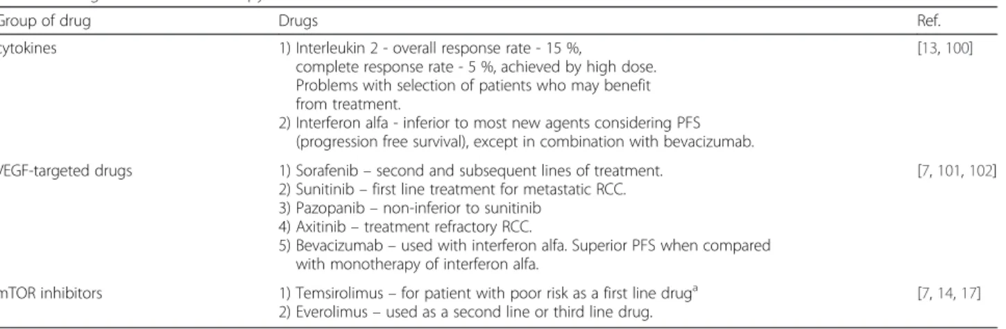
Unlike in most genes, promoter sequence lacks TATA
and CCAAT boxes that are usually required for efficient
transcription initiation. Instead of TATA box, promoter
is highly rich in GC base pairs. The initiator (INR) is
located approximately 1000 bp upstream of the coding
region. Specificity protein 1 (Sp1) is a key transcription
activator of the IGF1R gene that binds with high affinity
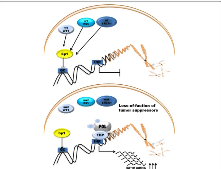
to GC boxes in promoter. The products of tumor
suppres-sor genes like BRCA1, p53, VHL acts through the Sp1
protein [52]. The schematic representation of promoter
and other transcription factor shows Fig. 2.
IGF1R activity deregulation is result of loss of tumor
suppressor genes or the action of oncogenes [53]. The
examples are p53, BRCA1, WT1 genes. p53 is the one of
the most frequently mutated tumor suppressor and is
well known gene associated with cancerogenesis.
Prod-uct of p53 gene accumulates in response of DNA
demage and results in arrest of cell cycle at G1 phase.
Fig. 2 Schematic representation of promoter region of IGF1R and its main transcription factors. Sp1, Specificity protein 1; HMGA1, High mobility group A1; KLF6, Krüppel-like factor 6; E2F1, E2F family of transcription factors; POL, RNA polymerase II; TBP, TATA-binding protein; GC, GC boxes; INR, initiator element
Fig. 1 Schematic representation of downstream signaling of IGF1R. AKT, protein kinase B; AMPK, AMP-activated protein kinase; Bcl-2, B-cell lymphoma 2; BAD, B-cell CLL/lymphoma 2 antagonist of cell death; ERK 1/2, extracellular-signal-regulated kinase 1/2, IGF1R, insulin-like growth factor 1 receptor; IR, insulin receptor; IRS1-4, insulin-like receptor substrate 1–4; MEK, mitogen-activated protein kinase kinase; mTOR, mammalian target of rapamycin; PI3K/AKT, phosphatidylinositol 3-kinase/AKT; PDK1, 3-phosphoinositide-dependent protein kinase; PIP2, phosphatidylinositol 4,5-bisphosphate; PIP3, phosphatidylinositol 3,4,5-trisphosphate; PTEN, phosphatase and tensin homolog; GLUT4, Glucose transporter type 4; HIF-1α, Hypoxia-inducible factor 1-alpha, VEGF, Vascular endothelial growth factor
Cell can repair DNA or activate apoptotic program.
Wild-type p53 supperess the activity of IGF1R promoter
whereas mutated p53 enhence promoter activity and
ac-celerate tumor growth [54]. Next antioncogene BRCA1
is linked to the etiology of hereditary of familial breast
and ovarian cancer. Wild-type BRCA1 reduce the
pro-moter activity of IGF1R whereas mutated BRCA1 lead to
enhance IGF1R promoter activity and expression of
pro-tein. BRCA1 and BRCA2 carries have an increased level
of IGF1R compared with those with sporadic cancers
[55, 56]. WT1 gene which product is a zinc-finger
tran-scription factor is associated with the etiology of Wilms
tumor a kind of pediatric kidney cancer [57]. All of these
genes products are able to suppress the activity of IGF1R
promoter, as well as the endogenous levels of IGF1R
mRNA [57–59]. IGF1R expression level is raised in
non-BRCA1-mutated ovarian cancer cells compared with
normal tissue and additionally IGF1R levels are
signifi-cantly increased in BRCA1-inactivation ovarian cancer
[59]. Moreover investigation in SNP in IGF-1 and IGFR-1
indicate that IGF1-pathway polymorphisms are potential
prognostic molecular markers in colorectal cancer and
pancreatic cancer. There are significantly associated with
progression-free survival and/or overall survival (OS) in
these cancers [60, 61]. SNP polymorphisms and mutation
of IGF1R that are associated with cancer represents
Table 2.
The mechanism of action of P53 and BRCA1 involves
interaction with Sp1 to repress the expression of IGF1R
[52]. BRCA1 can inhibit IGF1R expression regardless of
p53 expression level but not in mutant state of p53.
WT1 protein does not involve direct interaction with
promoter DNA sequence [62]. Loss-of-function of that
genes leads to up-regulation of IGF1R gene. Other
tumor suppressor gene VHL, Von Hippel–Lindau tumor
suppressor
also
inhibits
IGF1R promoter activity
through interaction with Sp1 protein. In RCC
inactiva-tion of VHL is associated with IGF1R up-regulainactiva-tion [63].
Schematic representation of main tumor suppressor
protein acting on promoter is shown on Fig. 3.
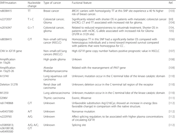
Table 2 SNP
’s and mutations of IGF1R gene associated with cancers
SNP/mutation Nucleotide change
Type of cancer Fuctional feature Ref.
rs8038415 TT Breast cancer BRCA1 carriers with homozigosity TT at this SNP site experience a 40 % higher risk of breast cancer.
[103]
rs2272037 T > C Colorectal cancer; Glioma
Significantly related with shorter OS in patients with metastatic colorectal cancer (mCRC). CT and TT associated with increased risk for glioma.
[60] [104] rs2016347 G > T Colorectal cancer,
glioma
Related to reduced responsiveness to cetuximab treatment. Shorter OS in patients with mCRC. G allele associated with increased risk for Glioma (3’UTR in 3129 site)
[104,
105]
rs8038415 C/T Non–small cell lung cancer (NSCLC)
Homozygous TT in this SNP had a significantly better OS compared with heterozygous individuals and a trend toward improved survival compared with patients that were homozygous for CC .
[106]
CNV inIGF1R gene Non–small-cell lung cancers (NSCLC)
HighIGF1R gene copy number harbors positive prognostic value in NSCLC [107] Amplification
in 15q26
High grade glioma Unkown [108]
Amplification in 15q25-26
Alveolar
Rhabdomyosarcoma
Related with the rearrangement ofPAX7 gene [109]
A1374V Lung squamous cell
carcinoma
Unknown; mutation occur in the C-terminal lobe of the kinase catalytic domain [110]
Deletion S1278 Renal clear cell carcinoma
Unknown; deletion occur in the C-terminal tail region of the receptor [110]
M1255I Lung adenocarcinoma Unknown; mutation occur in in the C-terminal lobe of the kinase catalytic domain [110]
G596V Thymic carcinoma Exonic, Missense [111]
rs61740868 C/T Unknown Unfavorable substitution Arg1216Cys; showed an increase in energy (less favorable change) in comparison with the native structure.
[112]
rs45437300 A/T Unknown Nonsense mutation [112]
rs2229765 A/G Unknown Affect splicing regulation; to be associated with higher plasma concentrations of circulating IGF1R [112] rs55895813; rs36108138; rs45495500 A/G; A/C; C/T
Expression of IGF-1 and IGF1R in kidney
Locally synthesize IGF-1 in kidney acts as a paracrine or
autocrine factors. Level of IGF-1 in venous renal blood
is higher than in renal arterial blood. There are no
strong proves on IGF-1 epithelial expression. Studies
in-dicates connective tissue adjacent to epithelial renal cells
to be responsible for local synthesis of IGF-1 [64, 65].
Research on human fetal kidney shows no expression of
mRNA of IGF-1 in nephrogenic zone but only probably
sequestration of IGF-1 peptide in proximal and distal
tubules. Studies on rats and mouse shows the IGF-1
(mRNA and peptide) expression (immunolabeling) in
collecting ducts. In situ hybridization studies indicate
the mRNA expression in medullary thick ascending limb
of henle’s loop [64, 66, 67]. IGF- 1 mediated decline in
renal vascular resistance, leads to elevated glomerular
perfusion, sodium and water resorption leading in turn to
soft tissue swelling and glomerular hypertrophy [65,
68]. IGF1R gene expression were detected in whole
nephogenic zone including the strong expression in
glomeruli and the tubular epithelium of medulla with
the least expression in proximal tubules [69]. In
con-trary Kamenicky et al. shows the strong expression of
mRNA of IGF1R in proximal tubule obtained from
murine cells by microdissection [65, 70].
Expression of IGF-1 and IGF1R in ccRCC
The type 1 insulin-like growth factor receptor (IGF1R) has
an influence on renal cells malignant transformation by
induction of cell proliferation, dedifferentiation and
anti-apoptotic effect [71, 72]. IGF-1 and IGF1R expression is
modulated in kidney development. In mouse model of
kidney development IGF-I mRNA is expressed in all cell
types with peak in the proximal tubules, peritubular
capil-laries of the outer medulla and inner cortex one week after
birth. The expression of IGF1R in normal proximal tubules
was similar to IGF-1 during kidney development until
birth [73], but it is poor prognosis factor in RCC [74].
Fig. 3 Regulation of promoter activity of IGF1R gene by tumor suppressor genes. POL, RNA polymerase II; TBP, TATA-binding protein; GC, GC boxes; INR, initiator element
Overexpression of IGF family members was reported
ob-served in oxidative stress (ferric nitrilotriacetate) induced
RCC in rats [75]. What is interesting RCC cells with high
expression of IGF1R are more resistant to chemotherapy
than cells with low expression of that receptor [2]. IGF-I
pretreatment levels in RCC patients was predictive to
im-paired response to interleukin-2 (IL-2) therapy [76]. In
SN12K1 cells (cell line derived from metastatic RCC) it
was shown that RCC express IGF-I and IGFBP-3, and
autocrine IGF-I and IGFBP-3 stimulate and inhibit growth
respectively. These cells are also are responsive to
exogen-ous IGF-I stimulation - DNA synthesis is increased. These
RCC cells are also responsive to exogenous IGF-I
stimula-tion [77]. Further experiments on RCC cell lines - Caki-2
(primary tumor) and SK-RC-52 (metastatic tumor)
– have
shown that IGF-I enhances transforming growth factor
(TGF)-beta signaling including phosphorylation and
nu-clear translocation of mothers against decapentaplegic
homolog 2 (Smad2). In turn TGF-beta promotes IGFBP-3
production [51]. In mice model injection of MZ4–71
-growth hormone-releasing hormone antagonist - reduced
the IGF-1 induced growth of Caki 1 RCC cell line derived
tumors [78].
In first clinical reports high serum IGF-I levels have been
associated with an increased risk of developing RCC [77].
At the same time in the analysis of 256 patients serum
IGF-1 was not correlated with tumor stage or grade, but
was independent favorable prognostic factors in a
multi-variable analysis [79]. More recently the prospective study
of 29 133 Finnish male smokers reported that men with
IGF-I levels >113 ng ml(−1) were 59 % less likely to
de-velop RCC than men with levels below or =113 ng ml(−1).
Among those 100 men with RCC the IGF binding
protein-3 (IGFBP-protein-3) levels did not alter the association [80]. In the
study of 90 patients with ccRCC and 20 normal renal
tissue samples, hyper-methylation of IGFBP-3 was not
found, but in 786–0, ACHN, HRC51 and HRC59 cell lines
methylation of IGFBP-3 was observed [81]. Moreover
IGF-I and its binding proteins IGF-IGFBP-3 and
−6 are
up-regulated in ccRCC tumor tissues [77]. IGF1R expression
was also associated with ccRCC and indicated molecular
prognostic marker and potential targets for therapeutic
intervention. Data of 280 patients who had ccRCC treated
with radical nephrectomy showed that IGF1R expression
had a 70 % increased risk of death than patients who had
tumors without IGF1R expression [82]. Finally nuclear
IGF-1R was detected in primary renal cancer tissues of
high proliferation rate and was associated with adverse
prognosis [43].
Pathological and clinical grading system and IGF1R status
Fuhrman nuclear grading system correlates with staging
of ccRCC. Studies on expression of IGF1R and Furman
score in 68 ccRCC showed the statistically significance
increasing correlation between Fuhrman grading and
IGF1R staining [83]. Also the high serum concentration
of IGF1R is associated with the risk of many cancers
such as breast, prostate, colorectal, and lung cancers
[71]. Overexpression of IGF1R is related with poor
progno-sis in many human cancers besides the renal cancer also in
breast and ovarian cancers [84, 85]. Antisense strategies
against the IGF1R suppresses the expression of IGF1R can
abolish cell transformation [71, 83]. No correlation was
observed between serum IGFBP-3 levels and RCC [79], but
high expression of IGFBP-3 was found in ccRCC tumors.
Fuhrman grades 3 and 4 ccRCC tumors showed higher
IGFBP-3 expression than low grade tumors [80, 86].
Although the IGF1R and Insulin receptor (IR) show
great homology and interaction they have different
relation to the prognosis in ccRCC. Lkhagvadrj et al.
investigated the role of IR expression in 126 ccRCC
cases. Researchers showed that there was no differences
of total IR protein between tumor and non-tumor but
immunoreactivity of IR in tumor was mostly observed in
nuclear but in non-tumor tissue in nuclear and
cyto-plasm. The expression of IR was elevated in low-grade
tumor but not in higher-grade. Also the IR expression
was inversely correlate with Furman nuclear grade and
TNM, pathologic T stage in ccRCC. Higher expression
of IR correlated with cystic changes in RCC which is
favorable prognostic factor. What is interesting IR
expression was not related to the diabetes presence. The
authors suggest that although IR and IGF1R share the
major down-stream signaling pathway, in RCC there are
specific substrates for each receptor [87].
Interaction of VHL and IGF1R
Hereditary RCC is commonly associated loss or mutation
of the Von Hippel-Lindau (VHL) tumor suppressor gene.
VHL mutation was first discovered in patients with VHL
disease by Latif F et al. [88] and VHL mutation or loss is
identified in 60-90 % of sporadic RCC cases [7, 89]. Next
20 % of ccRCC show silencing of VHL expression by
methylation in promoter region [90]. VHL gene encodes
protein with ubiquitin ligase E3 activity directing
hypoxia-inducible factor-1α (HIF-1α) for degradation [87]. HIF-1α
regulate hypoxia response and promote angiogenesis, cell
migration, and metabolism [7, 91] via VEGF, PDGF and
TGF [92]. Independently of oxygen status IGF1R activates
HIF-1α protein by suppressing VHL and promote RCC
development [63]. At the same time IGF-1-mediated
signaling is inhibited in the presence of wild-type VHL
and VHL decrease stability of IGF-1R [93]. In RCC cells
beta-domain of VHL interact with protein kinase C delta
(PKCD) and inhibits its association with IGF-IR and
subsequent downstream signaling [94]. It was also shown
that receptor for activated C kinase 1 (RACK1) serves as a
direct mediator between loss of pVHL function and
IGF-IR signaling in RCC cells. Upon IGF-1 stimulation,
pVHL-deficient RCC cells exhibit high rate of RACK1/
IGF-IR binding and up-regulated IGF-1R tyrosine
kinase activity, phosphoinositide
3-kinase/serine-threo-nine kinase Akt (PI3K/Akt) signaling and matrix
metalloproteinase-2 (MMP-2) activity and high cellular
invasiveness [95]. VHL protein has no influence on
ubiquitination of IGF1R. On the contrary the activity of
IGF1R expression is regulated by VHL at the
transcrip-tional level and is mediated by Sp1 transcription factor.
Sp1 protein is sequestered by VHL and act on promoter
of IGF1R. Loss of VHL gene increases therefore IGF1R
mRNA stability. The levels of IGF1R is higher in ccRCC
samples than in benign renal tumors which could be
associated with VHL mutation rate in RCC [63, 96].
At the same time depletion of IGF1R enhance the
chemosensitivity of ccRCC, but this effect is
signifi-cant in cells with no functional VHL. Depletion of
IGF1R changed sensitivity to mTOR inhibitors, 5-FU,
etoposide but not cisplatin [2].
Conclusions
The role of IGF-1/Insulin pathway in cancerogenesis
remain unclear. IGF-1 and insulin share major
down-stream regulation pathway and both are engaged in
cancerogenesis and diabetes [97]. There are evidences
that diabetic patients have risk of development of renal
cancer. Increase in mortality and incidence of renal cell
carcinoma among diabetic patients is linked to
hyperin-sulinemia and obesity. The interaction of hyperglycemia,
hyperinsulinemia causes the insulin resistant state and
contribute to central adiposity which result in chronic
inflammation. The adipose tissue-derived cytokines like
resistin, tumor necrosis factor
α (TNF-α) and interleukin
6 (IL-6) promotes persistent inflammation and result in
genetic instability, putting cells at risk to malignant
transformation. Nuclear factor-kB is strongly activated
by TNF-α what is associated with cell proliferation and
surviving malignant cells [97]. Associations between
hyperinsulinemia and increased circulating levels of
IGF-1 is proposed to be involved in carcinogenesis. IR and
IGF-1 has a nuclear translocation potential and were
postulated to be nonclassical transcription factors.
IGF1R physically interact with gDNA and also stimulates
itself expression. Study of nuclear IGF1R in primary
renal cancer cells revealed that IGF1R expression was
associated with poor prognosis in renal cancer [98]. Cell
culture experiments have proven that active IGF-IR is
necessary for cell transformation by multiple cellular
and viral oncogenes. IGF-IR signaling regulate the cell
cycle, cell survival/proliferation, cell-cell interactions,
ECM attachment, cell motility and metastatic spread. In
mice model IGF-IR overexpression promotes tumor
growth and metastases development, whereas
down-regulation of IGF leads to slower tumor development
[3]. In general anti-diabetic treatment may increase the
risk of RCC cancer development, but large prospective
data is not available at this point of time. Exception is
metformin an oral anti-diabetic drug has a
RCC-protective effect as it interacts with the IGF signaling
pathway which results in inhibition of proliferation and
apoptosis regardless the p53 status [99].
Abbreviations
ccRCC, Clear cell renal cell carcinoma; CTLA-4, cytotoxic T lymphocyte antigen 4; IGF-1, Insulin-like growth factor-1; IGF1R, IGF-1 receptor; IGFBPs, IGF-binding proteins; PD-1, programmed cell death 1; PDGFR, Platelet-derived growth factor receptors; RCC, renal cell carcinoma; VEGF, Vascular endothelial growth factor; VHL, Von Hippel–Lindau protein
Acknowledgements Not applicable. Funding
This work has been supported by National Science Centre (NCN) grant No. UMO-2012/05/D/NZ5/01844 and Military Institute Medicine Statutory Founding (355).
Availability of data and material Not applicable.
Authors' contributions
Conceptualizing and refining research ideas - AMC; Literature search - AMC, AT; Creating review design - AMC; Collection of literature data - AMC, AT; Data selection - AT, AMC; Interpreting analyses - AMC, AT, CP, CS; Drafting manuscript AT, AMC; Editing manuscript AMC. All authors read and approved the final manuscript.
Authors' information
AMC is MD PhD - expert in molecular biology and medical doctor - specializes in clinical oncology and has been trained at the Universite degli Studi di Palermo, Paracelsus Medizinische Privatuniversität and Emory University School of Medicine. CS– MD PhD - is professor of clinical oncology specialist since 1986, and has worked at Temple University School of Medicine, Jefferson Cancer Institute at Thomas Jefferson University, and in the last 10 years has participated in major renal cancer treatment clinical trials including AXIS, EU-ARCCS or TARGET and is an expert in renal cancer treatment. AT is specialist in Medical Biotechnology and is MD in training, researcher in the Laboratory of Molecular Oncology, Military institute of Medicine, Warsaw, Poland. CP - Professor of Medical Oncology at the University of Pavia, School of Biotechnology, Responsible of the Laboratory of Pre-Clinical Oncology and Developmental Therapeutics at the IRCCS San Matteo University Hospital in Pavia, Member of ESMO Scientific Committee, involved in Development of new anticancer drugs, e.g., WR2721 (Amifostine), Nolatrexed (Thymitaq), BBR3438, Rampirnase (Onconase), BAY 43–9006 (Sorafenib), SU011248 (Sunitinib), RAD001 (Everolimus), Perifosine, ARQ197, Pazopanib and Satraplatin.
Competing interests
The authors declare that they have no competing interests. Consent for publication
Not applicable.
Ethics approval and consent to participate Not applicable.
Author details
1Department of Oncology with Laboratory of Molecular Oncology, Military
Institute of Medicine, Szaserow 128, 04-141 Warsaw, Poland.2First Faculty of Medicine, Medical University of Warsaw, Warsaw, Poland.3Department of
Medical Oncology, IRCCS San Matteo University Hospital Foundation, Pavia, Italy.
Received: 11 October 2015 Accepted: 28 June 2016
References
1. Escudier B, Szczylik C, Porta C, Gore M. Treatment selection in metastatic renal cell carcinoma: expert consensus. Nat Rev Clin Oncol. 2012;9(6):327–37. 2. Yuen JS, Akkaya E, Wang Y, Takiguchi M, Peak S, Sullivan M, Protheroe AS,
Macaulay VM. Validation of the type 1 insulin-like growth factor receptor as a therapeutic target in renal cancer. Mol Cancer Ther. 2009;8(6):1448–59. 3. Gallagher EJ, LeRoith D. Minireview: IGF, Insulin, and Cancer. Endocrinology.
2011;152(7):2546–51.
4. Cardillo TM, Trisal P, Arrojo R, Goldenberg DM, Chang CH. Targeting both IGF-1R and mTOR synergistically inhibits growth of renal cell carcinoma in vitro. BMC Cancer. 2013;13:170.
5. Chang CH, Wang Y, Trisal P, Li R, Rossi DL, Nair A, Gupta P, Losman M, Cardillo TM, Rossi EA, et al. Evaluation of a novel hexavalent humanized anti-IGF-1R antibody and its bivalent parental IgG in diverse cancer cell lines. PLoS One. 2012;7(8):e44235.
6. ASCO. http://www.cancer.net/cancer-types/kidney-cancer/statistics. Accessed 28 April 2015.
7. Jonasch E, Gao J, Rathmell WK. Renal cell carcinoma. BMJ. 2014;349:g4797. 8. Cowey CL, Rathmell WK. VHL gene mutations in renal cell carcinoma: role as a biomarker of disease outcome and drug efficacy. Curr Oncol Rep. 2009; 11(2):94–101.
9. Zhang T, Niu X, Liao L, Cho EA, Yang H. The contributions of HIF-target genes to tumor growth in RCC. PLoS One. 2013;8(11):e80544. 10. Van Poppel H, Da Pozzo L, Albrecht W, Matveev V, Bono A, Borkowski A,
Colombel M, Klotz L, Skinner E, Keane T, et al. A prospective, randomised EORTC intergroup phase 3 study comparing the oncologic outcome of elective nephron-sparing surgery and radical nephrectomy for low-stage renal cell carcinoma. Eur Urol. 2011;59(4):543–52.
11. Vrdoljak E, Ciuleanu T, Kharkevich G, Mardiak J, Mego M, Padrik P, Petruzelka L, Purkalne G, Shparyk Y, Skrbinc B, et al. Optimizing treatment for patients with metastatic renal cell carcinoma in the Central and Eastern European region. Expert Opin Pharmacother. 2012;13(2):159–74.
12. Motzer RJ, Bacik J, Murphy BA, Russo P, Mazumdar M. Interferon-alfa as a comparative treatment for clinical trials of new therapies against advanced renal cell carcinoma. J Clin Oncol. 2002;20(1):289–96.
13. Fyfe G, Fisher RI, Rosenberg SA, Sznol M, Parkinson DR, Louie AC. Results of treatment of 255 patients with metastatic renal cell carcinoma who received high-dose recombinant interleukin-2 therapy. J Clin Oncol. 1995; 13(3):688–96.
14. Motzer RJ, Escudier B, Oudard S, Hutson TE, Porta C, Bracarda S, Grunwald V, Thompson JA, Figlin RA, Hollaender N, et al. Efficacy of everolimus in advanced renal cell carcinoma: a double-blind, randomised, placebo-controlled phase III trial. Lancet. 2008;372(9637):449–56.
15. Bellmunt J, Szczylik C, Feingold J, Strahs A, Berkenblit A. Temsirolimus safety profile and management of toxic effects in patients with advanced renal cell carcinoma and poor prognostic features. Ann Oncol. 2008;19(8):1387–92. 16. Escudier B, Eisen T, Stadler WM, Szczylik C, Oudard S, Staehler M, Negrier S, Chevreau C, Desai AA, Rolland F, et al. Sorafenib for treatment of renal cell carcinoma: Final efficacy and safety results of the phase III treatment approaches in renal cancer global evaluation trial. J Clin Oncol. 2009;27(20):3312–8. 17. Hudes G, Carducci M, Tomczak P, Dutcher J, Figlin R, Kapoor A,
Staroslawska E, Sosman J, McDermott D, Bodrogi I, et al. Temsirolimus, interferon alfa, or both for advanced renal-cell carcinoma. N Engl J Med. 2007;356(22):2271–81.
18. Sternberg CN, Davis ID, Mardiak J, Szczylik C, Lee E, Wagstaff J, Barrios CH, Salman P, Gladkov OA, Kavina A, et al. Pazopanib in locally advanced or metastatic renal cell carcinoma: results of a randomized phase III trial. J Clin Oncol. 2011;28(6):1061–8.
19. Rini BI, Escudier B, Tomczak P, Kaprin A, Szczylik C, Hutson TE, Michaelson MD, Gorbunova VA, Gore ME, Rusakov IG, et al. Comparative effectiveness of axitinib versus sorafenib in advanced renal cell carcinoma (AXIS): a randomised phase 3 trial. Lancet. 2012;378(9807):1931–9.
20. Escudier B, Pluzanska A, Koralewski P, Ravaud A, Bracarda S, Szczylik C, Chevreau C, Filipek M, Melichar B, Bajetta E, et al. Bevacizumab plus interferon alfa-2a for treatment of metastatic renal cell carcinoma: a randomised, double-blind phase III trial. Lancet. 2007; 370(9605):2103–11.
21. Yang JC, Hughes M, Kammula U, Royal R, Sherry RM, Topalian SL, Suri KB, Levy C, Allen T, Mavroukakis S, et al. Ipilimumab (anti-CTLA4 antibody) causes regression of metastatic renal cell cancer associated with enteritis and hypophysitis. J Immunother. 2007;30(8):825–30.
22. Brahmer JR, Tykodi SS, Chow LQ, Hwu WJ, Topalian SL, Hwu P, Drake CG, Camacho LH, Kauh J, Odunsi K, et al. Safety and activity of anti-PD-L1 antibody in patients with advanced cancer. N Engl J Med. 2012;366(26): 2455–65.
23. Motzer RJ, Escudier B, McDermott DF, George S, Hammers HJ, Srinivas S, Tykodi SS, Sosman JA, Procopio G, Plimack ER, et al. Nivolumab versus Everolimus in Advanced Renal-Cell Carcinoma. N Engl J Med. 2015;373(19): 1803–13.
24. Mahesh S, Kaskel F. Growth hormone axis in chronic kidney disease. Pediatr Nephrol. 2008;23(1):41–8.
25. Brahmkhatri VP, Prasanna C, Atreya HS. Insulin-like growth factor system in cancer: novel targeted therapies. BioMed Research International. 2015;2015:538019.
26. Renehan AG, Zwahlen M, Minder C, O'Dwyer ST, Shalet SM, Egger M. Insulin-like growth factor (IGF)-I, IGF binding protein-3, and cancer risk: systematic review and meta-regression analysis. Lancet. 2004;363(9418):1346–53. 27. Baker J, Liu JP, Robertson EJ, Efstratiadis A. Role of insulin-like growth factors
in embryonic and postnatal growth. Cell. 1993;75(1):73–82.
28. Roddam AW, Allen NE, Appleby P, Key TJ, Ferrucci L, Carter HB, Metter EJ, Chen C, Weiss NS, Fitzpatrick A, et al. Insulin-like growth factors, their binding proteins, and prostate cancer risk: analysis of individual patient data from 12 prospective studies. Ann Intern Med. 2008;149(7):461–71. W483-468. 29. Key TJ, Appleby PN, Reeves GK, Roddam AW. Insulin-like growth factor 1
(IGF1), IGF binding protein 3 (IGFBP3), and breast cancer risk: pooled individual data analysis of 17 prospective studies. Lancet Oncol. 2010;11(6): 530–42.
30. Rinaldi S, Cleveland R, Norat T, Biessy C, Rohrmann S, Linseisen J, Boeing H, Pischon T, Panico S, Agnoli C, et al. Serum levels of IGF-I, IGFBP-3 and colorectal cancer risk: results from the EPIC cohort, plus a meta-analysis of prospective studies. Int J Cancer. 2010;126(7):1702–15.
31. Clayton PE, Banerjee I, Murray PG, Renehan AG. Growth hormone, the insulin-like growth factor axis, insulin and cancer risk. Nat Rev Endocrinol. 2011;7(1):11–24.
32. Olivo-Marston SE, Hursting SD, Lavigne J, Perkins SN, Maarouf RS, Yakar S, Harris CC. Genetic reduction of circulating insulin-like growth factor-1 inhibits azoxymethane-induced colon tumorigenesis in mice. Mol Carcinog. 2009;48(12):1071–6.
33. Laron Z. Laron syndrome (primary growth hormone resistance or insensitivity): the personal experience 1958–2003. J Clin Endocrinol Metab. 2004;89(3):1031–44.
34. Steuerman R, Shevah O, Laron Z. Congenital IGF1 deficiency tends to confer protection against post-natal development of malignancies. Eur J Endocrinol. 2011;164(4):485–9.
35. Weinstein D, Sarfstein R, Laron Z, Werner H. Insulin receptor compensates for IGF1R inhibition and directly induces mitogenic activity in prostate cancer cells. Endocrine Connections. 2014;3(1):24–35.
36. Singh P, Alex JM, Bast F. Insulin receptor (IR) and insulin-like growth factor receptor 1 (IGF-1R) signaling systems: novel treatment strategies for cancer. Med Oncol. 2014;31(1):805.
37. Vigneri PG, Tirro E, Pennisi MS, Massimino M, Stella S, Romano C, Manzella L. The Insulin/IGF System in Colorectal Cancer Development and Resistance to Therapy. Frontiers in Oncology. 2015;5:230.
38. Frasca F, Pandini G, Scalia P, Sciacca L, Mineo R, Costantino A, Goldfine ID, Belfiore A, Vigneri R. Insulin receptor isoform A, a newly recognized, high-affinity insulin-like growth factor II receptor in fetal and cancer cells. Mol Cell Biol. 1999;19(5):3278–88.
39. Belfiore A, Frasca F, Pandini G, Sciacca L, Vigneri R. Insulin receptor isoforms and insulin receptor/insulin-like growth factor receptor hybrids in physiology and disease. Endocr Rev. 2009;30(6):586–623.
40. De Meyts P, Whittaker J. Structural biology of insulin and IGF1 receptors: implications for drug design. Nat Rev Drug Discov. 2002;1(10):769–83. 41. Tao Y, Pinzi V, Bourhis J, Deutsch E. Mechanisms of disease: signaling of the
insulin-like growth factor 1 receptor pathway–therapeutic perspectives in cancer. Nat Clin Pract Oncol. 2007;4(10):591–602.
42. Chitnis MM, Yuen JS, Protheroe AS, Pollak M, Macaulay VM. The type 1 insulin-like growth factor receptor pathway. Clin Cancer Res. 2008;14(20): 6364–70.
43. Aleksic T, Chitnis MM, Perestenko OV, Gao S, Thomas PH, Turner GD, Protheroe AS, Howarth M, Macaulay VM. Type 1 insulin-like growth factor receptor translocates to the nucleus of human tumor cells. Cancer Res. 2010;70(16):6412–9.
44. Werner H. Tumor suppressors govern insulin-like growth factor signaling pathways: implications in metabolism and cancer. Oncogene. 2012;31(22): 2703–14.
45. Gualberto A, Pollak M. Emerging role of insulin-like growth factor receptor inhibitors in oncology: early clinical trial results and future directions. Oncogene. 2009;28(34):3009–21.
46. Ouban A, Muraca P, Yeatman T, Coppola D. Expression and distribution of insulin-like growth factor-1 receptor in human carcinomas. Hum Pathol. 2003;34(8):803–8.
47. Hartog H, Wesseling J, Boezen HM, van der Graaf WT. The insulin-like growth factor 1 receptor in cancer: old focus, new future. Eur J Cancer. 2007;43(13):1895–904.
48. Pollak M. Insulin and insulin-like growth factor signalling in neoplasia. Nat Rev Cancer. 2008;8(12):915–28.
49. Carboni JM, Lee AV, Hadsell DL, Rowley BR, Lee FY, Bol DK, Camuso AE, Gottardis M, Greer AF, Ho CP, et al. Tumor development by transgenic expression of a constitutively active insulin-like growth factor I receptor. Cancer Res. 2005;65(9):3781–7.
50. Jones RA, Campbell CI, Gunther EJ, Chodosh LA, Petrik JJ, Khokha R, Moorehead RA. Transgenic overexpression of IGF-IR disrupts mammary ductal morphogenesis and induces tumor formation. Oncogene. 2007; 26(11):1636–44.
51. Rosendahl AH, Forsberg G. IGF-I and IGFBP-3 augment transforming growth factor-beta actions in human renal carcinoma cells. Kidney Int. 2006;70(9):1584–90.
52. Werner H, Sarfstein R. Transcriptional and epigenetic control of IGF1R gene expression: implications in metabolism and cancer. Growth Horm IGF Res. 2014;24(4):112–8.
53. Werner H, Shalita-Chesner M, Abramovitch S, Idelman G, Shaharabani-Gargir L, Glaser T. Regulation of the insulin-like growth factor-I receptor gene by oncogenes and antioncogenes: implications in human cancer. Mol Genet Metab. 2000;71(1–2):315–20.
54. Werner H, Karnieli E, Rauscher FJ, LeRoith D. Wild-type and mutant p53 differentially regulate transcription of the insulin-like growth factor I receptor gene. Proc Natl Acad Sci U S A. 1996;93(16):8318–23. 55. Maor S, Yosepovich A, Papa MZ, Yarden RI, Mayer D, Friedman E,
Werner H. Elevated insulin-like growth factor-I receptor (IGF-IR) levels in primary breast tumors associated with BRCA1 mutations. Cancer Lett. 2007;257(2):236–43.
56. Maor S, Papa MZ, Yarden RI, Friedman E, Lerenthal Y, Lee SW, Mayer D, Werner H. Insulin-like growth factor-I controls BRCA1 gene expression through activation of transcription factor Sp1. Horm Metab Res. 2007;39(3): 179–85.
57. Werner H, Rauscher 3rd FJ, Sukhatme VP, Drummond IA, Roberts Jr CT, LeRoith D. Transcriptional repression of the insulin-like growth factor I receptor (IGF-I-R) gene by the tumor suppressor WT1 involves binding to sequences both upstream and downstream of the IGF-I-R gene transcription start site. J Biol Chem. 1994;269(17):12577–82.
58. Ohlsson C, Kley N, Werner H, LeRoith D. p53 regulates insulin-like growth factor-I (IGF-I) receptor expression and IGF-I-induced tyrosine
phosphorylation in an osteosarcoma cell line: interaction between p53 and Sp1. Endocrinology. 1998;139(3):1101–7.
59. Liu B, Li D, Guan YF. BRCA1 regulates insulin-like growth factor 1 receptor levels in ovarian cancer. Oncol Lett. 2014;7(5):1733–7.
60. Winder T, Zhang W, Yang D, Ning Y, Bohanes P, Gerger A, Wilson PM, Pohl A, Mauro DJ, Langer C, et al. Germline polymorphisms in genes involved in the IGF1 pathway predict efficacy of cetuximab in wild-type KRAS mCRC patients. Clin Cancer Res. 2010;16(22):5591–602.
61. Dong X, Javle M, Hess KR, Shroff R, Abbruzzese JL, Li D. Insulin-like growth factor axis gene polymorphisms and clinical outcomes in pancreatic cancer. Gastroenterology. 2010;139(2):464–73. 473 e461-463.
62. Sarfstein R, Belfiore A, Werner H. Identification of Insulin-Like Growth Factor-I Receptor (IGF-IR) Gene Promoter-Binding Proteins in Estrogen Receptor (ER)-Positive and ER-Depleted Breast Cancer Cells. Cancers (Basel). 2010;2(2):233–61.
63. Yuen JS, Cockman ME, Sullivan M, Protheroe A, Turner GD, Roberts IS, Pugh CW, Werner H, Macaulay VM. The VHL tumor suppressor inhibits expression
of the IGF1R and its loss induces IGF1R upregulation in human clear cell renal carcinoma. Oncogene. 2007;26(45):6499–508.
64. Clemmons DR. Modifying IGF1 activity: an approach to treat endocrine disorders, atherosclerosis and cancer. Nat Rev Drug Discov. 2007;6(10): 821–33.
65. Kamenicky P, Mazziotti G, Lombes M, Giustina A, Chanson P. Growth hormone, insulin-like growth factor-1, and the kidney: pathophysiological and clinical implications. Endocr Rev. 2014;35(2):234–81.
66. Chin E, Zhou J, Bondy C. Anatomical relationships in the patterns of insulin-like growth factor (IGF)-I, IGF binding protein-1, and IGF-I receptor gene expression in the rat kidney. Endocrinology. 1992;130(6):3237–45. 67. Han VK, Hill DJ, Strain AJ, Towle AC, Lauder JM, Underwood LE, D'Ercole AJ.
Identification of somatomedin/insulin-like growth factor immunoreactive cells in the human fetus. Pediatr Res. 1987;22(3):245–9.
68. Guler HP, Schmid C, Zapf J, Froesch ER. Effects of recombinant insulin-like growth factor I on insulin secretion and renal function in normal human subjects. Proc Natl Acad Sci U S A. 1989;86(8):2868–72.
69. Chin E, Bondy C. Insulin-like growth factor system gene expression in the human kidney. J Clin Endocrinol Metab. 1992;75(3):962–8.
70. Kamenicky P, Viengchareun S, Blanchard A, Meduri G, Zizzari P, Imbert-Teboul M, Doucet A, Chanson P, Lombes M. Epithelial sodium channel is a key mediator of growth hormone-induced sodium retention in acromegaly. Endocrinology. 2008;149(7):3294–305.
71. Furstenberger G, Senn HJ. Insulin-like growth factors and cancer. Lancet Oncol. 2002;3(5):298–302.
72. Valentinis B, Baserga R. IGF-I receptor signalling in transformation and differentiation. Mol Pathol. 2001;54(3):133–7.
73. Lindenbergh-Kortleve DJ, Rosato RR, van Neck JW, Nauta J, van Kleffens M, Groffen C, Zwarthoff EC, Drop SL. Gene expression of the insulin-like growth factor system during mouse kidney development. Mol Cell Endocrinol. 1997; 132(1–2):81–91.
74. Parker AS, Cheville JC, Blute ML, Igel T, Lohse CM, Cerhan JR. Pathologic T1 clear cell renal cell carcinoma: insulin-like growth factor-I receptor expression and disease-specific survival. Cancer. 2004;100(12):2577–82. 75. Tanaka T, Kondo S, Iwasa Y, Hiai H, Toyokuni S. Expression of stress-response
and cell proliferation genes in renal cell carcinoma induced by oxidative stress. Am J Pathol. 2000;156(6):2149–57.
76. Lissoni P, Barni S, Ardizzoia A, Frigerio F, Paolorossi F, Cazzaniga M, Tancini G, Rocco F, Aapro M. Clinical efficacy of cancer subcutaneous immunotherapy with interleukin-2 in relation to the pretreatment levels of tumor growth factor insulin-like growth factor-1. Tumori. 1995;81(4):261–4.
77. Cheung CW, Vesey DA, Nicol DL, Johnson DW. The roles of IGF-I and IGFBP-3 in the regulation of proximal tubule, and renal cell carcinoma cell proliferation. Kidney Int. 2004;65(4):1272–9.
78. Jungwirth A, Schally AV, Pinski J, Groot K, Armatis P, Halmos G. Growth hormone-releasing hormone antagonist MZ-4-71 inhibits in vivo proliferation of Caki-I renal adenocarcinoma. Proc Natl Acad Sci U S A. 1997; 94(11):5810–3.
79. Rasmuson T, Grankvist K, Jacobsen J, Olsson T, Ljungberg B. Serum insulin-like growth factor-1 is an independent predictor of prognosis in patients with renal cell carcinoma. Acta Oncol. 2004;43(8):744–8.
80. Major JM, Pollak MN, Snyder K, Virtamo J, Albanes D. Insulin-like growth factors and risk of kidney cancer in men. Br J Cancer. 2010;103(1):132–5. 81. Perks CM, Holly JM. Epigenetic regulation of insulin-like growth factor
binding protein-3 (IGFBP-3) in cancer. J Cell Communication and Signaling. 2015;9(2):159–66.
82. Parker A, Cheville JC, Lohse C, Cerhan JR, Blute ML. Expression of insulin-like growth factor I receptor and survival in patients with clear cell renal cell carcinoma. J Urol. 2003;170(2 Pt 1):420–4.
83. Ahmad N, Keehn CA, Coppola D. The expression of insulin-like growth factor-I receptor correlates with Fuhrman grading of renal cell carcinomas. Hum Pathol. 2004;35(9):1132–6.
84. Belfiore A, Frasca F. IGF and insulin receptor signaling in breast cancer. J Mammary Gland Biol Neoplasia. 2008;13(4):381–406.
85. Beauchamp MC, Yasmeen A, Knafo A, Gotlieb WH. Targeting insulin and insulin-like growth factor pathways in epithelial ovarian cancer. J Oncol. 2008;2010:257058.
86. Chuang ST, Patton KT, Schafernak KT, Papavero V, Lin F, Baxter RC, Teh BT, Yang XJ. Over expression of insulin-like growth factor binding protein 3 in clear cell renal cell carcinoma. J Urol. 2008;179(2):445–9.
87. Lkhagvadorj S, Oh SS, Lee MR, Jung JH, Chung HC, Cha SK, Eom M. Insulin receptor expression in clear cell renal cell carcinoma and its relation to prognosis. Yonsei Med J. 2014;55(4):861–70.
88. Latif F, Tory K, Gnarra J, Yao M, Duh FM, Orcutt ML, Stackhouse T, Kuzmin I, Modi W, Geil L, et al. Identification of the von Hippel-Lindau disease tumor suppressor gene. Science. 1993;260(5112):1317–20.
89. Nickerson ML, Jaeger E, Shi Y, Durocher JA, Mahurkar S, Zaridze D, Matveev V, Janout V, Kollarova H, Bencko V, et al. Improved identification of von Hippel-Lindau gene alterations in clear cell renal tumors. Clin Cancer Res. 2008;14(15):4726–34.
90. Banks RE, Tirukonda P, Taylor C, Hornigold N, Astuti D, Cohen D, Maher ER, Stanley AJ, Harnden P, Joyce A, et al. Genetic and epigenetic analysis of von Hippel-Lindau (VHL) gene alterations and relationship with clinical variables in sporadic renal cancer. Cancer Res. 2006;66(4):2000–11.
91. Finley DS, Pantuck AJ, Belldegrun AS. Tumor biology and prognostic factors in renal cell carcinoma. Oncologist. 2011;16 Suppl 2:4–13.
92. Dorff TB, Goldkorn A, Quinn DI. Targeted therapy in renal cancer. Ther Adv Med Oncol. 2009;1(3):183–205.
93. Kim DS, Choi YB, Han BG, Park SY, Jeon Y, Kim DH, Ahn ER, Shin JE, Lee BI, Lee H, et al. Cancer cells promote survival through depletion of the von Hippel-Lindau tumor suppressor by protein crosslinking. Oncogene. 2011; 30(48):4780–90.
94. Datta K, Nambudripad R, Pal S, Zhou M, Cohen HT, Mukhopadhyay D. Inhibition of insulin-like growth factor-I-mediated cell signaling by the von Hippel-Lindau gene product in renal cancer. J Biol Chem. 2000;275(27): 20700–6.
95. He X, Wang J, Messing EM, Wu G. Regulation of receptor for activated C kinase 1 protein by the von Hippel-Lindau tumor suppressor in IGF-I-induced renal carcinoma cell invasiveness. Oncogene. 2011;30(5):535–47. 96. Gnarra JR, Tory K, Weng Y, Schmidt L, Wei MH, Li H, Latif F, Liu S, Chen F,
Duh FM, et al. Mutations of the VHL tumour suppressor gene in renal carcinoma. Nat Genet. 1994;7(1):85–90.
97. Gristina V, Cupri MG, Torchio M, Mezzogori C, Cacciabue L, Danova M. Diabetes and cancer: A critical appraisal of the pathogenetic and therapeutic links. Biomed Rep. 2014;3(2):131–6.
98. Sarfstein R, Werner H. Minireview: nuclear insulin and insulin-like growth factor-1 receptors: a novel paradigm in signal transduction. Endocrinology. 2013;154(5):1672–9.
99. Sarfstein R, Friedman Y, Attias-Geva Z, Fishman A, Bruchim I, Werner H. Metformin downregulates the insulin/IGF-I signaling pathway and inhibits different uterine serous carcinoma (USC) cells proliferation and migration in p53-dependent or -independent manners. PLoS One. 2013;8(4):e61537. 100. MRCRC Collaborators. Interferon-alpha and survival in metastatic renal
carcinoma: early results of a randomised controlled trial. Medical Research Council Renal Cancer Collaborators. Lancet. 1999;353(9146):14–7. 101. Motzer RJ, Hutson TE, Cella D, Reeves J, Hawkins R, Guo J, Nathan P,
Staehler M, de Souza P, Merchan JR, et al. Pazopanib versus sunitinib in metastatic renal-cell carcinoma. N Engl J Med. 2013;369(8):722–31. 102. Rini BI, Halabi S, Rosenberg JE, Stadler WM, Vaena DA, Ou SS, Archer L,
Atkins JN, Picus J, Czaykowski P, et al. Bevacizumab plus interferon alfa compared with interferon alfa monotherapy in patients with metastatic renal cell carcinoma: CALGB 90206. J Clin Oncol. 2008;26(33):5422–8. 103. Neuhausen SL, Brummel S, Ding YC, Singer CF, Pfeiler G, Lynch HT,
Nathanson KL, Rebbeck TR, Garber JE, Couch F, et al. Genetic variation in insulin-like growth factor signaling genes and breast cancer risk among BRCA1 and BRCA2 carriers. Breast Cancer Res. 2009;11(5):R76.
104. Lonn S, Rothman N, Shapiro WR, Fine HA, Selker RG, Black PM, Loeffler JS, Hutchinson AA, Inskip PD. Genetic variation in insulin-like growth factors and brain tumor risk. Neuro Oncol. 2008;10(4):553–9.
105. Diorio C, Brisson J, Berube S, Pollak M. Genetic polymorphisms involved in insulin-like growth factor (IGF) pathway in relation to mammographic breast density and IGF levels. Cancer Epidemiol Biomarkers Prev. 2008;17(4):880–8. 106. Reinmuth N, Kloos S, Warth A, Risch A, Muley T, Hoffmann H, Thomas M, Meister
M. Insulin-like growth factor 1 pathway mutations and protein expression in resected non-small cell lung cancer. Hum Pathol. 2014;45(6):1162–8. 107. Dziadziuszko R, Merrick DT, Witta SE, Mendoza AD, Szostakiewicz B,
Szymanowska A, Rzyman W, Dziadziuszko K, Jassem J, Bunn Jr. PA, et al. Insulin-like growth factor receptor 1 (IGF1R) gene copy number is associated with survival in operable non-small-cell lung cancer: a comparison between IGF1R fluorescent in situ hybridization, protein expression, and mRNA expression. J Clin Oncol. 2010;28(13):2174–80.
108. Bax DA, Mackay A, Little SE, Carvalho D, Viana-Pereira M, Tamber N, Grigoriadis AE, Ashworth A, Reis RM, Ellison DW, et al. A distinct spectrum of copy number aberrations in pediatric high-grade gliomas. Clin Cancer Res. 2010;16(13):3368–77.
109. Lynn M, Shah N, Conroy J, Ennis S, Morris T, Betts D, O'Sullivan M. A Study of Alveolar Rhabdomyosarcoma Copy Number Alterations by Single Nucleotide Polymorphism Analysis. Diagn Mol Pathol. 2014.
110. Craddock BP, Miller WT. Effects of somatic mutations in the C-terminus of insulin-like growth factor 1 receptor on activity and signaling. J Signal Transduct. 2012;2012:804801.
111. Shitara M, Okuda K, Suzuki A, Tatematsu T, Hikosaka Y, Moriyama S, Sasaki H, Fujii Y, Yano M. Genetic profiling of thymic carcinoma using targeted next-generation sequencing. Lung Cancer. 2014;86(2):174–9.
112. de Alencar SA, Lopes JC. A comprehensive in silico analysis of the functional and structural impact of SNPs in the IGF1R gene. J Biomed Biotechnol. 2010;2010:715139.
• We accept pre-submission inquiries
• Our selector tool helps you to find the most relevant journal • We provide round the clock customer support
• Convenient online submission • Thorough peer review
• Inclusion in PubMed and all major indexing services • Maximum visibility for your research
Submit your manuscript at www.biomedcentral.com/submit