SAPIENZA Università di Roma Facoltà di Farmacia e Medicina
Ph.D. in
MORPHOGENESIS AND TISSUE ENGINEERING XXXII Ciclo
(A.A. 2018/2019)
PKCθ as a target to manipulate satellite cell function
Ph.D. Student Anna Benedetti
Tutor Coordinator
INDEX
SUMMARY 7
1. INTRODUCTION 9
1.1 Satellite cells and muscle regeneration 9 1.1.1 Satellite cells in normal and regenerating muscle 9
1.1.2 Satellite cell self-renewal 12
1.1.3 Satellite cells in Duchenne Muscular Dystrophy 14 1.2 The immune response in skeletal muscle 15 1.2.1 The immune response after muscle injury 15 1.2.3 Satellite cell and immune cell crosstalk during injury 18
1.3 PKCθ in muscle 20
1.3.1 PKCθ in skeletal muscle 20
1.3.2 PKCθ and the immune system 23
1.3.3 Targeting PKCθ in Duchenne Muscular Dystrophy 26
2. AIMS OF THE THESIS 29
2.1 To investigate the direct role of PKCθ in satellite cell function 29 2.2 To investigate the indirect pathways by which PKCθ influences
the satellite cell compartment 29
3. RESULTS 31
3.1 PKCθ is expressed in satellite cells 31 3.1.1 PKCθ is expressed in freshly isolated and differentiating satellite cells, and it is downregulated in proliferating cells 31 3.1.2 Phospho-PKCθ is localized to the chromosomes, centrosomes and midbody of dividing satellite cells 34 3.1.3 Satellite cell proliferation is not influenced by PKCθ 36
3.2 Lack of PKCθ increases satellite cell self-renewal in vitro 38 3.2.1 Lack of PKCθ increases the number of ‘self renewing’ SCs
by promoting symmetric division 38
3.2.2 C20 treatment in cultured primary myoblasts increases the
fraction of reserve cells 41
3.3 Genetic and pharmacological ablation of PKCθ expands satellite cell reservoir after acute injury 45 3.3.1 The pool of quiescent satellite cells is amplified in PKCθ-/-
mice 28 days after injury 45
3.3.2 Pharmacological inhibition of PKCθ expands the pool of quiescent satellite cells after injury 49 3.3.3 The number of quiescent satellite cells increases in PKCθ-/-
mice after repeated injuries 52
3.3.4 Notch1 expression is similar in satellite cells isolated from
regenerating WT and PKCθ-/- muscles 54
3.4 Genetic ablation of PKCθ does not alter the inflammatory milieu after induction of acute injury 56 3.4.1 The quality and quantity of innate immune cells is not altered in PKCθ-/- regenerating muscle, in acute injury 56
4. DISCUSSION 61
5. MATERIALS AND METHODS 67
5.1 Animal models 67
5.2 Cardiotoxin injury 67
5.3 Single myofiber isolation and culture 67 5.4 Satellite cell isolation and culture 68
5.5 Immunofluorescence analysis 68
5.6 CSA analysis 70
5.7 Western Blot 70
5.8 Cell isolation and cytofluorimetric analysis 71
5.9 CFSE analysis 71
5.10 RNA isolation and Real Time PCR analysis 72
6. REFERENCES 75
SUMMARY
Skeletal muscle regeneration following injury depends on the ability of satellite cells (SCs) to proliferate, differentiate, self-renew, and eventually repair the damage. SC function declines with age and in certain pathological conditions, such as Duchenne Muscular Dystrophy (DMD). Previously we demonstrated that lack or inhibition of Protein kinase Cθ (PKCθ), in a mouse model of DMD (mdx mouse) is associated with a significant improvement in muscle regeneration, which is primarly due to reduced inflammation; hovever, it might also depend on a direct effect on SCs. Therefore, in this study we examined the potential role of PKCθ in SC function. We show that PKCθ is expressed in SCs and its active form is localized to the chromosomes during prophase, to the centrosomes during metaphase and to the midbody during cytokinesis. Interestingly, symmetric division of Pax7+/MyoD- SCs and the number of self-renewing SCs were significantly increased on single myofibers isolated from PKCθ-/- mice compared to wild type, after 2 and 3 days in culture. Genetic ablation of PKCθ or its pharmacological inhibition in vivo had no effect on SC number in healthy muscle. By contrast, after induction of muscle injury lack or inhibition of PKCθ resulted in a significant expansion of a population of self-renewing SCs known to contribute to the maintenance of the satellite cell pool. Lack of PKCθ had no effect on SC proliferation in vitro and in vivo and did not alter the inflammatory milieu after acute injury in muscle. Thus, the enhanced self-renewal ability of SCs in PKCθ-/- mice is not due to an increased proliferation or a different crosstalk between SCs and immune cells. Together, these results suggest that PKCθ absence or inhibition lead to an increased SC self-renewal by stimulating their expansion through symmetric division. In conclusion, PKCθ may be a promising target to manipulate satellite cell self-renewal in pathological conditions.
1. INTRODUCTION
1.1 Satellite cells and muscle regeneration
1.1.1 Satellite cells in normal and regenerating muscleSatellite cells (SCs) are the adult stem cells of skeletal muscle, first identified by Mauro [1]. They are so called because of their position between the sarcolemma and the basal lamina of the fiber [1]. During development, satellite cells originate from the dermomyotome, an epithelial structure on the dorsal part of the somites, from progenitor cells expressing the transcription factors Pax7 and Pax3. In the mouse embryo, during day 16.5 to 18.5, progenitor cells migrate to a position between the primitive basal lamina and the myotome. After birth, these cells begin to expand and acquire the adult phenotype [2]. Pax7 is the common biomarker used to identify satellite cells in adult muscle, because quiescent and proliferating satellite cells express it. Other important markers of satellite cells include, among others, M-cadherin, c-Met, α7-integrin, CD34, and syndecan 3 and 4 [3]. Satellite cells have long been considered a homogeneous population of progenitor cells. However, more and more studies support the findings that SCs are a heterogeneous population of cells based on their genetic signature, phenotype as well as stemness and myogenic propensity [4].
Satellite cells are mitotically quiescent (in G0 phase) under steady state, but following muscle injury they enter the cell cycle, expand and fuse to repair the muscle. At the same time, some of the satellite cells undergo self-renewal, a process necessary for the maintenance of the SC pool [5]. Quiescent SCs show a unique transcriptomic profile, with genes involved in transcriptomic control, negative regulators of cell cycle, cell-cell and cell- ECM adhesion and lipid transporter for metabolic control. At the epigenetic level half of the protein-coding genes show an active chromatin state, acquiring histone H3 Lys4 trimethylation
(H3K4me3). These findings suggest that quiescent SCs are primed for activation, to respond to an injury signal rapidly [6]. One of the key factors promoting SC quiescence is the transmembrane protein Notch. When Notch ligands Delta or Jagged bind to the receptor, the Notch intracellular domain Nicd is cleaved. Nicd then translocates to the nucleus where it interacts with downstream effectors such as recombining binding protein-Jκ (RBPJκ) [7]. RBPJκ and Nicd can bind the Pax7 promoter directly, thereby inducing its expression. Furthermore, Hes and Hey, two of the Notch target genes, reduce Myod mRNA, one of the main myogenic regulatory factors [8].
After muscle injury SCs are exposed to external stimuli that induce their activation, and cause a profound epigenetic transformation. Activated satellite cells express genes involved in cell cycle entry and progression, chemotaxis, and metabolic processes [9]. After activation, satellite cells start to express other myogenic factors such as Myf5 and MyoD, and enter cell cycle by 24-48h after injury. Activated satellite cells also start to migrate to reach the site of injury [5]. These cells can migrate along the same myofiber, or they can also migrate to other myofibers, by crossing the basal lamina [10].
MyoD is an important regulator of satellite cell differentiation. In the absence of MyoD, satellite cells do not differentiate and remain mostly in the proliferative state, with propensity for self-renewal [11].
After the proliferative phase, SCs enter the myogenic program and begin to fuse together or with preexisting fibers. The transcription factors regulating the switch from proliferation to differentiation are Myf6 (aka Mrf4), Mef2, and myogenin, which is mainly induced by MyoD [12]. These are master regulators that induce muscle-specific genes, such as myosin, actin and troponin. The transcriptional activity of these master transcription factors can be regulated at the post-transcriptional level: p38 α/β has been shown to phosphorylate and activate Mef2 and MyoD, stimulating their binding to promoters [13]. HDACs can either directly inhibit
MyoD transcriptional activity [14], or indirectly through a Mef2-dependent pathway [15].
Micro RNAs, have also been shown to regulate myogenic differentiation. MRFs induce the expression of microRNAs such as miR-1, miR-133 and miR-206, which regulate the expression of myogenic genes [16].
.E. Almada and A.J. Wagers, Mol Cell Biol (2016).
Figure 1: a: recapitulation of satellite cell activation and differentiation mechanism; b: Intrinsic factors expressed in quiescent, activated and self-renewing cells.
The process of myoblast fusion is regulated by different intracellular or membrane-bound proteins, such as β1-integrin, VLA-4 integrin, the receptor VCAM, and caveolin-3. These proteins promote cell recognition and adhesion. A first phase of myoblast fusion (primary fusion) forms small myotubes. Then, a second myoblast fusion wave incorporates new myoblasts to the nascent myofiber, thereby completing the myofiber growth. Newly formed myofibers can be recognized by their small size compared to normal fibers, by the centrally located nuclei, and the by expression of embryonic Myosin Heavy Chain (eMHC). With the progression of muscle regeneration, these fibers increase in size, the nuclei move to the periphery, and the muscle is fully regenerated by day 50 after injury [5].
1.1.2 Satellite cell self-renewal
As previously mentioned, like other adult stem cells, satellite cells are also able to self-renew, to maintain the muscle stem cell pool throughout life. Self-renewal can be achieved by symmetric or asymmetric division. In the first case a single cell gives rise to two identical cells that will both differentiate or go back to quiescence. In the asymmetric division, a cell generates two cells, one of which will differentiate and the other will self-renew [5]. Satellite cell ability to self renew was demonstrated with single myofiber transplantation experiments in scid-mdx (immunodeficient and dystrophic) mice. It was found that myofiber associated satellite cells after engraftment could undergo 10 fold expansion via self-renewal [17]. In addition experiments of single SC transplantation in scid-mdx mice, showed that transplanted SC could form new myofibers and also migrate to the quiescent niche [18]. Rudnicki's group demonstrated that the choice of SCs to undergo symmetric or asymmetric self-renewal, depends on the mitotic spindle orientation relative to the myofiber axix. Experiments conducted with Myf5Cre;R26R-loxP-stop-loxP-YFP mice, showed that asymmetric division is predominantly perpendicular to the myofiber axis. It gives rise to one SC ‘stem’ cell (Pax7+/Myf5-) that remains anchored to the basal lamina, and one cell (Pax7+/Myf5+) adjacent to the myofiber that will differentiate. Symmetric division happens when the mitotic spindle is parallel to the myofiber and can generate Pax7+/Myf5- or Pax7+/Myf5+ cells [19]. The Pax7+/Myf5- symmetric division is promoted by Wnt7a, or by the inhibition of JAK-STAT pathway during muscle regeneration [20], [21]. Pax7+/Myf5- SCs represent 10% of the total population of SCs. They are considered the true stem cells, because when transplanted into Pax7-/- mice they can reconstitute the stem cell compartment much more efficiently than Pax7+/Myf5+ cells [19].
A.E. Almada and A.J. Wagers, Mol Cell Biol (2016).
Figure 2: Satellite cell model of self-renewal: apico-basal division generates one differentiating cell and one self-renewing cell by asymmetric division. Planar division generates two self-renewing cells by symmetric division.
Another proposed mechanism governing self-renewal is lineage regression. Several studies have shown that while most of the Pax7+/MyoD+ cells undergo differentiation, some of them repress MyoD expression and keep Pax7 high, acquiring the quiescence marker Nestin [22].
The type of division can depend on the distribution of cellular components to one or the other side of the cell. For example during SC division, it has been shown that the DNA strand used for replication is segregated to the self-renewing cell [23]. Also p38 α/β shows an asymmetric distribution during SC division, with the differentiating cell acquiring p38 α/β [24]. Furthermore, during asymmetric SC division, the differentiating cell expresses higher level of Notch ligand Delta1, while the self-renewing SC expresses higher level of Notch3 [19].
During stem cell division the axis of polarity is already determined during interphase. The molecular machinery composed by Par-3, Par-6 and aPKC (PAR complex) is a well conserved mechanism to establish cell polarity in different organisms. In drosophila neuroblasts, it has been shown that the PAR complex is localized to the apical side of the cell cortex, from which the self-renewing cell will generate. PAR complex is responsible for the localization
of cell fate determinants to one or the other side of the dividing cell. One of these determinants is Numb, the Notch receptor antagonist, that is localized to the basal side of the cell cortex, giving rise to the differentiated cell [25]. On the contrary, in intestinal stem cells, PAR complex localizes to the side of the cell that generates the differentiating cell [26].
In satellite cells, PAR complex localises to the side of the future differentiated cell. The localization of Pard3 (the analogous of Par3 in mammals) is regulated by Mark2 (Par1 in Drosophila), which segregates the PAR complex to the opposite side of the cell cortex. Rudnicki's lab has shown that a protein localized to the cell membrane, called dystrophin, regulates Mark2 localization. When dystrophin is absent, Mark2 is dysregulated, and thus Pard3 polarization to the side of the cell that will differentiate, is broken. The aberrant localization of Pard3 leads to cell polarity defects, and impaired mitotic spindle orientation. As a consequence, the number of apico-basal divisions are reduced, and the number of simmetric divisions generating self-renewing cells increases [27]. Coherently, it has been shown that Pard3 knockdown causes loss of asymmetric division and impaired formation of progenitor cells [28].
1.1.3 Satellite cells in Duchenne Muscular Dystrophy
Duchenne Muscular Dystrophy (DMD) is an X-linked genetic disease caused by mutations in the dystrophin gene, a component of the dystrophin glycoprotein complex (DGC). This protein forms a link between the actin cytoskeleton of the muscle fiber and the ECM, protecting the fiber from breaking during muscle contraction. In the absence of dystrophin, muscle fibers are fragile, and muscle undergoes progressive degeneration [29]. Continuous cycles of muscle degeneration and regeneration cause chronic inflammation, which worsens the disease. Initially, skeletal muscle is able to regenerate, but eventually the regenerative ability decreases, and muscle is replaced by non-functional fibrotic tissue
[30]. Mdx mice also show defects in the satellite cell compartment, that contribute to the impaired regeneration. It has been suggested that dystrophic SCs undergo progressive exhaustion due to the continuous need for DMD muscle to regenerate [31], [32]. Moreover, one study showed that dystrophic SCs display reduced self-renewal ability, associated with attenuated Notch signaling. These evidence can further explain the reduction in stem cell pool observed in dystrophic muscle [33].
On the contrary, another study suggested that lack of dystrophin in satellite cells leads to an impairment in the asymmetric division, with an increased number of quiescent cells and a reduced number of myogenic precursors. This leads to a progressive increase in the number of quiescent satellite cells, which however, cannot participate to regeneration efficiently [27]. The mechanism by which dystrophin absence leads to increased number of quiescent cells is explained in the chapter above.
Although contrasting, all these studies agree with the fact that satellite cell defects contribute to the reduced regeneration in mdx mice. Thus, pharmacological therapies should target both the inflammation and the stem cell defects.
1.2 The immune response in skeletal muscle
1.2.1 The immune response after muscle injurySkeletal muscle regeneration after injury is a complex phenomenon involving different cell types: satellite cells, immune cells, and other cells in the niche. The first event to take place after muscle injury is necrosis, with the disruption of the muscle fiber integrity and the release of proteins and micro-RNAs that are normally confined inside the cell. During necrosis, an increase in Calcium release causes calcium-dependent proteolysis, exacerbating the degeneration of muscle fibers [34]. Following tissue damage, released chemotactic factors orchestrate the recruitment of circulating leukocytes to the injury site. Neutrophils (CD11b+
Ly6G+) are the first inflammatory cells to infiltrate damaged tissue, peaking between 1 and 6 h after damage. Resident macrophages (CD11b+, F4/80+), attract neutrophils to the injury site by producing neutrophil chemoattractant chemokine ligands CXCL1 and CCL2. Neutrophil number remains high until 24h after injury, and then rapidly decreases. These cells phagocytose cell debris and release free radicals, proteases and pro-inflammatory cytokines. Circulating monocytes extravasate and infiltrate the site of injury within the first 24-48h [35]. Pro-inflammatory cytokines such as Tumor Necrosis Factor α (TNF-α) and interferon γ (IFN-γ), drive the differentiation of recruited monocytes into pro-inflammatory macrophages (M1), which are responsible for the perpetuation of the inflammatory response. Phagocytosis promoted by M1 macrophages and neutrophils is an important process necessary for removing dead cells and debris and preparing the tissue for the following phase of regeneration. M1 produced cytokines IL-1β, IL-6 and TNF-α are important for the induction of satellite cell activation and proliferation [36], [37]. During the regeneration phase M1 macrophages undergo a transition to M2 anti-inflammatory macrophages (CD11b+, F4/80+CD206+). M1 and M2 macrophages represent the two extremes of different subpopulations of macrophages showing intermediate phenotype. M1 and M2 macrophages can also coexist in the same area after muscle injury. M2 macrophages peak around day 4, and remain high throughout the regenerative phase. They are activated by Th2 cytokines IL-4, IL-10 and IL-13. In particular, IL-10 plays a fundamental role in the switch from M1 to M2 macrophages in injured muscle in vivo. The ablation of this cytokine inhibits this switch, exacerbating tissue inflammation and altering muscle regeneration [38].
M2 macrophages are important to sustain muscle regeneration by secreting anti-inflammatory cytokines IL-4 and IGF-1 and TGF-β. They sustain ECM matrix deposition and suppress the inflammatory response.
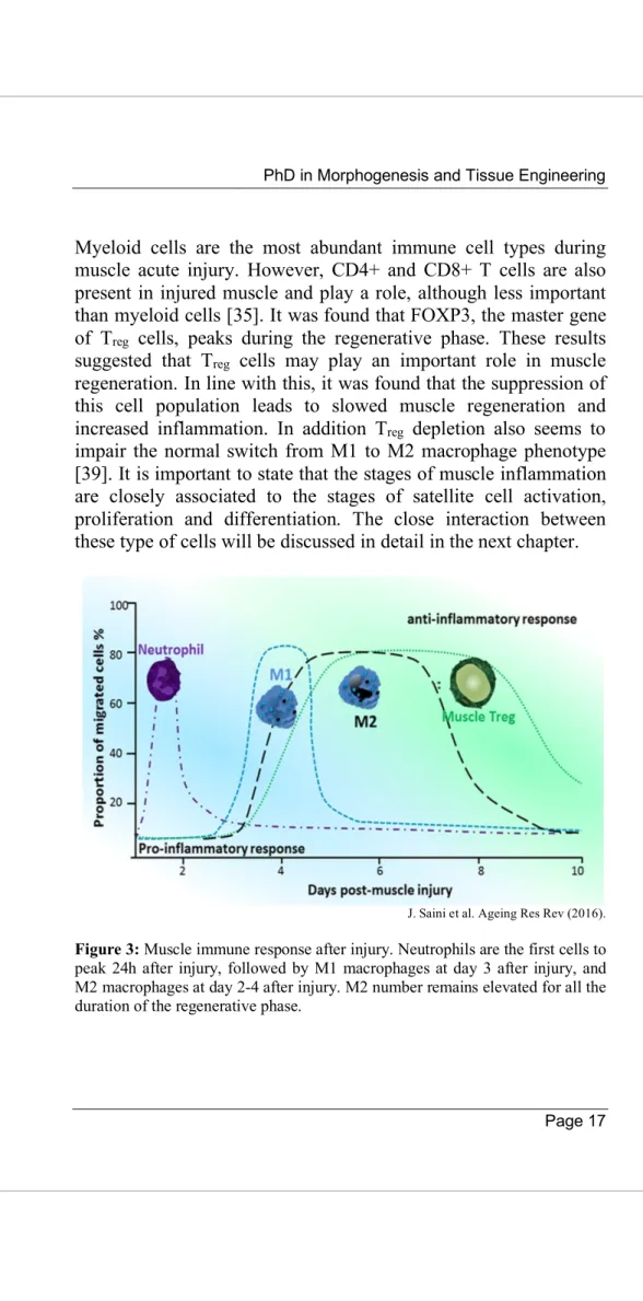
Myeloid cells are the most abundant immune cell types during muscle acute injury. However, CD4+ and CD8+ T cells are also present in injured muscle and play a role, although less important than myeloid cells [35]. It was found that FOXP3, the master gene of Treg cells, peaks during the regenerative phase. These results suggested that Treg cells may play an important role in muscle regeneration. In line with this, it was found that the suppression of this cell population leads to slowed muscle regeneration and increased inflammation. In addition Treg depletion also seems to impair the normal switch from M1 to M2 macrophage phenotype [39]. It is important to state that the stages of muscle inflammation are closely associated to the stages of satellite cell activation, proliferation and differentiation. The close interaction between these type of cells will be discussed in detail in the next chapter.
J. Saini et al. Ageing Res Rev (2016).
Figure 3: Muscle immune response after injury. Neutrophils are the first cells to peak 24h after injury, followed by M1 macrophages at day 3 after injury, and M2 macrophages at day 2-4 after injury. M2 number remains elevated for all the duration of the regenerative phase.
1.2.3 Satellite cell and immune cell crosstalk during injury After muscle injury, neutrophils are the first cells to arrive, followed by macrophages later, which begin to express the pro-inflammatory cytokine TNF-α. The level of this cytokine peaks 24h after injury, indicating its role in the early inflammatory response and the promotion of muscle fiber damage. TNF-α induces the production of NO by myeloid cells, that is linked to fiber damage. However, TNF-α level remains elevated for the following 2 weeks after injury, suggesting a role of this cytokine also in muscle regeneration [40]. These different roles of TNF-α may depend on the cell target. If the target is macrophages, TNF-α acts by inducing their pro-inflammatory phenotype, if the target is satellite cells TNF-α stimulates their activation and proliferation. In vivo studies of TNF-α-null mice showed a reduced expression of MyoD and Mef-2 compared to WT, suggesting a role of this cytokine in the induction of the early differentiation phase [41]. However, in vitro studies showed that TNF-α acts as an inhibitor of myoblast fusion, suggesting that this cytokine acts as a negative regulator for the switch from the early to the terminal differentiation phase of SCs[42]. TNF-α can also activate NF-κB transcription factor, which induces the expression of TNF-α and IL-1 further amplifying the inflammatory response. NF-κB acts by stimulating satellite cell proliferation and inhibiting their differentiation, by promoting Cyclin D1 expression and reducing MyoD expression in satellite cells [42], [43]. Furthermore NF-κB induces IL-6 expression, a pleiotropic cytokine that amplify the proliferative anti-differentiation effects of NF-κB. However, IL-6 can also influence the late phase of differentiation, contributing to muscle formation and growth [44].
IFN-γ is another critical cytokine for the coordination of inflammation and the early regeneration stage. Neutrophils and macrophages produce IFN-γ, which peaks within the first 24h after muscle injury. However, its levels remain high up to day 5 after injury. Together with TNF-α and IL-1β, IFN-γ induces monocyte differentiation to M1 macrophages [45]. IFN-γ also binds to
myoblasts, where it activates the JAK-STAT1 pathway, inhibiting their differentiation by reducing myogenin expression and inducing the expression of genes such as CIITA. IFN-γ is thus important for the clearance of cell debris, and satellite cell proliferation [46]. Around day 3-4 after injury, an increase in IL-10 level drives the switch of macrophages from M1 to M2 phenotype, and this corresponds to the transition from the proliferation to the differentiation phase of satellite cells. As discussed in the previous chapter, the M2 macrophage population is characterized by the production of IL-10 and TGF-β, and is essential to suppress inflammation and promote muscle regeneration. Ablation of IL-10 negatively affects the macrophage switch, prolonging the inflammatory phase and severely impairing muscle regeneration. M2 macrophages secrete numerous types of ECM proteins, fibronectin and Collagen VI [47]. Besides, IL-4 and IGF-1 released by M2 macrophages are essential to induce SC differentiation. IGF-1 is also produced by myofibers, satellite cells and endothelial cells. This cytokine binds to the IGF-1 receptor and induces AKT pathway to initiate protein synthesis and inhibit protein degradation, contributing to muscle growth [48].
In conclusion, the immune system and satellite cell crosstalk during muscle injury is a finely bidirectionally regulated process. Alterations in the balance of the type and level of cytokines translates in regeneration defects.
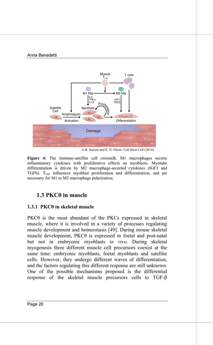
A.B. Aurora and E. N. Olson. Cell Stem Cell (2014).
Figure 4: The immune-satellite cell crosstalk. M1 macrophages secrete inflammatory cytokines with proliferative effects on myoblasts. Myotube differentiation is driven by M2 macrophage-secreted cytokines (IGF1 and TGFb). Tregs influences myoblast proliferation and differentiation, and are
necessary for M1 to M2 macrophage polarization.
1.3 PKCθ in muscle
1.3.1 PKCθ in skeletal musclePKCθ is the most abundant of the PKCs expressed in skeletal muscle, where it is involved in a variety of processes regulating muscle development and homeostasis [49]. During mouse skeletal muscle development, PKCθ is expressed in foetal and post-natal but not in embryonic myoblasts in vivo. During skeletal myogenesis three different muscle cell precursors coexist at the same time: embryonic myoblasts, foetal myoblasts and satellite cells. However, they undergo different waves of differentiation, and the factors regulating this different response are still unknown. One of the possible mechanisms proposed is the differential response of the skeletal muscle precursors cells to TGF-β
differentiation inhibitory effect. Embryonic myoblasts usually differentiate when treated with TGF-β. However, when PKCθ is expressed ectopically in these cells, they become sensitive to the differentiation inhibitory effect of TGF-β. This results suggest that PKCθ, together with TGF-β regulate the asynchronous differentiation of the muscle precursors [50].
Another role of PKCθ in fetal myogenic development is the regulation of the fetal muscle creatin kinase (MCK) expression. MCK transcription is directly mediated by MEF2A, which is expressed already in embryonic myoblasts, but is not active. Its activation is regulated by Nuclear Factor I X (Nfix), which is selectively expressed in fetal but not in embryonic myoblasts. The proposed mechanism for MCK expression is that MEF2A binds Nfix, which recruits PKCθ that in turn activates MEF2A by phosphorylating it. A study in C2C12 supports the regulation of MEFA activity by PKCθ, showing that PKCθ interacts with calcineurin to regulate HDAC shuttling and MEF2 activation [51]. Together these results show that PKCθ is involved in the fetal skeletal muscle myogenesis.
As mentioned before, PKCθ is expressed in neonatal muscle, suggesting its potential involvement in the regulation of postnatal muscle growth. Accordingly, we observed a delay in muscle and body growth in total and muscle specific PKCθ knock out mice, during the first weeks of life. Moreover, even muscle regeneration, after induction of freeze injury was delayed in PKCθ-/- mice, compared to WT. As a further confirmation of delayed regeneration, we observed reduced expression of myogenin, but not Pax7, indicating a role of PKCθ in the regulation of myogenic differentiation. In vitro, we found that PKCθ is expressed in satellite cells, and reaches a peak during the initial phase of myoblast fusion. Indeed, cultured myoblast derived from PKCθ-/-mice generated smaller myotubes with less myonuclei, compared to WT. This phenotype was associated with a reduced phosphorylation/ activation of FAK (focal adhesion kinase), and a reduced expression of FAK target genes caveolin-3 and β1D-
integrin (two genes upregulated during myoblast fusion). This results suggest that PKCθ regulates the myogenic differentiation program by activating FAK [52].
In another study, it was shown that silencing PKCθ in the myoblast cell line C2C12, led to opposite results. The authors showed that PKCθshRNA C2C12 cells had increased fusion rate, with bigger myotubes and an increased rate of protein synthesis. These effects were mediated by the modulation of IRS1 and ERK1/2 phosphorylation. The discrepancy between this results and our findings may depend on the different cell system used [53].
PKCθ is also localized at the Neuro Muscular Junction (NMJ), and its expression in muscle peaks concomitantly with the NMJ maturation. Thus, PKCθ may also be involved in nerve maturation. In line with this, it was found that synapse elimination, a process normally occurring during NMJ maturation, was altered in PKCθ-/- mice [54]. However, adult PKCθ-PKCθ-/- mice showed normal NMJ, indicating that PKCθ may not be required for its final maturation [55].
PKCθ also regulates other pathways involved in skeletal muscle homeostasis, such as ER stress induced autophagy in C2C12 cells [56]. Furthermore, PKCθ controls the sarcolemma electrical activity by regulating the CIC-1 chloride channel [57].
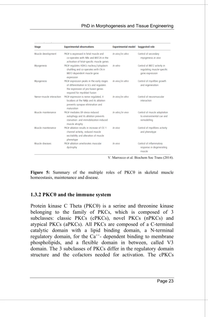
V. Marrocco et al. Biochem Soc Trans (2014).
Figure 5: Summary of the multiple roles of PKCθ in skeletal muscle homeostasis, maintenance and disease.
1.3.2 PKCθ and the immune system
Protein kinase C Theta (PKCθ) is a serine and threonine kinase belonging to the family of PKCs, which is composed of 3 subclasses: classic PKCs (cPKCs), novel PKCs (nPKCs) and atypical PKCs (aPKCs). All PKCs are composed of a C-terminal catalytic domain with a lipid binding domain, a N-terminal regulatory domain, for the Ca++- dependent binding to membrane phospholipids, and a flexible domain in between, called V3 domain. The 3 subclasses of PKCs differ in the regulatory domain structure and the cofactors needed for activation. The cPKCs
require for their activation both binding to DAG and Ca++. The nPKCs only require DAG but not Ca++ for the activation, while the aPKC do not require DAG nor Ca++ [58]. PKCθ belongs to the class of the novel PKCs, and its structure has been solved by Xu et al. These researchers showed that there are two main conformations for PKCθ: open/active and closed/ inactive. The switch from the closed state to the open state involves the biding to DAG, and the phosphorylation of Thr-538, which is fundamental for kinase activation [59]. PKCθ is mainly expressed in hematopoietic cells, in particular in T cells, but also in mast cells, natural killer cells and platelets [60], [61], [62]. PKCθ is also the most abundant PKC expressed in skeletal muscle [63], where it mediates insulin resistance associated with type 2 diabetes [64]. PKCθ best characterized role however, is that in T cells, where it regulates cell activation and proliferation. When a T cell recognizes an antigen presenting cell, a tight junction called immunological synapse forms at the site where the T cell receptor (TCR) binds to the peptide-MHC complex. PKCθ is selectively recruited to the immunological synapse in conventional and effector T cells, where it directs signal transduction cascade. PKCθ activation regulates the expression of NF-kB, AP-1, and NFAT, three genes fundamental for T cell function. In addition, PKCθ can regulate gene expression by physically associating with chromatin and inducing genes associated with the immune response [65]. PKCθ knock out mice (PKCθ-/-) are healthy and live normally, and show a normal development of T cells in the thymus [66]. However, PKCθ-/- mice show a severely impaired Th2 response. Following TCR binding to MHC, PKCθ-/- mice display a reduction in the production of IL-2, with a consequent defective proliferation of T cell, and impaired NF-kB and AP-1 activation [67]. Despite these findings, Berg-Brown et al. showed that PKCθ-/- mice could mount an immune response against vesicular stomatitis virus (VSV) infection. This, together with other studies led to the finding that PKCθ-/- is dispensable for the immune response against viruses and bacteria. This apparently contradictive
response can be explained by the fact that other mechanisms such as innate immune responses can compensate for the absence of PKCθ. Therefore, the innate immune response can overcome the need for a CD8+ T cell response [68].
Brezar et al. Front. In Immunol. (2015)
Figure 6: PKCθ localized to the immunological synapse in Teff cells, where it
regulates their activation and proliferation by inducing NF-kB, AP-1, and NFAT activation.
Also Treg cells express PKCθ. These cells are important regulators of CD4+, CD8+, NK, and B cell responses. PKCθ-/- mice showed impaired Treg development in the thymus, with a consequent reduced number of Treg in the periphery, although not completely suppressed [69][70]. However, Treg isolated from PKCθ-/- mice showed a normal suppressive activity in vitro. Although some studies identified PKCθ as a necessary enzyme for Treg differentiation, some other studies found that PKCθ inhibition actually enhances Treg cell suppressive activity [71]. These discrepancies have yet to be elucidated. However, what emerges from these findings is that Treg activity is not significantly
compromised following PKCθ inhibition. This, together with the finding that PKCθ is not necessary for Teff cell response against viruses and bacteria, makes PKCθ a promising target for the treatment of diseases requiring immune modulation.
1.3.3 Targeting PKCθ in Duchenne Muscular Dystrophy As discussed above, PKCθ plays an essential role in the immune response. Lack of PKCθ inhibits Teff cell function without significantly compromising Treg suppressive activity. For this reason PKCθ inhibition has been proposed for the treatment of immune diseases, such as autoimmune diseases, graft versus host disease, Th2 and Th17 mediated inflammatory diseases [58]. In our laboratory we tested the therapeutic effect of PKCθ inhibition for the tratment of a chronic muscle inflammation in Duchenne Muscular Dystrophy (DMD). The only available therapy for DMD is glucocorticoid treatment, general immunosuppressors which initially slow down the disease progression, but are associated with many adverse effects. Works from our lab in the mdx mouse, the animal model for DMD, showed that CD3+ T cells (CD4+ and CD8+) are among the first cells to infiltrate dystrophic muscle, prior to the onset of necrosis. These findings identified T cells as one of the main cell populations orchestrating the early acute inflammatory response in mdx mice. Pharmacological inhibition of PKCθ with C20, a very potent and selective inhibitor of PKCθ, led to a significant reduction in T cells, but also neutrophils, macrophages and eosinophils recruited to dystrophic muscle. Reduced inflammation was associated with reduced muscle degeneration, reduced collagen deposition and improved muscle performance [72], [73]. Similar results were obtained with the PKCθ-/- mouse model [74].
Interestingly, in our mdx PKCθ-/- mouse model, although muscle damage was reduced, we found that muscle regeneration was increased (Figure 7A). The increased muscle regeneration was
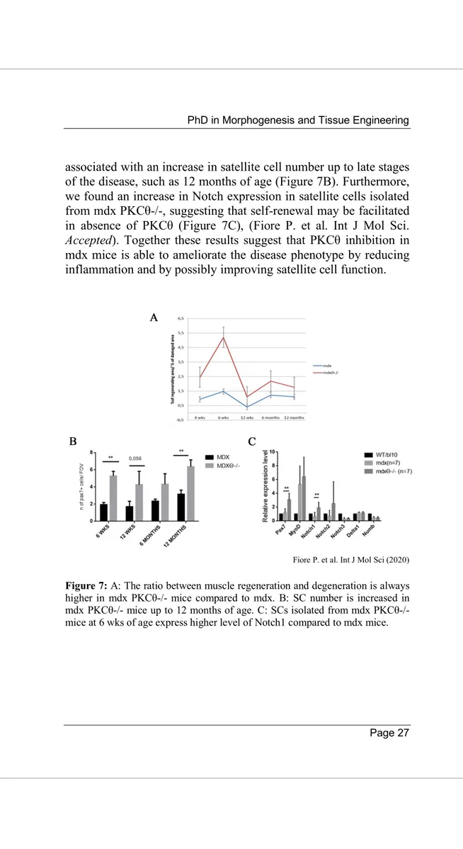
associated with an increase in satellite cell number up to late stages of the disease, such as 12 months of age (Figure 7B). Furthermore, we found an increase in Notch expression in satellite cells isolated from mdx PKCθ-/-, suggesting that self-renewal may be facilitated in absence of PKCθ (Figure 7C), (Fiore P. et al. Int J Mol Sci. Accepted). Together these results suggest that PKCθ inhibition in mdx mice is able to ameliorate the disease phenotype by reducing inflammation and by possibly improving satellite cell function.
Fiore P. et al. Int J Mol Sci (2020)
Figure 7: A: The ratio between muscle regeneration and degeneration is always higher in mdx PKCθ-/- mice compared to mdx. B: SC number is increased in mdx PKCθ-/- mice up to 12 months of age. C: SCs isolated from mdx PKCθ-/- mice at 6 wks of age express higher level of Notch1 compared to mdx mice.
2. AIMS OF THE THESIS
Our preliminary results in mdx PKCθ-/- mice suggest that lack of PKCθ improves regeneration and influences satellite cell function. Indeed, we observed an improved regenerative ability in mdx PKCθ-/- mice compared to control, even at later stages of the disease, which should be due predominantly to changes in the inflammatory response. However, there might be also a direct effect on SCs. Indeed, mdx PKCθ-/- mice show an increase in the number of satellite cells, which express higher level of Notch compared to mdx mice, indicating that self-renewal may be facilitated in the absence of PKCθ. Thus, we speculated that PKCθ may control satellite cell behavior via direct and/or indirect pathways (such as immune cell crosstalk).
2.1 To investigate the direct role of PKCθ in satellite cell function
To understand whether PKCθ influences SC function directly, we studied PKCθ expression kinetics and its localization in satellite cells. We then analyzed the effect of PKCθ absence/ inhibition on self-renewal in vitro, by studying satellite cell symmetric/asymmetric division and Pax7/MyoD expression on single myofibers. To understand whether PKCθ influences satellite cell self-renewal in vivo, we analyzed the effect of PKCθ absence/inhibition on satellite cell number in steady state and after muscle injury.
2.2 To investigate the indirect pathways by which PKCθ influences the satellite cell compartment
It is known that satellite cell behaviour is strongly influenced by the inflammatory environment [75]. PKCθ regulates T cell activation and proliferation, and in the context of chronic muscle inflammation is important for T cell recruitment. On the other hand, its role in neutrophil and macrophage responses, which are
known to affect SC ability to regenerate muscle, is less clear [58]. To understand whether PKCθ also influences myeloid cell infiltration we used a model of acute muscle injury, where neutrophils and monocytes but not T cells predominate during the first week following injury. Following intra-muscular injection of cardiotoxin (CTX) in WT and PKCθ-/- mice we analyzed the quality and quantity of the inflammatory infiltrate by flow-cytometry at 3 and 10 days after injury, during the inflammatory and the regenerative phase respectively. We also analyzed the production of inflammatory cytokines by Real Time PCR. These experiments helped to clarify whether PKCθ regulates the recruitment and activity of innate immune cells known to influence satellite cell ability to regenerate muscle.
3. RESULTS
3.1 PKCθ is expressed in satellite cells
3.1.1 PKCθ is expressed in freshly isolated and differentiating satellite cells, and it is downregulated in proliferating cells Satellite cells can exist in a quiescent state in physiological conditions, or as activated/committed progenitors during muscle regeneration. To elucidate the functional role of PKCθ in satellite cells, we first analyzed the kinetics of PKCθ expression by real time PCR (rtPCR) during myogenic differentiation. Satellite cells were isolated by magnetic bead labeling from WT hindlimb muscles, before or after CTX injury.
It is known that after isolation satellite cells become activated. Indeed, the isolation procedure leads to their dissociation from the fiber, causing profound transcriptomic and epigenetic modifications, compared to quiescent cells [76].
To investigate whether PKCθ is expressed in early activated cells, PKCθ was analyzed in freshly isolated satellite cells from uninjured muscle.
PKCθ expression was also analyzed in satellite cells isolated from regenerating muscle at 3, 5 and 7 days after CTX injury. Interestingly, rtPCR analysis showed that PKCθ expression is high in early activated cells and drastically decreases in cells isolated from 3 day-injured muscle, corresponding to the proliferation phase, in vivo. PKCθ expression then increases in cells isolated from 5 and 7 day-injured muscle, corresponding to the regeneration phase (Figure 8).
To confirm the differential expression observed by rtPCR, we examined PKCθ expression by western blot analysis in cultured myoblasts. Satellite cells were isolated by magnetic bead labeling and cultured in growth medium (GM) for 72h, and subsequently in differentiation medium (DM) for 24h, to analyze PKCθ expression
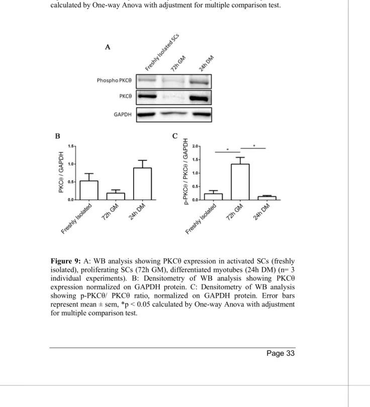
in proliferating and differentiating cells respectively. Another subset of satellite cells was collected for analysis immediately after isolation to analyze PKCθ expression in activated cells. Western Blot analysis showed that PKCθ is already expressed in activated cells, its expression declines in proliferating cells and increases again in differentiating cells (Figure 9A and B). The analysis of the phosphorylated/active form instead, revealed that the ratio between p-PKCθ and PKCθ was maximal at 72h in GM, during the proliferative phase (Figure 9A and C).
Figure 8: A: experimental design. B: rtPCR showing PKCθ expression in freshly isolated SCs (n = 9 mice) and SCs isolated from 3, 5 and 7 day-injured WT muscles (n ≥ 4 mice per group). Total RNA extract from WT tibialis muscle was used as positive control. GAPDH was used for normalization. Error bars represent mean ± sem, *p < 0.05, **p < 0.01, ***p < 0.001, ****p < 0.0001 calculated by One-way Anova with adjustment for multiple comparison test.
Figure 9: A: WB analysis showing PKCθ expression in activated SCs (freshly isolated), proliferating SCs (72h GM), differentiated myotubes (24h DM) (n= 3 individual experiments). B: Densitometry of WB analysis showing PKCθ expression normalized on GAPDH protein. C: Densitometry of WB analysis showing p-PKCθ/ PKCθ ratio, normalized on GAPDH protein. Error bars represent mean ± sem, *p < 0.05 calculated by One-way Anova with adjustment for multiple comparison test.
3.1.2 Phospho-PKCθ is localized to the chromosomes, centrosomes and midbody of dividing satellite cells
PKCθ shows two conformational states: one closed/inactive and one open/active [58]. The transition to the active state requires binding to the DAG and T538 phosphorylation. To understand the specific role of PKCθ in satellite cells, we assessed its activation and localization in cultured satellite cells.
To this aim we isolated satellite cells from WT mice and cultured them for 72h in GM. After 72h in culture we performed immunofluorescence analysis of phospho-PKCθ and α-Tubulin, to visualize the mitotic spindle.
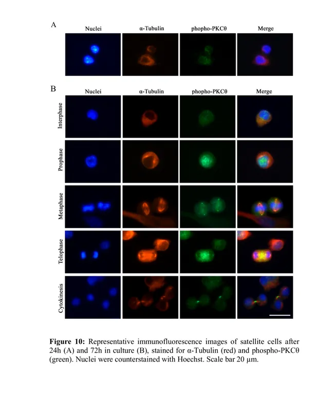
At 24h of culture in GM a diffused phospho-PKCθ immunostaining was visible on satellite cell membrane (Figure 10A). At 72h in culture in growth medium, satellite cells are proliferating. At this stage it was possible to distinguish the different phases of mitosis thanks to the nuclear shape.
The results showed a diffused membrane localization of phospho-PKCθ during interphase. During prophase phospho-phospho-PKCθ moves to the nucleus, where chromatin is condensating. During metaphase phospho-PKCθ immunostaining is detectable close to the centrosomes, and partially to the mitotic spindle. In telophase phospho-PKCθ is visible at the spindle midzone, and during cytokinesis it moves to the intercellular bridge of the midbody (Figure 10B).
These results suggest that PKCθ could be involved in the regulation of satellite cell division processes.
Figure 10: Representative immunofluorescence images of satellite cells after 24h (A) and 72h in culture (B), stained for α-Tubulin (red) and phospho-PKCθ (green). Nuclei were counterstained with Hoechst. Scale bar 20 µm.
A
3.1.3 Satellite cell proliferation is not influenced by PKCθ The localization of phospho-PKCθ to the chromatin, centrosomes and midbody of dividing satellite cells, suggested that PKCθ could be involved in the regulation of satellite cell division.
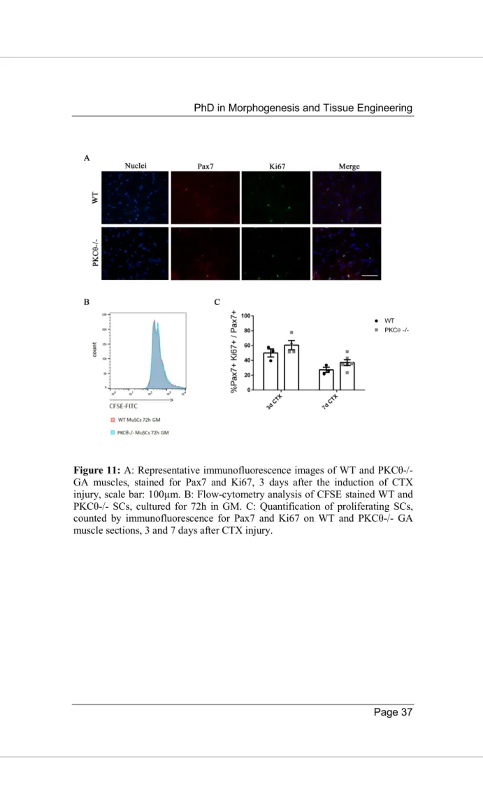
To understand whether satellite cell proliferation is affected by PKCθ, we analyzed proliferation both in vitro and in vivo. For the in vitro analysis, SCs were isolated from WT and PKCθ-/- muscles and cultured in growth medium in the presence of CellTrace CFSE Cell Proliferation Kit. CFSE is used to detect different generations of dividing cells by analyzing die dilution by flow-cytometry. After 72h of culture in GM with CFSE, satellite cell proliferation was analyzed. As shown, no difference was observed in CFSE fluorescence between WT and PKCθ-/- cells (Figure 11B), suggesting that the cells underwent similar rounds of cycling. Cell proliferation was also analyzed in regenerating gastrocnemius (GA) muscles in vivo, 3 and 7 days after CTX injury during the peak of SC proliferation and the initial phase of regeneration respectively, by immunofluorescence staining for Ki67 and Pax7. The number of cells double positive for Pax7 and Ki67 were counted, and the results were normalized to the total number of Pax7+ cells. As shown in Figure 11C, the percentage of proliferating SCs (Pax7+/Ki67+) was similar between WT and PKCθ-/- mice.
These results indicate that satellite cell proliferation is not influenced by PKCθ, neither in vitro nor in vivo.
Figure 11: A: Representative immunofluorescence images of WT and PKCθ-/- GA muscles, stained for Pax7 and Ki67, 3 days after the induction of CTX injury, scale bar: 100µm. B: Flow-cytometry analysis of CFSE stained WT and PKCθ-/- SCs, cultured for 72h in GM. C: Quantification of proliferating SCs, counted by immunofluorescence for Pax7 and Ki67 on WT and PKCθ-/- GA muscle sections, 3 and 7 days after CTX injury.
3.2 Lack of PKCθ increases satellite cell
self-renewal in vitro
3.2.1 Lack of PKCθ increases the number of ‘self renewing’ SCs by promoting symmetric division
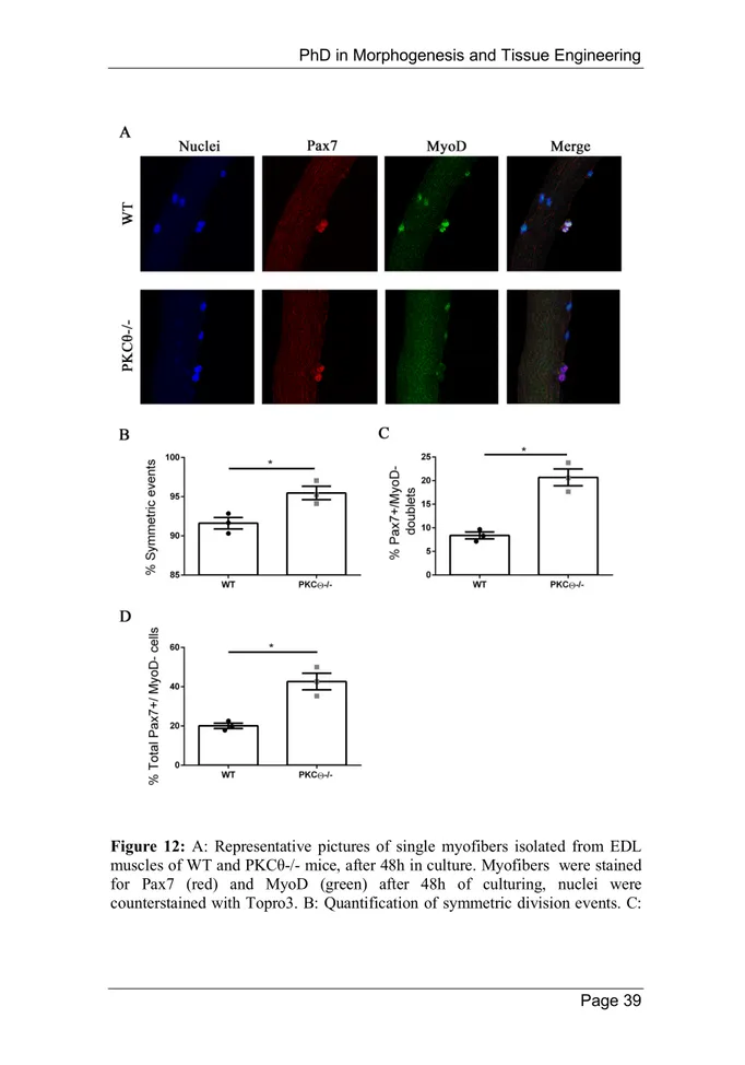
In stem cells, centrosomes and mitotic spindle orientation determine whether the outcome of a cell division is symmetric or asymmetric. In T cells, PKCθ promotes MTOC reorientation and cell polarization in the direction of the antigen presenting cell. This process may also promote asymmetric division, that is important for the acquisition of T cell memory [77]. The localization of phospho-PKCθ at the centrosomes and mitotic spindle of dividing satellite cells, led us to hypothesize that this protein may be involved in the regulation of symmetric/asymmetric division. To investigate this possibility we isolated single myofibers from WT and PKCθ-/- EDL muscles, and cultured them for 48h, the time necessary for the first cell division to occur. In this system satellite cells are not dissociated from their fiber, allowing to keep them in a niche-like environment. When myofibers are cultured satellite cells become activated, upregulate MyoD and enter cell cycle. The cells that undergo self-renewal will be positive for Pax7 and negative for MyoD, whilst satellite cells committed to differentiation will express MyoD. After 48h in culture cell doublets were visible, and symmetric/asymmetric division was studied by immunofluorescence staining for Pax7 and MyoD. We observed a significant increase in the number of symmetric satellite cell division in myofibers isolated from PKCθ-/- mice, compared to WT (Figure 12B). Within the symmetric division events, the number of ‘self-renewing’ Pax7+/MyoD- cell doublets was significantly higher in myofibers from PKCθ-/- mice compared to WT (Figure 12C).
Consistent with this result, also the number of total Pax7+/ MyoD- single cells was higher in PKCθ-/- myofibers, compared to the ones isolated from WT mice (Figure 12D).
Figure 12: A: Representative pictures of single myofibers isolated from EDL muscles of WT and PKCθ-/- mice, after 48h in culture. Myofibers were stained for Pax7 (red) and MyoD (green) after 48h of culturing, nuclei were counterstained with Topro3. B: Quantification of symmetric division events. C:
Quantification of Pax7+/MyoD- cell doublets, and D: quantification of total Pax7+/MyoD- cells in WT and PKCθ -/- single myofibers. (WT, n = 3 mice, PKCθ-/-, n = 3 mice, n > 20 myofibers analyzed per mouse). Error bars represent mean ± sem, *p < 0.05 calculated by Student’s t-test.
To understand whether the increased number of ‘self-renewing’ satellite cells is maintained after further divisions, we cultured single myofibers from WT and PKCθ-/- mice for 72h. After this time in culture satellite cells have proliferated, and cell clusters are visible on the myofiber surface. Pax7 and MyoD immunofluorescence revealed that the number of ‘self-renewing’ (Pax7+/MyoD-) cells was significantly higher in PKCθ-/- fibers compared to the WT ones. The fraction of Pax7+/MyoD+, and Pax7-/MyoD+ satellite cells, was reduced in PKCθ-/- myofibers instead (Figure 13B). The number of satellite cells per cluster was similar between the two genotypes, suggesting again that PKCθ does not affect satellite cell proliferation (Figure 13C).
Together, these results suggest that PKCθ absence increase satellite cell self-renewal in vitro by promoting symmetric cell division.
Figure 13: A: Representative pictures of single myofibers isolated from EDL muscles of WT and PKCθ -/- mice. Myofibers were stained for Pax7 and MyoD after 72h of culturing, nuclei were counterstained with Topro3. B: Quantification of SCs per fiber, single or double positive for Pax7 and/or MyoD. C: number of SCs per fiber in WT and PKCθ -/- single myofibers. (WT, n = 4 mice, PKCθ-/-, n = 4 mice, n = 100 cells analyzed per group). Error bars represent mean ± sem, *p < 0.05 calculated by Student’s t-test.
3.2.2 C20 treatment in cultured primary myoblasts increases the fraction of reserve cells
As described above, lack of PKCθ leads to increased satellite cell self-renewal in vitro. We next analyzed whether pharmacological inhibition of PKCθ with C20 might lead to similar results as
genetic ablation of PKCθ in cultured satellite cells. To answer this question, we cultured primary satellite cells from hind limb muscles of WT and PKCθ-/- mice, in presence of a PKCθ inhibitor, the C20. To analyze the effects of C20 throughout the different phases of myogenic progression, the cells were cultured for 4 days in growth medium (GM), and for 2 days in differentiation medium (DM). C20 was used at different concentration: 0.5 µM, 1 µM and 2 µM. These concentrations were already shown not to be toxic for in vitro and in vivo treatment [78], [72]. Control cultures were treated with DMSO at the same concentration used for C20 dilution. To analyze any possible indirect effect of C20, we treated PKCθ-/- satellite cells with C20, at the maximum concentration used for the experiment (2 µM). Treatment with C20 significantly increased the fraction of Pax7+/MyoD- reserve cells in a dose dependent manner (Figure 14A and B). In parallel, the fraction of cells committed to differentiation Pax7-/MyoD+ was reduced (Figure 14C). Fusion index was also reduced after treatment with C20, but the reduction was significant only at the highest dose of C20 used (Figure 14D). Similar results were obtained analyzing the phenotype of the PKCθ-/- cultured cells. Treatment of PKCθ-/- cells with C20, did not show any difference compared to the non-treated PKCθ-/- cells, instead (not shown). These results together suggest that pharmacological inhibition of PKCθ with C20 increases the fraction of ‘self-renewing’ satellite cells in culture.
PhD in Morphogenesis and Tissue Engineering
Figure 14: A: Representative pictures of PKCθ-/- and WT SCs, cultured or not with C20 at the concentration of 0.5, 1 and 2 µM (or DMSO as control). The cells were stained for Pax7 (red) and MyoD (green) after culturing for 4 days in GM and 2 days in DM. Nuclei were counterstained with Hoechst. B: percent of Pax7+/MyoD- SCs in WT and PKCθ-/- cultures, or in WT cultures treated with C20 (C). D: Percent of total MyoD+ SCs in WT and PKCθ-/- cultures, or in WT cultures treated with C20 (E). F: Fusion index of WT and PKCθ-/- myotubes, or WT myotubes treated with C20 (G) after culturing for 4 days in GM and 2 days in DM. (n = 3 replicate dishes per group). Error bars represent mean ± sem, *p < 0.05, **p < 0.01 calculated by One-way Anova with adjustment for multiple comparison test.
3.3 Genetic and pharmacological ablation of
PKCθ expands satellite cell reservoir after acute
injury
3.3.1 The pool of quiescent satellite cells is amplified in PKCθ-/- mice 28 days after injury
The immunofluorescence analysis performed on WT and PKCθ-/- myofibers in vitro, suggested that PKCθ is implicated in the regulation of satellite cell self-renewal. To understand whether PKCθ absence leads to similar results in vivo as well, we analyzed the number of satellite cells in WT and PKCθ-/- mice, before and after the induction of Cardiotoxin (CTX) injury.
To study satellite cell self-renewal in vivo we analyzed the number of satellite cells in WT and PKCθ-/- mice 7 and 28 days after injury, when the muscle is regenerating or is completely regenerated respectively. Contralateral uninjured muscle was used as control.
Immunofluorescence analysis of Pax7+ cells revealed that the number of satellite cells per mm2 and the number of satellite cells per fiber was similar in PKCθ-/- and WT gastrocnemius (GA) muscles in uninjured conditions. 7 days after injury, the number of Pax7+ cells was increased in both WT and PKCθ-/- mice, as a result of cell proliferation. However, the number of Pax7+ cells in PKCθ-/- mice was significantly higher compared to WT mice (Figure 15). 28 days after CTX injury when muscle is completely regenerated and satellite cells have returned to quiescence, the number of Pax7+ cells is significantly higher in PKCθ-/- muscle compared to the injured WT, with an increment of 64.4% (Figure 16B and C). To confirm that at this stage all of the satellite cells have gone back to quiescence, we analyzed their cycling status by immunofluorescence staining for Pax7 and Ki67. The result showed that more than 99% of the Pax7+ cells were negative for Ki67 in both WT and PKCθ-/- mice, indicating that they are not proliferating cells (Figure 16F). Moreover, all the cells analyzed 28
days after CTX were located in the final position of quiescent cells, beneath the basal lamina and the sarcolemma of muscle fibers (Figure 16A).
This observation demonstrates that satellite cells returned to homeostatic conditions 28 days after CTX in WT and PKCθ-/- mice, and the pool of quiescent satellite cells is amplified in the absence of PKCθ-/-.
To compare the regenerative ability of WT and PKCθ-/- mice, we analyzed myofiber CSA 28 days after injury (Figure 16 D and E), and we observed that the mean myofiber CSA, and the distribution of myofiber CSAs were similar in WT and PKCθ-/- mice. These results suggest that lack of PKCθ increases satellite cell self-renewal without affecting muscle regenerative ability after injury.
Figure 15: A: representative immunofluorescence pictures of WT and PKCθ-/- GA sections, 7 days after CTX injury. Sections were stained for Pax7 (red) and Laminin (green). Nuclei were counterstained with Hoechst. Scale bar: 100µm. B: quantification of the number of SCs per mm2 and C: number of SCs per fiber
in uninjured and 7 day-injured GA muscle, in WT and PKCθ-/- mice (WT, n = 4 mice, PKCθ-/-, n = 4 mice). Error bars represent mean ± sem, *p < 0.05, **p < 0.01, ***p < 0.001, **** p < 0.0001 calculated by Two-way Anova with adjustment for multiple comparison test.
Figure 16: A: representative immunofluorescence pictures of WT and PKCθ-/- GA sections, 28 days after CTX injury. Sections were stained for Pax7 (red) and Laminin (green). Nuclei were counterstained with Hoechst. Scale bar: 100µm. B: quantification of the number of SCs per mm2 and C: number of SCs per fiber
in uninjured and 28 day-injured GA muscle, in WT and PKCθ-/- mice. D: mean CSA and E: CSA distribution of muscle fibers in WT and PKCθ-/- GA sections, 28 days after injury. F: quantification of non proliferating SCs 28 days after CTX injury, in WT and PKCθ-/- GA, analyzed by immunofluorescence staining for Pax7 and Ki67. (WT, n = 4 mice, PKCθ-/-, n = 4 mice). Error bars represent mean ± sem, *p < 0.05, **p < 0.01, ***p < 0.001, **** p < 0.0001 calculated by Two-way Anova with adjustment for multiple comparison test.
3.3.2 Pharmacological inhibition of PKCθ expands the pool of quiescent satellite cells after injury
As we already demonstrated, lack of PKCθ leads to expansion of the satellite cell pool after induction of CTX injury.
To investigate whether pharmacological inhibition of PKCθ leads to similar results, we treated WT mice with the pharmacological inhibitor of PKCθ (C20), and we analyzed satellite cell number before and after CTX injury. C20 is a highly potent and selective inhibitor of PKCθ, which has been already used in our lab to test its ability to counteract the dystrophic phenotype in a mouse model of Duchenne Muscular Dystrophy, the mdx mouse [72], [73]. In those studies we showed that C20 treatment administered to mdx mice for 2 weeks , is able to successfully inhibit PKCθ, without causing any major collateral effect.
Thus, we injured GA muscles of WT mice by CTX injection, and we treated the mice with daily intra peritoneal injections of C20, at a dose of 5 mg/Kg (previously established to be effective in vivo, [78]). Control mice were treated with Vehicle (DMSO) at the same concentration used to dissolve C20. Treatment was started one day prior to the CTX injection, and was continued for 10 days following injury, during the phase of satellite cell activation, proliferation and differentiation (Figure 17A). Satellite cell number was then analyzed by Pax7 immunofluorescence 28 days after injury, in the injured GA and in the contralateral uninjured GA.
The results showed that the number of Pax7+ cells per mm2, and the number of Pax7+ cells per fiber was similar in uninjured GA in C20 and Vehicle treated mice. However, the number of satellite cells that returned to quiescence 28 days after injury, was significantly higher in mice treated with C20, compared to Vehicle treated ones, with an increment of 50% (Figure 17C and D). The analysis of the mean CSA and the CSA distribution of myofibers showed no significant differences between C20 and Vehicle treated mice, indicating that C20 treatment does not affect muscle regenerative ability (Figure 17E and F). These results suggest that C20 can be used in vivo to target satellite cell self-renewal.
Figure 17: A: experimental plan. B: representative immunofluorescence pictures of WT GA sections 28 days after CTX injury, treated with C20 or
vehicle. Sections were stained for Pax7 (red) and Laminin (green). Nuclei were counterstained with Hoechst. Scale bar: 100µm. C: quantification of the number of SCs per mm2 and D: number of SCs per fiber in uninjured and 28 day-injured
GA muscle, in WT mice treated with C20 or vehicle. E: mean CSA and F: CSA distribution of muscle fibers in WT mice treated with C20 or vehicle, 28 days after injury. (C20 treated WT, n = 4 mice, Vehicle treated mice n = 4 mice). Error bars represent mean ± sem, *p < 0.05, ***p < 0.001, **** p < 0.0001 calculated by Two-way Anova with adjustment for multiple comparison test.
3.3.3 The number of quiescent satellite cells increases in PKCθ-/- mice after repeated injuries
It has been shown that satellite cell population is maintained after repeated traumas, thanks to their ability to self-renew. To investigate the behavior of PKCθ-/- satellite cells following repeated injury, we induced 3 CTX injuries in WT and PKCθ-/- GA muscles. The three injuries were performed 20 days apart from each other, and the muscles were analyzed 30 days after each injury, when regeneration is completed (Fig. 18A). As shown above, one month after the first injury we observed a 64% increase in the number of SCs/mm2 in PKCθ-/- mice compared to WT. After 2 injuries this increase remained constant. However, one month after the third injury, while satellite cell number remained constant in WT mice, we observed an increase of 110% in the number of SCs/mm2 in PKCθ-/- mice compared to WT mice. Also, the number of satellite cells per fiber was significantly higher. Notably, this increase was significant also comparing the number of satellite cells in PKCθ-/- mice after 2 injuries.
The CSA of regenerated muscle fibers 30 days after the third injury was similar between WT and PKCθ-/- mice (Figure 18E and F), suggesting that although more satellite cells undergo self-renewal in PKCθ-/- mice, the myogenic potential is maintained.
Figure 18: A: Experimental plan. B: representative immunofluorescence pictures of WT and PKCθ-/- GA sections 30 days after 3 CTX injury, scale bar: 100 µm. Sections were stained for Pax7 (red) and Laminin (green). Nuclei were counterstained with Hoechst. C: Quantification of the number of SCs/mm2 D:
and the number of SCs per fiber in uninjured GA from WT and PKCθ-/- mice, or after 1, 2 or 3 injuries. E: mean CSA of regenerated fibers 30 days after the third injury in WT and PKCθ-/- mice. F: Frequency of myofiber CSA from GA muscles of WT and PKCθ-/- mice, 30 days after the third injury. Error bars represent mean ± sem, *p < 0.05,**p < 0.01, ***p < 0.001, **** p < 0.0001 calculated by Two-way Anova with adjustment for multiple comparison test.
3.3.4 Notch1 expression is similar in satellite cells isolated from regenerating WT and PKCθ-/- muscles
Notch is considered one of the key factors regulating satellite cell self-renewal. In our preliminary results obtained in dystrophic mice, we found an increased Notch1 expression in satellite cells derived from mdx PKCθ-/- mice, compared to the ones isolated from mdx mice (Fiore P. et al. Int J Mol Sci. Accepted). To investigate whether the increased satellite cell self-renewal observed in PKCθ-/- mice depends on changes in Notch1, we analyzed its expression in satellite cells isolated from injured or uninjured muscles. We induced a CTX injury in WT and PKCθ-/-muscle, and we isolated satellite cells at 3, 5 and 7 days after injury, during the phases of satellite cell activation/proliferation and differentiation respectively. The results obtained show that Notch1 expression level in SCs isolated from WT and PKCθ-/- muscles either uninjured or injured, was similar (Figure 19A). The expression level of Delta1 and Jagged1, two of the Notch-1 ligands, was also analyzed in WT and PKCθ-/- uninjured or injured muscles. While Jagged1 expression was similar in uninjured muscle, it was significantly increased in PKCθ-/- muscle 3 and 7 days after CTX injury, compared to WT (Figure 19C). Delta1 expression, although increased in PKCθ-/- muscle after injury, did not reach a significant difference compared to WT instead (Figure
19B). These results together indicate that the increased satellite cell self-renewal observed in PKCθ-/- mice is not dependent on higher Notch1 expression. However, the increased expression of Notch ligands Delta1 and Jagged1 in injured in PKCθ-/- muscle, suggests that PKCθ affects Notch signalling, and further experiments are required to investigate this aspect.
Figure 19: A: Notch1 expression level in satellite cells isolated from WT and PKCθ-/- uninjured TA muscles, or 3, 5 and 7 days after CTX injury, analyzed by RT-PCR. B: Delta1 and C: Jagged1 expression level in WT and PKCθ-/- uninjured TA muscles, or 3, 5 and 7 days after CTX injury, analyzed by RT-PCR. Error bars represent mean ± sem, *p < 0.05 calculated by Student’s t-test.
3.4 Genetic ablation of PKCθ does not alter the
inflammatory milieu after induction of acute
injury
3.4.1 The quality and quantity of innate immune cells is not altered in PKCθ-/- regenerating muscle, in acute injury
While PKCθ plays an important role in effector T cell recruitment in the context of chronic muscle inflammation as we reported previously, less is known about its role in myeloid cell recruitment. Myeloid cells such as neutrophils and monocytes are the principal immune cell players during acute muscle injury. They influence satellite cell behavior by stimulating their activation, proliferation, and differentiation following injury. To understand whether PKCθ indirectly affects satellite cell behavior via immune cells, we analyzed myeloid cell infiltration in WT and PKCθ-/- injured muscles. Therefore, we injected WT and PKCθ-/- GA muscles with CTX, and we analyzed immune cell infiltration by flow cytometry, at 3 and 10 days after injury. At 3 days after injury, inflammatory monocytes invade the injured muscle and drive satellite cell activation and proliferation. At 10 days after injury, the switch of the pro-inflammatory M1 to the anti-inflammatory M2 macrophages stimulates satellite cell differentiation and muscle regeneration.
At 3 days after CTX, flow-cytometry analysis showed no significant difference in myeloid cell infiltration between WT and PKCθ-/- mice. Total number of infiltrating mononuclear cells, CD45+ cells, and CD11b+ cells, was similar between the two genotypes. M1 and M2 macrophages were identified as CD11b+ F4/80+ Ly6c-hi cells, and CD11b+ F4/80+ Ly6c-lo cells, respectively. As it is shown in figure 20, total number of M1 and M2 macrophages did not change significantly between WT and PKCθ-/- muscles. Within the M2 macrophage population, we analyzed the expression of CD206 marker, which was also similar.