II
Sociale Europeo e della Regione Calabria. L’autore è il solo responsabile di questa tesi e la Commissione Europea e la Regione Calabria declinano ogni responsabilità
sull’uso che potrà essere fatto delle informazioni in essa contenute". ___________________________________________________________________
Index
ABSTRACT (Italian)
―Analisi del proteoma foliare delle seagrasses esposte a diversi regimi luminosi e a variazioni di salinità‖
Introduction
The context
Research aims
1.The biological systems
1.1.What are seagrasses? 1.2. Posidonia oceanica 1.3. Cymodocea nodosa
2. The –omics applied to seagrasses
2.1 What’s "OMICS" sciences? 2.2 Proteomics
2.3 Proteomics in seagrasses biology, ecology and threaten
2.4 Organelle proteomics: potential application in Seagrasses
2.5 Purity of Organelle or Compartment 2.6 Proteomics of the Chloroplast
2.6.1 Envelope proteins 2.6.2 Thylakoid lumen
2.6.3 The thylakoid membrane
2.6.4 The whole chloroplast experimental proteome
Pag. V 8 15 18 18 21 30 35 35 40 41 46 49 51 53 60 62 64
III
3. Environmental factors and cellular processes
3.1 Variations in light and temperature 3.2 Photosynthetic processes
3.3 Cellular energetic metabolism
3.4 Adapting to changes in salinity seagrasses 3.5 Adaptation of seagrasses to light changes 3.6 Adaptation of seagrasses to the depths
4. Materials and methods
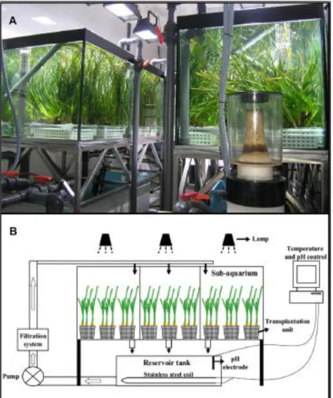
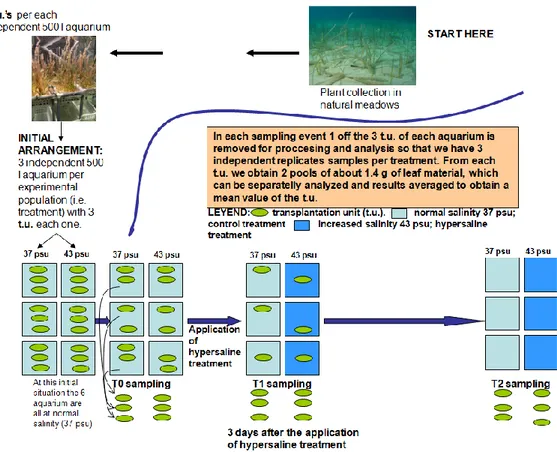
4.1 Culture and hypersaline treatments in the mesocosms
4.1.1 Field plant sampling 4.1.2 Mesocosm system 4.1.3 Aquarium culture 4.1.4 Salinity experiment
4.1.5 Extraction of total protein from leaf C. nodosa
4.1.6 Extraction and purification of proteins from the phenol phase
4.1.7 Electrophoresis of leaf proteins of C. nodosa 4.1.8 In-gel digestion , mass spectrometry,
bioinformatics analysis and identification of proteins of C.nodosa
4.1.9 Semi- quantitative analysis of proteins
4.2 Purification of chloroplasts and organelle sub- fractionation
4.2.1 Field plant sampling
4.2.2 Purification of chloroplasts
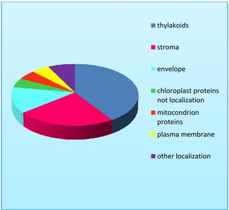
4.2.3 Electrophoresis of chloroplastic proteins on polyacrylamide gel with SDS (SDS -PAGE ) 4.2.4 Mass spectrometry , protein identification and sub-organellar localization
4.3 Study of protein expression as a function of depth
4.3.1 Field plant sampling
4.3.2 Extraction of leaf protein of P. oceanica
5. Results
5.1 Analysis of the leaf proteome of Cymodocea nodosa under salt stress
5.2 Extraction of chloroplasts and organelle sub-
68 69 70 75 76 81 82 85 85 85 86 88 89 90 91 92 93 95 95 95 96 97 98 100 100 101 103 103 109
IV
5.2.1 Extraction of intact chloroplasts
5.2.2 Extraction of the chloroplast proteins on SDS -PAGE
5.3 Protein expression as a function of depth
6. Discussion
- Expression proteomics of Cymodocea nodosa under salt stress
-Acclimation to the depth of Posidonia oceanica: a proteomic view
- Does Posidonia oceanica is a model plant for chloroplast sub-organelle proteomics in seagrasses?
References
Annex 1: Materials and buffers for chloroplast
Annex 2: Table of proteins from Cymodocea nodosa under
salt stress
Annex 3: Table of peptides from Cymodocea nodosa under
salt stress
Annex 4: Table chloroplastic proteins of P. oceanica
Annex 5. Peptide sequences assigned to each identified
chloroplast proteins
Annex 6: Identified proteins from P. oceanica chloroplasts Annex 7: Identified proteins from P. oceanica at different
depths
Appendix : Works submitted to Marine Ecology
109 110 113 118 120 127 131 135 154 155 160 178 185 198 203 225
V
ABSTRACT (Italian)
“Analisi del proteoma foliare delle seagrasses esposte a diversi regimi luminosi e a variazioni di salinità”
Le fanerogame marine, nel nostro studio limitate alle seagrasses, si sono adattate per occupare vaste estensioni dei fondi litorali e hanno dovuto sviluppare diversi adattamenti per poter vivere completamente sommerse. Le seagrasses non possono crescere in profondità dove non arriva almeno il 10% della luce in superficie, per questo si situano sempre sul piano infralitorale. In acque molto chiare, possono essere presenti fino a 70 m di profondità, però in mari con acque più torbide non superano i 15-20 m. Per tutte queste ragioni, queste formazioni vegetali sommerse rivestono un importante ruolo nella biologia e nella dinamica costiera.
Posidonia oceanica è una specie esclusiva del mar Mediterraneo. Mentre Cymodocea nodosa è, dopo Posidonia oceanica, la seconda seagrass del Mediterraneo per estensione delle sue praterie ed è una specie di origine tropicale, attualmente ambientata nel Mediterraneo e nell’Atlantico nordorientale, dal sud del Portogallo fino al Senegal, includendo le isole Canarie. Rispetto a P.oceanica presenta una maggiore tolleranza agli aumenti di salinità. In questo lavoro è stata analizzata l’espressione proteica in Posidonia oceanica e Cymodocea nodosa sottoposte a diversi regimi luminosi e concentrazioni saline. L’analisi ha riguardato specificamente il proteoma foliare e il sub-proteoma del cloroplasto, attraverso l’estrazione delle proteine, separazione elettroforetica, analisi delle sequenze in spettrometria di massa e identificazione proteica con software bioinformatici.
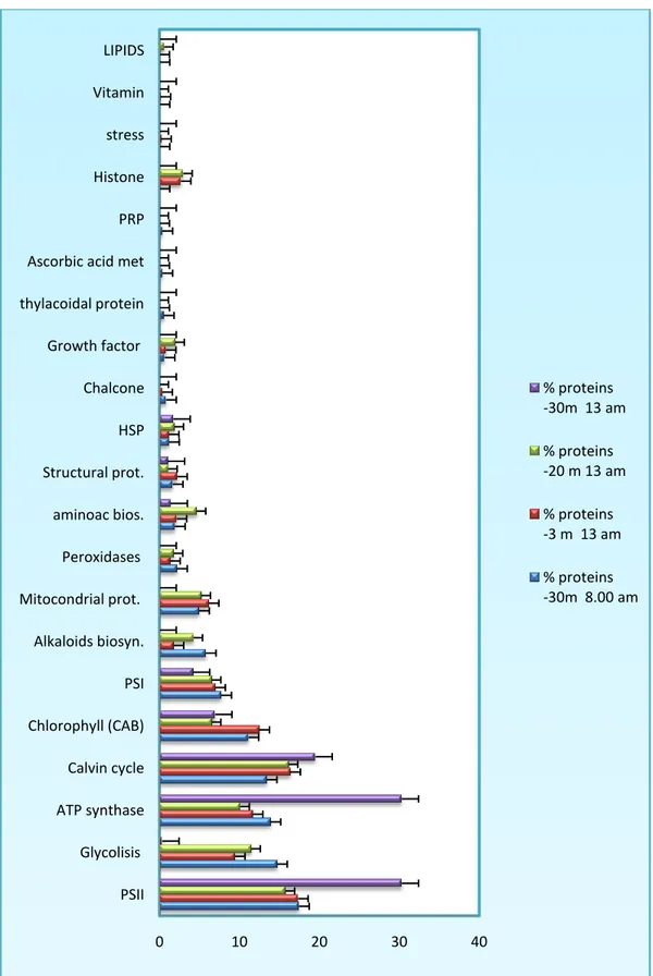
L’approccio proteomico così strutturato ha consentito di rilevare proteine differenzialmente espresse in popolazioni naturali adattate a tre diverse profondità. I risultati più evidenti riguardano proteine enzimatiche correlate al sistema fotosintetico PSII che risulta maggiormente espresso nelle praterie a 30 m di profondità alle 13:00, ora di massima disponibilità di luce. Altro dato rilevante è l’aumento dell’espressione degli enzimi del pathways metabolici che portano alla biosintesi di ATP, fotosfosforilazione cloroplastica e fosforilazione ossidativa mitocondriale. Sempre alla profondità di 30 m e alle 13:00, risultano overespressi gli enzimi del ciclo di Calvin-Benson rispetto ai livelli riscontrati nelle altre due profondità alla stesso tempo. Risultano invece poco espressi gli enzimi correlati alla glicolisi che raggiungono livelli molto elevati di espressione nel controllo,
VI
proteine correlate al PSI sono poco espresse in funzione delle profondità e raggiungono il minimo della loro espressione a 30 m nelle ore di massima illuminazione (13:00). Dato interessante e in apparente contraddizione con i dati di espressione dei gruppi funzionali correlati al processo fotosintetico, e la diminuzione dei livelli di espressione degli enzimi della via biosintetica delle clorofilla (a, b) alla profondità di 30 m associabili alla down-regolazione del fotosistema PSI.
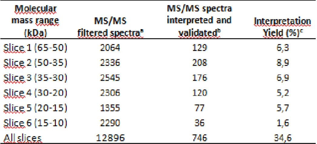
L’analisi delle proteine organellari ha consentito di creare un primo catalogo di proteine cloroplastiche di P. oceanica attraverso analisi dell’omologia di sequenza di proteine cloroplastiche di Arabidopsis e la loro localizzazione nei tre comparti sub-organellari (AT_CHLORO DATABASE). I cloroplasti intatti di P. oceanica sono stati ottenuti in accordo con quanto riportato in Rolland et al. 2003. Sono state identificate 74 proteine a cui è stata assegnata una diversa localizzazione e un numero di accesso corrispondente al database utilizzato. Il maggior numero di proteine identificate sono localizzate nei tilacoidi e nello stroma, mentre un numero minore di proteine sono localizzate nell’envelope. Inoltre l’8% delle proteine non hanno una esatta localizzazione nei compartimenti del cloroplasto.
Infine è stato analizzato il proteoma foliare di Cymodocea nodosa esposta a stress salino in condizioni controllate in mesocosmo, dove la parziale inibizione della fotosintesi, mediante la down-regulation delle proteine e degli enzimi sia del PSII che del PSI, e la ridotta attività respiratoria ottenuta dall’analisi proteomica permette alle piante di adattarsi a questa grave condizione di stress, ma presumibilmente con vitalità ridotta, dal momento che alcune delle risorse interne necessarie per la crescita e il mantenimento della biomassa devono essere riassegnati per far fronte allo stress metabolico. Nei trattamenti ipersalini sia a breve che a lungo termine troviamo gravi alterazioni dei metabolismi primari. Inoltre, i risultati di una bassa espressione della RuBisCo nei campioni ipersalini, in accordo con Beer et al . ( 1980), suggerisce che in condizioni di stress salino il bilancio del carbonio tende a favorire una maggiore produzione di carbonio inorganico ( Ci). Si verifica, poi, un aumento degli enzimi della glicolisi per controbilanciare la richiesta di energia e quindi produrre più molecole di ATP. Anche il metabolismo vacuolare è stato influenzato dal trattamento ipersalino , infatti, l’over-espressione dell’H(+)-PPasi suggerisce che i vacuoli sono coinvolti nel sequestro del Na+. Questo potrebbe essere quindi il meccanismo
VII
Introduction ________________________________________________________________________
8
Introduction The context
Mediterranean seagrasses form dense monospecific meadows across a wide bathymetric gradient (from shallow subtidal for shallow species and deep species till to 50-60 m depth in areas with very clear waters) (Borum and Greve 2004). Seagrass beds have an important ecological roles in costal ecosystem and provide high-value ecosystem services. The large-scale loss of seagrasses that occurred worldwide (29% of the known areal has disappeared, Wycott et al., 2009) had a serious effect on the ecosystem and on associated functions and services in the coastal zone (Duarte et al., 2004). For example the P. oceanica loss, like other seagrass ecosystems, have been attributed to a broad spectrum of causes, principally of anthropogenic origin, such as eutrophication, disturbance of sedimentary dynamics and mechanical destruction of the coastal area. Reported seagrass losses have led to increased awareness of the need for seagrass protection, monitoring, management and restoration (Borum et al., 2004; Orth et al., 2006; Larkum et al., 2006a; Bouderesque et al., 2006; Björk et al., 2008). common descriptor for monitoring programme are shoot density, leaf production and rhizome elongation, bathymetric position of the lower and/or upper depth limit, bottom cover, structure of the matte (see Pergent-Martini et al., 2005 for a synthesis), while additional
9
content and various trace metals (Casazza et al., 2006). However, these descriptors respond slowly to environmental change and don’t detect alterations of the costal water quality before that the effects become evident on the plant and/or on the whole meadow. New tools in monitoring such as genetic analysis could be very important to comprehend the evolutionary potential as well as resilience and resistance capacity under various forms of stress and to guide restoration initiatives of destructed .
Fig.1 From genomics to proteomics. integration of information from
the genome to the proteome for a better understanding of biological systems
Genomics has been the last specialty applied to study the mechanisms of acclimation of these plants to the submerged lifestyle (Wissler et
10
on how to incorporate the comparative gene expression studies with photosynthetic performance, carbon and nitrogen utilization and environmental adaptation, and how to combine the research related to mechanisms of carbon utilization, light requirements, temperature effects and natural
variation in pH and ocean acidification (Arnold et al., 2012; Hall-Spencer et al., 2008; The Royal Society, 2005).
Proteomics of marine plants is still at the early stage because of the poor information on genomics of most of the species. Among the aquatic plants, mangroves have been received attention for genomic and proteomic approaches because their constitute a model for salt-tolerant xylophytes (Huang et al., 2012).
On the side of seagrasses, proteomics gave first results regarding their acclimation mechanisms under chronic low light (Mazzuca et al., 2009; Serra and Mazzuca, 2011), different depths (Dattolo et al., 2013) and in response to salinity stress (Serra et al., 2012).
These –omics approaches have recently required to be coordinated to the research supported from the Cooperation in Science and Technology Action (COST) program of the European Science Foundation, to counteract the crisis of seagrasses conservation and their regression along the Mediterranean area. During this Action,
11
ecophysiological and physical approaches with the aim of understanding changes in seagrass productivity and metabolism in different conditions, thus to apply the potential of the data that come from this synergistic approach for seagrasses.
The research activities in the current thesis have been developed within the frame of this COST Action with the aim to correlate the proteomic approach to genomics, and ecophysiology of selected seagrass ecosystems.
The submerged lifestyle imposed many limiting factors to the growth and development of seagrasses that have been adapted their gene expression and physiological machineries to the marine conditions (Wissel et al., 2011). Few genes showed evidence for positive selection in seagrass branches indicating that photosynthesis, a few metabolic pathways, and ribosomes have strongly diverged after the split of the common ancestor of seagrasses from terrestrial monocots. In this context our proteomic studies have been address the following questions: how seagrasses exert their osmoregulatory capacity to tolerate high salinities, how CO2 is fixated, how their photosynthetic apparatus has evolved for under water light harvesting.
12
to their abundance in meadows, their sampling is not destructive and the physiology of plant is driving by the metabolisms that take place in leaves. Moreover, since leaf protein pattern is generally well known and many proteins have been identified (Saravanan & Rose, 2004), this overcomes the lack of completeness of the gene databases for these species that generally represent the greatest obstacle in using proteomic approaches for aquatic plants. On this basis we used the adult leaves to avoid the influence of tissues differentiation on the protein expression and yield.
We applied the analysis of leaf proteome thanks to the well developed and optimized protocol for protein extraction and purification from seagrass leaves (Spadafora et al., 2008) to look at the global protein expression of different species and conditions. A great challenge, working with non-model species, whose genomes are not completely sequenced, is to identify proteins by means of the classical bioinformatic engines that interrogate the public databases. To overcome this gap we used a combination of non-common software for proteomic analysis, that are easily customized, to identify much more proteins as possible against public databases and against local database of seagrasses created by the research team of University of
13
sequences thanks to next generation sequencing approaches.
Given that photosynthesis is the primary metabolism in leaf, we deeply investigate the sub-proteome of chloroplasts and the level of the expression of proteins that are involved in this process. It is well known that chloroplast proteomics describe both the metabolisms that are drive by their own genome to synthesize proteins for specific function and also those from the nuclear-encoded proteins (Salvi et al., 2007). Plant organelle proteomics should be limited mainly due to the inter-plant or inter-tissue complexity, to the difficulties in isolation of subcellular compartments and to their enrichment and purity. Despite these concerns, the field of organelle proteomics is growing in plants, such as Arabidopsis, Oriza sativa and Zea mais. The available data are beginning to help better understand organelles and their distinct and/or overlapping functions in different plant tissues, organs or cell types, and importantly, how protein components of organelles behave during development and with surrounding environments. As first the priority of seagrasses chloroplasts proteomics has been the isolation of organelles or sub-organellar compartments that provides a very direct method for confidently assigning proteins to specific localization, allowing to better understand known functions of the organelle or reveal novel ones. We used separation technologies in
14
to have opened up experimental possibilities to identify a more complete set of chloroplast proteins, the seagrass chloroplast proteome catalog, as well as their expression levels (van Wijk, 2000; Ferro et al., 2003; Baginsky & Gruissen, 2004; van Wijk, 2004, Rossignol et al., 2006).
15
One of the main purpose of the research project has been to
characterize the protein composition of the chloroplast of seagrasses adapted to different light regimes. Organelle proteomics is one of the latest applications in both the animal and plants, and has initiated the construction of several databases dedicated that are indispensable for the study of even complex proteomes from organs of species whose genomes have in part or in nothing sequenced. This is because the organelle proteins (eg, enzymes of the Calvin - Benson cycle) have high level of homology sequence among different species whose are genetically related or not. Nowadays dedicated databases are daily updated with results from model organisms and also from species whose genomes are not sequenced yet. In particular, we identified proteins from generalist databases (NCBI) and /or against the seagrasses-dedicate Dr Zompo; moreover we took protein details from the AT_Chloro database that contains information on subplastidial localization of proteins from envelope, stroma and thylakoids of Arabidobsis thaliana chloroplasts.
16
research project aims to i) obtain purify chloroplasts from adult leaves of seagrasses starting from most common available protocols and optimize the protocol to extract and purify the proteins from the three
organelle compartments, ii) applied the mono-dimensional
electrophoresis to separate protein mixture as the first step of the gel-based proteomics and obtain protein sequences through the mass spectroscopy analysis, using different ion sources (E.S.I., S.A.C.I, Orbitrap), iii) identity of the corresponding proteins from peptide sequences by database searching for homology; define the protein localization within the chloroplast compartments by means of the AT_Chloro database , iv) build a catalog of seagrass chloroplast proteins, v) compare the chloroplast protein expression levels in leaves growing at shallow and deep beds during the daily cycle.
The second main purpose of the research was to applied the expression proteomics to seagrass plants growing in different salinity conditions to detect the proteins that are differentially expressed during the acute stress, the acclimation and resilience. In order to define the impacts of hypersaline water, experiments have been undertaken in aquaria focuses on the leaves proteome under normal and hypersaline conditions.
17
Spanish Oceanography Institute, Oceanography Centre of Murcia, Spain, whose laboratories are well equipped for hypersaline experiments.
18
1.The biological systems
1.1.What are seagrasses?
The 60 species of seagrasses currently known in the world have had to develop different adaptations to live completely submerged, to tolerate high salinity of sea water, and to have an effective system of anchorage to the substrate and a sedimentary pollen, filamentous, capable of be transported in water (hydrophilic pollination) (Larkum et al., 2006).
The development of these adaptations led to a morphological model very uniform in all species of seagrasses , as the habitus that is very similar. They are rhizomatous plants (bearing a complex system of underground rhizomes) with clonal growth . The rhizomes may have a horizontal or vertical position. The former are responsible for the expansion of the bed and progressive employment space, while the latter prevents that plant has buried by sedimentation . The growth of horizontal rhizomes predominates in the edges of meadows, while the vertical development is more frequent in the central area. On the lower
19
part of the horizontal rhizomes a group of adventitious roots coming out, contributing to fix the plant and to absorb nutrients; on the upper part there are the short vertical rhizomes each developing a shoot with many leaves. In the rhizomes it can be distinguished nodes and internodes. Such as flowering plants, seagrasses can develop the inflorescence or flowers at certain times of the year, which are very noticeable and difficult to observe (Larkum et al., 2006). Currently the process of flowering is quite rare in many species of seagrasses, dominating the vegetative reproduction by means of clonal growth of rhizomes, than sexual. This has as a consequence that the genetic diversity in bed is very low, and therefore it is assumed that these can consist of a few clones. This low genetic diversity is supposed to be one of the causes of general regression and mass mortality that affect the meadows, which are not able to develop resistance against disturbances and threats.
The habitus of seagrasses is that of terrestrial monocots in which the plastocrone interval (the time interval between the onset of leaf bud in two consecutive nodes during the growth) is really short. The pattern of stem elongation and clonal growth are relatively constant
20
and specie-specific. One of the factors limiting the growth of these plants is the light (as for all photosynthetic organisms). The seagrasses cannot grow in depth where not arrive at least 10% of the light at the surface, for this reason they are located always in the upper part of the continental shelf (infralittoral). In very clear water , as in some tropical areas , the seagrasses may be present up to 70 m deep, but in seas with more turbid waters do not exceed 15-20 m. The meadows of seagrasses worldwide covering approximately 6000000 km2 of seabed submerged and are responsible for primary production, about 0.6 gigatons of carbon per year, and around 15% of CO2 absorption by all marine organisms. For all these reasons, these formations submerged plants play an important role in the biology and coastal systems:
1 . The density of the leaves in the bed promotes the deposition of particles in suspension and, therefore, the transparency of the water . 2 . Its complex network of rhizomes tend to consolidate and stabilize sediments.
3 . They attenuate the marine hydrodynamics and, as a result, prevent the coastal erosion.
4 . they are responsible for high production of oxygen and organic matter .
21
5 . they Provide habitat for many species, a lot of which use these environments as a hideout, as a breeding place and permanence of juvenile.
Along the coasts of Europe , there are four species of seagrasses, Zostera marina Linnaeus, Zostera noltii Hornemann, Cymodocea nodosa (Ucria) Ascherson and Posidonia oceanica (Linnaeus) Delile. Z. marina is along the coasts of the north Atlantic and Pacific, and has a very localized distribution in the Mediterranean and in the Alboran Sea, while Posidonia oceanica is endemic in the Mediterranean. Zostera noltii and Cymodocea nodosa are living both in the Atlantic coast to the Mediterranean coast and are the only species that are found in the Canary Islands.
1.2. Posidonia oceanica
Posidonia oceanica, a species exclusive to the Mediterranean Sea, which is distributed in both the eastern basin than in the West, as well as in most of the islands. This seagrass lives between the surface and a depth varying, depending on water clarity. It can grow on both substrates that soft or rock. Generally, it was observed that the growth
22
occurs on rocky seabed in shallow water and in open areas with less hydrodynamics, while large bays or deep waters, where the hydrodynamics is smaller, the growth occurs on sandy substrates. It is a plant stenoaline (i.e. unable to tolerate large variations in salinity) and cannot live with a lower salinity of 33 ‰ to 39 ‰ or higher, for this is not found in brackish or hypersaline lagoons. However, it tolerates a relatively wide temperature range from about 10°C to 28°C.
It is very sensitive to eutrophication and to the contaminants and does not tolerate high rates of sedimentation. These requirements explain his absence near the mouths of large rivers. In addition, it was
estimated that in areas with a high concentration of human activities, the Posidonia oceanica meadows occupy on average about 15 % of
23
the seabed in a bathymetric range 0-50 m, and close to 50% in a well-preserved and with very clear water. So that it can be considered an indicator of plant water clear, well-oxygenated and free from contamination.
The rhizomes of P.oceanica are particularly woody, can reach a thickness of about 1 cm, slightly laterally compressed and covered with scales that come from the bases of old leaves. Depart from the rhizomes some roots relatively short (normally not exceeding 10-15 cm), few in number, robust (thickness of about 2-4 mm) and that lignified very quickly. The roots have a role in anchoring the plant to the substrate and its quantity increases in the places with the most troubled waters. They form ribbon, about 1 cm in width and the length ranges from 20 to 140 cm, and they present 13-17 longitudinal veins. The growth of new leaves is a process more or less continuously over the year and their longevity varies from 4 to 11 months. The apices are rounded and they are often lost on wave action and of currents. The leaves are organized in bundles, which contain 6 or 7, with the older leaves that are outside and inside the youngest.
Leaves are divided into three categories (Fig.3):
1. Mature leaves: have a lamina with photosynthetic function and a base separated from the leaf blade from a concave structure called
24
ligule;
2. Intermediate leaves: they are avoid of the base; are photosynthetically active ;
3. Juvenile leaves: are conventionally length less than 50 mm, weakly pigmented.
In autumn, the plant loses its mature leaves, which become brown in color and are photosynthetically inactive and the new leaves are produced during the winter. Sexual reproduction takes place by producing flowers and fruits. The flowers are hermaphrodite and grouped in an inflorescence spike-shaped, green and enclosed in floral bracts. The floral axis attaches to the rhizome in the center of the beam. The gynoecium is formed by a unilocular ovary which continues with a stylus and ends with the stigma; the androecium consists of three stamens with anthers court. The flowering is regulated by environmental factors (light and temperature) and endogenous factors (age and size of the plant) and takes place in September-October in the shallow meadows, while it is shifted of two
25
months in the deeper meadows. The pollen within of the anther is spherical, but it becomes filamentous soon as it is released into the water. There are not mechanisms for recognition between pollen and stigma that prevent self-fertilization. Pollination is hydrophilic and can lead to the formation of the fruit, although some abortions before its maturation after six months. Once ripe, the fruits fall off and float to the surface. The fruit, slightly fleshy and commonly called "sea olive", is similar to a drupe and has a porous and rich pericarp of an oily substance that allows the waterline. When it degenerates the seed is released, coated by a thin membrane but without a real tegument, which falls to the bottom and if it finds the suitable conditions of depth, stability and sediment type, germinates and gives rise to a new plant. To start making roots, it is necessary to find a humified substrate. The humification is produced by the degradation of plant debris, so the plant can implant in "soil" previously colonized by other plants, such as macroalgae or other seagrasses. This generates a genuine ecological succession in which Posidonia is the last stage of succession. Germination begins with the protrusion of a small white root from the radical pole and a leaf from the apical pole. With sexual reproduction the plant colonizes new areas, meadows spread to other areas ensuring a genetic variability. The stolonization, which allows
26
the expansion of meadows, it's made by plagiotropic growth of rhizomes, which grow about to 7 cm/year and colonize new areas. A high accumulation of sediment and the decrease of the space available for the horizontal growth, stimulates the growth of the vertical rhizomes . So the vertical growth of the rhizomes leads to the formation of a structure called matte, consisting of a mesh of dead rhizomes and roots which remains trapped between the sediments. Only the top part of these structures is made up of alive plants. The formation of mattes depends mostly from the rhythms of sedimentation; the high sedimentation rate can lead to excessive silting of the rhizomes and then to their anoxia, on the contrary a too slow sedimentation can cause the weakening of the rhizomes and the regression of meadows. Since the rate of decomposition of the rhizomes is very slow they can stay inside the matte for millennia. The matte has a very slow rate of growth : its growth has been estimated at about 1 m per century. So that the meadows can accumulated organogenic structures that rise for meters above the base (Mateo et al., 1997). This accumulation of organic sediments not only represents a net sink of carbon and other elements, but can also attenuate wave action. It has been estimated that the removal of 1 m3 of matte, for example, can cause 20m of coastal regression (Jeudy De Grissac,
27
1984). Photosynthesis often depends on the light and decreases rapidly with increasing depth . Respiration, however, is independent of the light and in Posidonia oceanica is relatively high, since it has underground organs ( roots and rhizomes ) that are not photosynthetic, but have an important respiratory function. The growth dinamics and the large amount of biomass produced by Posidonia oceanica, are factors able to support the animal and plant communities with high biodiversity. We distinguish the community of epiphytes, ie bacteria, algae, bryozoans that colonize the surface of leaves and rhizomes, the animal communities and vagile and sessile communities of detritivore organisms.
Along the leaf there are several areas of differentiation that depend on the age of the leaf. Even epiphytic communities follow this zonation: at the base of the mature and
young leaves diatoms and
bacteria are implanted;
incrustations algae red and brown are implanted in the central part of the leaf, while
in the upper part the encrusting and filamentous algae are found. Fig.4 Posidonia oceanica leaves rich in epiphytes
28
Epiphytic communities are preyed by Molluscs, Gastropods, Crustaceans, Polychaetes and Amphipods play a very important role in the food chain of Posidonia oceanica meadows. There are few organisms that can directly feed the plant tissue, which is unwelcome to most herbivores due to the high content of structural carbohydrates, high values of C and N, and for the high concentration of phenolic compounds.
The epiphytes can also cause damage of Posidonia. Them, in fact, increasing the weight and can cause its premature fall; they can decrease the available light and also they hinder the gaseous exchanges and the absorption of nutrients through the leaves.
The fauna associated with Posidonia oceanica meadows consists of sessile animals that live coated on the substrate made from the leaves and rhizomes, and vagile animals ,capable of move within the meadows. Then there are organisms, which constitute the infauna, that live inside the matte and that are primarily detritivores.
Studies conducted by Gambi et al. in 1992 have demonstrated that approximately 70% of the total animal population of the Fig.5 Denizen habitual of Posidonia
29
meadows is constituted by herbivores. Between them, the most abundant are echinoderms, in particular the Paracentrotus lividus, one of the few organisms able to feed directly of the leaves of the plant. The carnivores are represented by fish, molluscs, polychaetes and decapods.
Between the molluscs, habitual and nearly exclusive inhabitant of the meadows is the Pinna nobilis, the bivalve largest in the Mediterranean and highly threatened from fisheries and pollution.
The fish population is constituted by a small number of species, principally labrids and sparids almost all carnivores. large fish are less frequent and during the year it witness to variations the abundance specific due to the referrals and migration.
In the shallow and secluded meadows, there is an abundance of Sarpa salpa, which represents 40-70% of the summer fish fauna.
The detritus constituted by the litter made from the remains of fallen leaves, is colonized by microorganisms and fungi.
A particular group of detritivores are polychaetes (Lysidice ninetta, Lysidice collaris and Nematonereis unicornis) and isopods (Idotea hectica, Limnoria mazzellae), called borers that dig tunnels inside the flakes (remains of leaf bases that remain attached to the rhizome all year) to feed themselves and to expand their habitat. The leaves,
30
degraded by wave and microorganisms, once beached, take the name of banquette and they serve as shelter and food for insects, amphipods and isopods.
1.3. Cymodocea nodosa
Cymodocea nodosa is the second seagrasses in the Mediterranean for extension of its meadows.
The Cymodocea nodosa is an aquatic plant of the spermatophyte family Cymodoceaceae. C. nodosa is a warm water species and is widely distributed throughout the Mediterranean, around the Canary Islands and down the North African coast, it can colonize the dead matte of Posidonia oceanica. The species does not extend further north than the southern coasts of Portugal. C. nodosa can be found from shallow subtidal areas to very deep waters (50-60 m). This
31
species has leaf bundles consisting of 2 to 5 leaves. The leaves are 2 to 4 mm wide and from 10 to 45 cm long.
The leaves resemble those of medium sized Zostera marina. However, the shoots are attached to vertical rhizomes with short rhizome segments which again are attached to a horizontal rhizome with 1-6 cm long segments. The apex forms vertical rhizomes and branches to new horizontal rhizomes. The rhizome may grow several meters per year, and C. nodosa is considered a pioneer species which can quickly colonize bare areas of the sea floor. C. nodosa can easily be identified by its vertical rhizomes and the long white to pink horizontal rhizome segments. The roots are dispersed along the vertical and horizontal rhizomes. Each rhizome segment only has one root which is often strongly branched and may be up to 3 mm thick and up to 35 cm long. The individuals are either male or female plants. The female flowers have two ovaries and the two lentil-shaped seeds produced from each flower are around 8 mm long and, hence, considerably larger than the seeds of the Zostera species.
Only C. nodosa shoots older than 1 year flower, and they do so between March and June. Fruit development takes 2-3 months,
32
although maximum density of shoots bearing fruits is observed in July-August. Afterwards, fruits detach from the mother shoot and, because they have negative-buoyancy, they are rapidly buried into the sediment nearby the mother plant. During events of intense sediment dynamics (e.g. strong storms), however, seeds may be transported across long distances, since there are meadows separated from the closest one by more than 300 km, and seeds of C. nodosa can be observed, although not very often, washed on the beaches. From April til June of the following year seeds germinate. C. nodosa clone formation rate has been estimated to be about 0.009 clones m-2 yr-1in an area with intense sexual reproduction. However, clone mortality rate is about 50-70 % during the first year of life, hence, decreasing substantially the success of sexual reproduction.
Reproductive effort and success in C. nodosa exhibits temporal and spatial heterogeneity. Flowering intensity, for instance, has been observed to increase in response to sand burial, like in other seagrasses. In addition, seed production in C. nodosa should be constrained by the spatial distribution and abundance of male and female clones. The consequences of clone sex composition on reproductive success are evident when examining C. nodosa meadow
33
genetic diversity. For instance, there is almost no genetic diversity in a C. nodosa meadow at the Algarve (Portugal), where no female flowers have been observed. The fast growth of C. nodosa clones and the relatively high patch formation rate of this species, when compared with the other European seagrasses, indicate that C. nodosa should be able to develop a meadow within a decade, if the colonisation process were initiated, on bare sediments. The time scales for meadow recovery if not all C. nodosa vegetation were lost should be even shorter. The rapid occupation of space by C. nodosa resulting from fast clonal growth, and the relatively high patch formation rate of this species explains the pioneering role that C. nodosa play during succession process in the Mediterranean.
Beds of C. nodosa are characteristic habitats for seahorses. C. nodosa growth ranks amongst the fastest ones across European seagrasses. The fast clonal growth of this species allows the clones to spread across 300 m2 after 7 years. The life span of C. nodosa modules and ramets is intermediate, average shoot population life-span varying between 4-22 months, and average leaf life-span ranging from 2 to 5 months. However its clones may live for at least 1 decade. The vegetative growth almost exclusively occurs during spring and
34
summer, exhibiting a substantial plasticity, which allows this species to survive disturbances. For instance, vertical and horizontal rhizome growth of C. nodosa is plastic enough for this species to colonize areas with intense sediment dynamics, such as bedforms with subaqueous dunes, with an average amplitude of 20 cm (range 7-65 cm) and wave length of 21 m (7-29 m), that migrate at average velocities of 13 m year-1. The close relationship between the growth of the rhizome and the vertical accumulation of sediment was used to quantify the dynamics of shallow coastal sediments, impossible to be measured with conventional sedimentary techniques. C. nodosa also exhibits substantial plasticity in response to ambient nutrient availability.
On the side of resistance C. nodosa can tolerate the anoxia and the presence of hydrogen sulphide in the soil. Its leaves are home to a rich epiphytic community almost as much as that of Posidonia.
35
2. The –omics applied to seagrasses
2.1 What’s "OMICS" sciences?
Omics technologies such as genomics and highthroughput DNA sequencing were introduced in parallel to the Human Genome Project since 1990s. According to one etymological analysis, the suffix 'ome' is derived from the latin omni- ("completeness and fullness") (Lederberg and McCray, 2001). By combining 'gene' and 'ome', Hans Winkler created the term genom(e), referring to "the haploid
36
chromosome set, which, together with the pertinent protoplasm, specifies the material foundations of the species [...]." (Winkler et al., 1920). Victor McKusick and Frank Ruddle added 'genomics' to the scientific lexicon as the title for the new journal they co-founded in 1987, with emphasis on linear gene mapping, DNA sequencing and comparison of genomes from different species (McKusick and Ruddle, 1987). Omics technologies and various neologisms that define their application contexts, however, are more than a simple play on
words. They substantially transformed both the through put and the design of scientific experiments. The omics technologies allow the generation of copious amounts of data at multiple levels of biology from gene sequence and expression to protein and metabolite patterns underlying variability in cellular networks and function of whole organ systems (Nicholson and Lindon, 2008). In fact this led to overabundance of data in biomedical experiments recently (Nicholson and Lindon, 2008). While the 1990s was named as the “decade of the brain”, we are now in the “decade of measurements”. This signals a new era in how we approach to scientific inquiries. In addition to amplified through put, the process of research is fundamentally altered in “omics science”. Ordinarily, scientists have accustomed to hypothesis-driven research wherein a clearly articulated scientific
37
question/hypothesis would be posed. Subsequently experiments would be carried out to obtain data in order to test the study hypothesis. With the omics approach, asking an initial research question is not always necessary or a pre-requisite. Genome or proteome wide data can be collected in an omics experiment without an existing hypothesis, followed by generation and testing of biological hypotheses.
During the last decades, the application of -omics technologies at ecological studies provided powerful tools for following the physiological acclimation in response to environmental variations (Feder and Walser, 2005; Foret et al., 2007; Gracey et al., 2007; Karr et al., 2008), and helped researchers to correlate the differences of gene’s expression profiles to changes in them a in ecological cues in many different organisms (Chevalier et al., 2004; Edge et al., 2008; Kassahn et al., 2009; Larsen et al., 2012; Richards et al., 2012). Despite their high ecological value, seagrasses are poorly understood for what concerns the genetic basis behind their physiological adaptation and plasticity (Procaccini et al., 2007).
It’s only recently that transcriptomic approaches were implemented for few species, to correlate seagrasses gene expression with ecological factors. In particular, transcriptomic response to
38
temperature changes and thermal stress was studies in the two congeneric species, Zostera marina and Zostera noltii (Maathuis et al., 2003; Reusch et al., 2008; Massa et al., 2011; Winters et al., 2011), while transcriptional (Bruno et al., 2010; Serra et al., 2012b) and proteomic approaches (Mazzuca et al., 2009) were applied to study light response in natural conditions in Posidonia oceanica. In P. oceanica, studies were hampered by the fact that available genomic and transcriptomic resources only consisted in a single Expressed Sequences Tags (EST) library, obtained from shoots collected along a depth range (from−5 to −30 m) in a single site (Wissler et al., 2009), and available in Dr.Zompo, a specific seagrasses database containing both P. oceanica and Z. marina EST sequences http://drzompo.uni-muenster.de/ (Wissler et al., 2009). Several approaches can be utilized for genomic studies in species for which the whole genome is not available (Hofmann et al., 2005; Stapley et al., 2010), most of them requiring high computational power and advanced bioinformatics resources (Morozova and Marra, 2008; Pop and Salzberg,2008; Metzker et al., 2010). Among the others, Suppressive Subtractive Hybridization (SSH)–EST library (Diatchenko et al., 1996) approach resulted especially powerful to identify differentially expressed genes in the presence of clear differences in physiological status (Jones et
39
al., 2006; Puthoff and Smigocki, 2007) and it was applied to study flowering (Matsumoto et al., 2006), senescence (Liu et al., 2008a,b), or salt-stress (Zouari et al., 2007) in terrestrial plants.
Previous studies have identified some differences in transcriptional and proteomic profiles in P. oceanica, correlated with its bathymetric distribution, with the ultimate goal to identify the metabolic pathways involved in acclimation. They also aimed to increase genomic resources in P. oceanica and to present a powerful approach for studying physiological response at a molecular level in organisms for which genomic resources are limited. In order to do that, a SSH-library was built between plants growing at two different depths in the same meadow, obtaining their protein identification using the innovative USIS mass, spectrometry methodology coupled with 1D-SDS electrophoresis. On the side of search engine against genome and proteome databases it has been used for proteins identifications the Global Proteome Machine (GPM) open-source system for analyzing, storing, and validating proteomics information derived from tandem mass spectrometry (Craig et al., 2004; Fenyö et al., 2010) and X!Tandem software (Craig and Beavis, 2003; Craig et al., 2005) that allowed to interface directly the mass spectrum data with a local database customized with the collection of each sequence stored in
40
the Dr.Zompo and UniProtKB databases for seagrasses and for plants among Liliopsida that are the closer terrestrial counterpart.
2.2 Proteomics
Proteomics is the large-scale study of proteins, particularly their structures and functions. Proteins are vital parts of living organisms, as they are the main components of the physiological metabolic pathways of cells. The term "proteomics" was first coined in 1997 to make an analogy with genomics, the study of the genes. The word "proteome" is a blend of "protein" and "genome", and was coined by Marc Wilkins in 1994 while working on the concept as a PhD student. The proteome is the entire complement of proteins and provides a direct measure of the quantity that are expressed in a cell at a time. Scientists are very interested in proteomics because it gives a much better understanding of an organism than genomics. First because the level of transcription of a gene gives only a rough estimate of its level of expression into a protein. An mRNA produced in abundance may be degraded rapidly or translated inefficiently, resulting in a small amount of protein. Second because , as mentioned above many proteins experience post-translational modifications that profoundly
41
affect their activities; for example some proteins are not active until they become phosphorylated. Third because, as it is well known the mRNA is not always translated into protein, and the amount of protein produced for a given amount of mRNA depends from the gene that it is transcribed and on the current physiological state of the cell. Even if it is studying a particular cell type, that cell may make different sets of proteins at different times, or under different conditions. Furthermore, as mentioned, any one protein can undergo a wide range of post-translational modifications. Therefore a "proteomics" study can become quite complex very quickly, even if the object of the study is very restricted.
2.3 Proteomics in seagrasses biology, ecology and threatens
Proteomics approach have been applied for the first time to Posidonia oceanica to understand the molecular bases of stress responses, resilience and acclimation to low light (Mazzuca et al., 2009; Serra and Mazzuca, 2011). In fact, P. oceanica beds have recently suffered from progressive die-offs attributed to lower light availability from elevated water turbidity. In addition P. oceanica meadows are extremely sensitive to moderate to high disturbance, and have suffered
42
substantial diebacks throughout the Mediterranean Sea due to anthropogenic disturbances affecting light and temperature regimes. The adaptive low-light responses of this seagrass have been highlighted by comparing the protein expression in plants collected from turbid waters (low-light) with plants collected from pristine-clear waters (high-light). Results summarized that enzymes involved in carbohydrate cleavage (1-fructose-bisphosphate aldolase, nucleoside diphosphate kinase, and beta-amylase) were upregulated in low-light conditions. Electron microscopy studies also revealed substantial changes in the stroma lamellae/grana ratios in chloroplasts receiving lowlight, possibly as a mechanism for re-establishing optimal PSI/PSII ratios. Furthermore, under low-light conditions, four components of the ubiquitin/mediated proteolysis pathway (26 S proteasome regulatory, proteasome beta type 1, proteasome 7 D beta type, and proteasome alpha 7), and the perchloric acid soluble
translation inhibitor protein, were upregulated. This suggests that, in P. oceanica leaves, enhanced protein turnover mediates acclimation to low-light conditions. Also, enzymes involved in defending against cellular stress (superoxide dismutase, pyridoxine, and 2-caffeic-acido-methyl transferase) were differentially expressed in low-light regime.
43
From this molecular approach it is possible to recognize new tools that may deserve the designation of “early-warning” markers for environmental stresses; the main goal will be to invert soon as possible the feedback mechanisms impose by stress that accelerate the decline of seagrass productivity, driving seagrass communities from autotrophic (where carbon is sequestered) to heterotrophic (where carbon is released). It is, therefore, important to understand how photosynthesis and carbon metabolism of meadows are affected by drivers of seagrass decline. The ecological status of P. oceanica is usually assessed by quantifying shoot densities, above-/below-ground biomass ratios, or growth rates. For example, the European Water Framework Directive (WFD, 2000/60/EC) uses seagrass taxonomic composition and abundance, determined by shoot density and spatial extent, to evaluate the ecological status of transitional or coastal water bodies. However, these descriptors respond slowly to environmental change. Once decline is apparent, it may be too late to implement a coastal management procedure that would allow an endangered meadow to recover. Therefore, early-warning indicators of seagrass health are necessary. Establishing the direct linkages between stressors and seagrass responses, and initializing the appropriate scales of spatial and temporal monitoring, will guide managers in
44
determining which actions are necessary to prevent further seagrass loss.
The primary cause of seagrass die-off is reductions in light due to increased turbidity and eutrophication, often attributed to anthropogenic activities along the coast (Short and Wyllie-Echeverria, 1996; Guidetti and Fabiano, 2000; Alcoverro et al., 2001; Ruitz and Romero, 2003). In low-turbid pristine P. oceanica beds, plants can flourish at a depth of 40 m, with high shoot densities, productivities, and growth (Duarte et al., 1991). Indeed, some authors have shown that, when PAR (Photosynthetically Active Radiation) lowers below 4.5%, light quality and quantity becomes insufficient to sustain the normal growth of P.oceanica (Zimmermann et al., 2006). Others have suggested that the photosynthetic activity of P. oceanica is regulated by depth rather than light intensity, as seagrass can acclimate to low-light conditions (Figueroa et al., 2002; Olsen et al., 2002; Lorenti et al., 2006). This implies that specie-specific determinants might explain differences in acclimation response. There was the need of new biomarkers, such as proteomics, to identify and quantify early alterations in the plant adaptive response. The application of proteomics in monitoring marine ecosystems is a relatively new tool focusing on gene function (Procaccini et al., 2007), and can be useful
45
in evaluating the response of an organism to environmental conditions (Andacht and Winn, 2006). Although proteomic bio-monitoring is a sensitive tool for studying the response of aquatic animals to environmental stress, only a few studies have applied proteomics to evaluate aquatic plants (Förster et al., 2006; Katz et al., 2007). Even if the plant's own genome was sequenced, proteomic analyses would remain unattractive because protein sequence analysis and identification are challenging. Unlike animal tissues, P. oceanica tissues are rich in compounds such as polysaccharides, lipids, phenols and other secondary metabolites that interfere with protein separation and analyses, (Agostini et al., 1998; Dumay et al., 2004; Cozza et al., 2004; Park et al., 2004). Furthermore, in comparison to animal tissues, plant tissues maintain lower protein concentrations (Tsugita and Kamo,1999). To increase the quality and yield of purified proteins from plant tissues, researchers have developed a number of alternative extraction procedures (Park et al., 2004; Saravan and Rose, 2004). While many of these procedures have proven useful in protein isolation and purification, newer techniques have demonstrated superior protein extraction in P. oceanica (Spadafora et al., 2008).
46
2.4 Organelle proteomics: potential application in seagrasses
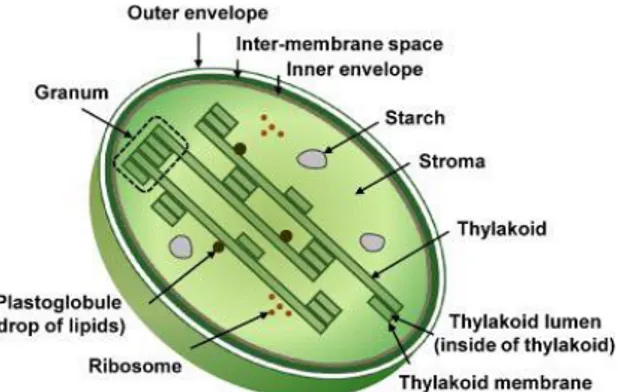
The organelle sub-proteomics is a new frontiers in the frame of plant proteomics (Rolland et al., 2012). The characterization of proteomes in different sub-cellular locations is of prime importance for a complete understanding of plant functions, biosynthetic and signaling pathways. Sub-cellular fractionation permits simplification of the proteome and, potentially, a gain in knowledge in that the sub-cellular localization of the proteins is revealed. The quality of the biological sample analyzed is often the limiting factor in both of these aims. The classical cell fractionation procedure generally consists of two major steps: (i) disruption of the CW and membrane and (ii) fractionation of the crude homogenate to purify the organelle of interest. Cell disruption has to be controlled to avoid excessive disruption of sub-cellular compartments. Protoplast preparation is perhaps the gentlest method and is a prerequisite for the purification especially for chloroplasts (Fig.8).
Fig. 8 The chloroplast. This plastid type
is essential to the complex process of
photosynthesis. Chloroplast is
surrounded by a lipidbilayer composite membrane with an inter-membrane space. Moreover, it has reticulations or many infoldings that fill the inner space, the stroma. Within the stroma are stacks of thylakoids, the suborganelles that are
the sites of photosynthesis. The
thylakoids are arranged in stacks called granum. The chloroplast also contains
plastidial DNA, plastoglobules,
ribosomes and starch (in Agrawal et al., 2009).
47
Chloroplasts are the organelle that permit the autotrophic life in plant, algae and bacteria. Assimilation of inorganic carbon (Ci) on air is promoted by diffusion of CO2 in plant tissue, or by converting it in carbonic acid (HCO3) by the specific enzyme Carbonic Anhydrase (CA) at plasma membrane (Ferro et al., 2003). In marine plants Ci assimilation depends on the pH of water that affects sensibly the conversion of CO2 in HCO3; so that the relative amount of these two molecules influence the photosynthetic performance and production of organic carbon. Chloroplasts have a part in the conversion process because of a carbonic anhydrases in its envelope, whose sequences were identified in a genomic (Procaccini et al.,2002) and proteomic studies (Serra and Mazzuca 2011).
Understanding of chloroplast metabolisms in marine plants are essential to clarify how these plants have been able to go back to the sea twice. (Wissler et al., 2011). The chloroplasts proteomics reached an advanced state of art, producing a lot of information on proteins expression and localization inside each sub-compartments. Starting from the protocol optimized for the model plants (A. thaliana, Spinacia oleracea) it has been evaluated the conditions that should work with aquatic or marine plants, that are living in extreme conditions of pH and salinity. In fact, the disruption medium can be
48
detrimental to organelle integrity and its composition is often modified for particular purposes. Thus, the osmoticum, buffering capacity, pH, ionic strength, reductant and presence of agents that
protect protein structure (bovine serum albumin (BSA),
polyvinylpyrrolidone (PVP) and protease inhibitors) have to be optimized. Fractionation, generally, is based on physical differences between organelles. A simple first step often involves filtration, by passing the homogenate through muslin and/or nylon mesh to remove large debris. This step is important and the pore size of the nylon mesh must be appropriate to the organelle to be isolated (usually 50 mm). A series of differential centrifugations can be used to enrich the target organelle and selectively eliminate other compartments and contaminants. The speed of centrifugation depends on size and density of the organelle to be purified. Larger and denser organelles are pelleted at lower centrifugal forces. By applying different centrifugation speeds to the cell homogenate, enriched fractions of the organelle of interest can be obtained. This enriched fraction can then be subjected to purification by density gradient centrifugation (usually on Percoll). A few protocols are schematically illustrated as examples in figure 9 that have been broadly used to purify nuclei, chloroplasts, mitochondria and vacuoles prior to proteomics characterization.
49
2.5 Purity of Organelle or Compartment
The current priority of organelle proteomics is to identify and characterize the protein complement of organelles and other functional compartments. There are at least three prerequisites:first, the organelle should be easily recognizable, second, the organelle can be purified, and third, the degree of enrichment can be critically assessed. The
Fig.9 . Schematic representation of protocols used to isolate organelles from higher
50
isolation of organelles or suborganellar compartments provides a very direct method for confidently assigning proteins to specific organelles, allowing researchers to better understand known functions of an organelle or reveal novel ones. However, the level of confidence depends largely on the degree of purification and the extent to which contamination can be recognized and reduced or avoided.
Success can often depend on the sensitivity with which one can detect both the target proteins and the contamination. A variety of methods have been developed to assess different steps of the subfractionation protocol, in terms of enrichment of the target organelle as well as the
Fig.10 Schematic representation of methods used for assessing organelle purity and
51
presence of contaminating proteins. A summary of these methods is given in figure 10.
2.6 Proteomics of the Chloroplast
New technologies, in combination with increasing amounts of plant genome sequence data, have opened up experimental possibilities to identify a more complete set of chloroplast proteins (the chloroplast proteome) as well as their expression levels and PTMs in a global manner (van Wijk et al., 2000). Complementary with the prediction of the complete plastid proteome through analysis of targeting signals, proteomics is expected to provide many new insights into chloroplast biogenesis, adaptation, and function. Organelle purification and subfractionation is essential for cataloging proteomes (Baginsky and Gruissen, 2004; van Wijk et al., 2004, Rossignol et al., 2006). Furthermore, due to the limits resulting from dissimilar physicochemical properties of soluble (stroma, thylakoid lumen) or membrane (envelope or thylakoid membranes) proteins (Sun, Emanuelsson, and van Wijk, 2004), different compartments of chloroplast have been investigated using a broad range of purification and solubilization techniques. Due to the low relative abundance of
52
chloroplast envelope proteins (less than 1% of chloroplast proteins) compared to other plastid compartments, the envelope fraction remained poorly characterized until the availability of Arabidopsis
genome information and development of proteomics-based
approaches targeted to this membrane system. Transcript levels were also relatively low and corresponding ESTs for many envelope proteins were also missing from the databases. Until recently, identifying the function of chloroplast envelope proteins mostly relied on classical biochemical approaches leading to the functional characterization of a relatively low number of enzymes involved in specific metabolisms, few transporters or ion channels and some members of the Toc and Tic translocons involved in the plastid targeting of nuclear-encoded chloroplast proteins (Joyard et al., 1998). One of the first efforts to develop proteomics-based analysis of the chloroplast envelope was based on classical 2-D gels. However, this study did not actually identify genuine envelope membrane proteins (Adessi et al., 1997) because most of the envelope membrane proteins are now known to be highly hydrophobic and basic proteins and would not appear on 2-D gels. The first data came from the use of organic solvents to obtain a specific enrichment of intrinsic proteins from the hydrophobic core of the membrane. This treatment combined
53
with 1-D SDS–PAGE successfully identified these hydrophobic proteins (Seigneurin-Berny et al., 1999; Ferro et al., 2000) including a small number of genuine envelope proteins, some of which were novel. Based on these observations, and on the optimization of various treatments used to remove highly abundant soluble contaminants from the neighboring soluble phase (the stroma), Many envelope components were known or predicted transporters.
2.6.1 Envelope proteins - Several specific physico-chemical properties were shared by most of these experimentally identified envelope proteins: (i) a high Res/TM (ratio of the number of Residues on the number of predicted TransMembrane helices), (ii) a pI ranging from 8.8 to more than 11 and (iii) included an N-terminal extension, which was predicted as a transit peptide using the ChloroP software (Emanuelsson, Nielsen, and Von Heijne, 1999). These stringent criteria were then used to predict a total of 136 chloroplast envelope proteins, likely transporters, encoded in the Arabidopsis genome (Ferro et al., 2002). At the same time, a purely theory-based in silico strategy was published that identified 541 potential inner envelope proteins (Koo and Ohlrogge, 2002). The selection criteria also relied on theprediction of chloroplast localization (3,665 proteins), the presence of transmembrane helices within the mature part of protein
54
(562 proteins) and the removal of 20 known thylakoid proteins (541 proteins). While excluding at least outer envelope proteins and proteins with incorrectly predicted primary sequence (lacking predicted transit peptides) and peripheral envelope membrane proteins (lacking predicted transmembrane helices), these two data sets clearly complemented proteomics based efforts in detection of minor envelope proteins or those not expressed in tissues selected for proteomics analysis (Table 1). Bioinformatics predictions were also
combined with the study of tissue-specific expression of corresponding genes (Koo and Ohlrogge, 2002), thus suggesting possible functions for these putative proteins. Multiple approaches towards identification of a more exhaustive list of experimentally Tab. 1 Prediction studies targeted to the chloroplast envelope membranes (in Agrawal et al., 2009)